Embed Size (px)
Citation preview

1 | P a g e
Overview: The Molecules of Life
All living things are made up of four classes of large biological molecules: carbohydrates,
lipids, proteins, and nucleic acids.
Macromolecules are large molecules composed of thousands of covalently connected
atoms, carbohydrates, , proteins, and nucleic acids …. lipids are not large enough to be
considered as macromolecule.
Concept 5.1: Macromolecules are polymers, built from monomers
A polymeris a long molecule consisting of many similar building or identical blocks linked
by covalent bond.
These small building-block molecules are called monomers. In addition to forming
polymers, some monomers have functions of their own.
Three of the four classes of life’s organic molecules are polymers:
Carbohydrates
Proteins
Nucleic acids
The Synthesis and Breakdown of Polymers
In cells, these processes are facilitated by enzymes.
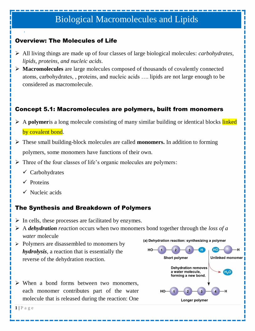
A dehydration reaction occurs when two monomers bond together through the loss of a
water molecule
Polymers are disassembled to monomers by
hydrolysis, a reaction that is essentially the
reverse of the dehydration reaction.
When a bond forms between two monomers,
each monomer contributes part of the water
molecule that is released during the reaction: One
Biological Macromolecules and Lipids
2 | P a g e
monomer provides a hydroxyl group ( - OH), while the other provides a hydrogen ( - H).
This reaction is repeated as monomers are added to the chain one by one, making a polymer
(also called polymerization or Condensation reaction)
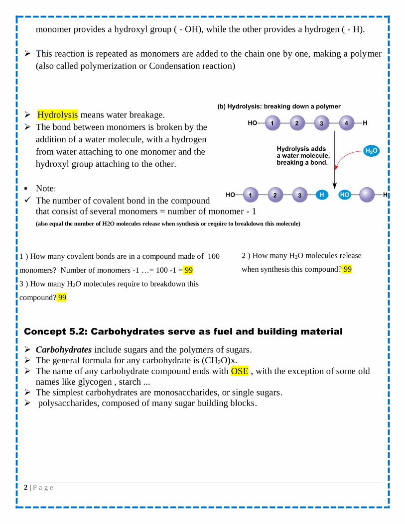
Hydrolysis means water breakage.
The bond between monomers is broken by the
addition of a water molecule, with a hydrogen
from water attaching to one monomer and the
hydroxyl group attaching to the other.
Note:
The number of covalent bond in the compound
that consist of several monomers = number of monomer - 1
(also equal the number of H2O molecules release when synthesis or require to breakdown this molecule)
Concept 5.2: Carbohydrates serve as fuel and building material
Carbohydrates include sugars and the polymers of sugars.
The general formula for any carbohydrate is (CH2O)x.
The name of any carbohydrate compound ends with OSE , with the exception of some old
names like glycogen , starch ...
The simplest carbohydrates are monosaccharides, or single sugars.
polysaccharides, composed of many sugar building blocks.
2 ) How many H2O molecules release
when synthesis this compound? 99
1 ) How many covalent bonds are in a compound made of 100
monomers? Number of monomers -1 …= 100 -1 = 99
3 ) How many H2O molecules require to breakdown this
compound? 99
3 | P a g e
Sugars
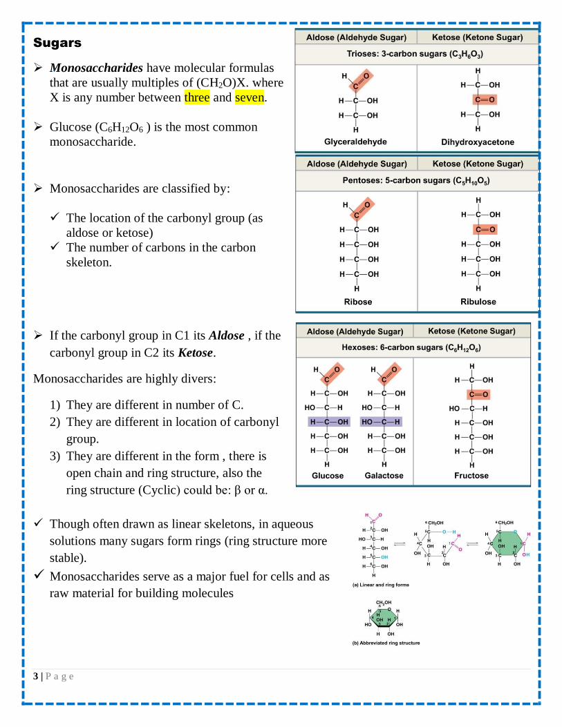
Monosaccharides have molecular formulas
that are usually multiples of (CH2O)X. where
X is any number between three and seven.
Glucose (C6H12O6 ) is the most common
monosaccharide.
Monosaccharides are classified by:
The location of the carbonyl group (as
aldose or ketose)
The number of carbons in the carbon
skeleton.
If the carbonyl group in C1 its Aldose , if the
carbonyl group in C2 its Ketose.
Monosaccharides are highly divers:
1) They are different in number of C.
2) They are different in location of carbonyl
group.
3) They are different in the form , there is
open chain and ring structure, also the
ring structure (Cyclic) could be: β or α.
Though often drawn as linear skeletons, in aqueous
solutions many sugars form rings (ring structure more
stable).
Monosaccharides serve as a major fuel for cells and as
raw material for building molecules

4 | P a g e
The carbonyl group of C1 react with the hydroxyl group of C5. This is the most stable ring
form.
The oxygen of the hydroxyl group of C5 joined the ring.
All OH groups on the right side of liner structure are written downwards.
All OH groups on the left side of liner structure are written upwards.
C6 is outside the ring.
approximately 80% of glucose present in the ring structure.
If the OH group where the ring is formed (C1 in glucose & C2 in fructose) in the right side it is α sugar
while If the OH group in the left side it is β sugar.
Monosaccharides functions:
1. particularly glucose, are major nutrients for cells. In the process known as cellular
respiration, cells extract energy from glucose molecules.
2. as raw material for the synthesis of other types of small organic molecules, such as amino
acids and fatty acids.
3. monomers for disaccharides or polysaccharides.
Disaccharide
A disaccharide is formed when a dehydration reaction joins two monosaccharides
This covalent bond is called a glycosidic linkage.
The molecular formula of disaccharide : (C6H12O6)*2 – H2O……(C12H22O11)
There are 3 examples of disaccharides:

5 | P a g e
1. Maltose is a disaccharide formed by the linking of two molecules of α glucose. Also
known as malt sugar
The type of bond in maltose is α-1,4-glycosidic linkage
2. The most prevalent disaccharide is sucrose, or table sugar. Its two monomers are glucose
and fructose.
The type of bond in sucrose is α-1,2-glycosidic linkage
3. Lactose, the sugar present in milk, is another disaccharide, in this case a glucose
molecule joined to a galactose molecule.
The type of bond in Lactose is α-1,4-glycosidic linkage.
Polysaccharides
Polysaccharides, the polymers of sugars, have storage and structural roles.
Maltose (malt sugar) Glucose + Glucose α-1,4-glycosidic linkage
Sucrose (table sugar) Glucose + Fructose
α-1,2-glycosidic linkage
Lactose (milk sugar) Glucose + Galactose α-1,4-glycosidic linkage
6 | P a g e
The structure and function of a polysaccharide are determined by its sugar monomers and
the positions of glycosidic linkages.
Storage Polysaccharides
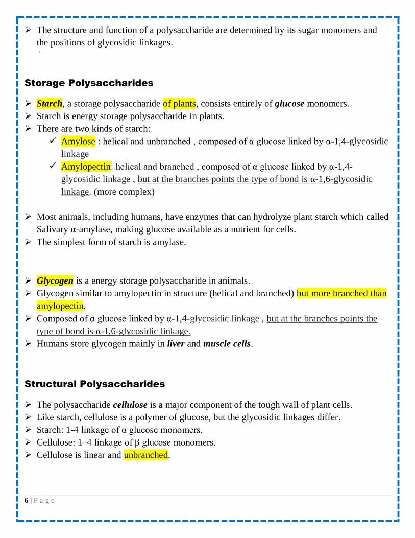
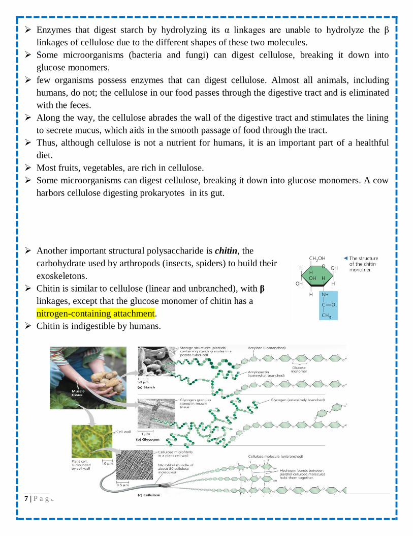
Starch, a storage polysaccharide of plants, consists entirely of glucose monomers.
Starch is energy storage polysaccharide in plants.
There are two kinds of starch:
Amylose : helical and unbranched , composed of α glucose linked by α-1,4-glycosidic
linkage
Amylopectin: helical and branched , composed of α glucose linked by α-1,4-
glycosidic linkage , but at the branches points the type of bond is α-1,6-glycosidic
linkage. (more complex)
Most animals, including humans, have enzymes that can hydrolyze plant starch which called
Salivary α-amylase, making glucose available as a nutrient for cells.
The simplest form of starch is amylase.
Glycogen is a energy storage polysaccharide in animals.
Glycogen similar to amylopectin in structure (helical and branched) but more branched than
amylopectin.
Composed of α glucose linked by α-1,4-glycosidic linkage , but at the branches points the
type of bond is α-1,6-glycosidic linkage.
Humans store glycogen mainly in liver and muscle cells.
Structural Polysaccharides
The polysaccharide cellulose is a major component of the tough wall of plant cells.
Like starch, cellulose is a polymer of glucose, but the glycosidic linkages differ.
Starch: 1-4 linkage of α glucose monomers.
Cellulose: 1–4 linkage of β glucose monomers.
Cellulose is linear and unbranched.
7 | P a g e
Enzymes that digest starch by hydrolyzing its α linkages are unable to hydrolyze the β
linkages of cellulose due to the different shapes of these two molecules.
Some microorganisms (bacteria and fungi) can digest cellulose, breaking it down into
glucose monomers.
few organisms possess enzymes that can digest cellulose. Almost all animals, including
humans, do not; the cellulose in our food passes through the digestive tract and is eliminated
with the feces.
Along the way, the cellulose abrades the wall of the digestive tract and stimulates the lining
to secrete mucus, which aids in the smooth passage of food through the tract.
Thus, although cellulose is not a nutrient for humans, it is an important part of a healthful
diet.
Most fruits, vegetables, are rich in cellulose.
Some microorganisms can digest cellulose, breaking it down into glucose monomers. A cow
harbors cellulose digesting prokaryotes in its gut.
Another important structural polysaccharide is chitin, the
carbohydrate used by arthropods (insects, spiders) to build their
exoskeletons.
Chitin is similar to cellulose (linear and unbranched), with β
linkages, except that the glucose monomer of chitin has a
nitrogen-containing attachment.
Chitin is indigestible by humans.

8 | P a g e
Concept 5.3: Lipids are a diverse group of hydrophobic molecules
Lipids are the one class of large biological molecules that do not form polymers
The unifying feature of lipids is having little or no affinity for water
Lipids are hydrophobic because they consist mostly of hydrocarbons, which form nonpolar
covalent bonds
The most biologically important lipids are fats, phospholipids, and steroids
Fats
fats are not polymers, they are large molecules assembled
from smaller molecules by dehydration reactions.
A fat is constructed from two kinds of smaller molecules:
glycerol and fatty acids.
Glycerol is an alcohol; each of its three carbons bears a
hydroxyl group.
A fatty acid has a long carbon skeleton, usually 16 or 18
carbon atoms in length.
The carbon at one end of the skeleton is part of a carboxyl
group, the functional group that gives these molecules the
name fatty acid.
The rest of the skeleton consists of a hydrocarbon chain.
The relatively nonpolar C-H bonds in the hydrocarbon chains of fatty acids are the reason
fats are hydrophobic.
In making a fat, three fatty acid molecules are each joined to glycerol by an covalent bond
called ester linkage, a bond formed by a dehydration reaction between a hydroxyl group and
a carboxyl group.
The resulting fat, also called a triacylglycerol, thus consists of three fatty acids linked to one
glycerol molecule. (Still another name for a fat is triglyceride).
Storage Polysaccharides
Structural Polysaccharides
Starch(Amylopectin & Amylopectin) Starch cellulose
Glycogen chitin
9 | P a g e
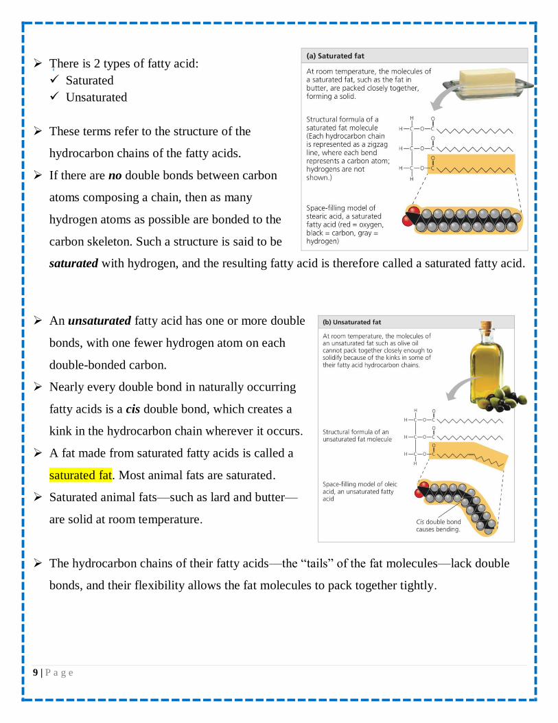
There is 2 types of fatty acid:
Saturated
Unsaturated
These terms refer to the structure of the
hydrocarbon chains of the fatty acids.
If there are no double bonds between carbon
atoms composing a chain, then as many
hydrogen atoms as possible are bonded to the
carbon skeleton. Such a structure is said to be
saturated with hydrogen, and the resulting fatty acid is therefore called a saturated fatty acid.
An unsaturated fatty acid has one or more double
bonds, with one fewer hydrogen atom on each
double-bonded carbon.
Nearly every double bond in naturally occurring
fatty acids is a cis double bond, which creates a
kink in the hydrocarbon chain wherever it occurs.
A fat made from saturated fatty acids is called a
saturated fat. Most animal fats are saturated.
Saturated animal fats—such as lard and butter—
are solid at room temperature.
The hydrocarbon chains of their fatty acids—the “tails” of the fat molecules—lack double
bonds, and their flexibility allows the fat molecules to pack together tightly.
10 | P a g e
In contrast, the fats of plants and fishes are generally unsaturated, meaning that they are built
of one or more types of unsaturated fatty acids.
Usually liquid at room temperature, plant and fish fats are referred to as oils
The kinks where the cis double bonds are located prevent the molecules from packing
together closely enough to solidify at room temperature.
A diet rich in saturated fats is one of several factors that may contribute to the cardiovascular
disease known as atherosclerosis.
The major function of fats is:
1. Energy storage (the hydrocarbon chains of fats are similar to gasoline molecules and just
as rich in energy).
2. Shocks absorption (adipose tissue cushions such vital organs as the kidneys).
3. Insulation (a layer of fat beneath the skin insulates the body).
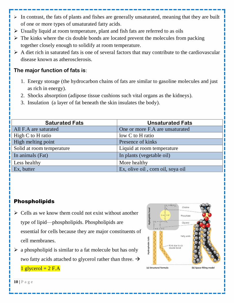
Phospholipids
Cells as we know them could not exist without another
type of lipid—phospholipids. Phospholipids are
essential for cells because they are major constituents of
cell membranes.
a phospholipid is similar to a fat molecule but has only
two fatty acids attached to glycerol rather than three.
1 glycerol + 2 F.A
Saturated Fats Unsaturated Fats All F.A are saturated One or more F.A are unsaturated
High C to H ratio low C to H ratio
High melting point Presence of kinks
Solid at room temperature Liquid at room temperature
In animals (Fat) In plants (vegetable oil)
Less healthy More healthy
Ex, butter Ex, olive oil , corn oil, soya oil

11 | P a g e
The third hydroxyl group of glycerol is joined to a phosphate group, which has a negative
electrical charge in the cell.
Typically, an additional small charged or polar molecule is also linked to the phosphate
group. Choline is one such molecule , but there are many others as well, allowing formation
of a variety of phospholipids that differ from each other.
The two ends of phospholipids show different behaviors with respect to water. The
hydrocarbon tails are hydrophobic and are excluded from water. However, the phosphate
group and its attachments form a hydrophilic head that has
an affinity for water.
When phospholipids are added to water, they self-
assemble into a double-layered sheet called a “bilayer”
that shields their hydrophobic fatty acid tails from water
At the surface of a cell, phospholipids are arranged in a similar bilayer.
The hydrophilic heads of the molecules are on the outside of the bilayer, in contact with the
aqueous solutions inside and outside of the cell.
The hydrophobic tails point toward the interior of the bilayer, away from the water.
Phospholipids Function
Major structural component of plasma membrane of all cells.
Saturated Phospholipids Unsaturated Phospholipids Solid Liquid
All F.A are saturated One or more F.A are unsaturated
High C to H ratio low C to H ratio
NO kinks Presence of kinks
12 | P a g e
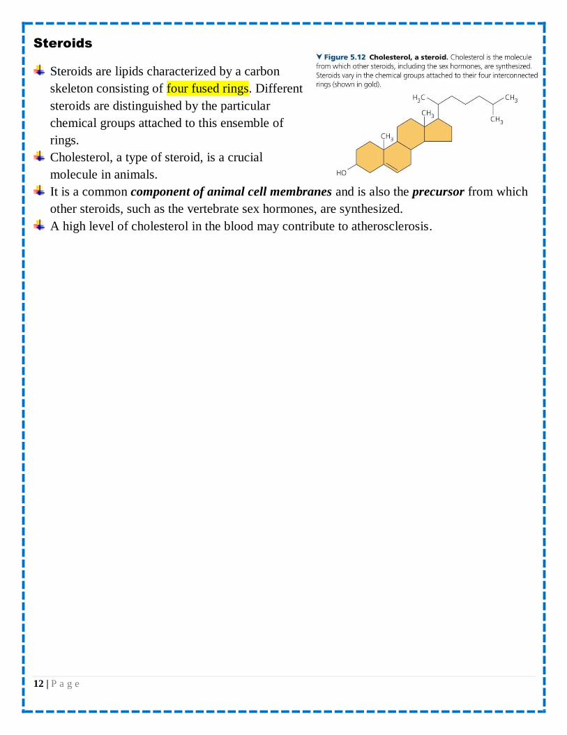
Steroids
Steroids are lipids characterized by a carbon
skeleton consisting of four fused rings. Different
steroids are distinguished by the particular
chemical groups attached to this ensemble of
rings.
Cholesterol, a type of steroid, is a crucial
molecule in animals.
It is a common component of animal cell membranes and is also the precursor from which
other steroids, such as the vertebrate sex hormones, are synthesized.
A high level of cholesterol in the blood may contribute to atherosclerosis.

13 | P a g e
14 | P a g e
Concept 5.4: Proteins include a diversity of structures, resulting in a
wide range of functions
Proteins are all constructed from the same set of 20 amino acids, linked in unbranched
polymers.
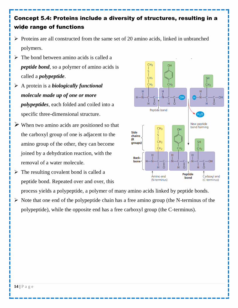
The bond between amino acids is called a
peptide bond, so a polymer of amino acids is
called a polypeptide.
A protein is a biologically functional
molecule made up of one or more
polypeptides, each folded and coiled into a
specific three-dimensional structure.
When two amino acids are positioned so that
the carboxyl group of one is adjacent to the
amino group of the other, they can become
joined by a dehydration reaction, with the
removal of a water molecule.
The resulting covalent bond is called a
peptide bond. Repeated over and over, this
process yields a polypeptide, a polymer of many amino acids linked by peptide bonds.
Note that one end of the polypeptide chain has a free amino group (the N-terminus of the
polypeptide), while the opposite end has a free carboxyl group (the C-terminus).
15 | P a g e
Amino Acid Monomers
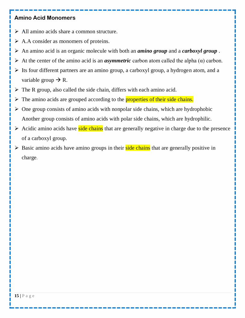
All amino acids share a common structure.
A.A consider as monomers of proteins.
An amino acid is an organic molecule with both an amino group and a carboxyl group .
At the center of the amino acid is an asymmetric carbon atom called the alpha (α) carbon.
Its four different partners are an amino group, a carboxyl group, a hydrogen atom, and a
variable group R.
The R group, also called the side chain, differs with each amino acid.
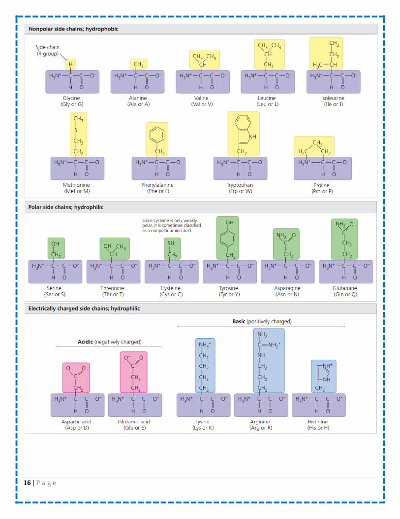
The amino acids are grouped according to the properties of their side chains.
One group consists of amino acids with nonpolar side chains, which are hydrophobic
Another group consists of amino acids with polar side chains, which are hydrophilic.
Acidic amino acids have side chains that are generally negative in charge due to the presence
of a carboxyl group.
Basic amino acids have amino groups in their side chains that are generally positive in
charge.
16 | P a g e
17 | P a g e
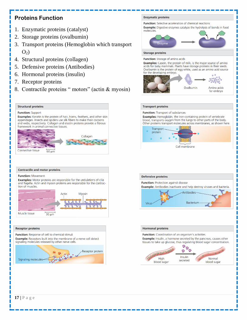
Proteins Function
1. Enzymatic proteins (catalyst)
2. Storage proteins (ovalbumin)
3. Transport proteins (Hemoglobin which transport
O2)
4. Structural proteins (collagen)
5. Defensive proteins (Antibodies)
6. Hormonal proteins (insulin)
7. Receptor proteins
8. Contractile proteins “ motors” (actin & myosin)
18 | P a g e
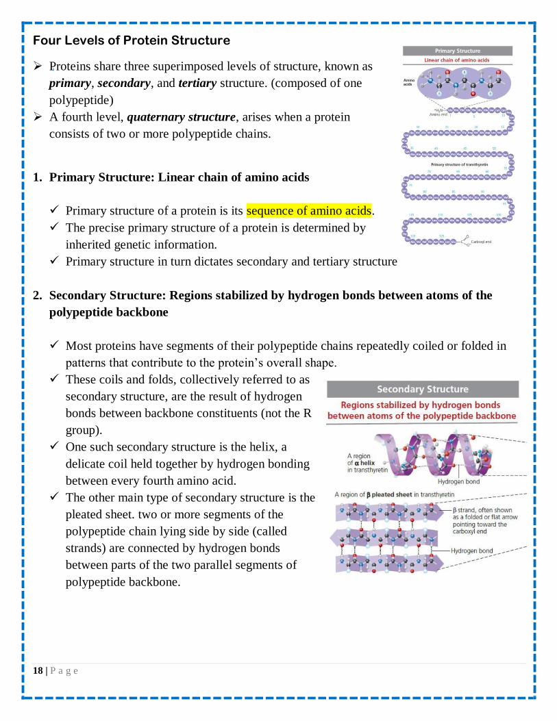
Four Levels of Protein Structure
Proteins share three superimposed levels of structure, known as
primary, secondary, and tertiary structure. (composed of one
polypeptide)
A fourth level, quaternary structure, arises when a protein
consists of two or more polypeptide chains.
1. Primary Structure: Linear chain of amino acids
Primary structure of a protein is its sequence of amino acids.
The precise primary structure of a protein is determined by
inherited genetic information.
Primary structure in turn dictates secondary and tertiary structure
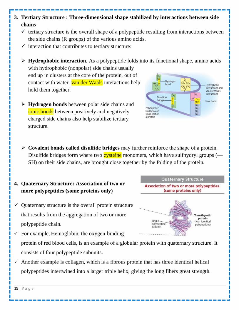
2. Secondary Structure: Regions stabilized by hydrogen bonds between atoms of the
polypeptide backbone
Most proteins have segments of their polypeptide chains repeatedly coiled or folded in
patterns that contribute to the protein’s overall shape.
These coils and folds, collectively referred to as
secondary structure, are the result of hydrogen
bonds between backbone constituents (not the R
group).
One such secondary structure is the helix, a
delicate coil held together by hydrogen bonding
between every fourth amino acid.
The other main type of secondary structure is the
pleated sheet. two or more segments of the
polypeptide chain lying side by side (called
strands) are connected by hydrogen bonds
between parts of the two parallel segments of
polypeptide backbone.
19 | P a g e
3. Tertiary Structure : Three-dimensional shape stabilized by interactions between side
chains
tertiary structure is the overall shape of a polypeptide resulting from interactions between
the side chains (R groups) of the various amino acids.
interaction that contributes to tertiary structure:
Hydrophobic interaction. As a polypeptide folds into its functional shape, amino acids
with hydrophobic (nonpolar) side chains usually
end up in clusters at the core of the protein, out of
contact with water. van der Waals interactions help
hold them together.
Hydrogen bonds between polar side chains and
ionic bonds between positively and negatively
charged side chains also help stabilize tertiary
structure.
Covalent bonds called disulfide bridges may further reinforce the shape of a protein.
Disulfide bridges form where two cysteine monomers, which have sulfhydryl groups (—
SH) on their side chains, are brought close together by the folding of the protein.
4. Quaternary Structure: Association of two or
more polypeptides (some proteins only)
Quaternary structure is the overall protein structure
that results from the aggregation of two or more
polypeptide chain.
For example, Hemoglobin, the oxygen-binding
protein of red blood cells, is an example of a globular protein with quaternary structure. It
consists of four polypeptide subunits.
Another example is collagen, which is a fibrous protein that has three identical helical
polypeptides intertwined into a larger triple helix, giving the long fibers great strength.

20 | P a g e
Denaturation of protein
• Is the disruption in protein bond resulting in loss of protein structure and function
• The denatured protein is biologically inactive.
• Factors that cause protein to denature:
Temperature (heating)
pH
When a protein denatured by heat or chemicals, it can sometimes return to its functional
shape (renaturation)when the denaturing agent is removed. (Sometimes this is not possible)
Concept 5.5. Nucleic acids
Nucleic acids (polynucleotides): many nucleotides linked by many covalent bond called
phosphodiester bond.
Nucleic acids are polymers made of monomers called nucleotides.
The two types of nucleic acids:
1. deoxyribonucleic acid (DNA)
2. ribonucleic acid (RNA)
A nucleotide, in general, is composed of three parts:
1. five-carbon sugar (a pentose)
2. nitrogen-containing (nitrogenous) base
3. one to three phosphate groups. The beginning
monomer used to build a polynucleotide has three
phosphate groups, but two are lost during the
polymerization process
The portion of a nucleotide without any
phosphate groups is called a nucleoside.
The nitrogenous bases
Each nitrogenous base has one or two rings
that include nitrogen atoms.
There are two families of nitrogenous bases: pyrimidines and purines.
A pyrimidine has one six-membered ring of carbon and nitrogen atoms. The members of the
pyrimidine family are cytosine (C), thymine (T), and uracil (U).

21 | P a g e
Purines are larger, with a six-membered ring fused to a five-membered ring. The purines are
adenine (A) and guanine (G).
Adenine, guanine, and cytosine are found in both DNA and RNA; thymine is found only in
DNA and uracil only in RNA.
In DNA the sugar is deoxyribose; in RNA it is ribose
The only difference between these two sugars is that deoxyribose lacks an oxygen atom on
the second carbon in the ring, hence the name
deoxyribose
The two free ends of the polymer are distinctly
different from each other. One end has a phosphate
attached to a 5′ carbon, and the other end has a
hydroxyl group on a 3′ carbon; we refer to these as
the 5′ end and the 3′ end, respectively.
We can say that a polynucleotide has a built-in
directionality along its sugar-phosphate backbone, from 5′ to 3′.