Embed Size (px)
Citation preview
Summary. Vasoactive intestinal peptide (VIP) is apotent inductor of cyclooxygenase-2 (COX-2)expression in human prostate cancer cell lines. There areconflicting data regarding the role of COX-2 in theprogression of this disease. Here we examined theexpression of VIP receptors (VPAC1 and VPAC2) andCOX-2 in prostate cancer specimens. Correlationsamong protein levels and various clinicopathologicalfactors and prognosis of patients were statisticallyanalyzed. For these purposes, formaldehyde-fixed,paraffin-embedded prostate tissue specimens from 63patients with prostate cancer and 9 control samples wereused. The expression of VPAC1 and VPAC2 receptorsand COX-2 was analyzed at mRNA levels byquantitative reverse transcriptase-PCR. Thecorresponding expression at protein level was studied byimmunohistochemistry, scored as negative, weak,moderate, or strong, and correlated with differentclinicopathological factors by means of multivariateanalysis. 88% of prostate cancer tissues overexpressedVPAC1-receptor at mRNA level, 72% VPAC2-receptorand 77% COX-2. Simultaneous overexpression of thethree genes was seen in 52% of patients. Similaroverexpression patterns were observed at protein level.The correlation between VPAC1 and VPAC2 receptorprotein levels was statistically significant. However, nosignificant correlations existed among protein levels ofVPAC receptors and COX-2 with patient age, prostate-specific antigen (PSA) levels, tumor stage, Gleasonscore and survival time. The overexpression of VPAC1and VPAC2 receptors and COX-2 in cancer tissue gives
them a potential role as targets for diagnosis of prostatecancer but results do not support a clear value asbiomarkers for the clinical prognosis of this disease.Key words: VIP, VPAC1 and VPAC2 receptors, COX-2,Prognostic marker, Prostate cancer
Introduction
Prostate cancer is the most common malignancy andthe second leading cause of cancer-related death amongmen in industrial western countries (Jemal et al., 2010).Various signaling pathways have been involved inprostate carcinogenesis and progression, but notmolecular biomarkers have yet been identified withcertainty to be correlated with clinical outcome of thedisease (Lopergolo and Zaffaroni, 2009).
Vasoactive intestinal peptide (VIP) is a neuropeptidepresent in the human prostate (Polak and Bloom, 1984;Fernández-Martínez et al., 2009). VIP exerts a widerange of biological effects which are initiated throughVIP receptors (VPAC1 and VPAC2 coupled to adenylatecyclase stimulation, as shown in many cells and tissues,including normal and cancer prostate gland as well asprostate cancer cell lines (Juarranz et al., 2001; García-Fernández et al., 2003). A previous study on prostatecancer tissue from a small number of patients indicated adecrease in the number of VIP receptors by means ofimmunochemistry, but RT-PCR and western-blotexperiments gave no conclusive differences incomparison with normal tissue samples (García-Fernández et al., 2003).
In prostate, VIP increases the expression of themajor angiogenic factor, vascular endothelial growth
Overexpression of vasoactive intestinal peptidereceptors and cyclooxygenase-2 in human prostatecancer. Analysis of potential prognostic relevanceAna B. Fernández-Martínez1, María J. Carmena1, M. Isabel Arenas2, Ana M. Bajo1, Juan C. Prieto1 and Manuel Sánchez-Chapado3,4Departments of 1Biochemistry and Molecular Biology, 2Cell Biology and Genetics, and 3Surgery, University of Alcalá and 4Department of Urology, Príncipe de Asturias Hospital, Alcalá de Henares, Spain
Histol Histopathol (2012) 27: 1093-1101
Offprint requests to: Prof. Juan C. Prieto, Department of Biochemistryand Molecular Biology, University of Alcalá, Campus Universitario, E-28871 Alcalá de Henares, Spain. e-mail: [email protected]
DOI: 10.14670/HH-27.1093
http://www.hh.um.es
Histology andHistopathologyCellular and Molecular Biology

factor (VEGF) and the proinflammatory enzymecyclooxygenase-2 (COX-2) (Collado et al., 2004;Fernández-Martínez et al., 2007). In addition, theneuropeptide induces neuroendocrine differentiation inandrogen-dependent prostate cancer LNCaP cells(Juarranz et al., 2001), promotes survival and stimulateshuman epidermal growth factor receptor-2 (HER2)transphosphorylation in androgen-independent prostatecancer PC3 cells (Gutiérrez-Cañas et al., 2003;Sotomayor et al., 2007) and behaves as a pro-metastaticfactor in both, LNCaP and PC3 cells (Fernández-Martínez et al., 2009). Angiogenesis, neuroendocrinedifferentiation and cell survival are steps of prostatecancer progression to androgen independence (Arya etal., 2006; Clarke et al., 2009). Thus, the consideration ofthe potential diagnostic and prognostic value of VIPreceptors in this disease is interesting.
The role of inducible cyclooxygenase (COX-2) inprostate carcinogenesis is still controversial, since thereare studies regarding the detection of enzyme expressionand activity in human tissue and cell lines which reportincreased or even absent expression of COX-2 inprostate cancer (Castelli et al., 2010; Abedinpour et al.,2011). Moreover, some studies describe thatproinflammatory atrophic lesions in prostate, which arethought to be precursors of prostate cancer, expressCOX-2 (Liu et al., 2000; Sotomayor et al., 2007). Thus,the association of COX-2 to prostate carcinogenesis orcancer progression has led to consider it as a rationaldrug target for prostate cancer prevention, although thesituation remains confusing.
Previous results from our laboratory on the effect ofVIP on COX-2 expression in human prostate non-neoplastic (RWPE-1), as well as cancer LNCaP and PC3cells, showed that VIP induced higher levels of COX-2protein expression in prostate cancer cells as comparedwith non-neoplastic cells (Fernández-Martínez et al.,2007). The relationship of VIP and COX-2 in a signalingnetwork in human prostate cancer suggest that theneuropeptide may induce promotion and progression ofprostate carcinoma through the activation ofproinflammatory and proangiogenic signals, such asthose deriving from the increased expression of COX-2enzyme. In order to clarify a diagnostic or prognosticvalue, here we compared the expression levels of VPAC1and VPAC2 receptors and COX-2 in human non-metastatic prostate cancers, as well as in control tissuesamples; then, we searched for correlations with variousclinicopathological factors.Materials and methods
Patients
Sixty three patients with prostate carcinoma,apparently limited to the prostate gland (aged from 47 to74 years) and subjected to radical prostatectomy withcurative purposes were included in this study (Table 1).The control group consisted of 9 patients (aged from 54
to 80 years) undergoing radical cystectomy-prostatectomy due to urothelial infiltrating carcinoma.None of the patients was treated with hormones or othertherapies before surgery. Clinicopathological data of thepatients (Table 1) included routine determination ofpreoperative serum prostate-specific antigen (PSA)levels. All prostate tumors were graded according to thesystem of Gleason (Helpap and Egevad, 2009). Thetumor pathological stage (pT) was also evaluated.Patients were regularly followed up and survival datawere ascertained through patient records. Writteninformed consent was obtained from all patients. Thestudy was approved by the Research Ethics Committee.Samples of the prostate proper zone and suspected tumorzones were delimited by two independent pathologistsand taken from resected tissues and immediately fixed in10 % (v/v) formaldehyde in PBS (pH 7.4) for 24 h,dehydrated, and embedded in paraffin (FFPE). Isolation of RNA and single-step real-time quantitativeRT-PCR
FFPE tissue samples were cut into 5 mm-thicksections on a microtome and subjected to RNA isolationwith the Absolutely RNA FFPE Kit (Stratagene, LaJolla, CA). Deparaffinization was first performed withxylene, followed by extraction in ethanol andhomogenization by overnight incubation in ProteinaseK. DNase I was then used to digest residual DNA and,finally, solubilized nucleic acids were bound to a glassfiber filter in the presence of guanidine salts. Filter-bound nucleic acids were washed and RNA was eluted.RNA concentration was determined with a NanodropND-100 spectrophotometer (Nanodrop Technologies,Wilmington, DE). Real-time quantitative RT-PCRanalysis was performed using SYBR Green PCR mastermix, in a one-step RT-PCR protocol according to themanufacturer’s instructions (Applied Biosystems, FosterCity, CA). Four nanograms of total RNA samples wereused for each PCR amplification with a primer set whichamplifies cDNAs for human VPAC1 receptor (sense 5’-CTG GGT CAG TCT GGT GGG-3’, antisense 5’-TCCGAG ACC TAG CAT TCG CT-3’), VPAC2 receptor(sense 5’-TCA GTG CTG GTC AAG GAC GAC-3’,antisense 5’- AAG ACC AGG CTC AGC TTG CA-3’),COX-2 (sense 5’- TGA CGG GGT CAC CCA CACTGT GCC CGT CTA-3’, antisense 5’- CTA GAA GCACGG TTG ACG ATG GAG GG-3’), and ß-actin (sense5’-AGA AGG ATT CCT ATG TGG GCG-3’, antisense5’- CAT GTC CCA GTT GGT GAC-3’). Thermalcycling parameters were 30 min at 48°C for RT and 10min at 95°C for activation of AmpliTaq Gold DNAPolymerase, followed by 50 cycles of 95°C for 15 s and60°C for 1 min. Negative controls with water instead ofcDNA were run in parallel to exclude contamination.The relative quantification was normalized to the ß-actingene expression level. PCR reactions were performedusing ABIPrism 7000 SDS (Applied Biosystems). Themean Ct (threshold cycle; cycle at which the increase in
1094VIP receptors/COX-2 in prostate cancer
signal associated with an exponential growth of PCRproduct is first detected) value of tumor samples wascompared to that of control samples using the Ct valueof ß-actin as an internal reference. ∆Ct was thedifference in Ct values derived from genes and ß-actingene, and ∆∆Ct represented the difference betweenpaired samples. The n-fold differential ratio wasexpressed as 2-∆∆Ct (Chang et al., 2002). It should benoted that ß-actin was similarly expressed in tumor andhealthy zones of tissue sections (data not shown).Immunohistochemistry
For immunohistochemistry studies, deparaffinizedsections of prostate tissue representative of the tumor (5µm thickness) were hydrated and incubated for 30 minin 3% H2O2 diluted in methanol to reduce endogenousperoxidase activity. For antigen retrieval, sections wereincubated with 0.1 mol/L citrate buffer (pH 6) for 2 minin a conventional pressure cooker. After rinsing in TBS,slides were incubated with normal donkey serum (NDS)at a 1:5 dilution in TBS (TBS/NDS) for 60 min, toprevent nonspecific binding of the primary antibody.Then, primary antibodies against VPAC1 and VPAC2receptors (Thermo Fisher Scientific, Rockford, IL) orCOX-2 (Cayman Chemical, Ann Arbor, MI) wereapplied at 1:500, 1:500 or 1:50 dilution, respectively,diluted in blocking solution 1:9, at 4°C overnight.Afterwards, sections were washed twice in TBS anddetection was done by the conventional labeled-streptavidin-biotin method (LSAB-kit, Dako, Barcelona,Spain). Peroxidase activity was detected using theglucose oxidase-3,3’-diaminobenzidine (DAB) nickelintensification method kit (Zymed Laboratories, SanFrancisco, CA). Sections were lightly counterstained
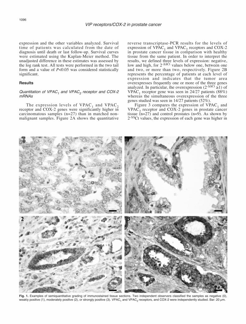
with hematoxylin, dehydrated and mounted in DePex(Probus, Barcelona, Spain). Sections of normal humantissues (skin, testes and cerebellum) that express VPAC1and VPAC2 receptors and COX-2 were used as positivecontrols. In negative control experiments, serial sectionsof each specimen were processed as described, but thecorresponding primary antibody was omitted.Immunoreactivity of each focus of interest wassemiquantitatively graded by two independent observersas negative (0), weakly positive (1), moderately positive(2), or strongly positive (3) (Fig. 1). In order tostrengthen the immunological score and gain inreproducibility, the samples were reviewed and scoredagain by two independent pathologists.Statistical analysis
The SPSS 17.0 software package (SPSS Inc.,Chicago, IL) was used for data retrieval and analysis. Toperform a differential analysis of the positive tissuespecimens for VPAC1 and VPAC2 receptors and COX-2,Dunn’s Multiple Comparison test was used. Univariateanalysis comparing categorical variables (VPAC1 andVPAC2 receptors and COX-2 expression, andclinicopathological data: patient age, tumor pathologicalstage, Gleason score, and pre-operatory PSA levels) wasperformed using chi-square tests. We tested for thepresence of a linear trend when there were more thantwo categories of staining using the Mantel-Haenszelchi-square test. Continuous variables were comparedusing the Mann-Whitney test. For all these tests wecomputed P-values using an exact method due to smallsample sizes. This analysis was completed with multipleregression analyses (Durbin-Watson test) to evaluate thepossible dependence between VPAC1 receptor
1095VIP receptors/COX-2 in prostate cancer
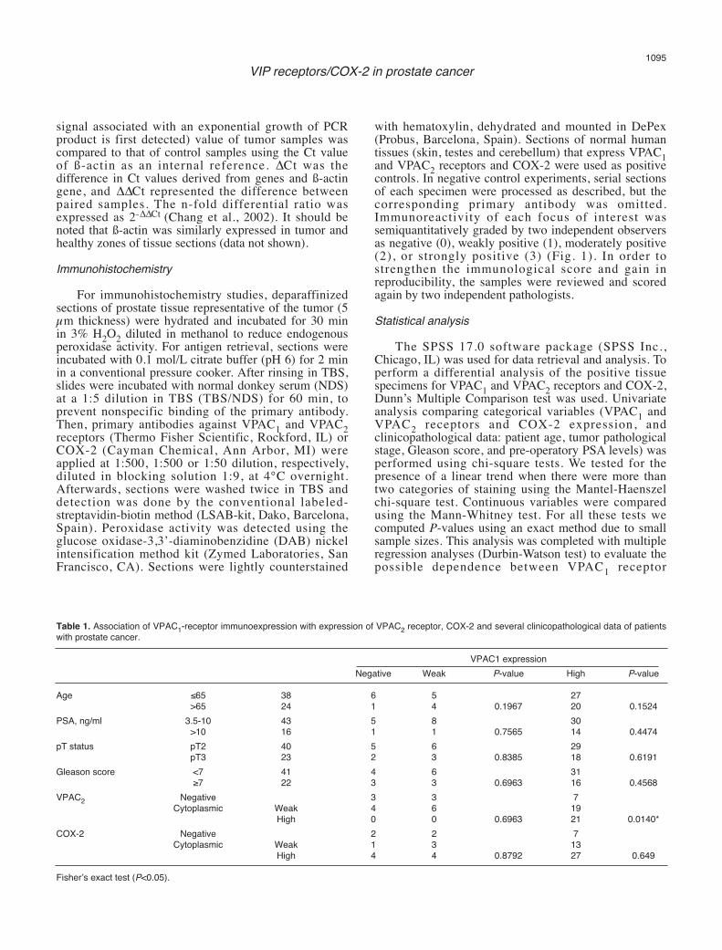
Table 1. Association of VPAC1-receptor immunoexpression with expression of VPAC2 receptor, COX-2 and several clinicopathological data of patientswith prostate cancer.
VPAC1 expressionNegative Weak P-value High P-value
Age ≤65 38 6 5 27>65 24 1 4 0.1967 20 0.1524
PSA, ng/ml 3.5-10 43 5 8 30>10 16 1 1 0.7565 14 0.4474
pT status pT2 40 5 6 29pT3 23 2 3 0.8385 18 0.6191
Gleason score <7 41 4 6 31≥7 22 3 3 0.6963 16 0.4568
VPAC2 Negative 3 3 7Cytoplasmic Weak 4 6 19
High 0 0 0.6963 21 0.0140*COX-2 Negative 2 2 7
Cytoplasmic Weak 1 3 13High 4 4 0.8792 27 0.649
Fisher’s exact test (P<0.05).
expression and the other variables analyzed. Survivaltime of patients was calculated from the date ofdiagnosis until death or last follow-up. Survival curveswere estimated using the Kaplan-Meier method. Theunadjusted difference in these estimates was assessed bythe log rank test. All tests were performed in the two tailform and a value of P<0.05 was considered statisticallysignificant.Results
Quantitation of VPAC1 and VPAC2 receptor and COX-2mRNAs
The expression levels of VPAC1 and VPAC2receptor and COX-2 genes were significantly higher incarcinomatous samples (n=27) than in matched non-malignant samples. Figure 2A shows the quantitative
reverse transcriptase-PCR results for the levels ofexpression of VPAC1 and VPAC2 receptors and COX-2in prostate cancer tissue in comparison with healthytissue from the same patient. In order to interpret theresults, we defined three levels of expression: negative,low and high, for 2-ΔΔCt values below one, between oneand two, or more than two, respectively. Figure 2Brepresents the percentage of patients at each level ofexpression and indicates that the tumor areaoverexpresses frequently one or more of the three genesanalyzed. In particular, the overexpression (2-ΔΔCt ≥1) ofVPAC1 receptor gene was seen in 24/27 patients (88%)whereas the simultaneous overexpression of the threegenes studied was seen in 14/27 patients (52%).
Figure 3 compares the expression of VPAC1 andVPAC2 receptor and COX-2 genes in prostate cancertissue (n=27) and control prostates (n=9). As shown by2-ΔΔCt values, the expression of each gene was higher in
1096VIP receptors/COX-2 in prostate cancer
Fig. 1. Examples of semiquantitative grading of immunostained tissue sections. Two independent observers classified the samples as negative (0),weakly positive (1), moderately positive (2), or strongly positive (3). VPAC1 and VPAC2 receptors, and COX-2 were independently studied. Bar: 20 µm.
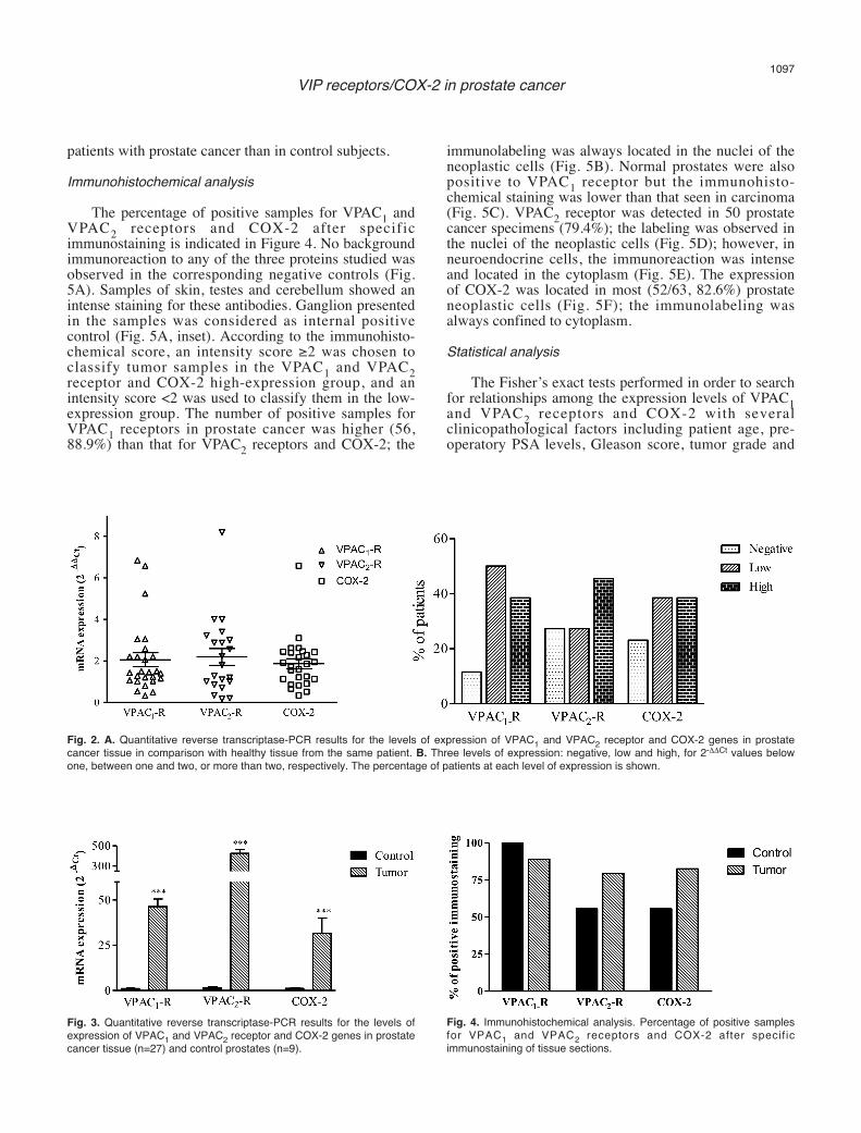
patients with prostate cancer than in control subjects.Immunohistochemical analysis
The percentage of positive samples for VPAC1 andVPAC2 receptors and COX-2 after specificimmunostaining is indicated in Figure 4. No backgroundimmunoreaction to any of the three proteins studied wasobserved in the corresponding negative controls (Fig.5A). Samples of skin, testes and cerebellum showed anintense staining for these antibodies. Ganglion presentedin the samples was considered as internal positivecontrol (Fig. 5A, inset). According to the immunohisto-chemical score, an intensity score ≥2 was chosen toclassify tumor samples in the VPAC1 and VPAC2receptor and COX-2 high-expression group, and anintensity score <2 was used to classify them in the low-expression group. The number of positive samples forVPAC1 receptors in prostate cancer was higher (56,88.9%) than that for VPAC2 receptors and COX-2; the
immunolabeling was always located in the nuclei of theneoplastic cells (Fig. 5B). Normal prostates were alsopositive to VPAC1 receptor but the immunohisto-chemical staining was lower than that seen in carcinoma(Fig. 5C). VPAC2 receptor was detected in 50 prostatecancer specimens (79.4%); the labeling was observed inthe nuclei of the neoplastic cells (Fig. 5D); however, inneuroendocrine cells, the immunoreaction was intenseand located in the cytoplasm (Fig. 5E). The expressionof COX-2 was located in most (52/63, 82.6%) prostateneoplastic cells (Fig. 5F); the immunolabeling wasalways confined to cytoplasm.Statistical analysis
The Fisher’s exact tests performed in order to searchfor relationships among the expression levels of VPAC1and VPAC2 receptors and COX-2 with severalclinicopathological factors including patient age, pre-operatory PSA levels, Gleason score, tumor grade and
1097VIP receptors/COX-2 in prostate cancer
Fig. 2. A. Quantitative reverse transcriptase-PCR results for the levels of expression of VPAC1 and VPAC2 receptor and COX-2 genes in prostatecancer tissue in comparison with healthy tissue from the same patient. B. Three levels of expression: negative, low and high, for 2-ΔΔCt values belowone, between one and two, or more than two, respectively. The percentage of patients at each level of expression is shown.
Fig. 3. Quantitative reverse transcriptase-PCR results for the levels ofexpression of VPAC1 and VPAC2 receptor and COX-2 genes in prostatecancer tissue (n=27) and control prostates (n=9).
Fig. 4. Immunohistochemical analysis. Percentage of positive samplesfor VPAC1 and VPAC2 receptors and COX-2 after specif icimmunostaining of tissue sections.

survival time (Tables 1-3) showed only a positiveassociation between the expression of VPAC1 andVPAC2 receptors. This association was confirmed withthe non-parametric Spearman correlation (r=0.494,P=0.014). Patient’s age was homogeneous andindependent of VPAC1-receptor results. VPAC1expression was not associated with pT status, Gleasonscore or pre-operatory PSA value. VPAC1 and VPAC2receptor and COX-2 expression levels were notcorrelated with survival time (Fig. 6).Discussion
It is important to find useful biological markers withthe potential to define the aggressiveness of prostatecancer and give prognostic information which will allowstratifying patients into appropriate treatment regimens(Sánchez-Chapado et al., 2003; Slater et al., 2003; Molet al., 2007; Fritzsche et al., 2008; Niu et al., 2008;Evans, 2009).
PSA is unquestionably the most commonly used
circulating biomarker for prostate cancer but itsrevolutionary role as screening tool is now subjected tocontroversy (Bensalah et al., 2008). In addition to PSA,a plethora of circulating prostate cancer biomarkers havebeen considered as promising candidates for prognosisand analysis of disease progression and response totherapy, including insulin-like growth factor-I (IGF-I),urokinase plasminogen activation system, transforminggrowth factor-ß (TGF-ß), interleukin-6 (IL-6),chromogranin A and prostate cancer autoantibodies(Bensalah et al., 2008). Among other molecules, COX-2,TGF-ß, IL-10, and Ki67 have evolved as potential tissuebiomarkers that can better identify the biological natureof prostate tumors and predict which will act moreaggressively; however, contradictory results warrantfurther studies (Howell and Rose-Zerilli, 2007; Evans,2009).
The expression of VIP receptors in tissue specimensfrom patients with prostate cancer has been describedpreviously by our group (García-Fernández et al., 2003).We have also shown that VIP action through these
1098VIP receptors/COX-2 in prostate cancer
Table 3. Association of COX-2 immunoexpression with several clinicopathological data of patients with prostate cancer.
COX-2 expressionNegative Weak P-value High P-value
Age ≤65 38 6 9 23>65 25 5 8 1.000 12 0.7216
PSA, ng/ml 3.5-10 43 8 14 21>10 16 1 3 1.000 12 0.2319
pT status pT2 40 7 13 20pT3 23 4 4 0.6715 15 1.000
Gleason score <7 41 8 13 20≥7 22 3 4 1.000 15 0.4865
Fisher’s exact test (P<0.05).
Table 2. Association of VPAC2-receptor immunoexpression with expression of COX-2 and several clinicopathological data of patients with prostatecancer.
VPAC2 expressionNegative Weak P-value High P-value
Age ≤65 38 7 17 14>65 25 6 12 1.000 7 0.4913
PSA, ng/ml 3.5-10 43 9 25 9>10 16 2 4 1.000 10 0.1213
pT status pT2 40 7 19 14pT3 23 6 10 0.5097 7 0.4913
Gleason score <7 41 10 18 13≥7 22 3 11 0.4852 8 0.4653
COX-2 Negative 5 2 3Weak 2 12 4High 5 16 0.0668 14 0.0852
Fisher’s exact test (P<0.05).

receptors induces the expression of COX-2 (Fernández-Martínez et al., 2007). In the present study, our aimswere to extend knowledge on the functional role of VIPin the etiopathogenesis of prostate cancer, as well as tocontribute to the identification of early diagnostic andprognostic markers for this disease. For these purposes,we studied the correlation of the expression of VPAC1and VPAC2 receptors and COX-2 in control and cancer
prostates as well as the possibility of clinicopathologicaland prognostic significance of the expression levels ofthese molecules.
Present quantitative RT-PCR results on matchedmalignant and normal prostate tissue samples show thatVPAC1 and VPAC2 receptors and COX-2 enzyme wereoverexpressed in a high number of cases (up to 88% forVPAC1 receptors or 52% for the combined three
1099VIP receptors/COX-2 in prostate cancer
Fig. 6. Kaplan-Meier analysis of the correlation of VPAC1 and VPAC2 receptor and COX-2 expression levels with survival time (n = 63 patients). Marksrepresent censored data. No statistically significant differences were found with the Log-Rank test, as shown by the corresponding P values.
Fig. 5. Immunohistochemical analysis. A. Negative control section of prostatic carcinoma was obtained when it was incubated without the primaryantibody. Inset: ganglion neurons were positive to VPAC1 receptor. B. Prostate cancer tissue displaying an intense immunoexpression to VPAC1receptor in the nucleus of neoplastic cells. C. VPAC1 receptor was also located in the nuclei of glands from control prostates. D. Tumoral prostatetissue displaying positive immunolabeling to VPAC2 receptor in the cellular nuclei. E. Neuroendocrine cells showing an intense cytoplasmic reaction toVPAC2 receptor; in this sample, the carcinomatous tissue showed no reaction for VPAC2 receptor antibody. F. A strong cytoplasmic immunoreaction toCOX-2 can be observed in neoplastic glands. Bar: 20 µm

molecules). These high levels of expression of the threegenes were confirmed when comparing cancer andnormal tissue pieces from different patients. Moreover,immunohistochemical studies on the correspondingprotein levels led to similar observations onoverexpression of VPAC1 and VPAC2 receptors andCOX-2. The detection of VPAC receptors in intracellularlocations is no surprise, since there is an increasingnumber of reports on the cytoplasmatic and/or nuclearpresence of plasma membrane receptors, includingVPAC receptors in human breast cancer cells (Valdehitaet al., 2010). The localization of VPAC receptors atintracellular levels in both normal and tumor prostatesamples does not discount their presence in the plasmamembrane. We have previously observed this dualpresence of VPAC receptors in the normal humanprostate epithelial cell line RWPE-1 after VIP-inducedmalignant transformation (Fernández-Martínez et al.,2010). Also, we have shown in breast carcinomasubcellular fractions that nuclear VPAC receptors arefunctional since VIP stimulated cAMP production(Valdehita et al., 2010). A previous study from ourlaboratory (García-Fernández et al., 2003) showed someincrease of immunostaining of VIP receptors but nodifferences in VIP receptor expression at mRNA andprotein levels as measured by means of RT-PCR andwestern-blot, respectively. Moreover, only a smallnumber of samples were studied so that they must beconsidered as inconclusive results. We have previouslyshown the overexpression of the main VIP receptor(VPAC1 receptor) and COX-2 proteins in a xenograftmodel of tumors derived from prostate cancer PC3 cells,as well as the blocking of VPAC1 receptor expression bya COX-2 inhibitor (Fernández-Martínez et al., 2009).Other studies have shown COX-2 overexpression inprostate cancer (Chang et al., 2002; Fujita et al., 2002;Dandekar and Lokeshwar, 2004; Evans, 2009) but thereare reports on high expression levels of this enzyme inbenign lesions of the prostate and normal levels inprostate carcinoma (Helpap and Egevad, 2009). Thus,our results support the consideration of VPAC1 andVPAC2 receptors and COX-2 among biomarkersassociated with prostate cancer growth.
In multivariate analysis, we could not find anysignificant association of the overexpression of VPAC1and VPAC2 receptors and COX-2 with clinico-pathological factors or prognosis of patients withprostate cancer. The intensity of immunostaining for thethree proteins in tumor areas was not significantlyassociated with patient age, preoperatory PSAcirculating levels, tumor stage, Gleason score andoverall survival time. Only the expression levels ofVPAC1 and VPAC2 receptors showed a statisticallysignificant correlation, which reinforces the role of VIPin prostate cancer (Juarranz et al., 2001; García-Fernández et al., 2003; Collado et al., 2004, 2006;Fernández-Martínez et al., 2007). Thus, our study doesnot support a value of VIP receptors as independentprognostic indicators in this disease. Something similar
occurred with the expression level of COX-2 that did notcorrelate significantly with the clinicopathologicalfeatures studied; however, the correlation between COX-2 levels and survival time approached the level ofstatistical significance (p=0.066). An association ofCOX-2 expression with Gleason score and tumor stage,but not with age or PSA, has been found in Chinesepatients with prostate cancer (Chang et al. 2002).Furthermore, overexpression of COX-2 protein has beenobserved in metastatic prostate tumors (Khor et al.,2007; Sooriakumaran et al., 2009) and in prostate cancerpatients who later metastasized (Evans, 2009). Incontrast, other reports have dismissed any associationwith prostate cancer progression (Izawa and Dinney,2001; Helpap and Egevad, 2009). Interobservervariations, low number of patients and othermethodological flaws, as well as race differences cancontribute to this controversy. In conclusion, the presentstudy indicates that VPAC1 and VPAC2 receptors andCOX-2 may be considered targets for diagnosis ofprostate cancer in view of their overexpression.However, it does not support their role as molecularbiomarkers for the clinical prognosis of this disease.Acknowledgements. This work was supported by the Ministerio deCiencia e Innovación (grant SAF2007-63794). A.B.F.M. was a fellowfrom the University of Alcalá.
References
Abedinpour P., Baron V.T., Welsh J. and Borgström P. (2011).Regression of prostate tumors upon combination of hormoneablation therapy and celecoxib in vivo. Prostate 71, 813-823.
Arya M., Bott S.R., Shergill I.S., Ahmed H.U., Williamson M. and PatelH. (2006). The metastatic cascade in prostate cancer. Surg. Oncol.15, 117-128.
Bensalah K., Lotan Y., Karam J.A. and Shariat S.F. (2008) Newcirculating biomarkers for prostate cancer. Prostate Cancer ProstaticDis. 11, 112-120.
Castelli T., Cimino S., Magno C. and Morgia G. (2010). Molecularmarkers for prostatic cancer. Front. Biosci. 2, 641-656.
Chang J.T., Chen I.H., Liao C.T., Wang H.M., Hsu Y.M., Hung K.F., LinC.J., Hsich L.L. and Cheng A.J. (2002). A reverse transcriptioncomparative real-time PCR method for quantitative detection ofangiogenic growth factors in head and neck cancer patients. Clin.Biochem. 35, 591-596.
Clarke N.W., Hart C.A. and Brown M.D. (2009). Molecular mechanismsof metastasis in prostate cancer. Asian J. Androl. 11, 57-67.
Collado B., Gutiérrez-Cañas I., Rodríguez-Henche N., Prieto J.C. andCarmena M.J. (2004). Vasoactive intestinal peptide increasesvascular endothelial growth factor expression and neuroendocrinedifferentiation in human prostate cancer LNCaP cells. Regul.Peptides 119, 69-75.
Collado B., Sánchez-Chapado M., Prieto J..C and Carmena M.J. (2006).Hypoxia regulation of expression and angiogenic effects ofvasoactive intestinal peptide (VIP) and VIP receptors in LNCaPprostate cancer cells. Mol. Cell. Endocrinol. 249, 116-122.
Dandekar D.S. and Lokeshwar B.L. (2004). Inhibition of cyclooxygenase
1100VIP receptors/COX-2 in prostate cancer
(COX)-2 expression by Tet-inducible COX-2 antisense cDNA inhormone-refractory prostate cancer significantly slows tumor growthand improves efficacy of chemotherapeutic drugs. Clin. Cancer Res.10, 8037-8047.
Evans C.P. (2009) Identification of molecular targets in urologiconcology. World J. Urol. 27, 3-8.
Fernández-Martínez A.B., Collado B., Bajo A.M., Sánchez-Chapado M.,Prieto J.C. and Carmena M.J. (2007). Vasoactive intestinal peptideinduces cyclooxygenase-2 expression through nuclear factor-ÎB inhuman prostate cell lines. Differential time-dependent responses incancer progression. Mol. Cell. Endocrinol. 270, 8-16.
Fernández-Martínez A.B., Bajo A.M., Sánchez-Chapado M., Prieto J.C.and Carmena M.J. (2009). Vasoactive intestinal peptide behaves asa pro-metastatic factor in human prostate cancer cells. Prostate 69,774-786.
Fernández-Martínez A.B., Bajo A.M., Arenas M.I., Sánchez-ChapadoM., Prieto J.C. and Carmena M.J. (2010). Vasoactive intestinalpeptide (VIP) induces malignant transformation of the humanprostate epithelial cell line RWPE-1. Cancer Lett. 299, 11-21.
Fritzsche F.R., Jung M., Tölle A, Kristiansen I., Lein M., Johannsen M.,Dietel M., Jung K. and Kristiansen G. (2008). ADAM9 expression isa significant and independent prognostic marker of PSA relapse inprostate cancer. Eur. Urol. 54, 1097-1106.
Fujita H., Koshida K., Keller E.T., Takahashi Y., Yoshimito T., NamikiM. and Mizokami A. (2002) Cyclooxygenase-2 promotes prostatecancer progression. Prostate 53, 232-240.
García-Fernández M.O., Solano R.M., Carmena M.J., Busto R., BodegaG., Ruíz-Villaespesa A., Prieto J.C. and Sánchez-Chapado M.(2003). Expression of functional PACAP/VIP receptors in humanprostate cancer and healthy tissue. Peptides 24, 893-902.
Gutiérrez-Cañas I., Rodríguez-Henche N., Bolaños O., Carmena M.J.,Prieto J.C. and Juarranz M.G. (2003). VIP and PACAP are autocrinefactors that protect the androgen-independent prostate cancer cellline PC-3 from apoptosis induced by serum withdrawal. Br. J.Pharmacol. 139, 1050-1058.
Helpap B. and Egevad L. (2009). Modified Gleason grading. An updatedreview. Histol. Histopathol. 24, 661-666.
Howell W.M. and Rose-Zeri l l i M.J. (2007) Cytokine genepolymorphisms, cancer susceptibility, and prognosis. J. Nutr. 137,194S-199S.
Izawa J.I. and Dinney C.P. (2001). The role of angiogenesis in prostateand other urologic cancers: a review. Can. Med. Assoc. J. 164, 662-670.
Jemal A., Siegel R., Xu J. and Ward E. (2010). Cancer statistics, 2010.CA Cancer J. Clin. 60, 277-300.
Juarranz M.G., Bolaños O., Gutiérrez-Cañas I., Lerner E.A., RobberechtP., Carmena M.J., Prieto J.C., Rodríguez-Henche N. (2001).
Neuroendocrine differentiation of the LNCaP prostate cancer cellline maintains the expression and function of VIP and PACAPreceptors. Cell. Signal. 13, 887-894.
Khor L.Y., Bae K., Pollack A., Hammond M.E., Grignon D.J.,Venkatesan V.M., Rosenthal S.A., Ritter M.A., Sandler H.M., HanksG.E., Shipley W.U. and Dicker A.P. (2007). COX-2 expressionpredicts prostate-cancer outcome: analysis of data from the RTOG92-02 trial. Lancet Oncol. 8, 912-920.
Liu X.H., Kirschenbaum A., Yao S., Lee R., Holland J.F. and LevineA.C. (2000). Inhibit ion of cyclooxygenase-2 suppressesangiogenesis and the growth of prostate cancer in vivo. J. Urol. 164,820-825.
Lopergolo A. and Zaffaroni N. (2009). Biological markers of outcomeprediction in prostate cancer. Cancer 115 (Suppl 13), 3058-3067.
Mol A.J., Geldof A.A., Meijer G.A., van der Poel H.G. and vanMoorselaar R.J. (2007). New experimental markers for earlydetection of high-risk prostate cancer: role of cell-cell adhesion andcell migration. J. Cancer Res. Clin. Oncol. 133, 687-695.
Niu Z., Ren G. and Song S. (2008). Diagnosis and treatment forprostate cancer. Chinese German J. Clin. Oncol. 7, 492-494, 2008.
Polak J.M. and Bloom S.R. (1984). Localisation and measurement ofVIP in the genitourinary system of man and animals. Peptides 5,225-230.
Sánchez-Chapado M., Olmedilla G., Cabeza M., Donat E. and Ruíz A.(2003). Prevalence of prostate cancer and prostatic intraepithelialneoplasia in Caucasian Mediterranean males: an autopsy study.Prostate 54, 238-247.
Slater M.D., Lauer C., Gidley-Baird A. and Barden J.A. (2003). Markersfor the development of early prostate cancer. J. Pathol. 199, 368-377.
Sooriakumaran P., Coley H.M., Fox S.B., Macanas-Pirard P., LovellD.P., Henderson A., Eden C.G., Miller P.D., Langley S.E., LaingR.W. (2009). A randomized controlled trial investigating the effectsof celecoxib in patients with localized prostate cancer. AnticancerRes. 29, 1483-1488.
Sotomayor S., Carmena M.J., Schally A.V., Varga J.L., Sánchez-Chapado M., Prieto J.C. and Bajo A.M. (2007). Transactivation ofHER2 by vasoactive intestinal peptide in experimental prostatecancer: Antagonistic action of an analog of growth-hormone-releasing hormone. Int. J. Oncol. 31, 1223-1230.
Valdehita A., Bajo A.M., Fernández-Martínez A.B., Arenas M.I., VacasE., Valenzuela P., Ruíz-Villaespesa A., Prieto J.C. and CarmenaM.J. (2010). Nuclear localization of vasoactive intestinal peptide(VIP) receptors in human breast cancer. Peptides 31, 2035-2045.
Accepted March 14, 2012
1101VIP receptors/COX-2 in prostate cancer