Embed Size (px)
Citation preview
O
O
Ra
b
a
ARAA
KCHMOST
I
stBAi2eo
dctusF
l
0
Revista Brasileira de Farmacognosia 26 (2016) 15–22
www . j ourna l s .e lsev i er .com/rev is ta -bras i le i ra -de- farmacognos ia
riginal Article
vary and fruit morphology and anatomy of Amphilophium crucigerum
osana Casoti a, Melânia Palermo Manfrona,∗, João Marcelo Santos de Oliveirab
Laboratório de Farmacognosia, Universidade Federal de Santa Maria, Santa Maria, RS, BrazilLaboratório de Botânica Estrutural, Universidade Federal de Santa Maria, Santa Maria, RS, Brazil
r t i c l e i n f o
rticle history:eceived 2 April 2015ccepted 6 August 2015vailable online 28 September 2015
eywords:rystalsistochemicalorphoanatomyvary
a b s t r a c t
Amphilophium crucigerum (L.) L.G. Lohmann, known as “pente-de-macaco” is a species of Bignoniaceaenative to Brazil, and whose seeds are used in folk medicine. This study aimed to describe morphoanatomi-cal features of this species of fruit to aid in its correct identification and pharmacognostic analysis. Samplesof ovary, pericarp and seed were fixed with 3% glutaraldehyde, sectioned on a rotary microtome and ana-lyzed by stereomicroscope. The results are shown in three parts: (1) The ovary presents peltate trichomes,long non-glandular trichomes and emergences in the epidermis; it is 2-carpellate and unilocular with twointruding parietal placenta; ovules are numerous on the placenta; it presents a large quantity of crystals.(2) The pericarp is woody, densely echinate and elliptic shape; it presents a 2-valved capsule and is septi-cidal; it presents emergences, stomata, lenticels, crystals and a large quantity of clustered stones cells. (3)Seeds are alate, exalbuminate and exotestal; there is a large amount of crystals in the exotestal region; it
eedrichomes presents an endothelium and remnant endosperm. Histochemical tests showed the presence of lipophilicsubstances, polysaccharides, phenolic substances, alkaloids and a small quantity of starch. These phar-macobotanical features described for A. crucigerum are essential for the pharmacognostic analysis of thedrug plant.
© 2015 Sociedade Brasileira de Farmacognosia. Published by Elsevier Editora Ltda. All rights reserved.
ntroduction
The Bignoniaceae family is composed of approximately 406pecies, predominantly neotropical (Lohmann, 2015). Only threeribes of this family occur in Brazil: Tecomeae, Crescentieae andignonieae (Sandwith and Hunt, 1974; Von Poser et al., 2000).mphilophium crucigerum (L.) L.G. Lohmann belongs to the Bignon-
eae tribe, whose genus is composed of 28 liana species (Lohmann,015). A. crucigerum presents synonymy with Pithecocteniumchinatum and Pithecoctenium crucigerum among several other syn-nyms (Lohmann, 2015).
This species frequently grows on forest clearings and on the bor-ers of highways. It blooms from October to December. In Brazil, A.rucigerum is popularly known as “pente-de-macaco” and is cul-ivated as ornamental (Sandwith and Hunt, 1974). Its fruits aresed in folk medicine to treat neuralgia (Bye, 1979), inflammations,kin infections and headaches and as a calming agent (Franco and
ontana, 2005).The Bignoniaceae species typically present iridoids, alka-oids, flavones, naphthaquinones, anthraquinones, tannins, and
∗ Corresponding author.E-mail: [email protected] (M.P. Manfron).
http://dx.doi.org/10.1016/j.bjp.2015.08.006102-695X/© 2015 Sociedade Brasileira de Farmacognosia. Published by Elsevier Editora
anthocyanins (Fischer et al., 2004). Iridoid glycosides were isolatedfrom stems of A. crucigerum, showing an antioxidant potentialagainst DPPH, by bioautography, and against acetylcholinesteraseinhibitors (Martin et al., 2007).
Despite the pharmacological potential attributed to A. cru-cigerum, there are no reports to date describing diagnostic featuresfor discriminating this species. Delimiting the generic level of theBignonieae tribe has always been a problem according to Lohmann(2006), due to the lack of diagnostic features and because of over-lapping patterns of morphological variation which make it difficultto identify. The present study aims to characterize the morpho-logical and anatomical features of the ovary, pericarp and seedof A. crucigerum, describing useful structural features in order toimprove its description and identification, as well to present ahistochemical analysis. These features are essential for the phar-macognostic analysis of the drug plant.
Material and methods
Plant material
Amphilophium crucigerum (L.) L.G. Lohmann, Bignoniaceae, wasobtained from the Southern region of Brazil, at 29◦41′02′′ S and53◦48′25′′ W. The flowers and mature fruits, from seven individuals,
Ltda. All rights reserved.
16 R. Casoti et al. / Revista Brasileira de Farmacognosia 26 (2016) 15–22
ynoec
wwrB
M
Msvw12psahdrSrzPBafc
H
dId
Fm
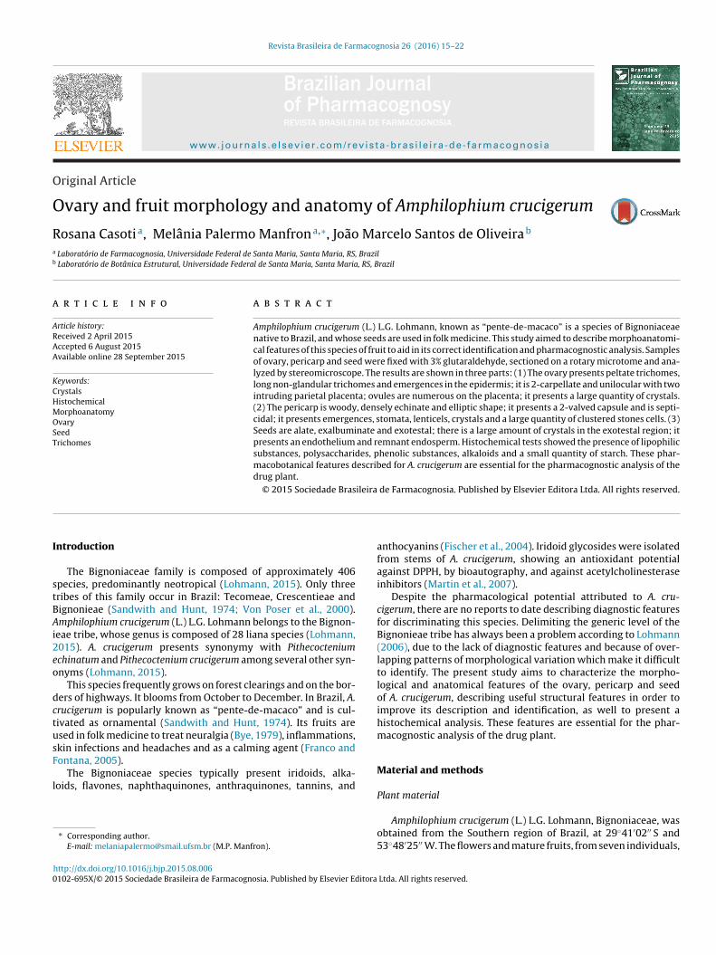
Fig. 1. Dissected flower of Amphilophium crucigerum. (A) General aspect of the g
ere collected from November to March. The collected materialas identified by Gilberto Dolejal Zanetti and the voucher was
egistered under number 12872 SMDB at the herbarium of theiology Department at the Federal University of Santa Maria.
orphological and anatomical characterization
After dissection with stereomicroscope SZH10 (Olympus®) and80 (Leica®), the samples were fixed in 3% glutaraldehyde in 0.1 M
odium phosphate buffer, pH 7.2 (Gabriel, 1982) and submitted toacuum for 6 h for improved infiltration. Subsequently, the samplesere washed in 0.1 M sodium phosphate buffer, pH 7.2 (Gabriel,
982), and then in distilled water. Tween 20 was utilized during4 h for extraction of epicuticular waxes. Subsequently, the sam-les underwent dehydration in an ethyl alcohol series, followed byolutions of chloroform and pure ethanol (1:3, 1:1, 3:1, 1:1, 1:3),nd finally of pure ethanol. The samples were pre-infiltrated in a 2-ydroxyethyl methacrylate (HEMA) and pure ethanol solution (1:1)uring 12 h, followed in pure HEMA and embedding in the sameesin, in a Teflon holder until reaching polymerization (Gerrits andmid, 1983). Sections of 5 �m thickness were made using a RM2245otary microtome (Leica®). Toluidine blue O in 0.05% sodium ben-oate buffer, pH 4.4 was used for staining (Feder and O’Brien, 1968).ermanent slides were deposited in the collection at the Structuralotany Laboratory of the Biology Department, UFSM. Observationsnd photomicrographs in bright field and polarized light were per-ormed using a DM 2000 microscope (Leica®) with a DFC 295 imageapture system (Leica®).
istochemical analysis
Hand sections of seeds were prepared for histochemical tests forifferent purposes: Lugol’s solution for starch (Jensen, 1962); Sudan
II to detect lipophilic substances (Brundrett et al., 1991); Dragen-orff to detect alkaloids (Furr and Mahlberg, 1981); PAS (periodic
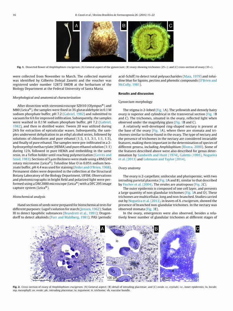
ig. 2. Cross-section of ovary of Amphilophium crucigerum. (A) General aspect; (B) detailp, mesophyll; ov, ovule; plc, intruding placentae; te, tegument; tr, trichome; vb, vascul
ium; (B) ovary showing trichomes (25×); and (C) cross-section of ovary (35×).
acid-Schiff) to detect total polysaccharides (Maia, 1979) and tolui-dine blue for lignins, pectins and phenolic compounds (O’Brien andMcCully, 1981).
Results and discussion
Gynoecium morphology
The stigma is 2-lobed (Fig. 1A). The yellowish and densely hairyovary is superior and cylindrical in the transversal section (Fig. 1Band C). The trichomes, situated in the ovary, reflected light whenobserved under the magnifying glass (Fig. 1B and C).
A relatively well-developed ring-shaped nectary is present atthe base of the ovary (Fig. 1A), where there are stomata and tri-chomes similar to those found in the ovary. The type of nectary andthe presence of trichomes in the nectary are considered invariablefeatures, making them important in the determination of species ofdifferent genera, including Amphilophium (Rivera, 2000). Some ofthe features described above were also described for genus deter-mination by Sandwith and Hunt (1974), Galetto (1995), Nogueiraet al. (2013) and Lohmann and Taylor (2014).
Ovary anatomy
The ovary is 2-carpellate, unilocular and plurispermic, with twointruding parietal placenta (Fig. 2A and B), similar to that describedby Fischer et al. (2004). The ovules are anatropous (Fig. 2C).
The outer epidermis is composed of one cell layer, and presentsa large quantity of non-glandular trichomes (Fig. 3A and D). Thesetrichomes are multicellular, long and non-branched. Studies carriedout by Nogueira et al. (2013), in leaves of A. crucigerum, showed the
presence of branched non-glandular trichomes. In the nectary wasobserved stomata (Fig. 3E).In the ovary, emergences were also observed, besides a rela-tively fewer number of glandular trichomes at different stages of
of intruding placentae; and (C) ovule. cr, crystals; i.e., inner epidermis; lo, locule;ar bundle.

R. Casoti et al. / Revista Brasileira de Farmacognosia 26 (2016) 15–22 17
F aspecw landu
dadtTt
stota
(rnb
Fpcc
those usually described for Bignoniaceae and Bignonieae (Corner,1976; Armstrong, 1985; Fischer et al., 2004, Lohmann and Taylor,
ig. 3. Longitudinal section of the ovary of Amphilophium crucigerum. (A) General
ith crystals; and (E) stomata. cr, crystals; em, emergence; ep, epidermis cells; gt, g
evelopment (Fig. 3B and D). The emergences possess wide basesnd the subepidermal cells are only part of the base, possiblyerived from one or two subepidermal cells (Fig. 3C). A large por-ion of this structure is made of cells derived from the epidermis.he arrangement and size of these cells are irregular. In addition,he emergences present a sharp apex defined by a single cell.
The glandular trichome is a peltate morphotype, composed ofix cells located on the head of the trichome and a single cell onhe stalk (Fig. 3B). According to Nogueira et al. (2013), the presencef peltate trichomes is very common in species of the Bignonieaeribe. The presence of trichomes is often associated with desiccationnd/or protection against herbivores (Wagner et al., 2004).
The mesophyll is composed of parenchymatous tissueFigs. 2A and 3A). The marginal vascular bundles bifurcate
adially with half of the bundle facing toward a central positionear the placentae. Thus, the ovary has two ventral large vascularundles and two large dorsal vascular bundles on the carpel wall,ig. 4. Longitudinal section of the ovary of Amphilophium crucigerum. (A) Detail ofarenchymatic and vascular tissues with crystal accumulation (bright field) and (B)rystals in the parenchymatic tissue and in vascular bundles after polarized light.r, crystals; vb, vascular bundle.
t of ovary; (B) outer epidermis; (C) trichome and emergence; (D) outer epidermislar trichomes; mp, mesophyll; ne, nectary; ov, ovule; st, stomata; tr, trichomes.
besides many small lateral vascular bundles found on the ovarywall (Figs. 2A and 3A).
Numerous crystalliferous cells occur in the mesophyll (Fig. 2B).There is also a large accumulation of calcium oxalate crystalson the vascular tissue, which apparently obliterate some ves-sel elements, and therefore, without an identifiable form or type(Figs. 2B and 4A and B). The inner epidermis presents isodiametriccells that develop vacuoles. The epidermis, which covers the pla-centa, presents cells with dense cytoplasm that are smaller thanthe locular cells (Fig. 2B). The ovules are anatropous, unitegmicand tenuinucellate. They are located on the placenta and arenumerous (Fig. 3A). Part of the results is in agreement with
2014).
Fig. 5. Fruit of Amphilophium crucigerum. (A) pericarp: echinate projections; (B)seminiferous column and (C) seeds. sec, seminiferous column.
18 R. Casoti et al. / Revista Brasileira de Farmacognosia 26 (2016) 15–22
F (B) ge(
F
P
itfla
F(v
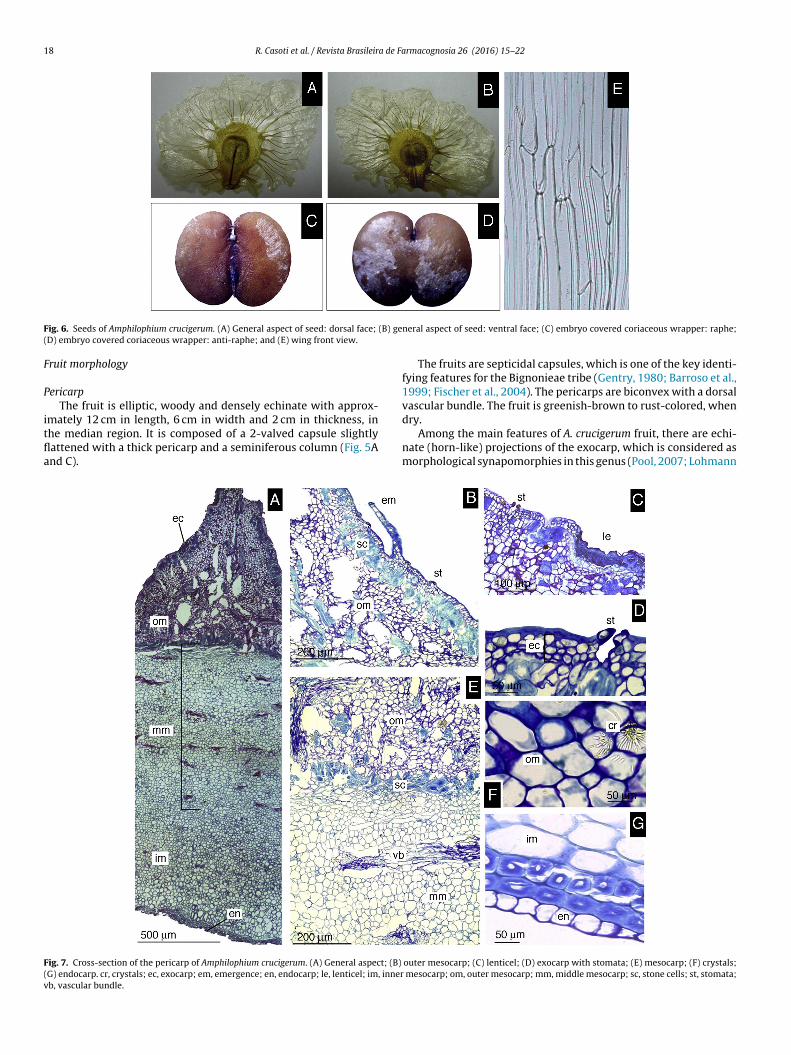
ig. 6. Seeds of Amphilophium crucigerum. (A) General aspect of seed: dorsal face;
D) embryo covered coriaceous wrapper: anti-raphe; and (E) wing front view.
ruit morphology
ericarpThe fruit is elliptic, woody and densely echinate with approx-
mately 12 cm in length, 6 cm in width and 2 cm in thickness, inhe median region. It is composed of a 2-valved capsule slightlyattened with a thick pericarp and a seminiferous column (Fig. 5And C).
ig. 7. Cross-section of the pericarp of Amphilophium crucigerum. (A) General aspect; (B)
G) endocarp. cr, crystals; ec, exocarp; em, emergence; en, endocarp; le, lenticel; im, innerb, vascular bundle.
neral aspect of seed: ventral face; (C) embryo covered coriaceous wrapper: raphe;
The fruits are septicidal capsules, which is one of the key identi-fying features for the Bignonieae tribe (Gentry, 1980; Barroso et al.,1999; Fischer et al., 2004). The pericarps are biconvex with a dorsalvascular bundle. The fruit is greenish-brown to rust-colored, when
dry.Among the main features of A. crucigerum fruit, there are echi-nate (horn-like) projections of the exocarp, which is considered asmorphological synapomorphies in this genus (Pool, 2007; Lohmann
outer mesocarp; (C) lenticel; (D) exocarp with stomata; (E) mesocarp; (F) crystals; mesocarp; om, outer mesocarp; mm, middle mesocarp; sc, stone cells; st, stomata;

R. Casoti et al. / Revista Brasileira de Farmacognosia 26 (2016) 15–22 19
Fig. 8. Longitudinal sections through the lateral plane of the seeds of Amphilophium crucigerum. (A) General aspect; (B) testa; (C) endosperm and endothelium; (D) detailof the cotyledon node; (E) hypocotyl radicle axis; and (F) calazal region. cn, cotyledon node; co, cotyledon; de, cotyledon dorsal epidermis; eb, embryo; ed, endosperm; eh,h
aja
S
faTwlTmtc
Abtceb2c2
ypocotyl radicle axis; et, endothelium; ex, exotesta; mt, mesotesta; t, testa.
nd Taylor, 2014). According to Gentry (1973), these echinate pro-ections in the valves may serve as protection against predators tollow the ripening of fruits and seeds.
eed morphology and classification of the mature embryo
A. crucigerum presents winged seeds along the length of theruit and occupying the entire locule (Fig. 5C). The seeds present
brownish-gold color and an evident seminal nucleus (Fig. 6A–D).hey have bright translucent wings and papyraceous consistencyith approximately 5 cm in width and 2.5 cm in height. The papil-
ate seed coat is another synapomorphy of the genus (Lohmann andaylor, 2014). The wings consist of lateral expansion of the tegu-ent and chalazal region. The number of cell layers reduces toward
he edge of the seminal nucleus and of the wing, where only oneell layer occurs (Fig. 6E).
According to Fischer et al. (2004), in Pithecoctenium,nemopaegma and Jacaranda, the seminal body is envelopedy a large wing. The same can be observed for other taxa fromhis family, such as Tabebuia ochracea (Sampaio et al., 2007), T.hrysotrica (Souza et al., 2005) and Macfadyena unguis-cati (Souzat al., 2008), where the chalazal portion of the seed expands and
ecomes part of the wing, except in T. caraiba (Ferreira and Cunha,000). However, in some Bignoniaceae there is an almost vestigialhalazal expansion of the wing, as in Tecoma stans (Renò et al.,007).Fruit anatomy and histology
PericarpThe pericarp is composed of three different portions (Fig. 7A).
The outer portion comprises the exocarp. Below exocarp occurs themesocarp, divided into three regions. The inner portion comprisesthe endocarp.
The exocarp is composed of one to three cell layers. Lenticelsand emergences are present (Fig. 7B–D). The outer periclinal wallof the cells is thick and lignified (Fig. 7D). Stomatal guard cells occurslightly above the surface of the exocarp (Fig. 7D).
The outer mesocarp is formed during fruit differentiationthrough the proliferation of outer tissue, resulting in horn-like pro-jections (Fig. 7A). The outer mesocarp presents parenchymatouscells and clustered stones cells (Fig. 7B) that form an almost con-tinuous layer. At the base of the horn-like projections, there arelayers of sclerified cells, demarcating the outer mesocarp from themiddle mesocarp (Fig. 7A and E). Needle-like crystals are presenton the outer mesocarp in a radial organization (Fig. 7F).
The middle mesocarp is composed of vascular bundles, andparenchymatous tissue with thin sclerified walls (Fig. 7E). Theinner mesocarp is predominantly composed of tissue similar to that
found in the middle mesocarp, but there are no vascular bundles(Fig. 7A). In addition, the inner mesocarp presents two isodiamet-ric cell layers with thick walls. The endocarp is formed by a singlelayer, with thin-walled cells (Fig. 7G).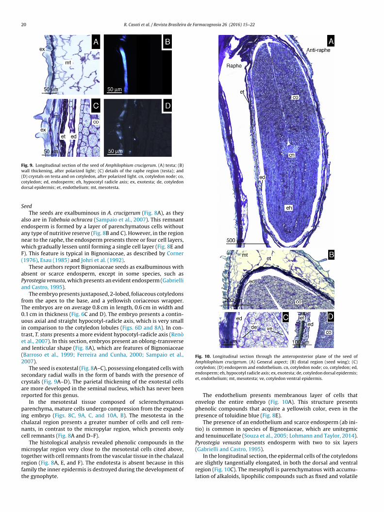
20 R. Casoti et al. / Revista Brasileira de Farmacognosia 26 (2016) 15–22
Fig. 9. Longitudinal section of the seed of Amphilophium crucigerum. (A) testa; (B)wall thickening, after polarized light; (C) details of the raphe region (testa); and(D) crystals on testa and on cotyledon, after polarized light. cn, cotyledon node; co,cd
S
aeanwF(
aPa
fT0uitea(2
scar
picnc
mtrft
Fig. 10. Longitudinal section through the anteroposterior plane of the seed ofAmphilophium crucigerum. (A) General aspect; (B) distal region (seed wing); (C)
In the longitudinal section, the epidermal cells of the cotyledons
otyledon; ed, endosperm; eh, hypocotyl radicle axis; ex, exotesta; de, cotyledonorsal epidermis; et, endothelium; mt, mesotesta.
eedThe seeds are exalbuminous in A. crucigerum (Fig. 8A), as they
lso are in Tabebuia ochracea (Sampaio et al., 2007). This remnantndosperm is formed by a layer of parenchymatous cells withoutny type of nutritive reserve (Fig. 8B and C). However, in the regionear to the raphe, the endosperm presents three or four cell layers,hich gradually lessen until forming a single cell layer (Fig. 8E and
). This feature is typical in Bignoniaceae, as described by Corner1976), Esau (1985) and Johri et al. (1992).
These authors report Bignoniaceae seeds as exalbuminous withbsent or scarce endosperm, except in some species, such asyrostegia venusta, which presents an evident endosperm (Gabriellind Castro, 1995).
The embryo presents juxtaposed, 2-lobed, foliaceous cotyledonsrom the apex to the base, and a yellowish coriaceous wrapper.he embryos are on average 0.8 cm in length, 0.6 cm in width and.1 cm in thickness (Fig. 6C and D). The embryo presents a contin-ous axial and straight hypocotyl-radicle axis, which is very small
n comparison to the cotyledon lobules (Figs. 6D and 8A). In con-rast, T. stans presents a more evident hypocotyl-radicle axis (Renòt al., 2007). In this section, embryos present an oblong-transversend lenticular shape (Fig. 8A), which are features of BignoniaceaeBarroso et al., 1999; Ferreira and Cunha, 2000; Sampaio et al.,007).
The seed is exotestal (Fig. 8A–C), possessing elongated cells withecondary radial walls in the form of bands with the presence ofrystals (Fig. 9A–D). The parietal thickening of the exotestal cellsre more developed in the seminal nucleus, which has never beeneported for this genus.
In the mesotestal tissue composed of sclerenchymatousarenchyma, mature cells undergo compression from the expand-
ng embryo (Figs. 8C, 9A, C, and 10A, B). The mesotesta in thehalazal region presents a greater number of cells and cell rem-ants, in contrast to the micropylar region, which presents onlyell remnants (Fig. 8A and D–F).
The histological analysis revealed phenolic compounds in theicropylar region very close to the mesotestal cells cited above,
ogether with cell remnants from the vascular tissue in the chalazal
egion (Fig. 8A, E, and F). The endotesta is absent because in thisamily the inner epidermis is destroyed during the development ofhe gynophyte.cotyledon; (D) endosperm and endothelium. cn, cotyledon node; co, cotyledon; ed,endosperm; eh, hypocotyl radicle axis; ex, exotesta; de, cotyledon dorsal epidermis;et, endothelium; mt, mesotesta; ve, cotyledon ventral epidermis.
The endothelium presents membranous layer of cells thatenvelop the entire embryo (Fig. 10A). This structure presentsphenolic compounds that acquire a yellowish color, even in thepresence of toluidine blue (Fig. 8E).
The presence of an endothelium and scarce endosperm (ab ini-tio) is common in species of Bignoniaceae, which are unitegmicand tenuinucellate (Souza et al., 2005; Lohmann and Taylor, 2014).Pyrostegia venusta presents endosperm with two to six layers(Gabrielli and Castro, 1995).
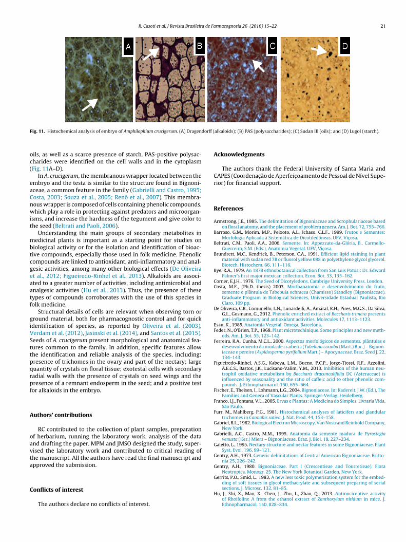
are slightly tangentially elongated, in both the dorsal and ventralregion (Fig. 10C). The mesophyll is parenchymatous with accumu-lation of alkaloids, lipophilic compounds such as fixed and volatile
R. Casoti et al. / Revista Brasileira de Farmacognosia 26 (2016) 15–22 21
F dorff (
oc(
eaCnwit
mbtcgeaatf
giVSttpqrpf
A
oavta
C
ig. 11. Histochemical analysis of embryo of Amphilophium crucigerum. (A) Dragen
ils, as well as a scarce presence of starch. PAS-positive polysac-harides were identified on the cell walls and in the cytoplasmFig. 11A–D).
In A. crucigerum, the membranous wrapper located between thembryo and the testa is similar to the structure found in Bignoni-ceae, a common feature in the family (Gabrielli and Castro, 1995;osta, 2003; Souza et al., 2005; Renò et al., 2007). This membra-ous wrapper is composed of cells containing phenolic compounds,hich play a role in protecting against predators and microorgan-
sms, and increase the hardness of the tegument and give color tohe seed (Beltrati and Paoli, 2006).
Understanding the main groups of secondary metabolites inedicinal plants is important as a starting point for studies on
iological activity or for the isolation and identification of bioac-ive compounds, especially those used in folk medicine. Phenolicompounds are linked to antioxidant, anti-inflammatory and anal-esic activities, among many other biological effects (De Oliveirat al., 2012; Figueiredo-Rinhel et al., 2013). Alkaloids are associ-ted to a greater number of activities, including antimicrobial andnalgesic activities (Hu et al., 2013). Thus, the presence of theseypes of compounds corroborates with the use of this species inolk medicine.
Structural details of cells are relevant when observing torn orround material, both for pharmacognostic control and for quickdentification of species, as reported by Oliveira et al. (2003),erdam et al. (2012), Jasinski et al. (2014), and Santos et al. (2015).eeds of A. crucigerum present morphological and anatomical fea-ures common to the family. In addition, specific features allowhe identification and reliable analysis of the species, including:resence of trichomes in the ovary and part of the nectary; largeuantity of crystals on floral tissue; exotestal cells with secondaryadial walls with the presence of crystals on seed wings and theresence of a remnant endosperm in the seed; and a positive testor alkaloids in the embryo.
uthors’ contributions
RC contributed to the collection of plant samples, preparationf herbarium, running the laboratory work, analysis of the datand drafting the paper. MPM and JMSO designed the study, super-ised the laboratory work and contributed to critical reading ofhe manuscript. All the authors have read the final manuscript andpproved the submission.
onflicts of interest
The authors declare no conflicts of interest.
alkaloids); (B) PAS (polysaccharides); (C) Sudan III (oils); and (D) Lugol (starch).
Acknowledgments
The authors thank the Federal University of Santa Maria andCAPES (Coordenac ão de Aperfeic oamento de Pessoal de Nível Supe-rior) for financial support.
References
Armstrong, J.E., 1985. The delimitation of Bignoniaceae and Scrophulariaceae basedon floral anatomy, and the placement of problem genera. Am. J. Bot. 72, 755–766.
Barroso, G.M., Morim, M.P., Peixoto, A.L., Ichaso, C.L.F., 1999. Frutos e Sementes:Morfologia Aplicada à Sistemática de Dicotiledôneas. UFV, Vic osa.
Beltrati, C.M., Paoli, A.A., 2006. Semente. In: Appezzato-da-Glória, B., Carmello-Guerreiro, S.M. (Eds.), Anatomia Vegetal. UFV, Vic osa.
Brundrett, M.C., Kendrick, B., Peterson, C.A., 1991. Efficient lipid staining in plantmaterial with sudan red 7B or fluorol yellow 088 in polyethylene glycol glycerol.Biotech. Histochem. 66, 111–116.
Bye, R.A., 1979. An 1878 ethnobotanical collection from San Luis Potosi: Dr. EdwardPalmer’s first major mexican collection. Econ. Bot. 33, 135–162.
Corner, E.J.H., 1976. The Seed of Dicotyledons. Cambrige University Press, London.Costa, M.E., (Ph.D. thesis) 2003. Morfoanatomia e desenvolvimento do fruto,
semente e plântula de Tabebuia ochracea (Chamisso) Standley (Bignoniaceae).Graduate Program in Biological Sciences, Universidade Estadual Paulista, RioClaro, 109 pp.
De Oliveira, C.B., Comunello, L.N., Lunardelli, A., Amaral, R.H., Pires, M.G.S., Da Silva,G.L., Gosmann, G., 2012. Phenolic enriched extract of Baccharis trimera presentsanti-inflammatory and antioxidant activities. Molecules 17, 1113–1123.
Esau, K., 1985. Anatomía Vegetal. Omega, Barcelona.Feder, N., O’Brien, T.P., 1968. Plant microtechinique. Some principles and new meth-
ods. Am. J. Bot. 55, 123–142.Ferreira, R.A., Cunha, M.C.L., 2000. Aspectos morfológicos de sementes, plântulas e
desenvolvimento da muda de craibeira (Tabebuia caraiba (Mart.) Bur.) – Bignon-iaceae e pereiro (Aspidosperma pyrifolium Mart.) – Apocynaceae. Braz. Seed J. 22,134–143.
Figueiredo-Rinhel, A.S.G., Kabeya, L.M., Bueno, P.C.P., Jorge-Tiossi, R.F., Azzolini,A.E.C.S., Bastos, J.K., Lucisano-Valim, Y.M., 2013. Inhibition of the human neu-trophil oxidative metabolism by Baccharis dracunculifolia DC (Asteraceae) isinfluenced by seasonality and the ratio of caffeic acid to other phenolic com-pounds. J. Ethnopharmacol. 150, 655–664.
Fischer, E., Theisen, I., Lohmann, L.G., 2004. Bignoniaceae. In: Kadereit, J.W. (Ed.), TheFamilies and Genera of Vascular Plants. Springer-Verlag, Heidelberg.
Franco, I.J., Fontana, V.L., 2005. Ervas e Plantas: A Medicina do Simples. Livraria Vida,São Paulo.
Furr, M., Mahlberg, P.G., 1981. Histochemical analyses of laticifers and glandulartrichomes in Cannabis sativa. J. Nat. Prod. 44, 153–158.
Gabriel, B.L., 1982. Biological Electron Microscopy. Van Nostrand Reinhold Company,New York.
Gabrielli, A.C., Castro, M.M., 1995. Anatomia da semente madura de Pyrostegiavenusta (Ker.) Miers – Bignoniaceae. Braz. J. Biol. 18, 227–234.
Galetto, L., 1995. Nectary structure and nectar features in some Bignoniaceae. PlantSyst. Evol. 196, 99–121.
Gentry, A.H., 1973. Generic delimitations of Central American Bignoniaceae. Britto-nia 25, 226–242.
Gentry, A.H., 1980. Bignoniaceae. Part I (Crescentieae and Tourretieae). FloraNeotropica. Monogr. 25. The New York Botanical Garden, New York.
Gerrits, P.O., Smid, L., 1983. A new less toxic polymerization system for the embed-
ding of soft tissues in glycol methacrylate and subsequent preparing of serialsections. J. Microsc. 132, 81–85.Hu, J., Shi, X., Mao, X., Chen, J., Zhu, L., Zhao, Q., 2013. Antinociceptive activityof Rhoifoline A from the ethanol extract of Zanthoxylum nitidum in mice. J.Ethnopharmacol. 150, 828–834.

2 a de Fa
J
J
J
L
L
L
M
MN
O
O
2 R. Casoti et al. / Revista Brasileir
asinski, V.C.G., Silva, R.Z.D., Pontarolo, R., Budel, J.M., Campos, F.R., 2014.Morpho-anatomical characteristics of Baccharis glaziovii in support of its phar-macobotany. Rev. Bras. Farmacogn. 24, 609–616.
ensen, W.A., 1962. Botanical Histochemistry: Principles and Practice. W.H. Freemanand Co., San Francisco.
ohri, B.M., Ambergaokar, K.B., Srivastava, P.S., 1992. Comparative Embryology ofAngiosperms. Springer-Verlag, Berlin.
ohmann, L.G., 2015. Bignoniaceae. In: Lista de espécies da Flora do Brasil. JardimBotânico Rio de Janeiro, http://floradobrasil.jbrj.gov/2010/FB112461 (accessedFebruary, 2015).
ohmann, L.G., 2006. Untangling the phylogeny of neotropical lianas (Bignonieae,Bignoniaceae). Am. J. Bot. 93, 304–318.
ohmann, L.G., Taylor, C.M., 2014. A new generic classification of tribe Bignonieae(Bignoniaceae). Ann. Missouri Bot. Gard. 99, 348–489.
artin, F., Hay, A.E., Corno, L., Gupta, M.P., Hostettmann, K., 2007. Iridoid glycosidesfrom the stems of Pithecoctenium crucigerum (Bignoniaceae). Phytochemistry68, 1307–1311.
aia, V., 1979. Técnica Histológica. Atheneu, São Paulo.ogueira, A., El-Otra, J.H.L., Guimarães, E., Machado, S.R., Lohmann, L.G., 2013.
Trichome structure and evolution in Neotropical lianas. Ann. Bot. 112,1331–1350.
’Brien, T.P., McCully, M.E., 1981. The Study of Plant Structure: Principles andSelected Methods. Temarcarphi Press, Melbourne.
liveira, T.B., Neto, B.H.J.C., Xaveier, M.A., Garrote, C.F.D., Asquieri, E.R., Rezende,M.H., Ferreira, H.D., Paula, J.R., 2003. Estudo Farmacognóstico das raízes deJacaranda decurrens Cham (carobinha). Rev. Bras. Farmacogn. 13, 54–55.
rmacognosia 26 (2016) 15–22
Pool, A., 2007. A revision of the genus Pithecoctenium (Bignoniaceae). Ann. MissouriBot. Gard. 94, 622–642.
Renò, L.R., Mosqueta, I.S., Braccini, A.L., 2007. Morfo-anatomia do fruto e sementesde amarelinho (Tecoma stans (L.) Kunth – Bignoniaceae). Braz. Seed J. 29, 18–30.
Rivera, G.L., 2000. Nuptial nectary structure of Bignoniaceae of Argentina. Darwini-ana 38, 227–239.
Sampaio, D.S., Costa, M.E., Paoli, A.A.S., 2007. Ontogenia da semente de Tabebuiaochracea (Cham.) Standl (Bignoniaceae). Braz. J. Biol. 30, 289–302.
Sandwith, N.Y., Hunt, D.R., 1974. Flora Catarinense. I Parte: As Plantas. Bign. Itajaí,Fascicle.
Santos, V.L., Franco, C.R., Amano, E., Messias-Reason, I.J., Budel, J.M., 2015. Anatom-ical investigations of Piper amalago (jaborandi-manso) for the quality control.Rev. Bras. Farmacogn. 25, 85–91.
Souza, L.A., Iwazaki, M.C., Moscheta, I., 2005. Morphology of the pericarp and seedof Tabebuia chrysotricha (Mart. ex DC.) Standl (Bignoniaceae). Braz. Arch. Biol.Technol. 48, 407–418.
Souza, L.A., Oyama, S.D.O., Muneratto, J.C., 2008. Morphology and anatomy of thedeveloping fruit of Macfadyena unguis-cati (l.) A.H. Gentry, Bignoniaceae. ActaBot. Venez. 31, 1–14.
Verdam, M.C.S., Ohana, D.T., Araújo, M.G.P., Guilhon-Simplicio, F., De-Mendonc a,M.S., Pereira, M.M., 2012. Morphology and anatomy of Justicia acuminatissima
leaves. Rev. Bras. Farmacogn. 22, 1212–1218.Von Poser, G.L., Schripsema, J., Henriques, A.T., Jensen, S.R., 2000. The distribution ofiridoids in Bignoniaceae. Biochem. Syst. Ecol. 28, 351–366.
Wagner, G.J., Wang, E., Shepherd, R.W., 2004. New approaches for studying andexploiting an old protuberance, the plant trichome. Ann. Bot. 93, 3–11.