Embed Size (px)
Citation preview
Outpatient EEG for the
Diagnosis of Epilepsy December 2, 2011
Elson L. So, MD
Professor of Neurology
EEG & Epilepsy
Mayo Clinic
Rochester, MN
American Epilepsy Society | Annual Meeting
Disclosures
American Epilepsy Society | Annual Meeting
• Speakers Bureau – none
• Industry consultantship – none
• Non-FDA labeled drug use – none
• Intellectual Property – Epilepsy Treatment Planning Software, no financial proceeds
• Editorial Boards – Epilepsia
– Epilepsy Research
– Journal of Clinical Neurophysiology

Objectives
• Recognize the role and limitations of the outpatient EEG in epilepsy diagnosis and management
• Avoid the pitfalls of outpatient EEG
• Apply measures to increase yield of the outpatient EEG in epilepsy practice
American Epilepsy Society | Annual Meeting

EEG Issues in Sandy
• How useful is a “routine” outpatient EEG?
• How can we increase the yield of outpatient EEGs?

EEG Issues in Sandy
• How useful is a standard outpatient EEG?
• How can we increase the yield of outpatient EEGs?

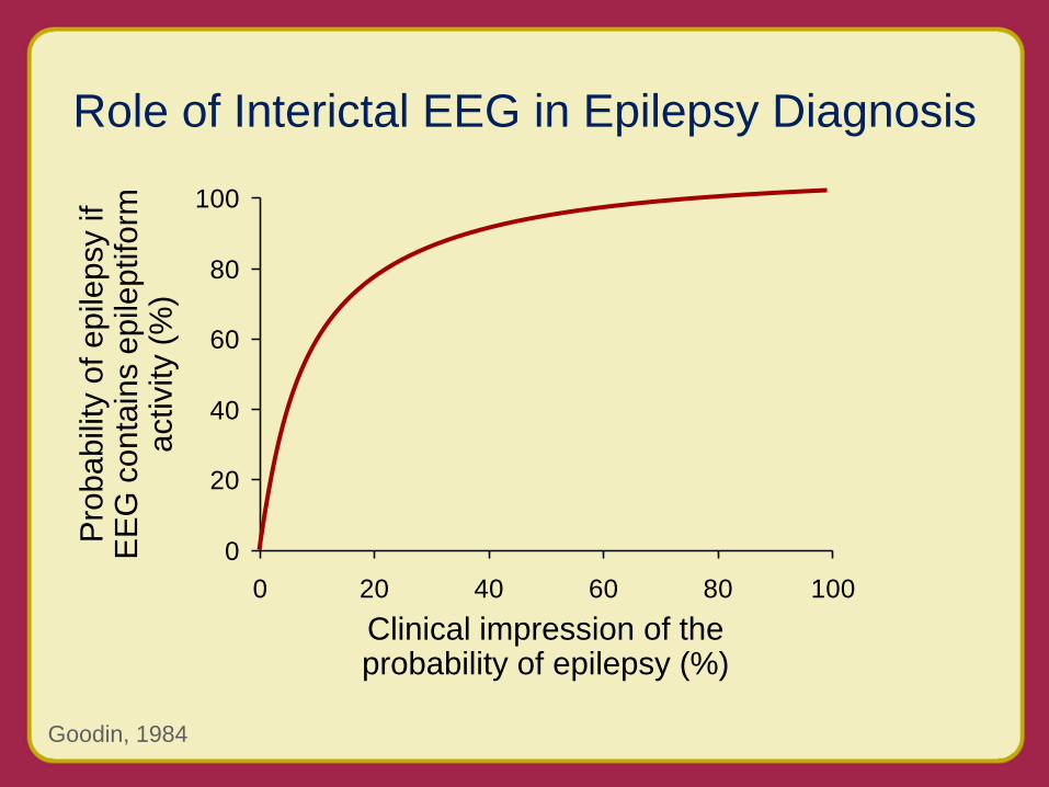
Role of Interictal EEG in Epilepsy Diagnosis
0
20
40
60
80
100
0 20 40 60 80 100
Goodin, 1984
Clinical impression of the probability of epilepsy (%)
Pro
ba
bili
ty o
f e
pile
psy if
EE
G c
on
tain
s e
pile
ptifo
rm
activity (
%)
Role of Interictal EEG in Epilepsy Diagnosis
0
20
40
60
80
100
0 20 40 60 80 100
Goodin, 1984
Clinical impression of the probability of epilepsy (%)
Pro
ba
bili
ty o
f e
pile
psy if
EE
G c
on
tain
s e
pile
ptifo
rm
activity (
%)

Role of Interictal EEG in Epilepsy Diagnosis
0
20
40
60
80
100
0 20 40 60 80 100
Goodin, 1984
Clinical impression of the probability of epilepsy (%)
Pro
ba
bili
ty o
f e
pile
psy if
EE
G c
on
tain
s e
pile
ptifo
rm
activity (
%)

Role of Interictal EEG in Epilepsy Diagnosis
0
20
40
60
80
100
0 20 40 60 80 100
Goodin, 1984
Clinical impression of the probability of epilepsy (%)
Pro
ba
bili
ty o
f e
pile
psy if
EE
G c
on
tain
s e
pile
ptifo
rm
activity (
%)

Limitation of the Standard Outpatient EEG
Rates of interictal epileptiform discharges in epilepsy patients vary between ~ 30% to 70%
Gilbert, 2002; Ajmone-Marsan, 1970; Salinsky, 1987

EEG Issues in Sandy
• How useful is a standard outpatient EEG?
• How can we increase the yield of outpatient EEGs?

You, the EEG reader The EEG record
The patient

You, the EEG reader
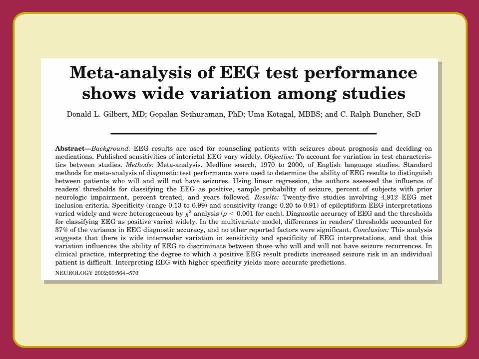
Meta-Analysis of EEG Test Performance Shows Wide Variation Among Studies
Differences in readers’ threshold was the only significant factor
Gilbert, 2002

-0.5
0.0
0.5
1.0
1.5
2.0
2.5
3.0
3.5
-6 -4 -2 0 2 4 6
Epileptiform
Gilbert, 2002
Threshold for positive EEG
Dia
gn
ostic a
ccu
racy
Diagnostic accuracy = 0.75 + -0.25
r2 = 0.37


Wicket Waves The Most Frequent Misleading Mimicker
Krauss, 2005; Benbadis, 2008

Gilbert, 2002
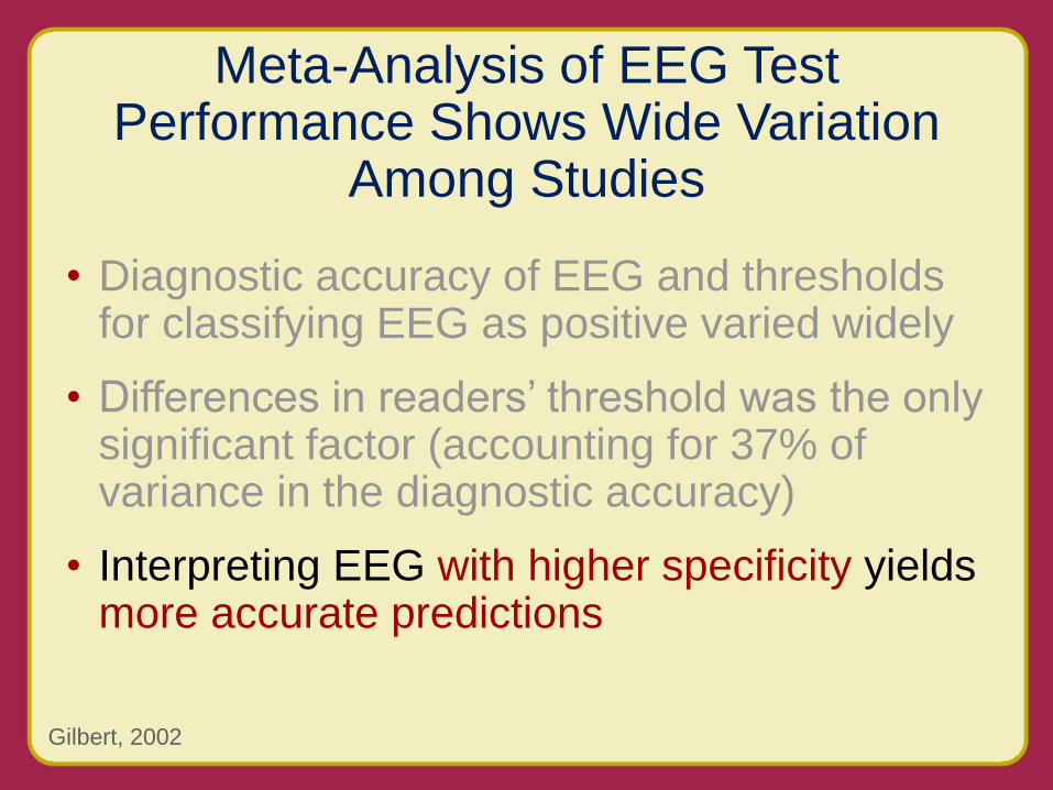
Meta-Analysis of EEG Test Performance Shows Wide Variation
Among Studies
• Diagnostic accuracy of EEG and thresholds for classifying EEG as positive varied widely
• Differences in readers’ threshold was the only significant factor (accounting for 37% of variance in the diagnostic accuracy)
• Interpreting EEG with higher specificity yields more accurate predictions

Westmoreland, 2003
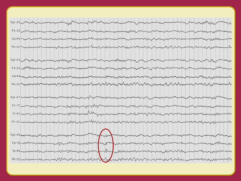

Benign Sporadic Sleep Spikes (BSSS)
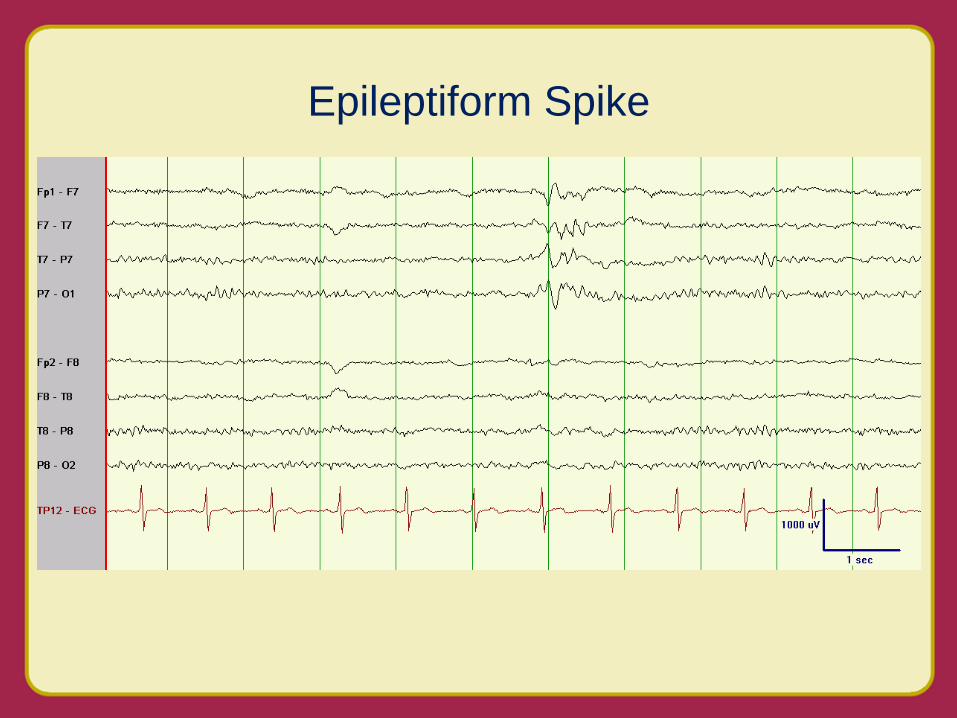
Epileptiform Spike
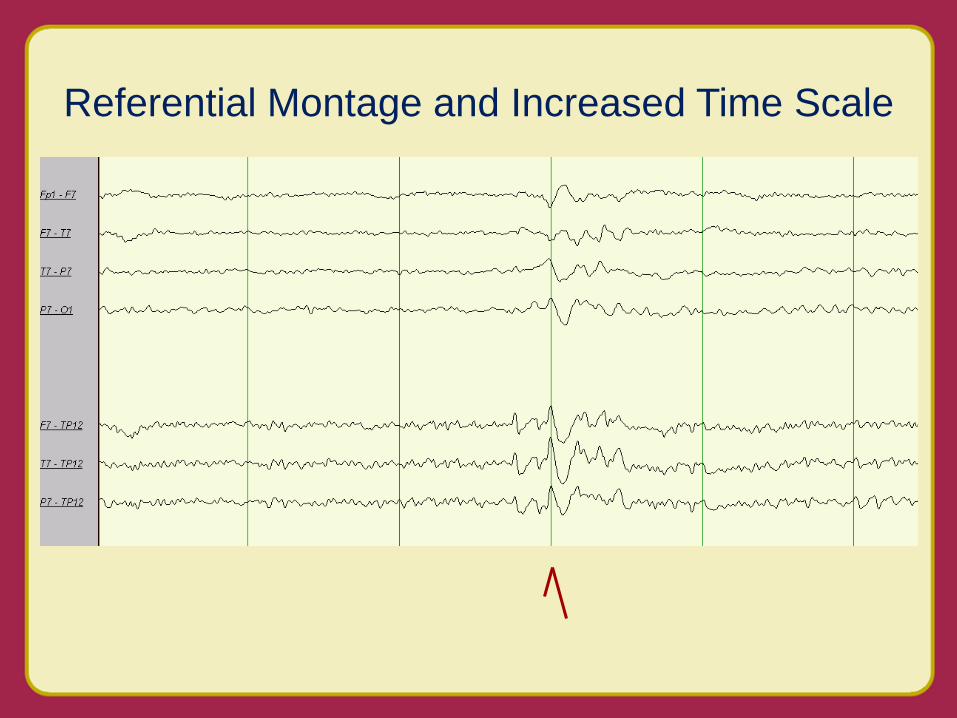
Referential Montage and Increased Time Scale

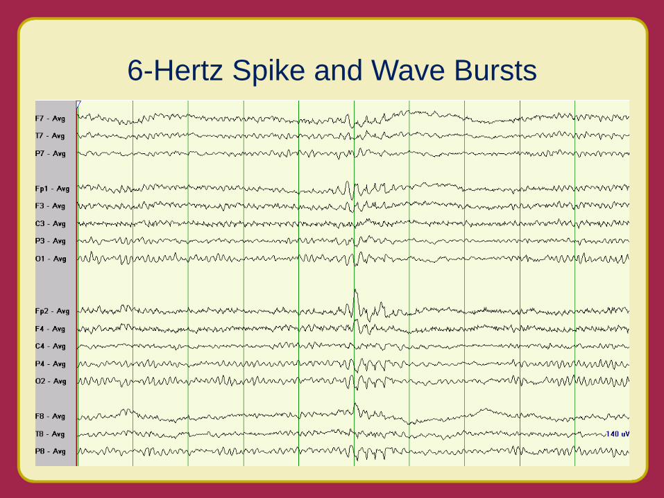
6-Hertz Spike and Wave Bursts

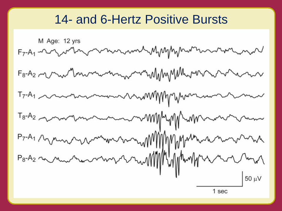
14- and 6-Hertz Positive Bursts


Lateral Rectus Muscle Spike

Mimickers of Epileptiform Discharges
Physiologic cerebral activities
• Benign variants
• Drowsiness and sleep activities – Hypnagogic hypersynchrony, POSTS, V waves especially
in children
– Quiet sleep sharp activities in neonates
• Neonatal EEGs – eg, frontal sharp transients
Artifacts
• Physiologic – eg, EKG, muscle
• Non-physiologic – eg, electrical, mechanical

The patient


Yield of Initial EEG and Repeat Sleep-Deprived EEG
King et al, 1998 (Excludes many acute seizure patients)
Clinically diagnosed epilepsy syndrome
Initial EEG epileptiform
Sleep-deprived EEG
epileptiform Total
epileptiform
Generalized 17/25 (68%) 6/8 (75%) 23/25 (92%)
Partial 51/116 (44%) 19/60 (32%) 70/116 (60%)
Unclassified 61/159 (38%) 30/90 (33%) 91/159 (57%)
Total 129/300 (43%) 55/158 (35%) 184/300 (61%)


Sleep Deprivation (SD) + Chloral Hydrate (CH) versus Sleep Deprivation Only
Britton, 2010
SD + CH (%)
SD only (%)
P
Sleep recorded 85 86 NS
Awake epileptiform 27 35 NS
Sleep epileptiform 18 17 NS
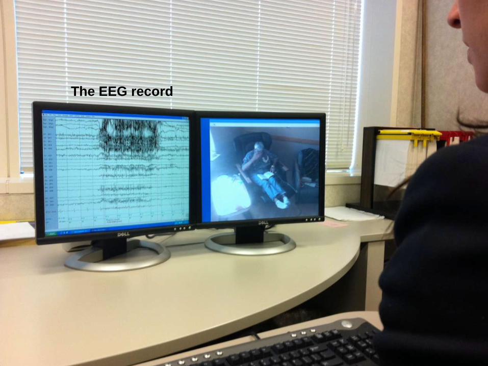
The EEG record

Proportion of Children Attaining Sleep During EEG Procedure
IPS = intermittent photic stimulation; HV = hyperventilation Kaleyias, 2006
6.2
35.5
23.0
0
20
40
60
80
100
%
EEG procedure
Group I n=48
Group II n=48
Group III n=48
Start End
Group I IPS HV
Group II HV IPS
Group III HV and IPS

0.0
0.2
0.4
0.6
0.8
1.0
0 15 30 45 60 75 90
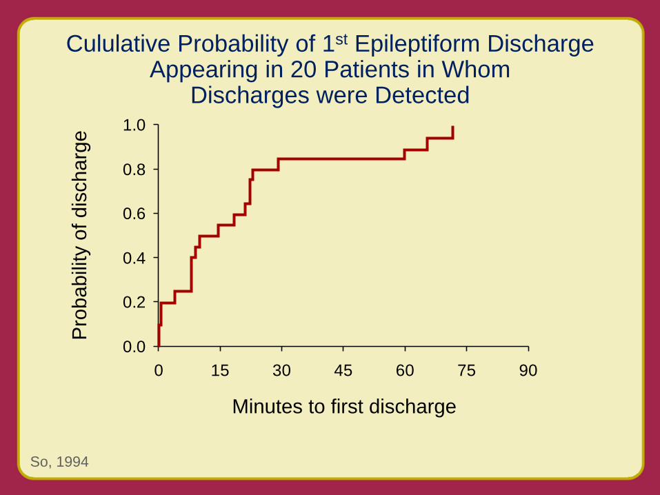
Cululative Probability of 1st Epileptiform Discharge Appearing in 20 Patients in Whom
Discharges were Detected
So, 1994
Minutes to first discharge
Pro
ba
bili
ty o
f d
isch
arg
e

51
34
0
20
40
60
80
100
<24 hours >24 hours
EEG After Presenting Seizure
King, 1998
Timing of recording
With
ep
ilep
tifo
rm
dis
ch
arg
e (
%)
95% CI for difference in proportion = 6-28%
Excludes acute seizures, but includes patients with previous seizures
80/156
49/144

Before seizure began
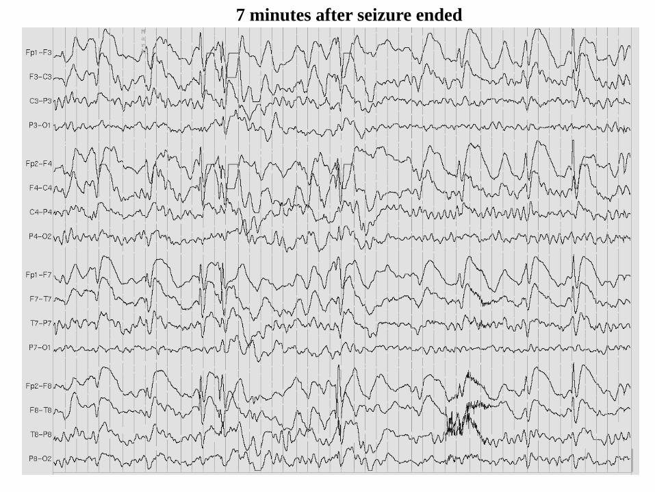
7 minutes after seizure ended

Relationships Between Interictal Spiking and Seizures Human and Experimental Evidence
• Seizures are often followed by long-lasting increases in spiking
• Increases in spiking before seizures have not been clearly documented
• Decreases in antiepileptic medication do not result directly in increased spiking
Gotman, 1991

How about Ambulatory EEG?

Yield of Ambulatory EEG
• 24 hour Ambulatory EEG versus Routine EEG in 46 patients; EEGer’s blinded to clinical information
• Epileptiform activity seen 33% of Ambulatory versus 24% of Routine EEGs
• Seizures in 15% of AEEG, none in REEG
Slide courtesy of S. Rao Liporace et al, 1998

Fountain, 2011; Britton, 2011
Summary of Epilepsy Metrics
3. At least one EEG needs to
have been done or reviewed…


0.0
0.2
0.4
0.6
0.8
1.0
0 12 24 36 48 60 72 84
Cumulative Percent of Remaining Seizure Free After 1st Unprovoked Seizure in Children
Scotini, 2004
Months
Normal EEG
Abnormal EEG

Rates of Seizure Recurrence in Children 2 Years After a 1st Unprovoked Seizure
Berg, 1991
%
Abnormal EEG alone 48
Remote symptomatic etiology 48
If both absent 24
If both present 65

• “Routine EEG as part of the diagnostic evaluation was recommended.”
• EEG was the only test with Class I evidence for usefulness
Hirtz, 2000
Practice Parameter: Evaluating a First Nonfebrile Seizure in Children

0.0
0.1
0.2
0.3
0.4
0.5
0.6
0 6 12 18 24
Recurrence After 1st Unprovoked Seizure in Adults with Normal versus Abnormal EEG
Schreiner, 2003
Follow-up (months)
Normal EEG
Abnormal EEG
Cu
mu
lative
re
cu
rre
nce
ris
k
P<0.001

• “EEG should be considered as part of the routine neurodiagnostic evaluation…”
• EEG has Level B evidence
Krumholz, 2000
Practice Parameter: Evaluating an apparent unprovoked first seizure in adults (an evidence-based review)
Report of the Quality Standards Subcommittee of the American Academy of Neurology and the American Epilepsy Society
Tips for Increasing Yield of
Outpatient EEG in Epilepsy
• Avoid over-reading EEGs; beware of benign variants and artifacts
• Sleep deprive patient whenever possible; no need to sedate in most cases
• Perform EEG sooner than later after an episode
• Perform hyperventilation early in the procedure in children
• Extend EEG up to 1 hour if needed

References
Ajmone Marsan C, Zivin L. Factors related to the occurrence of typical paroxysmal abnormalities in the EEG records of epileptic patients. Epilepsia 1970;11:361-81
Benbadis S, Lin K. Errors in EEG interpretation and misdiagnosis of epilepsy. Which EEG patterns are over-read? Eur Neurol 2008;59:267-71
Britton J, Kosa S. The clinical value of chloral hydrate in the routine electroencephalogram. Epilepsy Research 2010;88:215-20
Geyer J, Bilir E, Faught R, Kuzniecky R, Gilliam F. Significance of interictal temporal lobe delta activity for the localization of the primary epileptogenic region. Neurology 1999;52:202-5
Gilbert DL, Sethuraman G, Kotagal U, Buncher CR. Meta-analysis of EEG test performance shows wide variation among studies. Neurology 2003;60:564-570
Goodin D, Aminoff M. Does the interictal EEG have a role in the diagnosis of epilepsy? Lancet 1984;1:1837-9
Gotman J. Relationships between interictal spiking and seizures: human and experimental evidence. Canadian Journal of Neurological Sciences 1991;18(4 Suppl):573-6

References
Hirtz D, Ashwal S, Berg A, Bettis D, Camfield C, Camfield P, et al. Practice parameter: evaluating a first nonfebrile seizure in children: report of the quality standards subcommittee of the American Academy of Neurology, The Child Neurology Society, and The American Epilepsy Society. Neurology 2000 Sep 12;55(5):616-23
Kaleyias J, Kothare SV, Pelkey M, Harrison G, Legido A, Khurana DS. Achieving sleep state during EEG in children; sequence of activation procedures. Clinical Neurophysiology 2006 Jul;117(7):1582-4
King M, Newton M, Jackson G, Fitt G, Mitchell L, Silvapulle M. Epileptology of the first seizure presentation: a clinical electroencephalographic, and magnetic resonance imaging study of 300 consecutive patients. Lancet 1998;352:1007-1011
Krauss G, Abdallah A, Lesser R, al. e. Clinical and EEG features of patients with EEG wicket rhythms misdiagnosed with epilepsy. Neurology 2005;64:1879-83
Krumholz A, Wiebe S, Gronseth G, Shinnar S, Levisohn P, Ting T, et al. Practice Parameter: evaluating an apparent unprovoked first seizure in adults (an evidence-based review): report of the Quality Standards Subcommittee of the American Academy of Neurology and the American Epilepsy Society. Neurology 2007 Nov 20;69(21):1996

References
Liporace J, Tatum Wt, Morris GL, 3rd, French J. Clinical utility of sleep-deprived versus computer-assisted ambulatory 16-channel EEG in epilepsy patients: a multi-center study. Epilepsy Research 1998;32:357-362
Neugebauer R, Paik M, Hauser W, Nadel E, Leppik I, Susser M. Stressful life events and seizure frequency in patients with epilepsy. Epilepsia 1994;35:336–43
Reiher J, Beaudry M, Leduc C. Temporal internittent rhythmic delta acitivity (TIRDA) in the diagnosis of complex partial epilepsy: sensitivity, specificity, and predictive value. Can J Neurol Sci 1989;16:398-401
Sadleir LG, Scheffer IE. Optimizing electroencephalographic studies for epilepsy diagnosis in children with new-onset seizures. Archives of Neurology Nov;67(11):1345-9
Salinsky M, Kanter R, Dasheiff R. Effectiveness of multiple EEGs in supporting diagnosis of epilepsy: an operational curve. Epilepsia 1987;28:331-334
Schreiner A, Pohlmann-Eden B. Value of the early electroencephalogram after a first unprovoked seizure. Clinical Electroencephalography 2003 Jul;34(3):140-4
References
Scotini A, Manreza M, Guerreiro M. Recurrence after a first unprovoked cryptogenic/idiopathic seizure in children: a prospective study from Sao Paulo, Brazil. Epilepsia 2004;45:166-70
Sharbrough F, Chatrian G, Lesser R, Luders H, Nuwer M, Picton T. American Electroencephalographic Society guidelines for standard electrode position nomenclature. 1991; 8: 200–2. Guidelines for Standard Electrode Position Nomenclature. J Clin Neurophysiol 1991;8:200-2
So E, Ruggles K, Ahmann P. Yield of Sphenoidal Recordings in Sleep-Deprived Outpatients. J Clin Neurophysiol 1994;11:226-30
Westmoreland B. Benign electroencephalographic variants and patterms of uncertain significance. In: Ebersole J, ed. Current Practice of Clinical Electroencephalography. Philadelphia: Lippincott Williams & Wilkins 2003:235-45
Additional References:
Britton JW Do you measure up?Further steps toward standardization and measuring quality in neurologic care. Neurology. 2011;76:16-7.
Fountain N, al e. Quality improvement in neurology: AAN epilepsy quality measures. Report of the Quality Measurement and Reporting Subcommittee of the American Academy of Neurology. Neurology. 2011;76:94-9.