Embed Size (px)
Citation preview
OUR HIDDEN AGEING:
TIME TO LISTEN TO THE HEART
Foreword 2
Abbreviations 3
Executive Summary 4
Key Findings 6
1. Heart valve disease as part of the current burden of cardiovascular disease 8
2. Clinical aspects of heart valve disease 18
3. Cardiovascular ageing and valve disease 22
4. Management of Heart valve disease 26
5. Economic and societal costs of heart valve disease in the elderly 32
6. Cost-effectiveness of interventions for heart valve disease 42
7. Case studies 52
8. Conclusion 54
Calls to Action 56
References 58
Appendix 64
CONTENTS
If you’re over 65, ask your doctor to listen to your heart
A thorough clinical exam is critical to
detecting heart valve disease
You may be familiar with heart attack and stroke but few know
about heart valve disease
Ageing blood vessels can damage vital organs and place
stress on heart valves
Heart valve disease can cause serious
complications including heart failure, stroke,
blood clots and death
More than a quarter of a
million Australians have heart valve disease & don’t
know it
Heart valve disease is rising rapidly and it’s serious. However, it is
often treatableMinimally-invasive valve
replacement for more people over 65 could
save $117 million in a year
Authors Prof Tom Marwick, Baker Heart and Diabetes Institute, Melbourne.
Prof Seana Gall, Dr Marie-Jeanne Buscot, Dr Rachel Climie, Dr Hoang Phan, Menzies Institute for Medical Research, University of Tasmania
Prof Marj Moodie, Dr Lan Gao, Dieu Nguyen, Deakin University, Melboune
Prof Ajay Mahal, Teralynn Michelle Ludwick, Marie Ishida, University of Melbourne
Reviewers Prof David Kaye, Alfred Hospital, Melbourne, VIC
Prof David Celermajer, Royal Prince Alfred Hospital, Sydney, NSW
Prof Derek Chew, Flinders Medical Centre, Adelaide, SA
Prof Greg Scalia, The Prince Charles Hospital, Brisbane, QLD
Prof David Playford, The Mount Hospital, Perth, WA
Dr Peter French & Dr Ren Tan, Canberra Hospital, ACT
Dr Paul Macintyre, Royal Hobart Hospital, TAS
Prof Liza Thomas, Westmead Hospital, NSW
Reference: Our Hidden Ageing: Time to Listen to the Heart
The Baker Heart and Diabetes Institute acknowledges the provision of an unrestricted educational grant from Edwards Lifesciences, used for the analyses and preparation of this report.
$
1
FOREWORD
The vascular system is the main transport system in our body. It permits blood to circulate and transport nutrients, oxygen, carbon dioxide, hormones, and blood cells to and from the cells in the body to provide nourishment, to help fight disease, and to maintain stability of the body’s metabolism.
The heart valves have a critical role to play in ensuring the flow of blood through the heart. A number of age-related factors increase mechanical stress on the heart valves, and the same processes that cause abnormalities in the blood vessels can damage the coverings of the valves. Complications of heart valve disease include mortality, heart failure, stroke, blood clots, and heart rhythm abnormalities.
The incidence and prevalence of aortic and mitral valve disease both increase with age. Therefore, it’s not surprising that, as the population ages, heart valve disease is emerging as a serious and increasingly common health issue. About half a
million Australians already have valve disease - aortic valve disease is the most frequent cause of significant disease - with this figure projected to grow over the next three decades. Perhaps more concerning, more than a quarter of a million Australians have undiagnosed heart valve disease.
The availability of non-surgical valve replacement has reduced barriers to interventions in the elderly. Increasing access will increase the number of these procedures, with overall higher healthcare costs. However, it also delivers greater benefits because earlier diagnosis and treatment reduces the long-term consequences of these diseases, and preserves productivity, which remains important in the elderly. Early intervention in the form of non-surgical valve replacement could prevent productivity losses of up to $117 million in a single year.
Although heart valve conditions are serious, they are eminently and increasingly treatable. That’s why this whitepaper, led by experts from the Baker Heart and Diabetes Institute together with specialists from The University of Melbourne, University of Tasmania and Deakin University, is so important and timely.
ABBREVIATIONS
AQoL – And indices of Quality of Life
AHA - American Heart Assosciation
AS – Aortic Stenosis
AVR – Aortic Valve Replacement
BAV – Bicuspid Aortic Valve
CVD – Cardiovascular disease
ECG – Electrocardiogram
GDP – Gross Domestic Product
HF – Heart Failure
HILDA – Household, Income and Labour Dynamics in Australia
ICER – Incremental Cost Effectiveness Ratio
LV – Left Ventricular
MAS – Moderate Aortic Stenosis
MR – Mitral Regurgitation
MS – Mitral Stenosis
MVD – Mitral Vascular Disease
MVR – Mitral Valve Replacement
PNMA – Productive Non-Market Activities
QALY – Quality Adjusted Life Years
RV - Right Ventricular
SAS – Severe Aortic Stenosis
SD – Standard Deviation
TAVI – Transcatheter Aortic Valve Implantation
T2D – Type 2 diabetes
VHD – Valvular Heart Disease
WTP – Willingness To Pay
WW – Worried Well
OUR HIDDEN AGEING: TIME TO LISTEN TO THE HEART 2 3
Heart valve disease has flown under the radar for far too long. That’s why we have examined this issue in detail. Urgent attention is critical, and we recommend:
• Individual and social marketing campaigns to increase awareness of heart valve disease and other manifestations of cardiovascular ageing, particularly amongst GPs, healthcare and health advocacy groups. A benefit might be that more patients get heart murmurs checked.
• Strategies involving primary care. These might include educational updates and upskilling. People >65 years should have heart checks during GP visits for other problems. For people 65 and older who are not engaged with primary care, an extension of the current preventive cardiology item numbers (MBS 699 and 177) to include cardiac auscultation (listening to heart murmurs) should be considered.
• Support for emerging technologies. Development of translational research streams to more rapidly evaluate novel technologies for management of structural
heart disease. Investment and clinical application of AI-supported and hand-held echo will require adjustments in current funding arrangements, which preclude these approaches from reimbursement.
• Health service design, including improving access to echocardiography. These steps might involve early detection and out-reach echocardiography programs in rural areas.
• Policy. Dedicated funding for service level interventions that improve access and equity to transcatheter valvular interventions (minimally-invasive interventions). This will require planning, training and resourcing, along with financial incentives to drive clinical change.
• Guidelines. Development of national heart valve disease guidelines to facilitate decision-making.
When it comes to cardiovascular issues, many people are familiar with heart attack, stroke, heart failure, and coronary artery disease. We appreciate that the heart has an essential role, including sending blood around our body, providing tissue with the oxygen and nutrients they need. Each day, the heart pumps about 7500 litres of blood via a blood vessel system stretching more than 100,000km.
The mechanics of the circulation are important to the smooth operation of this system.
Much like an important part of a machinery that can break down, the heart can malfunction if all parts aren’t working well. This includes small but incredibly important components such as the valves between the atria and ventricles that make sure blood flows in one direction through the heart. Valves are also located at the “exits” or “doorways” of the heart for this same reason—to make sure blood flows in one direction. In a lifetime, these valves will open and close more than two billion times.
When the large blood vessels are functioning well, they optimise the efficiency of pumping blood around the body. However, ageing causes the blood vessels to progressively lose elasticity and become stiff, impacting the vascular structure and function. Arterial damage increases mechanical stress on the valves, which are also susceptible to the same threats as the arteries. Heart valve disease can cause many serious complications, including heart failure, stroke, blood clots, and heart rhythm abnormalities.
In the context of a rapidly ageing Australian population, recent overseas reports of large, age-related increases in the incidence and prevalence of aortic and mitral valve disease are important. In Australia between 1990 and 2017, increases in the numbers of people, deaths and disability-adjusted life years from non-rheumatic valve diseases have ranged from 50-170%. Valve disease is often unrecognised until it provokes a crisis.
There are 500-600,000 Australians living with heart valve disease in 2021; it is estimated that there are also 254,000 with undiagnosed disease.
This number will grow substantially to 336,000 in 2031 and to 435,000 in 2051. In particular, the numbers with moderate to severe narrowing of the aortic valve – arguably the most treatable valve lesion because of the development of non-surgical valve replacement - will continue to climb to 200,000 in 2031 and 266,000 in 2051. Moderate to severe leakage from the mitral valve is present in about 150,000 Australians in 2021, and will increase to 200,000 in 2051, but it is currently less amenable to intervention. Thus, a primary barrier to addressing this problem is timely recognition, followed by access to interventions, especially for the elderly.
Most of us know that high blood pressure and high cholesterol levels are risk factors for heart disease but few of us are aware of the importance of ageing on the cardiovascular system. With an ageing population in Australia and in many countries globally, it is critical that we change that.
So what can we do?
Regular physical activity, because it helps to maintain the elasticity of the arteries, may slow down vascular ageing. Other lifestyle strategies such as smoking, controlling blood pressure, and
poor diet are also important. Nonetheless, there is no strong evidence that these strategies prevent valvular heart disease.
However, while we know heart valve conditions are serious, they are increasingly treatable.
From a clinical perspective, the most important thing is to recognise valvular disease so that the patient doesn’t end up presenting in a crisis. In addition to a primary care physician or nurse checking for cardiac symptoms (breathing problems, chest pain, dizzy spells, blackouts), a careful physical exam of the cardiovascular system (including listening with a stethoscope)
should be part of the annual review of patients over the age of 65 years. Abnormalities can be evaluated further with echocardiography – the test of choice for valvular disease.
We also know the social and economic costs of valvular disease are significant. Offering transcatheter aortic valve implantation (TAVI) for people 65 years and above could potentially prevent the productivity loss of $117 million due to withdrawal from productive activities in a single year. Our analyses found exposure to heart disease was associated with a decline of up to 24% in the likelihood of people participating in any employment, while earning losses were estimated to be as high as $19,000 per person, depending on heart disease severity.
EXECUTIVE SUMMARY
OUR HIDDEN AGEING: TIME TO LISTEN TO THE HEART 4 5
The disease burden of degenerative valve disease far exceeds other causes of valve disease - despite the high ongoing prevalence of rheumatic valve disease in the Aboriginal community. There were 50-170% increases in the numbers of people, deaths and loss of disability adjusted life years from non-rheumatic valve disease between 1990 and 2017 in Australia.
Although heart valve conditions are serious, they are eminently and increasingly treatable.
Aortic valve disease is the most frequent cause of severe valvular heart disease. Its most common manifestation is aortic stenosis (AS), which is present in around 3% in those aged >65 years. There are currently 150,000 people in Australia with moderate to severe aortic stenosis, and this is likely to climb to 200,000 in 2031 and 266,000 in 2051.
There are large increases in the incidence and prevalence of aortic and mitral valve disease with increasing age. Therefore, the magnitude of this problem will increase with the ageing of the Australian population.
Heart valve disease can cause many complications, including heart failure, stroke, blood clots, and heart rhythm abnormalities, so early detection is critical.
Heart valve interventions involve replacement, either with an operation, or – increasingly – using a catheter procedure, repairing valve leaflets (usually for regurgitant valves) or splitting a stenotic valve with a balloon (valvuloplasty).
The low risk and high tolerability of TAVI is enabling this intervention to be undertaken earlier in the course of disease.
Increasing the proportion of patients receiving timely TAVI will result in greater benefits including more quality of life (QALY) gained and fewer life years lost and cases of heart failure, but with higher healthcare costs compared to the current approach of ‘watchful waiting’. The increment in cost for each year gained at full QALY is low from a healthcare system perspective at around $10,000 per QALY gained.
The avoidance of cardiac symptoms (especially heart failure) is also economically beneficial in the elderly because of curtailment of losses in annual value of earnings from work, as well as childcare and volunteering activities.
The common symptoms of heart valve disease – especially exercise intolerance – are often misattributed to ‘old age’. Timely diagnosis is based on awareness and clinical examination – especially listening to the heart sounds.
Access to echocardiography is a vital component in managing valvular disease in the community.
Primordial prevention of valve disease (like other forms of cardiovascular disease) is focused on healthy living.
In the patients in whom valvular heart disease is recognised before intervention is required, frequent medical follow-up is essential.
In 2021, 500-600,000 Australians were living with heart valve disease. There are estimated to be 254,000 Australians with undiagnosed heart valve disease. This is projected to grow to 336,000 in 2031 and 435,000 in 2051.
Mitral regurgitation (MR) is the most common specific type of heart valve disease, and is strongly age-related, with a prevalence of 1-2% in those aged <60 years and 9-11% in those aged >70 years. The 520,000 Australians with MR in 2021 will increase to 670,000 in 2051, with 30% having moderate to severe disease.
Key Findings
OUR HIDDEN AGEING: TIME TO LISTEN TO THE HEART 6 7

Valvular heart disease as part of the current burden of cardiovascular disease
Heart valves – What they are and why they’re there
CHAPTER ONE
The circulation takes oxygenated blood from the lungs, distributes it to every organ of the body and returns it to the lungs for re-oxygenation. The contraction of pumping chambers (ventricles) powers this process. Each ventricle has a valve at the inlet and the outlet, to stop the blood going backwards; on the left side of the heart, these are the aortic and mitral valves, with the pulmonary and tricuspid valve on the right side (Figure 1). During ventricular filling, the aortic and pulmonary valves are closed, so that blood can enter from the atrium without being mixed with arterial blood from the previous contraction. During contraction, the mitral and tricuspid valves close so that blood only goes forwards into the arteries and not backward into the atria and veins.
These heart valves are made from similar tissues to the blood vessels - fibrous tissue which is covered by the endothelium. A number of processes may damage these valves, leading them to become narrowed, limiting the progression of blood, or leaky, causing blood to go backwards. In Australia, the most common causes of heart disease are age-related, and there is evidence of a growing age-related burden of heart valve disease in Australia.
Recent modelling by the 2017 Global Burden of Disease study investigators demonstrated 50-170% increases in the numbers of people, deaths and disability adjusted life years from non-rheumatic valve disease between 1990 and 2017 in Australia (1).
FIGURE 1. Location of the left- and right-sided heart valves.
7
CHAPTER ONE
Valvular heart disease as part of the current burden of cardiovascular disease
Heart valves – What they are and why they’re there
The circulation takes oxygenated blood from the lungs, distributes it to every organ of the
body and returns it to the lungs for re-oxygenation. The contraction of pumping chambers
(ventricles) powers this process. Each ventricle has a valve at the inlet and the outlet, to stop
the blood going backwards; on the left side of the heart, these are the aortic and mitral
valves, with the pulmonary and tricuspid valve on the right side (Figure 1). During ventricular
filling, the aortic and pulmonary valves are closed, so that blood can enter from the atrium
without being mixed with arterial blood from the previous contraction. During contraction, the
mitral and tricuspid valves close so that blood only goes forwards into the arteries and not
backward into the atria and veins.
Figure 1. Location of the left- and right-sided
heart valves.
Figure 2. Heart valve structure. [E] endothelium, [F] lamina fibrosa, [S] fibro-elastic supporting layer, [M] myocardium.
These heart valves are made from similar tissues to the blood vessels - fibrous tissue which
is covered by the endothelium. A number of processes may damage these valves, leading
them to become narrowed, limiting the progression of blood, or leaky, causing blood to go
backwards. In Australia, the most common causes of heart disease are age-related, and
there is evidence of a growing age-related burden of heart valve disease in Australia. Recent
modelling by the 2017 Global Burden of Disease study investigators demonstrated 50-170%
increases in the numbers of people, deaths and disability adjusted life years from non-
rheumatic valve disease between 1990 and 2017 in Australia (1).
OUR HIDDEN AGEING: TIME TO LISTEN TO THE HEART 8 9
Types and causes of heart valve diseaseThere are three potential problems with heart valves – narrowing, leaking, or less commonly, absence (atresia).
Narrowing (stenosis) may occur because the valve is structurally abnormal from birth, the most frequent manifestation being the bicuspid aortic valve. The other major driver of valve narrowing is degenerative, largely age-related damage to the valve leaflets which produces scarring and calcification. This is most common in the aortic valve, where it is identified in 3% of people >65 years (2). Stenosis may less commonly occur because of an inflammatory process such as rheumatic fever or connective tissue diseases. The narrowed or stiffened valve produces an increased workload on the relevant pumping chamber of the heart, leading to thickening of the muscle, scarring, enlargement, and eventually failure. At some stage, this narrowing process exhausts the ability of the heart to compensate, at which stage the amount of blood flow through the valve is reduced. The most common causes of heart valve narrowing are bicuspid aortic valve (BAV) and degenerative aortic valve disease (calcific aortic stenosis).
Leakage (regurgitation) may occur because the valve leaflets have stretched or ruptured, or are too small to close the orifice. When regurgitation occurs due to a valve leaflet problem, it is described as primary regurgitation. The most common manifestation is the stretching of the valve leaflets or supporting structures, which most frequently occurs in mitral valve prolapse, a degenerative disease representing a weakness in
the fibrous tissue of the valve. Leaflets can be too small to close the valve orifice if they have shrunk due to scarring, or if the valve annulus has been stretched, usually because of enlargement of a connected chamber, typically the ventricle but also the atria. This mechanism is known as secondary regurgitation.
The most common sites of valvular disease are on the aortic and mitral valves, respectively presenting as stenosis and regurgitation (Table 1).
The most common causes of heart valve disease in affluent societies are congenital and degenerative. The most common congenital heart disease is bicuspid aortic valve, present in 1-2% of the population. The two major causes of degeneration are age-related changes (often described as “calcific”, even though calcium is a consequence of scarring rather than the primary cause) and myxomatous degeneration (a weakening of connective tissue in the mitral valve that affects 2% to 3%).
Secondary mitral regurgitation occurs due to left ventricular damage from coronary artery disease or other causes of heart failure, including hypertension. Secondary tricuspid regurgitation is due to increased pressure in the pulmonary circulation, or right heart failure, most commonly caused by left heart failure. Less common causes of both stenosis and regurgitation are inflammatory diseases that cause damage the heart valve such as infectious endocarditis or the effects of radiation therapy, or damage of the annulus including connective tissue diseases and aortic aneurysms.
The current burden of cardiovascular disease in AustraliaCardiovascular disease (CVD) is the leading cause of death in Australia and worldwide.
In Australia, CVD accounts for 30% of all deaths, causing one death every 12 minutes (3). In 2017-18, it was estimated that 1.2 million (or 5.6%) Australian adults aged 18 years or over had one or more conditions related to CVD (4). CVD is Australia’s second largest direct health care cost, amounting to $10.4 billion annually (5).
The major causes of death and disability due to CVD include coronary artery disease, stroke and heart failure. The Australian Burden of Disease Study (6) found that in 2015 coronary heart disease was the leading disease in males and females, accounting for 7% of the total burden (9% for males and 5% for females). Stroke was the ninth leading disease, accounting for 3% of total disease burden. In 2017-18, there were 1.2 million hospitalisations (or 11%) in Australia where CVD was recorded as the principal or additional diagnosis (7). Important risk factors for CVD, including high blood pressure, obesity and type 2 diabetes, are highly prevalent in the Australian population, but modifiable risk factors remain poorly managed (8).
In general, CVD has a greater impact on males, the elderly, Indigenous Australians and people living in remote or socioeconomically disadvantaged areas. In 2017-18, the prevalence of CVD was higher among men (7%) than women (5%) and increased with age – 26% of those aged 75 years had CVD. 7% of those living in the most socioeconomically disadvantaged areas had CVD compared to 5% in least disadvantaged areas. Furthermore, 5% of Indigenous Australians had CVD in 2017-18, compared to 4% of non-Indigenous Australians (9).
Heart valve disease is much less common than other types of cardiovascular disease – it only reaches the “top ten” for disease burden and mortality in elderly women (6). However, although heart valve conditions are serious, they are eminently and increasingly treatable.
Burden of heart valve disease - 2021, 2031 and beyondDiseases of the aortic valve are most prevalent, followed by those of the mitral and tricuspid valves (10). Of note, it is common for people to have disease of more than one heart valve. Sometimes this is a consequence of the primary culprit - for example, secondary mitral regurgitation is a common consequence of the remodelling of the cardiac chambers due to aortic valve disease. Likewise, tricuspid valve problems are present in 10-20% of people with mitral or aortic disease undergoing valve repair or replacement (11). It is estimated that there are currently between 500,000 and 600,000 people in Australia with heart valve disease including narrowing (stenosis) or leakage (regurgitation) (Figure 2) (12).
Increases in the burden of heart valve disease are strongly driven by the ageing of our population.
Studies from the US and Europe demonstrate large increases in the incidence and prevalence of aortic and mitral valve disease with increasing age (10, 13).
The increase in the burden of these diseases with age likely reflects accumulation of exposure to risk factors that cause stenosis and regurgitation of the heart valves (14).
It is important to recognise that many people have undiagnosed heart valve disease. In the UK-based OxVALVE study, investigators imaged the hearts of a representative sample of the older population to find new heart valve disease (16). They found that the number of people with undiagnosed moderate to severe heart valve disease - the group that may be candidates for heart valve repair or replacement - overwhelmingly outnumbered those with diagnosed disease.
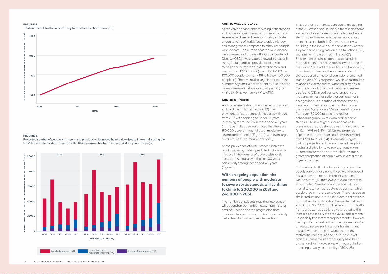
It is estimated that there are 254,000 Australians with undiagnosed heart valve disease in 2021. It is projected that this number will grow substantially in 2031 to 336,000 and in 2051 to 435,000 (Figure 3).
As Australians increasingly have greater access to heart imaging that can diagnose heart valve disease, there will be a large increase in the numbers of people eligible for intervention.
TABLE 1. Causes of stenosis and regurgitation at each heart valve. Common causes are shown in bold.
Aortic Mitral Pulmonary Tricuspid
Stenosis Bicuspid (BAV)
Degenerative AS
Rheumatic Congenital Rare
Regurgitation Bicuspid (BAV)
Degenerative AR
MVP
Degenerative MVD
Secondary
Congenital Secondary
OUR HIDDEN AGEING: TIME TO LISTEN TO THE HEART 1110
AORTIC VALVE DISEASE
Aortic valve disease (encompassing both stenosis and regurgitation) is the most common cause of severe valve disease. There is arguably a greater understanding of its risk factors, epidemiology and management compared to mitral or tricuspid valve disease. The burden of aortic valve disease has increased in Australia - the Global Burden of Disease (GBD) investigators showed increases in the age-standardised prevalence of aortic stenosis or regurgitation in Australian men and women from 1990 to 2017 (men – 169 to 203 per 100,000 people; women – 118 to 148 per 100,000 people) (1). There were also large increases in the numbers of years lived with disability due to aortic valve disease in Australia over that period (men – 4215 to 7542; women – 2991 to 6115).
AORTIC STENOSIS
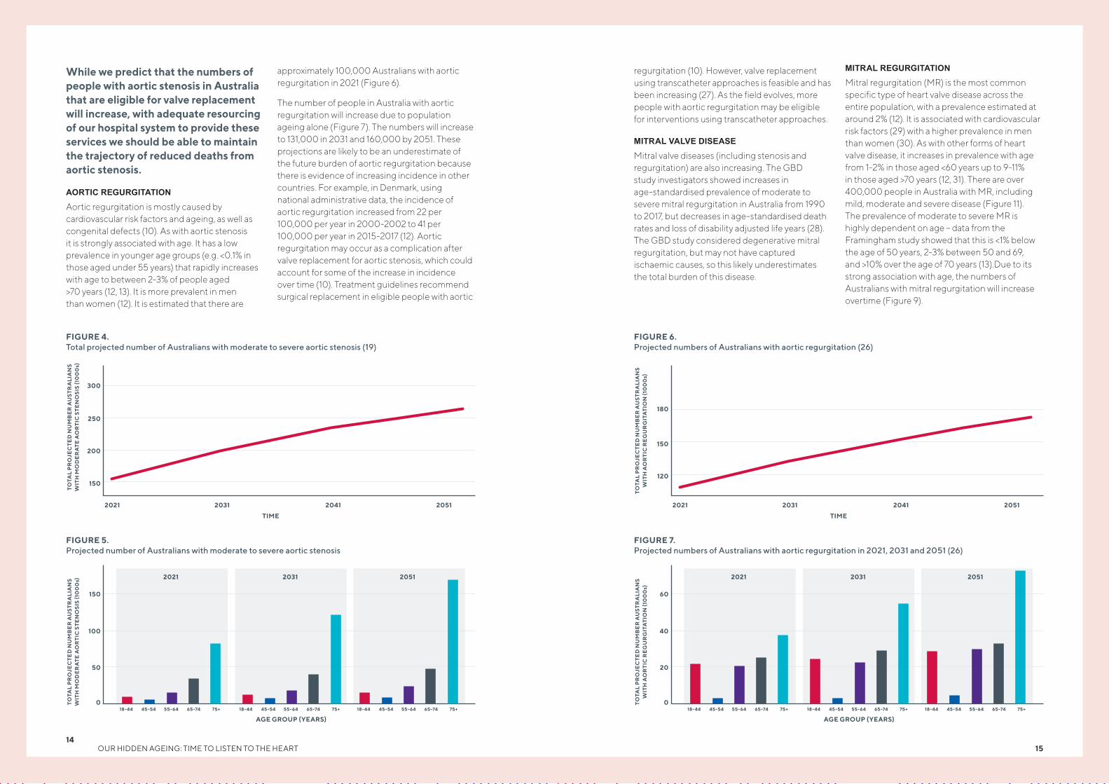
Aortic stenosis is strongly associated with ageing and cardiovascular risk factors (10). The prevalence of aortic stenosis increases with age from <0.1% of people aged under 55 years increasing to around 2% in those aged >75 years (4). In 2021, it has been estimated that there are 150,000 people in Australia with moderate to severe aortic stenosis (Figure 4), with even larger numbers reported internationally (18).
As the prevalence of aortic stenosis increases rapidly with age, there is predicted to be a large increase in the number of people with aortic stenosis in Australia over the next 30 years, particularly among those aged >75 years (Figure 5).
With an ageing population, the numbers of people with moderate to severe aortic stenosis will continue to climb to 200,000 in 2031 and 266,000 in 2051.
The numbers of patients requiring intervention will depend on co-morbidities, symptom status, cardiac function and the progression from moderate to severe stenosis – but it seems likely that at least half will require intervention.
These projected increases are due to the ageing of the Australian population but there is also some evidence of an increase in the incidence of aortic stenosis over time – due to better recognition, more disease or both. In Denmark, there was doubling in the incidence of aortic stenosis over a 15-year period using data on hospitalisations (20), with similar increases cited in France (21). Smaller increases in incidence, also based on hospitalisations, for aortic stenosis were noted in the United States of America (22) and Canada (21). In contrast, in Sweden, the incidence of aortic stenosis based on hospital admissions remained stable over a 20-year period, which was attributed to good risk factor control with similar trends in the incidence of other cardiovascular diseases also found (23). In addition to changes in the incidence or hospitalisation for aortic stenosis, changes in the distribution of disease severity have been noted. In a single hospital study in the United States over a 17-year period, records from over 130,000 people referred for echocardiography were examined for aortic stenosis. The investigators found that while prevalence of aortic stenosis was similar over time (6.4% in 1995 to 5.5% in 2012), the proportion of people with severe aortic stenosis increased from 19.3% to 35.2% (24).These data suggest that our projections of the numbers of people in Australia eligible for valve replacement are an underestimate, with a potential shift towards a greater proportion of people with severe disease in years to come.
Fortunately, deaths due to aortic stenosis at the population-level or among those with diagnosed disease have decreased in recent years. In the United States, (17) from 2008 to 2018, there was an estimated 1% reduction in the age-adjusted mortality rate from aortic stenosis per year, which accelerated in more recent years. There have been similar reductions in in-hospital deaths of patients hospitalised for aortic valve diseases from 4.5% in 2000 to 3.5% in 2012 (18). The reduction in deaths from aortic stenosis are largely attributed to the increased availability of aortic valve replacements – especially transcatheter replacements. However, it is important to realise that unrecognised and/or untreated severe aortic stenosis is a malignant disease, with an outcome worse than many metastatic cancers. Indeed, the outcomes of patients unable to undergo surgery have been unchanged for five decades, with recent studies reporting a two-year mortality of 50% (25).
Newly diagnosed HVD
FIGURE 2. Total number of Australians with any form of heart valve disease (15)
TIME
AGE GROUP (YEARS)
2021 2031 2041 2051
65-69 70-74 75-79 80-84 85+ 65-69 70-74 75-79 80-84 85+ 65-69 70-74 75-79 80-84 85+
2021 2031 2051
600
0
25000
50000
75000
75000
125000
800
1000
1200
TOTA
L P
RO
JEC
TE
D N
UM
BE
R A
US
TR
AL
IAN
S W
ITH
AN
Y H
VD
(10
00
s)P
RO
JEC
TE
D N
UM
BE
R O
F A
US
TR
AL
IAN
S W
ITH
HE
AR
T V
ALV
E D
ISE
AS
E (1
00
0s)
FIGURE 3. Projected number of people with newly and previously diagnosed heart valve disease in Australia using the OXValve prevalence data. Footnote: The 85+ age group has been truncated at 95 years of age.(17)
New diagnosed moderate or severe HVD
Previously diagnosed HVD
OUR HIDDEN AGEING: TIME TO LISTEN TO THE HEART 1312
regurgitation (10). However, valve replacement using transcatheter approaches is feasible and has been increasing (27). As the field evolves, more people with aortic regurgitation may be eligible for interventions using transcatheter approaches.
MITRAL VALVE DISEASE
Mitral valve diseases (including stenosis and regurgitation) are also increasing. The GBD study investigators showed increases in age-standardised prevalence of moderate to severe mitral regurgitation in Australia from 1990 to 2017, but decreases in age-standardised death rates and loss of disability adjusted life years (28). The GBD study considered degenerative mitral regurgitation, but may not have captured ischaemic causes, so this likely underestimates the total burden of this disease.
MITRAL REGURGITATION
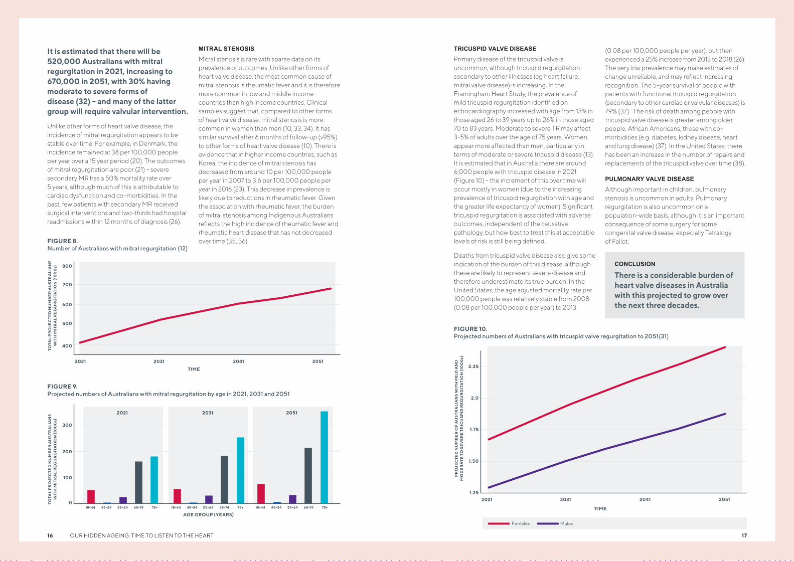
Mitral regurgitation (MR) is the most common specific type of heart valve disease across the entire population, with a prevalence estimated at around 2% (12). It is associated with cardiovascular risk factors (29) with a higher prevalence in men than women (30). As with other forms of heart valve disease, it increases in prevalence with age from 1-2% in those aged <60 years up to 9-11% in those aged >70 years (12, 31). There are over 400,000 people in Australia with MR, including mild, moderate and severe disease (Figure 11). The prevalence of moderate to severe MR is highly dependent on age – data from the Framingham study showed that this is <1% below the age of 50 years, 2-3% between 50 and 69, and >10% over the age of 70 years (13).Due to its strong association with age, the numbers of Australians with mitral regurgitation will increase overtime (Figure 9).
While we predict that the numbers of people with aortic stenosis in Australia that are eligible for valve replacement will increase, with adequate resourcing of our hospital system to provide these services we should be able to maintain the trajectory of reduced deaths from aortic stenosis.
AORTIC REGURGITATION
Aortic regurgitation is mostly caused by cardiovascular risk factors and ageing, as well as congenital defects (10). As with aortic stenosis it is strongly associated with age. It has a low prevalence in younger age groups (e.g. <0.1% in those aged under 55 years) that rapidly increases with age to between 2-3% of people aged >70 years (12, 13). It is more prevalent in men than women (12). It is estimated that there are
approximately 100,000 Australians with aortic regurgitation in 2021 (Figure 6).
The number of people in Australia with aortic regurgitation will increase due to population ageing alone (Figure 7). The numbers will increase to 131,000 in 2031 and 160,000 by 2051. These projections are likely to be an underestimate of the future burden of aortic regurgitation because there is evidence of increasing incidence in other countries. For example, in Denmark, using national administrative data, the incidence of aortic regurgitation increased from 22 per 100,000 per year in 2000-2002 to 41 per 100,000 per year in 2015-2017 (12). Aortic regurgitation may occur as a complication after valve replacement for aortic stenosis, which could account for some of the increase in incidence over time (10). Treatment guidelines recommend surgical replacement in eligible people with aortic
TIME TIME
2021 2031 2041 2051 2021 2031 2041 2051
150120
200150
250180
300
TOTA
L P
RO
JEC
TE
D N
UM
BE
R A
US
TR
AL
IAN
S W
ITH
MO
DE
RA
TE
AO
RT
IC S
TE
NO
SIS
(10
00
s)
TOTA
L P
RO
JEC
TE
D N
UM
BE
R A
US
TR
AL
IAN
S W
ITH
AO
RT
IC R
EG
UR
GIT
AT
ION
(10
00
s)
AGE GROUP (YEARS) AGE GROUP (YEARS)
18-44 45-54 55-64 65-74 75+ 18-44 45-54 55-64 65-74 75+18-44 45-54 55-64 65-74 75+ 18-44 45-54 55-64 65-74 75+
2021 20212031 20312051 2051
0 0
50 20
100 40
150 60
TOTA
L P
RO
JEC
TE
D N
UM
BE
R A
US
TR
AL
IAN
S W
ITH
MO
DE
RA
TE
AO
RT
IC S
TE
NO
SIS
(10
00
s)
TOTA
L P
RO
JEC
TE
D N
UM
BE
R A
US
TR
AL
IAN
S W
ITH
AO
RT
IC R
EG
UR
GIT
AT
ION
(10
00
s)
18-44 45-54 55-64 65-74 75+ 18-44 45-54 55-64 65-74 75+
FIGURE 5. Projected number of Australians with moderate to severe aortic stenosis
FIGURE 6. Projected numbers of Australians with aortic regurgitation (26)
FIGURE 7. Projected numbers of Australians with aortic regurgitation in 2021, 2031 and 2051 (26)
FIGURE 4. Total projected number of Australians with moderate to severe aortic stenosis (19)
OUR HIDDEN AGEING: TIME TO LISTEN TO THE HEART 1514
TRICUSPID VALVE DISEASE
Primary disease of the tricuspid valve is uncommon, although tricuspid regurgitation secondary to other illnesses (eg heart failure, mitral valve disease) is increasing. In the Framingham Heart Study, the prevalence of mild tricuspid regurgitation identified on echocardiography increased with age from 13% in those aged 26 to 39 years up to 26% in those aged 70 to 83 years. Moderate to severe TR may affect 3-5% of adults over the age of 75 years. Women appear more affected than men, particularly in terms of moderate or severe tricuspid disease (13). It is estimated that in Australia there are around 6,000 people with tricuspid disease in 2021 (Figure 10) – the increment of this over time will occur mostly in women (due to the increasing prevalence of tricuspid regurgitation with age and the greater life expectancy of women). Significant tricuspid regurgitation is associated with adverse outcomes, independent of the causative pathology, but how best to treat this at acceptable levels of risk is still being defined.
Deaths from tricuspid valve disease also give some indication of the burden of this disease, although these are likely to represent severe disease and therefore underestimate its true burden. In the United States, the age adjusted mortality rate per 100,000 people was relatively stable from 2008 (0.08 per 100,000 people per year) to 2013
(0.08 per 100,000 people per year), but then experienced a 25% increase from 2013 to 2018 (26). The very low prevalence may make estimates of change unreliable, and may reflect increasing recognition. The 5-year survival of people with patients with functional tricuspid regurgitation (secondary to other cardiac or valvular diseases) is 79% (37). The risk of death among people with tricuspid valve disease is greater among older people, African Americans, those with co-morbidities (e.g. diabetes, kidney disease, heart and lung disease) (37). In the United States, there has been an increase in the number of repairs and replacements of the tricuspid valve over time (38).
PULMONARY VALVE DISEASE
Although important in children, pulmonary stenosis is uncommon in adults. Pulmonary regurgitation is also uncommon on a population-wide basis, although it is an important consequence of some surgery for some congenital valve disease, especially Tetralogy of Fallot.
CONCLUSION
There is a considerable burden of heart valve diseases in Australia with this projected to grow over the next three decades.
It is estimated that there will be 520,000 Australians with mitral regurgitation in 2021, increasing to 670,000 in 2051, with 30% having moderate to severe forms of disease (32) – and many of the latter group will require valvular intervention.
Unlike other forms of heart valve disease, the incidence of mitral regurgitation appears to be stable over time. For example, in Denmark, the incidence remained at 38 per 100,000 people per year over a 15 year period (20). The outcomes of mitral regurgitation are poor (21) – severe secondary MR has a 50% mortality rate over 5 years, although much of this is attributable to cardiac dysfunction and co-morbidities. In the past, few patients with secondary MR received surgical interventions and two-thirds had hospital readmissions within 12 months of diagnosis (26).
MITRAL STENOSIS
Mitral stenosis is rare with sparse data on its prevalence or outcomes. Unlike other forms of heart valve disease, the most common cause of mitral stenosis is rheumatic fever and it is therefore more common in low and middle income countries than high income countries. Clinical samples suggest that, compared to other forms of heart valve disease, mitral stenosis is more common in women than men (10, 33, 34). It has similar survival after 6 months of follow-up (>95%) to other forms of heart valve disease (10). There is evidence that in higher income countries, such as Korea, the incidence of mitral stenosis has decreased from around 10 per 100,000 people per year in 2007 to 3.6 per 100,000 people per year in 2016 (23). This decrease in prevalence is likely due to reductions in rheumatic fever. Given the association with rheumatic fever, the burden of mitral stenosis among Indigenous Australians reflects the high incidence of rheumatic fever and rheumatic heart disease that has not decreased over time (35, 36).
AGE GROUP (YEARS)
18-44 45-54 55-64 65-74 75+ 18-44 45-54 55-64 65-74 75+
2021 2031 2051
0
100
200
300
TOTA
L P
RO
JEC
TE
D N
UM
BE
R A
US
TR
AL
IAN
S W
ITH
MIT
RA
L R
EG
UR
GIT
AT
ION
(10
00
s)
18-44 45-54 55-64 65-74 75+
FIGURE 9. Projected numbers of Australians with mitral regurgitation by age in 2021, 2031 and 2051
FIGURE 8. Number of Australians with mitral regurgitation (12)
Females Males
TIME
2021 2031 2041 20511.25
1.50
1.75
2.25
2.0
PR
OJE
CT
ED
NU
MB
ER
OF
AU
ST
RA
LIA
NS
WIT
H M
ILD
AN
D
MO
DE
RA
TE
TO
SE
VE
RE
TR
ICU
SP
ID R
EG
UR
GIT
AT
ION
(10
00
s)
TIME
2021 2031 2041 2051
400
500
600
700
800
TOTA
L P
RO
JEC
TE
D N
UM
BE
R A
US
TR
AL
IAN
S W
ITH
MIT
RA
L R
EG
UR
GIT
AT
ION
(10
00
s)
FIGURE 10. Projected numbers of Australians with tricuspid valve regurgitation to 2051(31)
OUR HIDDEN AGEING: TIME TO LISTEN TO THE HEART 1716

Clinical aspects of heart valve disease
Signs and Symptoms
CHAPTER TWO
There is very limited awareness of valvular heart disease in the community. A 2019 European Heart Health survey of people aged over 60 across 11 European countries found only a quarter were familiar with VHD (39).
This is not helped by the fact that common symptoms of heart valve disease – especially exercise intolerance – are often misattributed to ‘old age’.
Timely diagnosis is based on awareness and clinical examination – especially listening to the heart sounds.
The signs and symptoms of valvular heart disease are not specific to these entities, and reflect the consequences of valve disease on cardiac output,
maintaining normal cardiac and circulatory pressure, or the consequences of increased workload (Table 2).
Symptoms due to reduced cardiac output, including dizziness, blackouts and fatigue, are most commonly seen with aortic stenosis, but may occur with other advanced valvular diseases where the forward stroke volume is compromised because compensatory mechanisms have been exhausted.
The most common symptom due to congestion is shortness of breath, particularly during or immediately after activity or when lying down, which reflects pulmonary congestion. While this may be a direct consequence of stenosis or regurgitation involving the mitral valve (which is directly connected with the pulmonary circulation), this may occur with any left-sided
TABLE 2. Signs and symptoms of valvular heart disease.
Symptom Cause Valve lesion
Fatigue
Dizziness, blackouts
Inadequate cardiac output Stenosis
Shortness of breath, cough
Swelling of ankles and feet
Abdominal swelling
Congestion Regurgitation
Heart failure
Chest pain Increased workload Aortic stenosis
Palpitations Enlargement of heart chambers Regurgitation (esp. mitral)
Mitral stenosis
Stroke Blood clots Mitral
OUR HIDDEN AGEING: TIME TO LISTEN TO THE HEART 18 19
heart disease because of increased workload on the left ventricle. On the right side of the heart, congestion is manifest by swelling of the ankles and feet, or abdominal swelling, reflecting congestion of the liver and fluid within the abdominal cavity.
Chest pain occurs when the workload on the heart exceeds the delivery of blood through the coronary arteries. While this may be due to narrowing of the coronaries (atherosclerotic cardiovascular disease is also disease of the elderly), it often reflects increased workload of the left ventricle, and/or changes in the ventricular pressure that compromises blood supply to the heart muscle.
Heart valve disease can cause many complications (including heart failure, stroke, blood clots, and heart rhythm abnormalities) and mortality.
Some symptoms of valvular heart disease arise from these complications. For example, enlargement of the left atrium may lead to an irregular heartbeat (atrial fibrillation) which the patient may experience as palpitations. Atrial fibrillation and left atrial enlargement may also lead to the development of blood clots within the heart.
Clinical signs are features identifiable by a clinician doing a physical examination. Each of the heart valve lesions has a characteristic murmur that can be heard with a stethoscope or digital stethoscope.
They can be characterised by their timing during the cardiac cycle, their location of the chest, and the pitch of the sound. In addition, evidence of enlargement of different cardiac chambers and congestion of the lungs or body are also markers the presence and severity of these valve lesions.
DiagnosisThe physical examination is the cornerstone of detecting heart valve disease.
Symptoms such as chest pain and shortness of breath should be reported to a primary care physician, and in this instance, a physical examination should be performed. More problematic are patients who develop significant heart valve disease in the absence of symptoms. This may occur either because the patient is very inactive, or because there is adequate compensation of cardiac function, but this is especially the case in a significant number of patients with regurgitant valve lesions, which are well tolerated until late in the course.
Therefore, more effective surveillance of heart valve disease in the community is linked very closely to vulnerable patients, particularly the elderly, having a complete physical examination.
Unfortunately, the process of delivering this step is more complicated than it might seem. Adequate physical examination requires a significant degree of undressing of the patient, and may especially pose a barrier in women – in the setting of a busy primary care practice, an adequate examination is hard to achieve in a short consultation. The environment for adequate examination is also important. Significant extraneous noise from a busy primary care clinic is incompatible with recognition of abnormal heart sounds of murmurs, which may be subtle. Various technical developments such as the electronic stethoscope and handheld ultrasound could compensate for some of these problems, but their uptake has been limited and slow. Finally, physical examination skills have attenuated over the last few decades, and there is widespread recognition that physical signs are insensitive and non-specific. The use of investigations is therefore important in screening.
An electrocardiogram (ECG) shows the electrical activity of the heart. While the primary use of this test is used to check for abnormal heart rhythms, the nature of ECG waveforms changes as a particular cardiac chambers enlarge or show signs of increased workload. In the absence of other causes such as high blood pressure, ECG abnormalities can be a warning sign to the presence of a heart valve lesion.
The chest X-ray provides useful information about the size and configuration of the heart, the presence of valvular calcification, and congestion of the lung fields. For this reason, it is a simple and inexpensive addition to the physical examination which can provide clues about valvular heart disease. Unfortunately, many of these changes are seen in the setting of advanced valve disease and therefore perhaps less useful for screening for asymptomatic disease.
Echocardiography is an ultrasound test that is used to image the heart valves and chambers and to measure flow within the heart. It is the most commonly performed test for the assessment and follow-up of valvular heart disease.
Access to echocardiography is a vital component in managing valvular disease in the community, and current efforts to use artificial intelligence to better acquire, measure and analyse these images will be of value.
At the moment, the main focus of echocardiography is in patients with symptoms or clinically-suspected disease, but there may be a time where a routine echocardiogram is used for screening people at the age of 70 or 75 years, for example.
Stress tests may be very useful to identify the association of symptoms with activity, and to define the functional, as opposed to an anatomic severity of valve lesions. Stress testing is particularly useful in conjunction with echocardiography, and probably underutilised in the follow-up of heart palpitations.
Cardiac catheterization is an invasive test where catheters are used to measure pressure and flow of blood within the heart, as well as inject dye to image the coronary arteries and cardiac chambers. These imaging steps are often being undertaken with cardiac computed tomography, but invasive catheterisation remains helpful for physiologic assessment.
Cardiac magnetic resonance imaging provides imaging structure and function analogous to echocardiography, but consistently of high quality, whereas ultrasound may be compromised by overlying lung tissue or fat. The unique aspect of MRI is its ability to characterise tissue and recognise fibrosis, which impacts on the likelihood of responsiveness to valvular interventions.
OUR HIDDEN AGEING: TIME TO LISTEN TO THE HEART 2120

Cardiovascular ageing and valvular disease
Valves
CHAPTER THREE
Aortic stenosis prevents blood from being pumped effectively, creating a pressure gradient between the aorta and the left ventricle. To compensate, the left ventricle walls thicken (a process called myocardial hypertrophy) to maintain adequate systolic function.
Age-related valvular changes predominantly include degeneration and scarring (i.e. valvular sclerosis). Aortic valve sclerosis is evident in approximately 30–80% of elderly individuals (40). While aortic valve sclerosis does not obstruct blood flow, it can progress to aortic stenosis when severe thickening, stiffening and calcification of the leaflet obstruct the aortic valve. Increased leaflet calcification and decreased leaflet mobility may be early warning signs of progression to aortic stenosis. Individuals with left ventricular hypertrophy, hypertension, hyperlipidaemia, end-stage renal disease, congenital bicuspid aortic valves or smokers are at an increased risk of progression of aortic valve sclerosis to aortic stenosis (41). Furthermore, those with aortic valve sclerosis have an increased risk of cardiovascular events and mortality (42, 43) .
Aortic regurgitation most commonly arises from degeneration, thickening and retraction of valve cusps in the 7th and 8th decade (44), or from the same processes occurring at an earlier age (5th and 6th decade) in congenitally abnormal (most commonly bicuspid) valves. Other causes include destruction of valve leaflets by infection, or enlargement of the aortic annulus or aortic root. Aortic regurgitation means that the aortic valve does not close properly and the flow of backwards blood from the aorta to the left ventricle in diastole results in increased work for the left ventricle and an increase in size.
Mitral valve regurgitation results when the mitral valve fails to seal completely resulting in a backwards flow of blood into the left ventricle and inadequate supply of blood to the rest of the body. Mitral annular calcification involves fibrosis of the annulus of the mitral value and is associated with ageing. It commonly occurs alongside aortic valve sclerosis, given their overlapping pathology (45). Hypertension, end-stage renal disease, aortic stenosis and mitral valve prolapse are risk factors for mitral annular calcification. Those with mitral annular calcification are at an increased risk of mitral stenosis and regurgitation, heart failure, atrial fibrillation, conduction system diseases, stroke, coronary and vascular diseases, cardiovascular events and mortality (40).
OUR HIDDEN AGEING: TIME TO LISTEN TO THE HEART 22 23
Heart muscle It is very important to consider valvular heart disease as an entity that often involves both the valves and heart muscle (myocardium).
Myocardial dysfunction and heart failure may arise as a consequence of either pressure or volume loading due to valvular disease.
Regurgitation at the atrio-ventricular valves may occur secondary to myocardial disease leading to ventricular enlargement and restriction of leaflets, causing them to fail to close and therefore leak. These heart muscle diseases (cardiomyopathies) can be classified into three types (dilated, hypertrophic and restrictive) based on anatomical appearance and abnormal physiology (46).
In dilated cardiomyopathy, enlargement of all four cardiac chambers is typical, but sometimes the dilation and/or reduced contraction can be limited to the left or right side of the heart. An increase in thickness of the ventricular walls may occur but chamber dilation is generally out of proportion to any hypertrophy. Myocyte damage due to genetic, inflammatory, toxic and metabolic causes contribute to the development of this cardiomyopathy (46). Ventricular stroke volume and cardiac output decline due to impaired myocyte contractility. To compensate, ventricular diastolic volume increases thereby subsequently increasing stroke volume and sympathetic nervous activity increases resulting in an increase in heart rate, buffering any decline in cardiac output. Dilated cardiomyopathy is primarily associated with secondary (or functional) mitral or tricuspid regurgitation.
Mitral regurgitation in hypertrophic cardiomyopathy is associated with functional influences on the mitral valve (due to obstruction of systolic outflow), associated structural abnormalities of the mitral valve apparatus, or enlargement of the left atrium, which can stretch the valve annulus. Hypertrophic cardiomyopathy is a familial disease in which inheritance follows an autosomal dominant pattern (46).
Restrictive cardiomyopathy is an uncommon disease, characterised by abnormally rigid myocardium due to fibrosis or infiltration. The reduced compliance of the ventricles leads to abnormally high filling pressure, atrial stretch and mitral regurgitation. The most recognised cause of restrictive cardiomyopathy is amyloidosis (46) – although often seen as a problem associated with haematologic malignancy, one of the forms of amyloidosis is associated with ageing and aortic stenosis.
VasculatureVascular ageing refers to the deterioration in vascular structure and function over time, which ultimately leads to end-organ damage in the heart, brain and kidney.
Vascular ageing commences in early life and is a normal ageing phenomenon. However, pathological vascular ageing, as evident in conditions such as hypertension, results in accelerated changes related to atherosclerosis. Exposure to adverse environmental and genetic factors as early as during childhood or even during foetal life promotes the development and accumulation of subclinical vascular changes that may be an important contributor to early vascular ageing (47). Vascular age encompasses the cumulative effect of all cardiovascular risk factors on the arterial wall over the life course. This contrasts with more classical risk factors (such as BP) which may vary with time (48), so vascular age may help to identify those at elevated CVD risk.
Vascular ageing involves both arteriosclerosis and atherosclerosis (49). Arteriosclerosis is the thickening, hardening, and loss of elasticity of the arterial walls. While the process of atherosclerosis principally takes place in inner lining (intima), the ageing process affects the entire arterial wall. This process includes dysfunction of the inner lining (endothelium), decrease in nitric oxide (NO) production and local inflammation in the intima (50). There is a reduction of elastic tissue and a scarring process (evidenced by a relative increase in collagen content) in the middle of the vessel (media) (51). In the adventitia (outer covering), there is impairment of nerve control, a loss of function of the blood vessels that supply
the artery wall (52) and development of fat deposits that may increase local inflammation and adversely impact vasodilation (53). This leads to structural changes in the arterial wall (54-57), accompanied by lumen enlargement (55-57) and increased stiffness in the large, proximal elastic arteries (58) but not in distal muscular arteries. Importantly, the vascular ageing process involves the entire vascular system including remodelling of the small arteries. The valves are involved because the same pathologic processes occur in valve tissue, and because arterial damage increases mechanical stress on the valves.
In a healthy cardiovascular system, the compliant properties of the large arteries ensure that pulsations in pressure and flow generated by cyclic left ventricular contraction are dampened at the aorta into a continuous pressure (and flow) downstream at the smaller vessels. This allows for steady blood flow to the organs and protects the small vessels from the damaging effects of pulsatile pressure (59). However, in response to ageing (60, 61), high blood pressure and other disease states such as type 2 diabetes (T2D) (62-64), arterial stiffening limits the buffering capacity of the elastic arteries, which has a number of adverse consequences for cardiovascular health.
Alterations in vascular structure and function have been observed in patients with prediabetes or impaired fasting glucose (65, 66) and in patients with overt T2D (67, 68) or CVD (69), while hypertension is related to increased stiffness of the aorta for any given level of BP (70). This suggests that the presence of CVD risk factors progressively worsens vascular health.
OUR HIDDEN AGEING: TIME TO LISTEN TO THE HEART 2524
Management of valvular heart disease
Prevention
CHAPTER FOUR
HEALTHY LIVING
We increasingly understand the role of ‘traditional’ risk factors for cardiovascular diseases such as smoking, hypertension, diabetes and obesity (71-73). Population level changes in these risk factors may impact the incidence and outcomes of heart valve diseases. While healthy living is critical to the primordial prevention of heart disease, the extent to which this is specifically protective against degenerative valve changes is undefined, and probably mediated by effects on risk factors. Hypertension is associated with an increased risk of MR; for every 20mmHg increase in systolic BP, there is a 26% increased risk of MR (74). Obesity, which is present in 41% of adults aged 65-74 years (75) is associated with increased CVD risk (76), including aortic stenosis (77). Likewise, despite the generic effects of healthy diets on CVD (78-80), their protective effect on degenerative valve lesions has only been shown in experimental models. Low to moderate alcohol consumption is associated with lower odds of having aortic valve sclerosis (81). Smoking cessation has well-established benefits on CVD mortality (82), but little evidence of benefit for valvular disease. Low cardiorespiratory fitness is one of the strongest and most important risk factors for CVD morbidity and mortality (83, 84), including in people with valvular heart disease. Whether fitness influences the development or progression of valve disease is unknown.
PREVENTION OF RHEUMATIC FEVER AND ENDOCARDITIS
The heart valves may be damaged specifically by infections and inflammatory diseases. Rheumatic valve disease occurs as a result of an immune response to Streptococcal infection of the throat
or skin, leading to valve scarring and leakage. There has been a reduction of rheumatic fever in Australia due to improvements in hygiene and availability of antibiotics. However, there is an ongoing high burden of rheumatic heart disease in Indigenous Australians, with a consequent major large burden of heart valve disease (14) that is probably the highest in the developed world. Rheumatic valve disease is a relatively small contributor to the burden of valvular heart disease in Australia because the at-risk population is small, but should attain more attention because it is preventable.
Endocarditis describes direct infection of the heart valves - generally by bacteria – that causes destruction of valve tissue and therefore valvular regurgitation. The most common portals for bacterial entry are dental, surgical and related to intravenous drug use. Although an uncommon disease, its high mortality (>10% acutely and about 30% within a year) has defied advances in medical and surgical management. The best opportunity for prevention is antibiotic cover (generally penicillin) for dental procedures, but this necessitates treatment of large numbers of people – with consequent antibiotic side-effects and antibiotic resistance. For these reasons, guidelines for prophylaxis were made more restrictive over a decade ago. A study in the UK showed a reduction of about 8000 prescriptions/month was temporally associated with an increment of endocarditis of about 35 per month (85). While these data do not establish a causal association, they emphasise the ongoing opportunity for prevention of a subgroup of valvular heart disease.
OUR HIDDEN AGEING: TIME TO LISTEN TO THE HEART 26 27
Early detection – Putting heart valve disease on the agendaSome patients with heart disease remain asymptomatic, or are symptomatic but the valve disease is unrecognised, until a crisis occurs - often heart failure or atrial fibrillation - and urgent intervention is required. This particularly occurs in the elderly, and other individuals where functional capacity is restricted, and insufficient exercise is undertaken to generate symptoms. As acute presentations with heart failure or atrial fibrillation incur cost and risk, it would certainly be preferable if patients were recognised at an earlier stage in the disease, at which stage they may be asymptomatic or minimally symptomatic. In rheumatic heart disease, early detection permits the initiation of antibiotic therapy to prevent recurrent Streptococcal infections.
Detection of asymptomatic valvular disease requires routine physical examination (discussed in Chapter 2) or screening echocardiography. While echocardiographic screening is a cornerstone of detecting early rheumatic heart disease in at-risk communities (86), its use for detecting advanced degenerative valvular heart disease in the broader population is controversial. Once clinical evidence of valve disease is detected, then a full echocardiogram should be obtained to confirm the diagnosis, estimate the severity of the valuation, and evaluate the level of cardiac reserve.
Established disease and interventionSome patients with valvular heart disease are recognised before intervention is required.
In these individuals, frequent medical follow-up is essential, the frequency of which will vary according to severity of the valve lesion and comorbidities. In general, mild lesions may be followed in primary care every 2-3 years, but as the lesion moves to moderate or severe, follow-up evaluations may need to occur with a cardiologist, annually or more frequently. During this phase, standard risk factor control is important, including weight control and following a healthy diet. As always, smoking cessation is important, particularly so if operative intervention will be required. Recent changes to the Medical Benefits Scheme, based on AHA recommendations, stipulate different follow-up for different levels of disease severity. For mild to moderate disease, a repeat echocardiogram is justified every 3–5 years for mild disease and 1–2 years for moderate disease. Severe disease can be monitored in line with the guidelines.
At present, no medications have been shown to alter the natural history of either stenotic or regurgitant lesions. Nonetheless, in the pre-intervention stage, diuretics may be used to reduce fluid retention, and vasodilators and ACE inhibitors may be used to improve cardiac loading conditions - these are particularly beneficial in secondary mitral regurgitation. Digoxin or beta blockers may be prescribed in patients with atrial fibrillation or heart failure.
In general, heart valve interventions involve splitting a stenotic valve with a balloon (valvuloplasty), repairing valve leaflets (usually for regurgitant valves) or replacement.
Replacements of the mitral (MVR) or aortic valve (AVR) may be made of biological tissue (animal, cadaveric, or autotransplanted), or mechanical. Mechanical valves are highly durable and have been reported to last for 2-3 decades, but they require anticoagulation with warfarin.
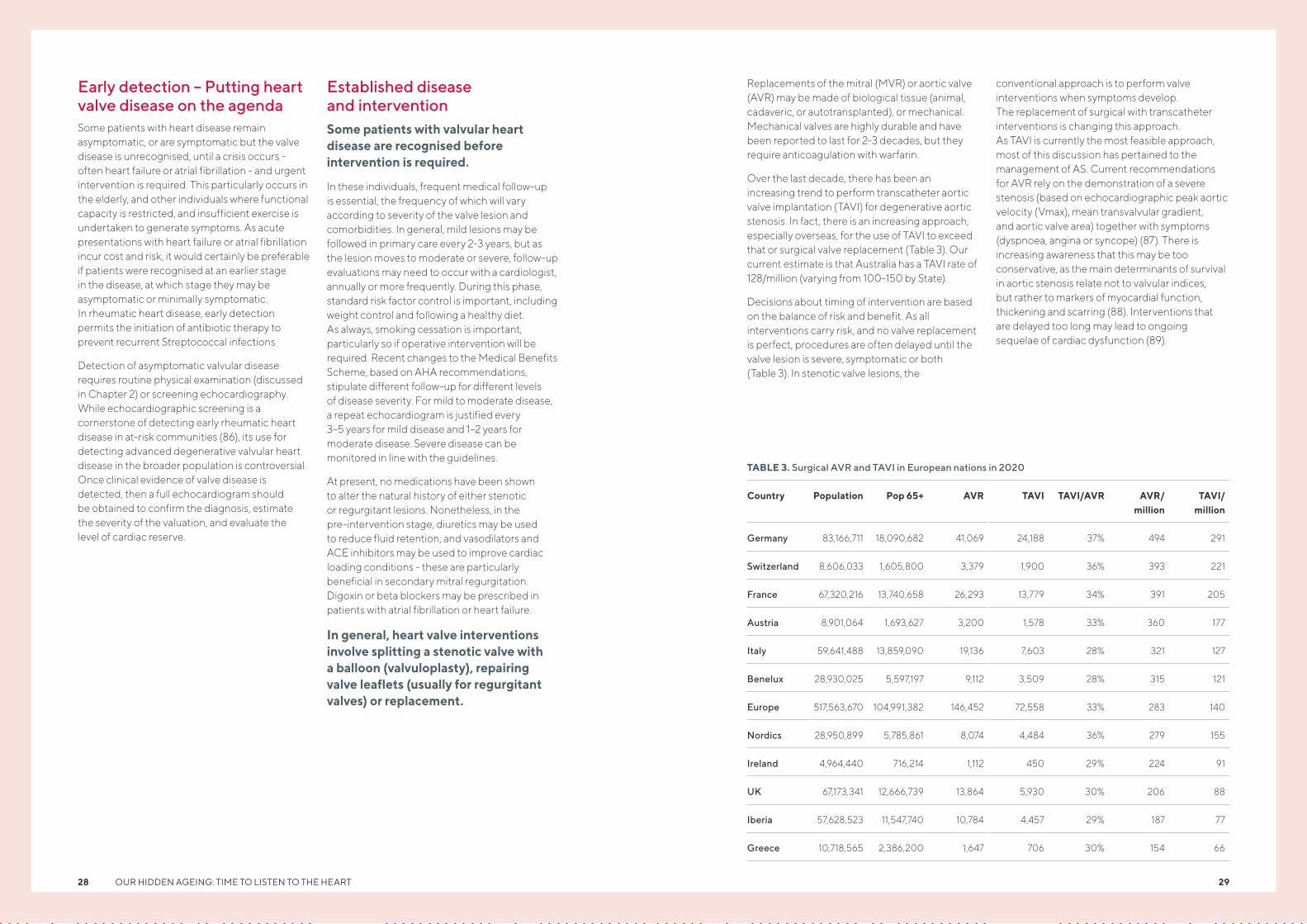
Over the last decade, there has been an increasing trend to perform transcatheter aortic valve implantation (TAVI) for degenerative aortic stenosis. In fact, there is an increasing approach, especially overseas, for the use of TAVI to exceed that or surgical valve replacement (Table 3). Our current estimate is that Australia has a TAVI rate of 128/million (varying from 100-150 by State).
Decisions about timing of intervention are based on the balance of risk and benefit. As all interventions carry risk, and no valve replacement is perfect, procedures are often delayed until the valve lesion is severe, symptomatic or both (Table 3). In stenotic valve lesions, the
conventional approach is to perform valve interventions when symptoms develop. The replacement of surgical with transcatheter interventions is changing this approach. As TAVI is currently the most feasible approach, most of this discussion has pertained to the management of AS. Current recommendations for AVR rely on the demonstration of a severe stenosis (based on echocardiographic peak aortic velocity (Vmax), mean transvalvular gradient, and aortic valve area) together with symptoms (dyspnoea, angina or syncope) (87). There is increasing awareness that this may be too conservative, as the main determinants of survival in aortic stenosis relate not to valvular indices, but rather to markers of myocardial function, thickening and scarring (88). Interventions that are delayed too long may lead to ongoing sequelae of cardiac dysfunction (89).
TABLE 3. Surgical AVR and TAVI in European nations in 2020
Country Population Pop 65+ AVR TAVI TAVI/AVR AVR/million
TAVI/million
Germany 83,166,711 18,090,682 41,069 24,188 37% 494 291
Switzerland 8,606,033 1,605,800 3,379 1,900 36% 393 221
France 67,320,216 13,740,658 26,293 13,779 34% 391 205
Austria 8,901,064 1,693,627 3,200 1,578 33% 360 177
Italy 59,641,488 13,859,090 19,136 7,603 28% 321 127
Benelux 28,930,025 5,597,197 9,112 3,509 28% 315 121
Europe 517,563,670 104,991,382 146,452 72,558 33% 283 140
Nordics 28,950,899 5,785,861 8,074 4,484 36% 279 155
Ireland 4,964,440 716,214 1,112 450 29% 224 91
UK 67,173,341 12,666,739 13,864 5,930 30% 206 88
Iberia 57,628,523 11,547,740 10,784 4,457 29% 187 77
Greece 10,718,565 2,386,200 1,647 706 30% 154 66
OUR HIDDEN AGEING: TIME TO LISTEN TO THE HEART 2928
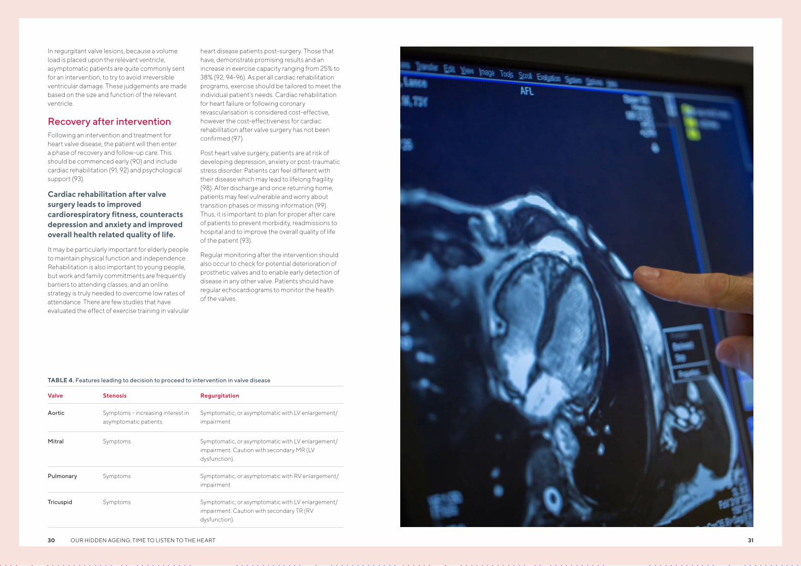
TABLE 4. Features leading to decision to proceed to intervention in valve disease
Valve Stenosis Regurgitation
Aortic Symptoms – increasing interest in asymptomatic patients.
Symptomatic, or asymptomatic with LV enlargement/impairment
Mitral Symptoms Symptomatic, or asymptomatic with LV enlargement/impairment. Caution with secondary MR (LV dysfunction).
Pulmonary Symptoms Symptomatic, or asymptomatic with RV enlargement/impairment
Tricuspid Symptoms Symptomatic, or asymptomatic with LV enlargement/impairment. Caution with secondary TR (RV dysfunction).
In regurgitant valve lesions, because a volume load is placed upon the relevant ventricle, asymptomatic patients are quite commonly sent for an intervention, to try to avoid irreversible ventricular damage. These judgements are made based on the size and function of the relevant ventricle.
Recovery after interventionFollowing an intervention and treatment for heart valve disease, the patient will then enter a phase of recovery and follow-up care. This should be commenced early (90) and include cardiac rehabilitation (91, 92) and psychological support (93).
Cardiac rehabilitation after valve surgery leads to improved cardiorespiratory fitness, counteracts depression and anxiety and improved overall health related quality of life.
It may be particularly important for elderly people to maintain physical function and independence. Rehabilitation is also important to young people, but work and family commitments are frequently barriers to attending classes, and an online strategy is truly needed to overcome low rates of attendance. There are few studies that have evaluated the effect of exercise training in valvular
heart disease patients post-surgery. Those that have, demonstrate promising results and an increase in exercise capacity ranging from 25% to 38% (92, 94-96). As per all cardiac rehabilitation programs, exercise should be tailored to meet the individual patient’s needs. Cardiac rehabilitation for heart failure or following coronary revascularisation is considered cost-effective, however the cost-effectiveness for cardiac rehabilitation after valve surgery has not been confirmed (97).
Post heart valve surgery, patients are at risk of developing depression, anxiety or post-traumatic stress disorder. Patients can feel different with their disease which may lead to lifelong fragility (98). After discharge and once returning home, patients may feel vulnerable and worry about transition phases or missing information (99). Thus, it is important to plan for proper after care of patients to prevent morbidity, readmissions to hospital and to improve the overall quality of life of the patient (93).
Regular monitoring after the intervention should also occur to check for potential deterioration of prosthetic valves and to enable early detection of disease in any other valve. Patients should have regular echocardiograms to monitor the health of the valves.
OUR HIDDEN AGEING: TIME TO LISTEN TO THE HEART 30 31
Economic and societal costs of heart valve disease in the elderly
Introduction
CHAPTER FIVE
The epidemiology of valve disease points towards this as an epidemic of the elderly. An influential Productivity Commission (2013) report estimated that expenditures on pensions, healthcare and old age care in Australia will rise to an additional 5.7% of gross domestic product (GDP) over the next 50 years (100-103), with no major corresponding benefits for GDP. A tacit conclusion might be that additional healthcare expenditure to address valvular heart disease might not be justified on the basis of economic benefit. A key driver is the implicitly held view that the elderly contribute relatively little to the economy given their low participation rates in paid work. This view, specifically, neglects the participation of the elderly to society through other means, including volunteer work, childcare and informal provision of adult care.
Recent estimates suggest that unpaid work accounts for about 10% to 40% of GDP in Organization for Economic Cooperation and Development (OECD) countries, including significant contributions by the elderly (104). Data from the Australian Bureau of Statistics’ Survey of Disability, Ageing and Carers (SDAC) suggest that individuals 55 years and over account for about 45% of all providers of informal adult care (with individuals 65 years and over comprising one-quarter of all informal care providers) (105). Studies also show that grandparents are a major provider of support for younger children in Australia (106). Estimates from de Vaus et al (107) suggest that the unpaid contributions of individuals over 55 years of age, in the form of childcare, household support and adult care, contributed more than 12% of Australia’s GDP (7% of GDP in the case of Australians 65 years and over).
A recognition of the value of unpaid services provided to society by the elderly also implies that policy evaluations should extend assessments to go beyond traditional indicators to include their contribution to non-market activities. In the context of interventions to improve health, this would mean extending measures of cost-effectiveness and cost-benefit ratios to go beyond traditional approaches that focus on outcomes such as beneficiaries’ quality of life, or their paid work contributions.
This chapter highlights the above issues by exploring the potential consequences of heart disease for the economic value of elderly contributions in Australia – as workers and as providers of unpaid services, or productive non-market activities (PNMA). Our primary motivation for analysing the impact of heart disease was to assess how aortic stenosis could influence the social value (in terms of work (market) activities and PNMA) produced, thereby providing some insight into the potential social gains from interventions such as heart valve replacement. As is well understood, AS is associated with a significant worsening of the quality of life, including physical functioning, and high mortality risk (108-110). Because most datasets on time use, work status and other characteristics of individuals do not have the level of detail required to identify patients with aortic stenosis, our strategy was to infer the potential impact of aortic stenosis from analyses that assessed the association of heart disease (as self-reported by survey respondents) of varying levels of severity with the monetary value of elderly contributions to market-based activities and PNMA.
OUR HIDDEN AGEING: TIME TO LISTEN TO THE HEART 32 33
Data and Approach Two main datasets provided most of the information used for the analysis that were carried out. The Household, Income and Labour Dynamics in Australia (HILDA) Survey is a household-level, nationally representative, longitudinal survey that gathers information from the same individuals annually through interviews and self-completion questionnaires, periodically adding new individuals to the survey. The analysis reported here used longitudinal data from a sample of individuals aged 55 years and over from wave 13 (2013) – the baseline survey, and wave 17 (2017) to capture changes in heart disease status of individuals over time. After dropping observations with missing data, excluding individuals who reported heart disease in wave 13, and non-working individuals, a balanced panel of 1,539 individuals was used for the final analysis.
The Australian Longitudinal Study on Women’s Health (ALSWH) is a longitudinal population-based survey which collects information on social, behavioural, and economic factors affecting choices related to lifestyle, family, and workforce participation. Launched in 1996, ALSWH surveys women in three age groups (18-23 years (born 1973-78); 45-50 years (born 1946-51); and 70-75 years (born 1921-26) to represent women at different life stages. Data from two rounds – 2007 and 2016 – were used for a cohort of women who were of retirement age, 64-70 years, at the time of the 2016 survey. In our analysis, only those survey participants who were employed at the time of the 2007 (baseline) survey and who did not have heart disease at that time were included, resulting in a balanced panel of 5,227 individuals.
Both HILDA and the ALSWH included information on socio-economic status and demographic characteristics (e.g. marital status, education level, location, country of origin); health status (presence of heart disease, self-rated health, and SF-36 scores, an internationally recognized scale that assesses physical functioning); paid employment (e.g. hours worked, wages earned) and whether participating in self-employment; and productive, non-market activities (e.g. caring for grand/children or sick, frail, and disabled adults; other voluntary work). While information included was similar across both datasets, the ‘core data’ release for ALSWH lacked information on Aboriginal status, rural location, and number of hours spent on particular non-market activities, including caring for grand/children, domestic tasks, and other voluntary work. This limited its usefulness in calculating an aggregate monetary
measure of PNMA, while permitting assessments of some components of it.
Monetary values per unit time were assigned for work (whether undertaken for wages, or as self-employed), and to productive non-market activities (PNMA). The latter specifically referred to time spent caring for adults, childcare and voluntary work. Appendix Table 6 summarizes the methods used to assign monetary values per hour for paid work, self-employment and PNMA. Valuation of PNMA was based on methods used in Bloom et al (111).
Self-oriented household activities (e.g., cooking, cleaning) were excluded from this valuation. Information on self-reported heart disease from the two sets of surveys was combined with data on self-reported health and measures of physical functioning based on responses to SF-36 questionnaires administered to respondents in the two surveys to generate indicators of severity of heart disease.
Associations between indicators of heart disease and four types of outcomes were assessed, using methods described in Box 1: (a) the monetary values of work and PNMA; (b) whether participating in work; and (c) whether participating in PNMA; and (d) the hours devoted to work or PNMA. The analyses were limited to individuals who reported working in the baseline round of each longitudinal survey, provided they did not also report heart disease. Employed individuals were considered, because non-participation in work at baseline could be driven by a range of factors not necessarily captured by the surveys.
Table 5 summarises the socioeconomic and demographic composition of the HILDA and ALSWH respondents from the most recent waves of the two datasets, along with the proportion of respondents reporting heart disease. The data for HILDA respondents corresponds to those aged 55 years and over, and for ALSWH for ages 65 and over. Reflecting the urban concentration of Australia’s population, the sample is dominated by respondents living in urban areas, although the ALSWH does oversample from rural populations. The proportion of respondents reporting heart disease is rising in age, and the proportion is also higher among Indigenous Australians, among men, and urban residents. The data also suggest that indicators of poor health (using either the physical functioning criterion from SF-36 responses, or self-reported health indicators) are positively correlated with self-reported heart disease.
Measuring the Association between Heart Disease, Work and PNMA: Estimation Strategy
Associations of two types were estimated: (a) between aggregate monetary values of work (PNMA) and their components (e.g., value of adult-care, the value of childcare, or value of paid (unpaid) work) and heart disease; and (b) between participation in work or its components (PNMA and its components) and the natural log of hours spent in work or its components (PNMA or its components) and heart disease. Paid work or self -employment and the term “market activities” will be used interchangeably.
ASSOCIATION BETWEEN MONETARY VALUE OF WORK AND PNMA WITH HEART DISEASE
The monetary contribution of the elderly to market activities (whether from paid employment, or self-employment) and PNMA was obtained by separately estimating two sets of equations (one for market activities, and one for PNMA), with the monetary value of the individual’s contribution as the dependent variable and a range of socioeconomic, locational and demographic explanatory variables, an indicator for heart disease (by varying levels of severity) and a baseline indicator of health status. A baseline indicator of health status was used as a control to account for pre-existing differences in health that could account for differential earnings from work or value of PNMA. A similar strategy was used to estimate the association of the value of time spent on components of work (e.g., paid or as self-employed), and components of PNMA (e.g., childcare, adult-care, volunteering), with indicators of heart disease.
ASSOCIATION BETWEEN PARTICIPATION IN WORK (PNMA) WITH HEART DISEASE
The main weakness of the approach measuring the association between monetary value of work (PNMA) and heart disease is its inability to provide much information on whether the direction of association is being driven by participation or changes in hours, conditional on participation. For example, ill health can lead an individual to exit the labour market, or
to work fewer hours. Either can lower the value of market-based activities, provided earnings per hour are unchanged. Similarly, the value of PNMA is determined by whether an individual decides to pursue a particular activity, and the number of hours they devote to it. To explore this issue further, two-part models were estimated with outcomes being participation, and separately, the natural log of hours of participation (conditional on participating), for each of five activities: paid work or self-employment, paid work only, childcare, adult-care, and voluntary work. As under the first method, explanatory variables included a set of socioeconomic, locational, and demographic explanatory variables, an indicator for heart disease (by varying levels of severity) and a baseline indicator of health status. A baseline indicator of health status was used as a control to account for pre-existing differences in health (that could account for differential participation rates in activities and in hours devoted to activities.
REGRESSION MODEL AND INTERPRETATION OF COEFFICIENTS
For HILDA data, the association was estimated using a multilevel model version of longitudinal data analysis with three levels, with the first level being the survey wave-specific observation of the individual, the second level being the individual, and the third-level being the primary sampling unit (census district), with sample weighting. For ALSWH data, the longitudinal data analysis permitted a two-level multi-level model estimation, with sample weighting, the first level being the survey wave-specific observation of the individual, the second level being the individual themselves.
Technical Note: For regression models using a semi-log specification (e.g., equation with log of hours of work as the dependent variable), the coefficient ßβ on the heart disease variable was translated into a percentage change measure (denoted by g), where g=expβ(βß)-1.
OUR HIDDEN AGEING: TIME TO LISTEN TO THE HEART 3534
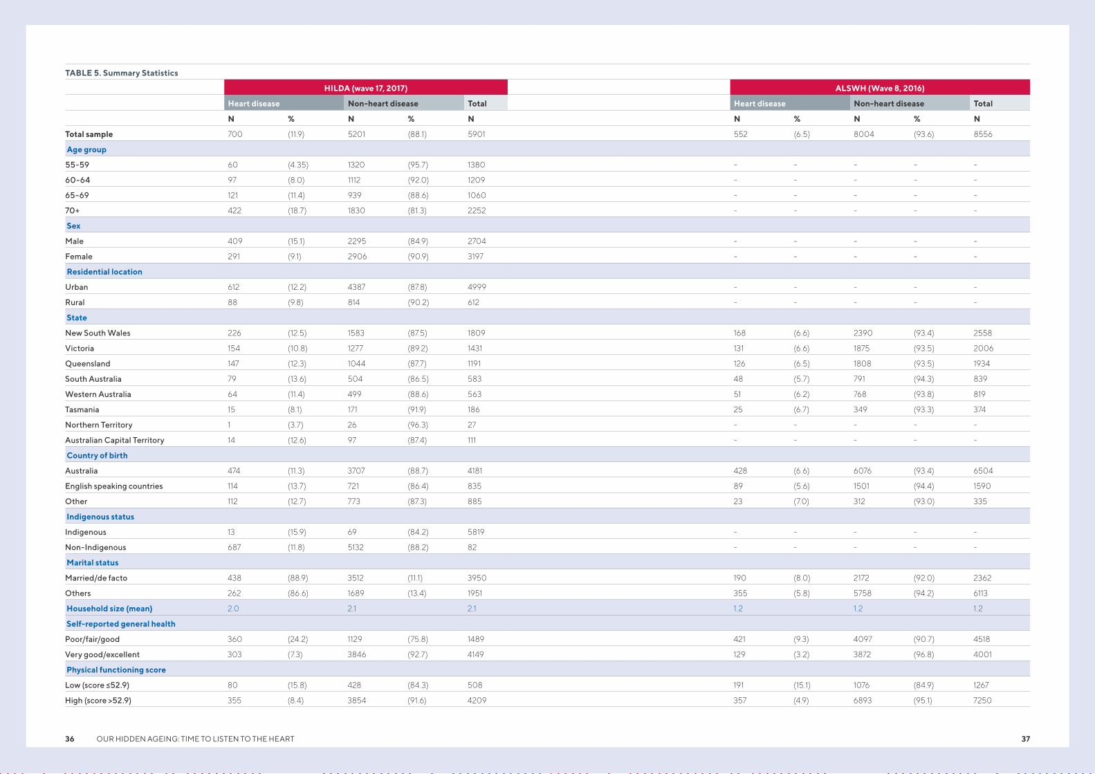
TABLE 5. Summary Statistics
HILDA (wave 17, 2017) ALSWH (Wave 8, 2016)
Heart disease Non-heart disease Total Heart disease Non-heart disease Total
N % N % N N % N % N
Total sample 700 (11.9) 5201 (88.1) 5901 552 (6.5) 8004 (93.6) 8556
Age group
55-59 60 (4.35) 1320 (95.7) 1380 - - - - -
60-64 97 (8.0) 1112 (92.0) 1209 - - - - -
65-69 121 (11.4) 939 (88.6) 1060 - - - - -
70+ 422 (18.7) 1830 (81.3) 2252 - - - - -
Sex
Male 409 (15.1) 2295 (84.9) 2704 - - - - -
Female 291 (9.1) 2906 (90.9) 3197 - - - - -
Residential location
Urban 612 (12.2) 4387 (87.8) 4999 - - - - -
Rural 88 (9.8) 814 (90.2) 612 - - - - -
State
New South Wales 226 (12.5) 1583 (87.5) 1809 168 (6.6) 2390 (93.4) 2558
Victoria 154 (10.8) 1277 (89.2) 1431 131 (6.6) 1875 (93.5) 2006
Queensland 147 (12.3) 1044 (87.7) 1191 126 (6.5) 1808 (93.5) 1934
South Australia 79 (13.6) 504 (86.5) 583 48 (5.7) 791 (94.3) 839
Western Australia 64 (11.4) 499 (88.6) 563 51 (6.2) 768 (93.8) 819
Tasmania 15 (8.1) 171 (91.9) 186 25 (6.7) 349 (93.3) 374
Northern Territory 1 (3.7) 26 (96.3) 27 - - - - -
Australian Capital Territory 14 (12.6) 97 (87.4) 111 - - - - -
Country of birth
Australia 474 (11.3) 3707 (88.7) 4181 428 (6.6) 6076 (93.4) 6504
English speaking countries 114 (13.7) 721 (86.4) 835 89 (5.6) 1501 (94.4) 1590
Other 112 (12.7) 773 (87.3) 885 23 (7.0) 312 (93.0) 335
Indigenous status
Indigenous 13 (15.9) 69 (84.2) 5819 - - - - -
Non-Indigenous 687 (11.8) 5132 (88.2) 82 - - - - -
Marital status
Married/de facto 438 (88.9) 3512 (11.1) 3950 190 (8.0) 2172 (92.0) 2362
Others 262 (86.6) 1689 (13.4) 1951 355 (5.8) 5758 (94.2) 6113
Household size (mean) 2.0 2.1 2.1 1.2 1.2 1.2
Self-reported general health
Poor/fair/good 360 (24.2) 1129 (75.8) 1489 421 (9.3) 4097 (90.7) 4518
Very good/excellent 303 (7.3) 3846 (92.7) 4149 129 (3.2) 3872 (96.8) 4001
Physical functioning score
Low (score ≤52.9) 80 (15.8) 428 (84.3) 508 191 (15.1) 1076 (84.9) 1267
High (score >52.9) 355 (8.4) 3854 (91.6) 4209 357 (4.9) 6893 (95.1) 7250
OUR HIDDEN AGEING: TIME TO LISTEN TO THE HEART 3736
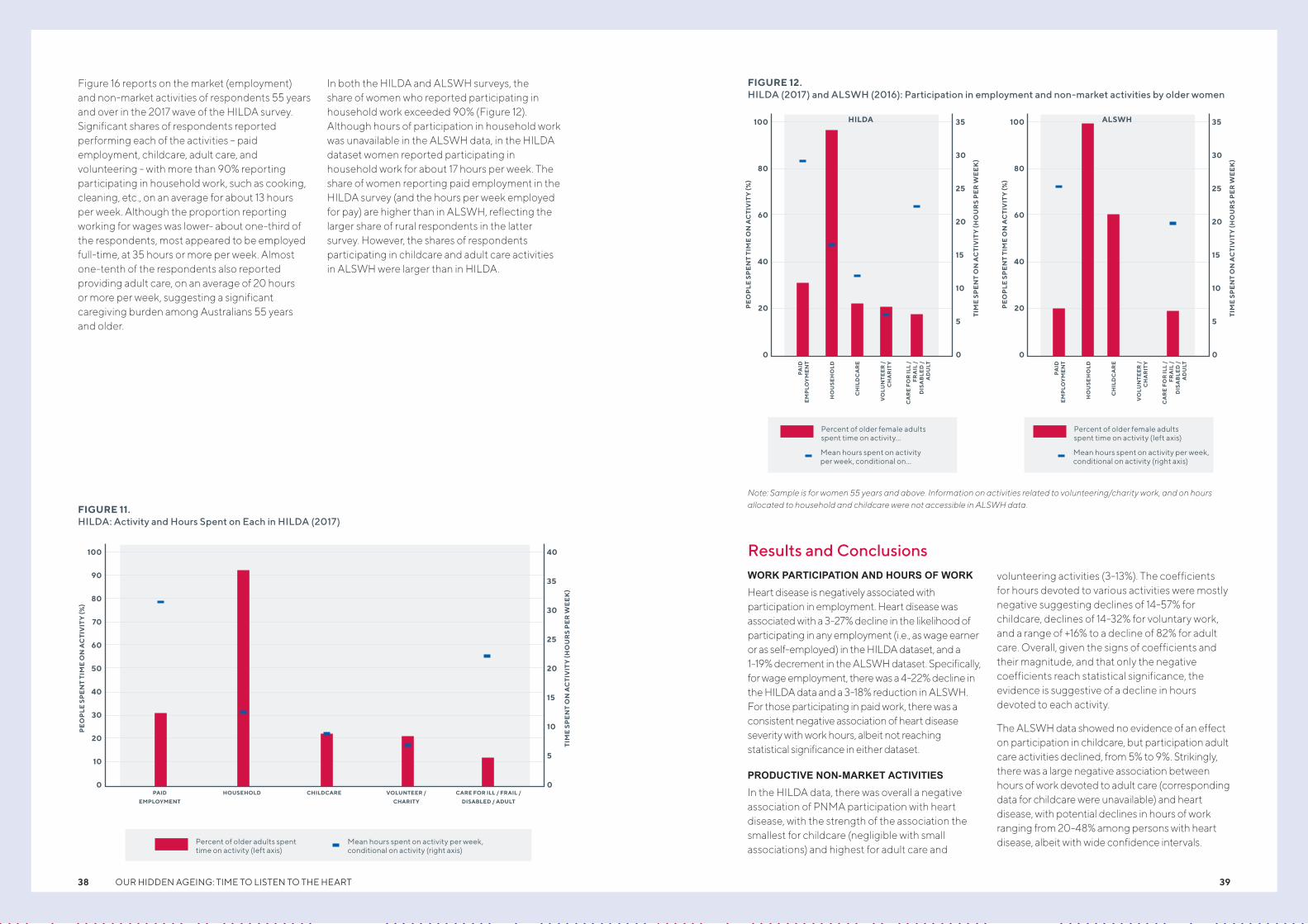
Figure 16 reports on the market (employment) and non-market activities of respondents 55 years and over in the 2017 wave of the HILDA survey. Significant shares of respondents reported performing each of the activities – paid employment, childcare, adult care, and volunteering - with more than 90% reporting participating in household work, such as cooking, cleaning, etc., on an average for about 13 hours per week. Although the proportion reporting working for wages was lower- about one-third of the respondents, most appeared to be employed full-time, at 35 hours or more per week. Almost one-tenth of the respondents also reported providing adult care, on an average of 20 hours or more per week, suggesting a significant caregiving burden among Australians 55 years and older.
In both the HILDA and ALSWH surveys, the share of women who reported participating in household work exceeded 90% (Figure 12). Although hours of participation in household work was unavailable in the ALSWH data, in the HILDA dataset women reported participating in household work for about 17 hours per week. The share of women reporting paid employment in the HILDA survey (and the hours per week employed for pay) are higher than in ALSWH, reflecting the larger share of rural respondents in the latter survey. However, the shares of respondents participating in childcare and adult care activities in ALSWH were larger than in HILDA.
Results and ConclusionsWORK PARTICIPATION AND HOURS OF WORK
Heart disease is negatively associated with participation in employment. Heart disease was associated with a 3-27% decline in the likelihood of participating in any employment (i.e., as wage earner or as self-employed) in the HILDA dataset, and a 1-19% decrement in the ALSWH dataset. Specifically, for wage employment, there was a 4-22% decline in the HILDA data and a 3-18% reduction in ALSWH. For those participating in paid work, there was a consistent negative association of heart disease severity with work hours, albeit not reaching statistical significance in either dataset.
PRODUCTIVE NON-MARKET ACTIVITIES
In the HILDA data, there was overall a negative association of PNMA participation with heart disease, with the strength of the association the smallest for childcare (negligible with small associations) and highest for adult care and
volunteering activities (3-13%). The coefficients for hours devoted to various activities were mostly negative suggesting declines of 14-57% for childcare, declines of 14-32% for voluntary work, and a range of +16% to a decline of 82% for adult care. Overall, given the signs of coefficients and their magnitude, and that only the negative coefficients reach statistical significance, the evidence is suggestive of a decline in hours devoted to each activity.
The ALSWH data showed no evidence of an effect on participation in childcare, but participation adult care activities declined, from 5% to 9%. Strikingly, there was a large negative association between hours of work devoted to adult care (corresponding data for childcare were unavailable) and heart disease, with potential declines in hours of work ranging from 20-48% among persons with heart disease, albeit with wide confidence intervals.
Note: Sample is for women 55 years and above. Information on activities related to volunteering/charity work, and on hours allocated to household and childcare were not accessible in ALSWH data.
Percent of older adults spent time on activity (left axis)
Percent of older female adults spent time on activity…
Percent of older female adults spent time on activity (left axis)
FIGURE 12. HILDA (2017) and ALSWH (2016): Participation in employment and non-market activities by older women
0
0 0
0
0 0
20
20 20
10
10 10
105
5 5
40
40 40
20
20 20
30
15
15 15
60
60 60
30
30 30
50
25
25 25
100
100 100
40
35 35
90
80
80 80
70
35
PE
OP
LE
SP
EN
T T
IME
ON
AC
TIV
ITY
(%)
PE
OP
LE
SP
EN
T T
IME
ON
AC
TIV
ITY
(%)
PE
OP
LE
SP
EN
T T
IME
ON
AC
TIV
ITY
(%)
TIM
E S
PE
NT
ON
AC
TIV
ITY
(HO
UR
S P
ER
WE
EK
)
TIM
E S
PE
NT
ON
AC
TIV
ITY
(HO
UR
S P
ER
WE
EK
)
TIM
E S
PE
NT
ON
AC
TIV
ITY
(HO
UR
S P
ER
WE
EK
)
PAID
EMPLOYMENT
HOUSEHOLD CHILDCARE VOLUNTEER /
CHARITY
CARE FOR ILL / FRAIL /
DISABLED / ADULTP
AID
E
MP
LOY
ME
NT
HO
US
EH
OL
D
CH
ILD
CA
RE
VO
LUN
TE
ER
/
CH
AR
ITY
CA
RE
FO
R IL
L /
FR
AIL
/
DIS
AB
LE
D /
A
DU
LT
PA
ID
EM
PLO
YM
EN
T
HO
US
EH
OL
D
CH
ILD
CA
RE
VO
LUN
TE
ER
/
CH
AR
ITY
CA
RE
FO
R IL
L /
FR
AIL
/
DIS
AB
LE
D /
A
DU
LT
FIGURE 11. HILDA: Activity and Hours Spent on Each in HILDA (2017)
Mean hours spent on activity per week, conditional on activity (right axis)
Mean hours spent on activity per week, conditional on…
Mean hours spent on activity per week, conditional on activity (right axis)
OUR HIDDEN AGEING: TIME TO LISTEN TO THE HEART 3938
HILDA ALSWH
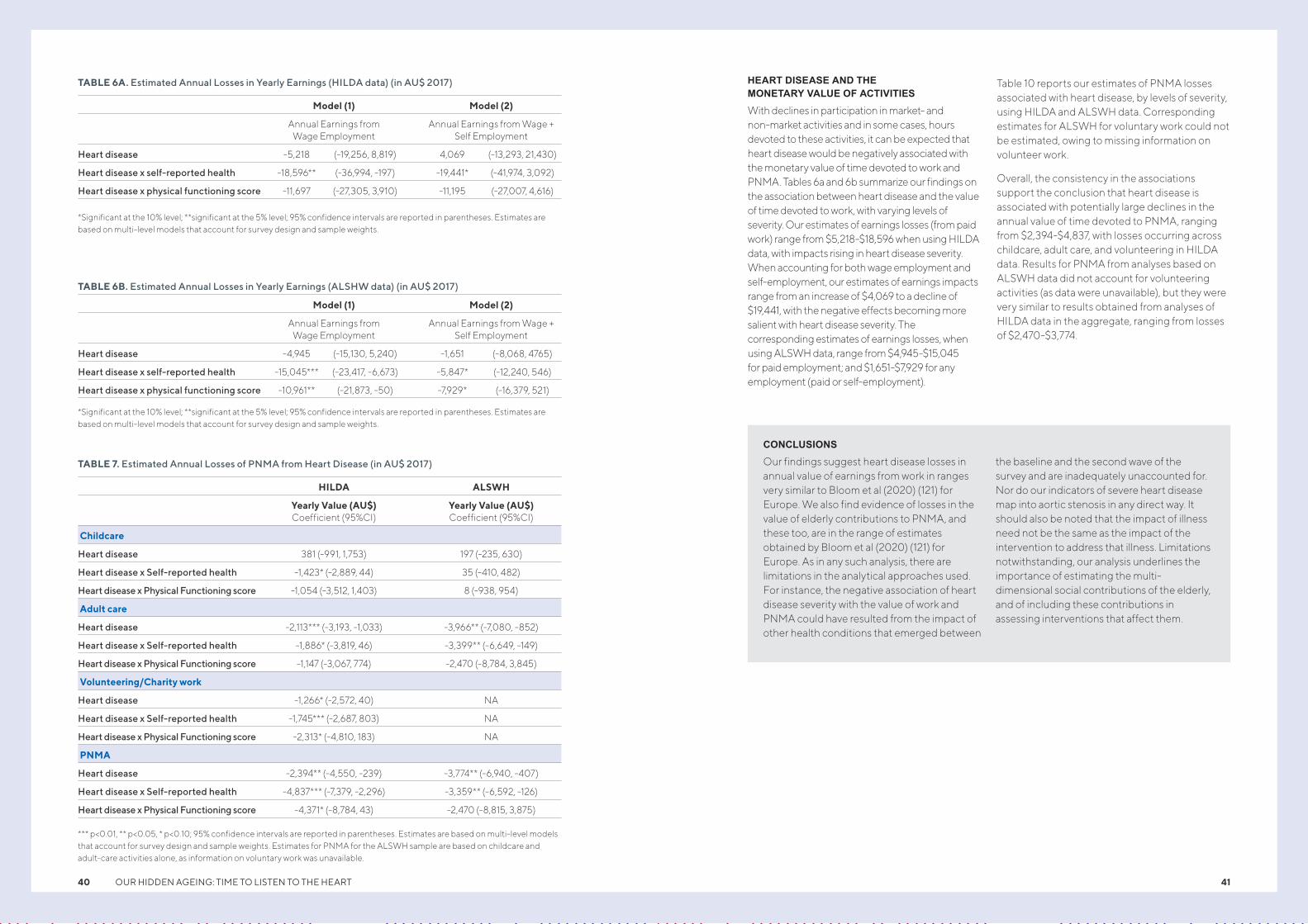
HEART DISEASE AND THE MONETARY VALUE OF ACTIVITIES
With declines in participation in market- and non-market activities and in some cases, hours devoted to these activities, it can be expected that heart disease would be negatively associated with the monetary value of time devoted to work and PNMA. Tables 6a and 6b summarize our findings on the association between heart disease and the value of time devoted to work, with varying levels of severity. Our estimates of earnings losses (from paid work) range from $5,218-$18,596 when using HILDA data, with impacts rising in heart disease severity. When accounting for both wage employment and self-employment, our estimates of earnings impacts range from an increase of $4,069 to a decline of $19,441, with the negative effects becoming more salient with heart disease severity. The corresponding estimates of earnings losses, when using ALSWH data, range from $4,945-$15,045 for paid employment; and $1,651-$7,929 for any employment (paid or self-employment).
Table 10 reports our estimates of PNMA losses associated with heart disease, by levels of severity, using HILDA and ALSWH data. Corresponding estimates for ALSWH for voluntary work could not be estimated, owing to missing information on volunteer work.
Overall, the consistency in the associations support the conclusion that heart disease is associated with potentially large declines in the annual value of time devoted to PNMA, ranging from $2,394-$4,837, with losses occurring across childcare, adult care, and volunteering in HILDA data. Results for PNMA from analyses based on ALSWH data did not account for volunteering activities (as data were unavailable), but they were very similar to results obtained from analyses of HILDA data in the aggregate, ranging from losses of $2,470-$3,774.
TABLE 7. Estimated Annual Losses of PNMA from Heart Disease (in AU$ 2017)
HILDA ALSWH
Yearly Value (AU$) Coefficient (95%CI)
Yearly Value (AU$) Coefficient (95%CI)
Childcare
Heart disease 381 (-991, 1,753) 197 (-235, 630)
Heart disease x Self-reported health -1,423* (-2,889, 44) 35 (-410, 482)
Heart disease x Physical Functioning score -1,054 (-3,512, 1,403) 8 (-938, 954)
Adult care
Heart disease -2,113*** (-3,193, -1,033) -3,966** (-7,080, -852)
Heart disease x Self-reported health -1,886* (-3,819, 46) -3,399** (-6,649, -149)
Heart disease x Physical Functioning score -1,147 (-3,067, 774) -2,470 (-8,784, 3,845)
Volunteering/Charity work
Heart disease -1,266* (-2,572, 40) NA
Heart disease x Self-reported health -1,745*** (-2,687, 803) NA
Heart disease x Physical Functioning score -2,313* (-4,810, 183) NA
PNMA
Heart disease -2,394** (-4,550, -239) -3,774** (-6,940, -407)
Heart disease x Self-reported health -4,837*** (-7,379, -2,296) -3,359** (-6,592, -126)
Heart disease x Physical Functioning score -4,371* (-8,784, 43) -2,470 (-8,815, 3,875)
*** p<0.01, ** p<0.05, * p<0.10; 95% confidence intervals are reported in parentheses. Estimates are based on multi-level models that account for survey design and sample weights. Estimates for PNMA for the ALSWH sample are based on childcare and adult-care activities alone, as information on voluntary work was unavailable.
CONCLUSIONS
Our findings suggest heart disease losses in annual value of earnings from work in ranges very similar to Bloom et al (2020) (121) for Europe. We also find evidence of losses in the value of elderly contributions to PNMA, and these too, are in the range of estimates obtained by Bloom et al (2020) (121) for Europe. As in any such analysis, there are limitations in the analytical approaches used. For instance, the negative association of heart disease severity with the value of work and PNMA could have resulted from the impact of other health conditions that emerged between
the baseline and the second wave of the survey and are inadequately unaccounted for. Nor do our indicators of severe heart disease map into aortic stenosis in any direct way. It should also be noted that the impact of illness need not be the same as the impact of the intervention to address that illness. Limitations notwithstanding, our analysis underlines the importance of estimating the multi-dimensional social contributions of the elderly, and of including these contributions in assessing interventions that affect them.
TABLE 6B. Estimated Annual Losses in Yearly Earnings (ALSHW data) (in AU$ 2017)
Model (1) Model (2)
Annual Earnings from
Wage Employment Annual Earnings from Wage +
Self Employment
Heart disease -4,945 (-15,130, 5,240) -1,651 (-8,068, 4765)
Heart disease x self-reported health -15,045*** (-23,417, -6,673) -5,847* (-12,240, 546)
Heart disease x physical functioning score -10,961** (-21,873, -50) -7,929* (-16,379, 521)
*Significant at the 10% level; **significant at the 5% level; 95% confidence intervals are reported in parentheses. Estimates are based on multi-level models that account for survey design and sample weights.
TABLE 6A. Estimated Annual Losses in Yearly Earnings (HILDA data) (in AU$ 2017)
Model (1) Model (2)
Annual Earnings from
Wage Employment Annual Earnings from Wage +
Self Employment
Heart disease -5,218 (-19,256, 8,819) 4,069 (-13,293, 21,430)
Heart disease x self-reported health -18,596** (-36,994, -197) -19,441* (-41,974, 3,092)
Heart disease x physical functioning score -11,697 (-27,305, 3,910) -11,195 (-27,007, 4,616)
*Significant at the 10% level; **significant at the 5% level; 95% confidence intervals are reported in parentheses. Estimates are based on multi-level models that account for survey design and sample weights.
OUR HIDDEN AGEING: TIME TO LISTEN TO THE HEART 4140
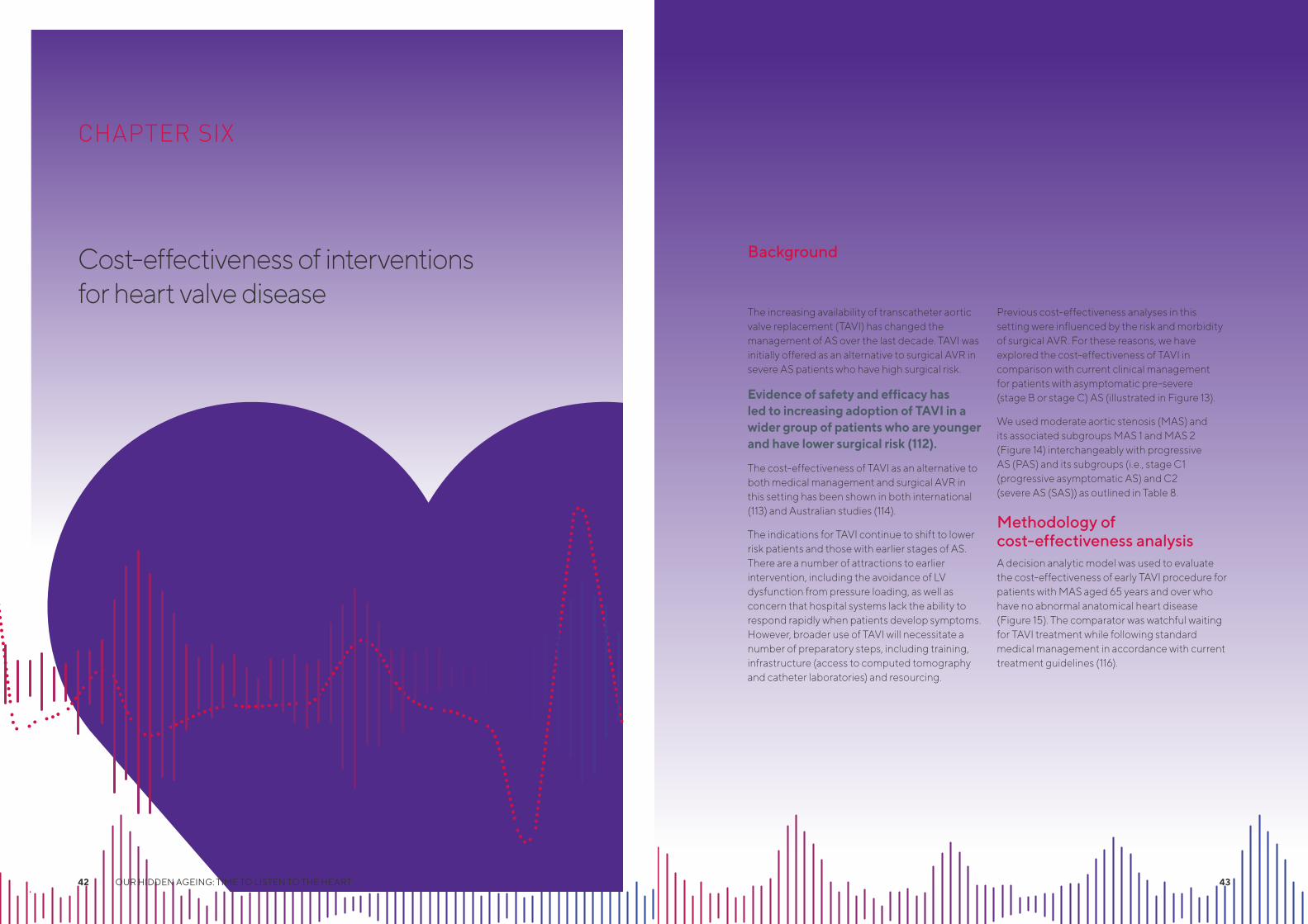
Cost-effectiveness of interventions for heart valve disease
Background
CHAPTER SIX
The increasing availability of transcatheter aortic valve replacement (TAVI) has changed the management of AS over the last decade. TAVI was initially offered as an alternative to surgical AVR in severe AS patients who have high surgical risk.
Evidence of safety and efficacy has led to increasing adoption of TAVI in a wider group of patients who are younger and have lower surgical risk (112).
The cost-effectiveness of TAVI as an alternative to both medical management and surgical AVR in this setting has been shown in both international (113) and Australian studies (114).
The indications for TAVI continue to shift to lower risk patients and those with earlier stages of AS. There are a number of attractions to earlier intervention, including the avoidance of LV dysfunction from pressure loading, as well as concern that hospital systems lack the ability to respond rapidly when patients develop symptoms. However, broader use of TAVI will necessitate a number of preparatory steps, including training, infrastructure (access to computed tomography and catheter laboratories) and resourcing.
Previous cost-effectiveness analyses in this setting were influenced by the risk and morbidity of surgical AVR. For these reasons, we have explored the cost-effectiveness of TAVI in comparison with current clinical management for patients with asymptomatic pre-severe (stage B or stage C) AS (illustrated in Figure 13).
We used moderate aortic stenosis (MAS) and its associated subgroups MAS 1 and MAS 2 (Figure 14) interchangeably with progressive AS (PAS) and its subgroups (i.e., stage C1 (progressive asymptomatic AS) and C2 (severe AS (SAS)) as outlined in Table 8.
Methodology of cost-effectiveness analysisA decision analytic model was used to evaluate the cost-effectiveness of early TAVI procedure for patients with MAS aged 65 years and over who have no abnormal anatomical heart disease (Figure 15). The comparator was watchful waiting for TAVI treatment while following standard medical management in accordance with current treatment guidelines (116).
OUR HIDDEN AGEING: TIME TO LISTEN TO THE HEART 42 43
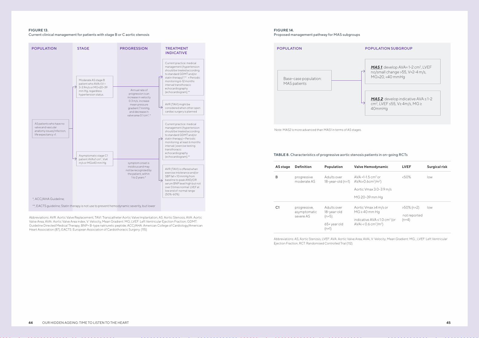
FIGURE 13. Current clinical management for patients with stage B or C aortic stenosis
FIGURE 14. Proposed management pathway for MAS subgroups
Abbreviations: AVR: Aortic Valve Replacement; TAVI: Transcatheter Aortic Valve Implantation; AS: Aortic Stenosis; AVA: Aortic Valve Area; AVAi: Aortic Valve Area index; V: Velocity, Mean Gradient: MG; LVEF: Left Ventricular Ejection Fraction; GDMT: Guideline Directed Medical Therapy; BNP= B-type natriuretic peptide; ACC/AHA: American College of Cardiology/American Heart Association (87); EACTS: European Association of Cardiothoracic Surgery. (115)
Note: MAS2 is more advanced than MAS1 in terms of AS stages.
TABLE 8. Characteristics of progressive aortic stenosis patients in on-going RCTs
Abbreviations: AS; Aortic Stenosis, LVEF: AVA: Aortic Valve Area; AVAi, V: Velocity; Mean Gradient: MG, ; LVEF: Left Ventricular Ejection Fraction, RCT: Randomised Controlled Trial (112).
AS stage Definition Population Valve Hemodynamic LVEF Surgical risk
B progressive moderate AS
Adults over 18-year-old (n=1)
AVA =1-1.5 cm2 or AVAi>0.6cm2/m2)
Aortic Vmax 3.0–3.9 m/s
MG 20–39 mm Hg
<50% low
C1 progressive, asymptomatic severe AS
Adults over 18-year-old (n=5)
65+ year old (n=1)
Aortic Vmax >=4 m/s or MG >= 40 mm Hg
indicative AVA =< 1.0 cm2 (or AVAi < 0.6 cm2/m2)
>50% (n=2)
not reported (n=4)
low
TREATMENT INDICATIVE
PROGRESSION POPULATION SUBGROUPSTAGEPOPULATION POPULATION
Annual rate of progression is an
increase in velocity 0.3 m/s, increase
mean pressure gradient 7 mmHg,
and decrease in valve area 0.1 cm2, *
AS patients who have no valve and vascular anatomy issues/infection, life expectancy >1.
Base-case population: MAS patients
MAS 1: develop AVA= 1-2 cm2, LVEF no/small change >55, V=2-4 m/s, MG>20, <40 mmHg
MAS 2: develop indicative AVA =< 1-2 cm2, LVEF =<55, V>= 4m/s, MG >= 40mmHg
*, ACC/AHA Guideline;
**, EACTS guideline; Statin therapy is not use to prevent hemodynamic severity, but lower
Moderate AS stage B patient who AVA>1,V = 3-3.9m/s or MG=20–39 mm Hg, regardless hypertension status.
Asymptomatic stage C1 patient AVA=<1 cm2 , V>=4 m/s or MG>=40 mm Hg symptom onset is
insidious and may not be recognized by
the patient, within 1 to 2 years *
Current practice: medical management (hypertension should be treated according to standard GDMT and/or statin therapy)*,** + Periodic monitoring 6-12 months interval transthoracic echocardiography (echocardiogram),**
AVR (TAVI) might be considered when other open cardiac surgery is planned
Current practice: medical management (hypertension should be treated according to standard GDMT and/or statin therapy) + Periodic monitoring at least 6 months interval ( exercise testing transthoracic echocardiography (echocardiogram),**
AVR (TAVI) is offered when exercise intolerance and/or SBP fall < 10 mmHg from baseline to peak AND/OR serum BNP level high but not over 3 times normal LVEF at low end of normal range (50%-60%)
OUR HIDDEN AGEING: TIME TO LISTEN TO THE HEART 4544
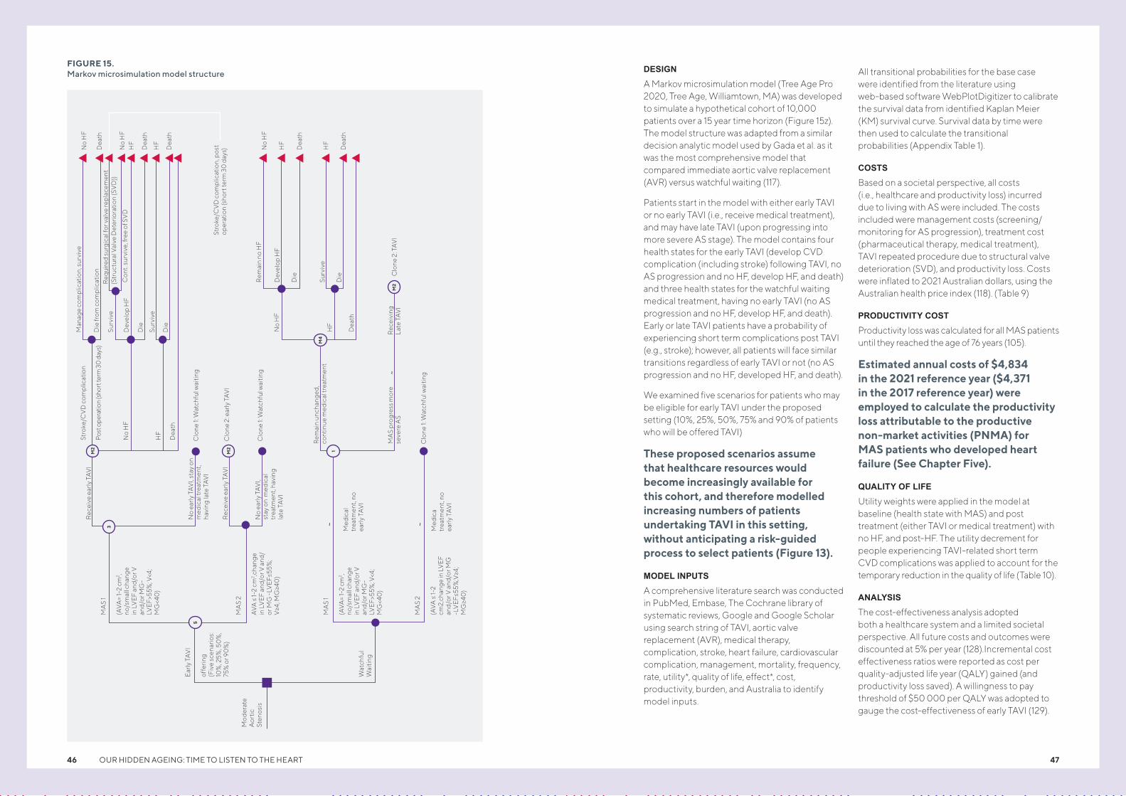
DESIGN
A Markov microsimulation model (Tree Age Pro 2020, Tree Age, Williamtown, MA) was developed to simulate a hypothetical cohort of 10,000 patients over a 15 year time horizon (Figure 15z). The model structure was adapted from a similar decision analytic model used by Gada et al. as it was the most comprehensive model that compared immediate aortic valve replacement (AVR) versus watchful waiting (117).
Patients start in the model with either early TAVI or no early TAVI (i.e., receive medical treatment), and may have late TAVI (upon progressing into more severe AS stage). The model contains four health states for the early TAVI (develop CVD complication (including stroke) following TAVI, no AS progression and no HF, develop HF, and death) and three health states for the watchful waiting medical treatment, having no early TAVI (no AS progression and no HF, develop HF, and death). Early or late TAVI patients have a probability of experiencing short term complications post TAVI (e.g., stroke); however, all patients will face similar transitions regardless of early TAVI or not (no AS progression and no HF, developed HF, and death).
We examined five scenarios for patients who may be eligible for early TAVI under the proposed setting (10%, 25%, 50%, 75% and 90% of patients who will be offered TAVI)
These proposed scenarios assume that healthcare resources would become increasingly available for this cohort, and therefore modelled increasing numbers of patients undertaking TAVI in this setting, without anticipating a risk-guided process to select patients (Figure 13).
MODEL INPUTS
A comprehensive literature search was conducted in PubMed, Embase, The Cochrane library of systematic reviews, Google and Google Scholar using search string of TAVI, aortic valve replacement (AVR), medical therapy, complication, stroke, heart failure, cardiovascular complication, management, mortality, frequency, rate, utility*, quality of life, effect*, cost, productivity, burden, and Australia to identify model inputs.
All transitional probabilities for the base case were identified from the literature using web-based software WebPlotDigitizer to calibrate the survival data from identified Kaplan Meier (KM) survival curve. Survival data by time were then used to calculate the transitional probabilities (Appendix Table 1).
COSTS
Based on a societal perspective, all costs (i.e., healthcare and productivity loss) incurred due to living with AS were included. The costs included were management costs (screening/monitoring for AS progression), treatment cost (pharmaceutical therapy, medical treatment), TAVI repeated procedure due to structural valve deterioration (SVD), and productivity loss. Costs were inflated to 2021 Australian dollars, using the Australian health price index (118). (Table 9)
PRODUCTIVITY COST
Productivity loss was calculated for all MAS patients until they reached the age of 76 years (105).
Estimated annual costs of $4,834 in the 2021 reference year ($4,371 in the 2017 reference year) were employed to calculate the productivity loss attributable to the productive non-market activities (PNMA) for MAS patients who developed heart failure (See Chapter Five).
QUALITY OF LIFE
Utility weights were applied in the model at baseline (health state with MAS) and post treatment (either TAVI or medical treatment) with no HF, and post-HF. The utility decrement for people experiencing TAVI-related short term CVD complications was applied to account for the temporary reduction in the quality of life (Table 10).
ANALYSIS
The cost-effectiveness analysis adopted both a healthcare system and a limited societal perspective. All future costs and outcomes were discounted at 5% per year (128).Incremental cost effectiveness ratios were reported as cost per quality-adjusted life year (QALY) gained (and productivity loss saved). A willingness to pay threshold of $50 000 per QALY was adopted to gauge the cost-effectiveness of early TAVI (129).
FIGURE 15. Markov microsimulation model structure
Mod
erat
e A
ortic
St
enos
is
Ear
ly T
AV
I
offe
ring
(F
ive
scen
ario
s:
10%
, 25%
, 50
%,
75%
or 9
0%
)
MA
S 1
(AV
A=
1-2
cm2 ,
no/s
mal
l cha
nge
in L
VE
F an
d/or
V
and/
or M
G-
LVE
F>55
%; V
<4;
MG
<40
)
Rec
eive
ear
ly T
AV
ISt
roke
/CV
D c
omp
licat
ion
Man
age
com
plic
atio
n, s
urvi
ve
Die
from
com
plic
atio
nR
equi
red
surg
ical
for v
alve
rep
lace
men
t
No
HF
No
HF
No
HF
HF
HF
HF
Dea
th
Dea
th
Dea
th
Dea
th
HF
Dea
th
Stro
ke/C
VD
com
plic
atio
n, p
ost
oper
atio
n (s
hort
term
30
day
s)
(Str
uctu
ral V
alve
Det
erio
ratio
n (S
VD
))
Con
t. su
rviv
e, fr
ee o
f SV
D
Rem
ain
no H
F
Dev
elop
HF
Die
Die C
lone
2: T
AV
I
Clo
ne 1:
Wat
chfu
l wai
ting
Clo
ne 2
: ear
ly T
AV
I
Clo
ne 1:
Wat
chfu
l wai
ting
Surv
ive
Surv
ive
Dev
elop
HF
Die
Die
No
HF
HF
Dea
th
Rec
eivi
ng
Late
TA
VI
Surv
ive
Pos
t ope
ratio
n (s
hort
term
30
day
s)
No
HF
HF
Dea
th
Rem
ain
unch
ange
d,
cont
inue
med
ical
trea
tmen
t
MA
S p
rogr
ess
mor
e se
vere
AS
~
~ ~C
lone
1: W
atch
ful w
aitin
g
No
early
TA
VI,
stay
on
med
ical
trea
tmen
t, ha
ving
late
TA
VI
Rec
eive
ear
ly T
AV
I
No
early
TA
VI,
stay
on
med
ical
tr
eatm
ent,
havi
ng
late
TA
VI
Med
ical
tr
eatm
ent,
no
early
TA
VI
Med
ica
trea
tmen
t, no
ea
rly T
AV
I
MA
S 1
(AV
A=
1-2
cm2 ,
no/s
mal
l cha
nge
in L
VE
F an
d/or
V
and/
or M
G-
LVE
F>55
%; V
<4;
MG
<40
)
MA
S 2
AV
A =<
1-2
cm2 ,c
hang
e in
LV
EF
and/
or V
and
/or
MG
-LV
EF
=<55%
; V
>=4; M
G>=4
0)
MA
S 2
(AV
A =<
1-2
cm2,
chan
ge in
LV
EF
and/
or V
and
/or M
G
-LV
EF
=<55%
;V>=4
; M
G>=4
0)
Wat
chfu
l W
aitin
g
1M2
M2
3
5
M4
M2
OUR HIDDEN AGEING: TIME TO LISTEN TO THE HEART 4746
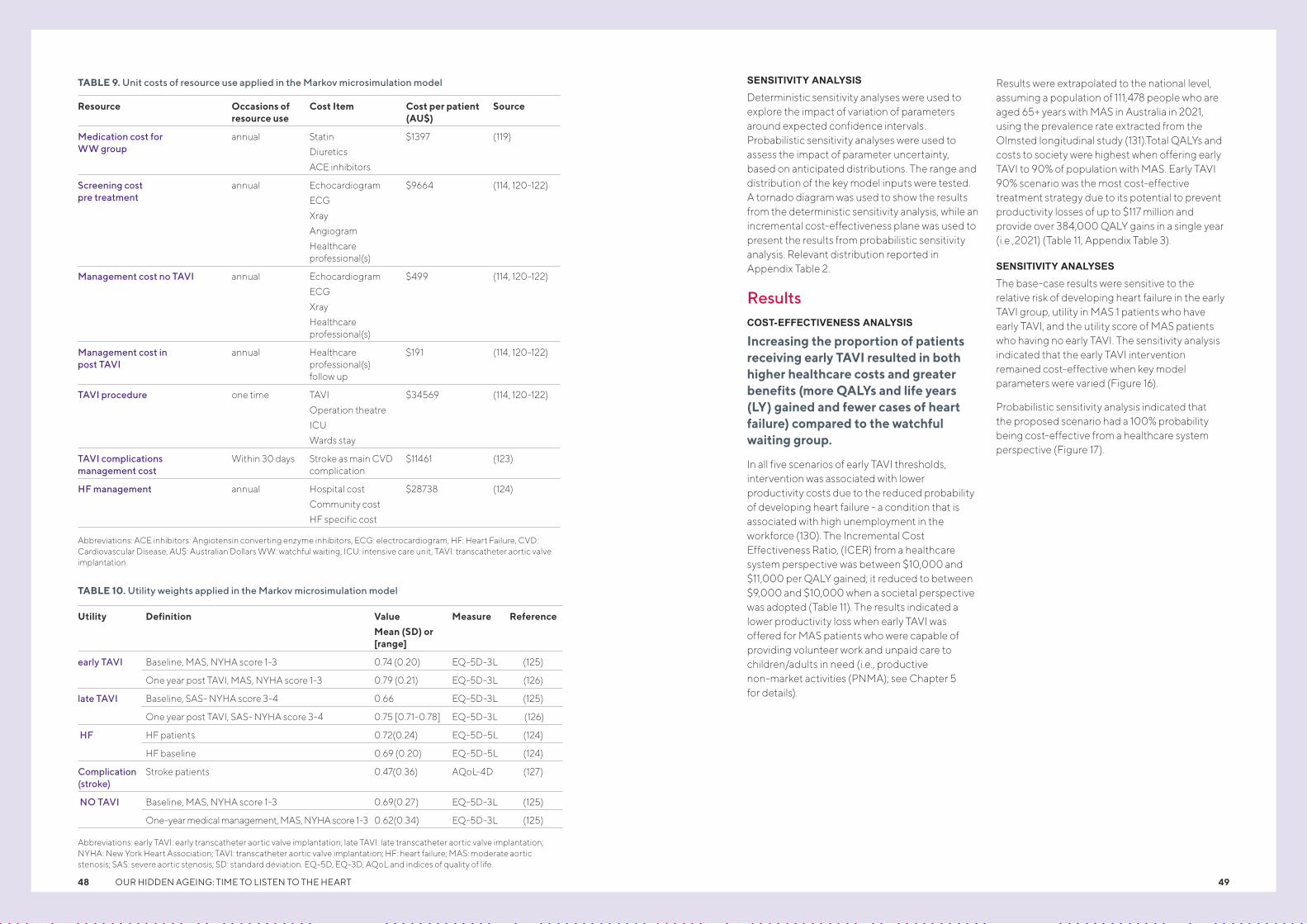
SENSITIVITY ANALYSIS
Deterministic sensitivity analyses were used to explore the impact of variation of parameters around expected confidence intervals. Probabilistic sensitivity analyses were used to assess the impact of parameter uncertainty, based on anticipated distributions. The range and distribution of the key model inputs were tested. A tornado diagram was used to show the results from the deterministic sensitivity analysis, while an incremental cost-effectiveness plane was used to present the results from probabilistic sensitivity analysis. Relevant distribution reported in Appendix Table 2.
ResultsCOST-EFFECTIVENESS ANALYSIS
Increasing the proportion of patients receiving early TAVI resulted in both higher healthcare costs and greater benefits (more QALYs and life years (LY) gained and fewer cases of heart failure) compared to the watchful waiting group.
In all five scenarios of early TAVI thresholds, intervention was associated with lower productivity costs due to the reduced probability of developing heart failure - a condition that is associated with high unemployment in the workforce (130). The Incremental Cost Effectiveness Ratio, (ICER) from a healthcare system perspective was between $10,000 and $11,000 per QALY gained; it reduced to between $9,000 and $10,000 when a societal perspective was adopted (Table 11). The results indicated a lower productivity loss when early TAVI was offered for MAS patients who were capable of providing volunteer work and unpaid care to children/adults in need (i.e., productive non-market activities (PNMA); see Chapter 5 for details).
Results were extrapolated to the national level, assuming a population of 111,478 people who are aged 65+ years with MAS in Australia in 2021, using the prevalence rate extracted from the Olmsted longitudinal study (131).Total QALYs and costs to society were highest when offering early TAVI to 90% of population with MAS. Early TAVI 90% scenario was the most cost-effective treatment strategy due to its potential to prevent productivity losses of up to $117 million and provide over 384,000 QALY gains in a single year (i.e.,2021) (Table 11, Appendix Table 3).
SENSITIVITY ANALYSES
The base-case results were sensitive to the relative risk of developing heart failure in the early TAVI group, utility in MAS 1 patients who have early TAVI, and the utility score of MAS patients who having no early TAVI. The sensitivity analysis indicated that the early TAVI intervention remained cost-effective when key model parameters were varied (Figure 16).
Probabilistic sensitivity analysis indicated that the proposed scenario had a 100% probability being cost-effective from a healthcare system perspective (Figure 17).
TABLE 9. Unit costs of resource use applied in the Markov microsimulation model
TABLE 10. Utility weights applied in the Markov microsimulation model
Abbreviations: ACE inhibitors: Angiotensin converting enzyme inhibitors, ECG: electrocardiogram, HF: Heart Failure, CVD: Cardiovascular Disease, AU$: Australian Dollars WW: watchful waiting, ICU: intensive care unit, TAVI: transcatheter aortic valve implantation.
Abbreviations: early TAVI: early transcatheter aortic valve implantation; late TAVI: late transcatheter aortic valve implantation; NYHA: New York Heart Association; TAVI: transcatheter aortic valve implantation; HF: heart failure; MAS: moderate aortic stenosis; SAS: severe aortic stenosis; SD: standard deviation. EQ-5D, EQ-3D, AQoL and indices of quality of life.
Resource Occasions of resource use
Cost Item Cost per patient (AU$)
Source
Medication cost for WW group
annual Statin
Diuretics
ACE inhibitors
$1397 (119)
Screening cost pre treatment
annual Echocardiogram
ECG
Xray
Angiogram
Healthcare professional(s)
$9664 (114, 120-122)
Management cost no TAVI annual Echocardiogram
ECG
Xray
Healthcare professional(s)
$499 (114, 120-122)
Management cost in post TAVI
annual Healthcare professional(s) follow up
$191 (114, 120-122)
TAVI procedure one time TAVI
Operation theatre
ICU
Wards stay
$34569 (114, 120-122)
TAVI complications management cost
Within 30 days Stroke as main CVD complication
$11461 (123)
HF management annual Hospital cost
Community cost
HF specific cost
$28738 (124)
Utility Definition Value
Mean (SD) or [range]
Measure Reference
early TAVI Baseline, MAS, NYHA score 1-3 0.74 (0.20) EQ-5D-3L (125)
One year post TAVI, MAS, NYHA score 1-3 0.79 (0.21) EQ-5D-3L (126)
late TAVI Baseline, SAS- NYHA score 3-4 0.66 EQ-5D-3L (125)
One year post TAVI, SAS- NYHA score 3-4 0.75 [0.71-0.78] EQ-5D-3L (126)
HF HF patients 0.72(0.24) EQ-5D-5L (124)
HF baseline 0.69 (0.20) EQ-5D-5L (124)
Complication (stroke)
Stroke patients 0.47(0.36) AQoL-4D (127)
NO TAVI Baseline, MAS, NYHA score 1-3 0.69(0.27) EQ-5D-3L (125)
One-year medical management, MAS, NYHA score 1-3 0.62(0.34) EQ-5D-3L (125)
OUR HIDDEN AGEING: TIME TO LISTEN TO THE HEART 4948
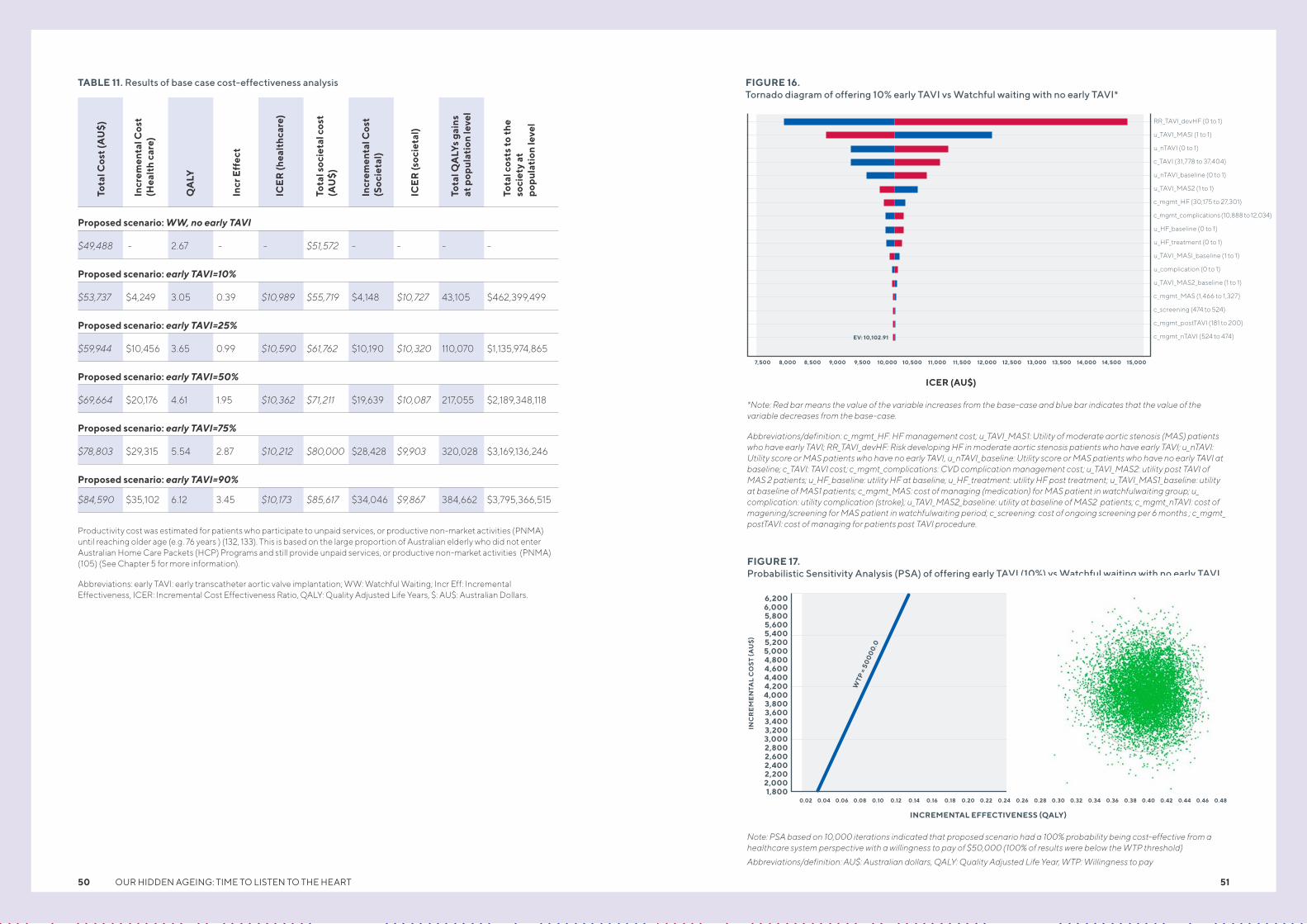
FIGURE 16. Tornado diagram of offering 10% early TAVI vs Watchful waiting with no early TAVI*
FIGURE 17. Probabilistic Sensitivity Analysis (PSA) of offering early TAVI (10%) vs Watchful waiting with no early TAVI
*Note: Red bar means the value of the variable increases from the base-case and blue bar indicates that the value of the variable decreases from the base-case.
Abbreviations/definition: c_mgmt_HF: HF management cost; u_TAVI_MAS1: Utility of moderate aortic stenosis (MAS) patients who have early TAVI; RR_TAVI_devHF: Risk developing HF in moderate aortic stenosis patients who have early TAVI; u_nTAVI: Utility score or MAS patients who have no early TAVI, u_nTAVI_baseline: Utility score or MAS patients who have no early TAVI at baseline; c_TAVI: TAVI cost; c_mgmt_complications: CVD complication management cost; u_TAVI_MAS2: utility post TAVI of MAS 2 patients; u_HF_baseline: utility HF at baseline; u_HF_treatment: utility HF post treatment; u_TAVI_MAS1_baseline: utility at baseline of MAS1 patients; c_mgmt_MAS: cost of managing (medication) for MAS patient in watchfulwaiting group; u_complication: utility complication (stroke); u_TAVI_MAS2_baseline: utility at baseline of MAS2 patients; c_mgmt_nTAVI: cost of magening/screening for MAS patient in watchfulwaiting period; c_screening: cost of ongoing screening per 6 months ; c_mgmt_postTAVI: cost of managing for patients post TAVI procedure.
Note: PSA based on 10,000 iterations indicated that proposed scenario had a 100% probability being cost-effective from a healthcare system perspective with a willingness to pay of $50,000 (100% of results were below the WTP threshold)
Abbreviations/definition: AU$: Australian dollars, QALY: Quality Adjusted Life Year, WTP: Willingness to pay
TABLE 11. Results of base case cost-effectiveness analysis
Proposed scenario: WW, no early TAVI
$49,488 - 2.67 - - $51,572 - - - -
Proposed scenario: early TAVI=10%
$53,737 $4,249 3.05 0.39 $10,989 $55,719 $4,148 $10,727 43,105 $462,399,499
Proposed scenario: early TAVI=25%
$59,944 $10,456 3.65 0.99 $10,590 $61,762 $10,190 $10,320 110,070 $1,135,974,865
Proposed scenario: early TAVI=50%
$69,664 $20,176 4.61 1.95 $10,362 $71,211 $19,639 $10,087 217,055 $2,189,348,118
Proposed scenario: early TAVI=75%
$78,803 $29,315 5.54 2.87 $10,212 $80,000 $28,428 $9,903 320,028 $3,169,136,246
Proposed scenario: early TAVI=90%
$84,590 $35,102 6.12 3.45 $10,173 $85,617 $34,046 $9,867 384,662 $3,795,366,515
Tota
l Cos
t (A
U$)
Incr
emen
tal C
ost
(Hea
lth
care
)
QA
LY
Incr
Eff
ect
ICE
R (h
ealt
hcar
e)
Tota
l soc
ieta
l cos
t (A
U$)
Incr
emen
tal C
ost
(Soc
ieta
l)
ICE
R (s
ocie
tal)
Tota
l QA
LYs
gain
s at
pop
ulat
ion
leve
l
Tota
l cos
ts to
the
soci
ety
at
pop
ulat
ion
leve
lProductivity cost was estimated for patients who participate to unpaid services, or productive non-market activities (PNMA) until reaching older age (e.g. 76 years ) (132, 133). This is based on the large proportion of Australian elderly who did not enter Australian Home Care Packets (HCP) Programs and still provide unpaid services, or productive non-market activities (PNMA) (105) (See Chapter 5 for more information).
Abbreviations: early TAVI: early transcatheter aortic valve implantation; WW: Watchful Waiting; Incr Eff: Incremental Effectiveness, ICER: Incremental Cost Effectiveness Ratio, QALY: Quality Adjusted Life Years, $: AU$: Australian Dollars.
RR_TAVl_devHF (0 to 1)
u_TAVI_MASl (1 to 1)
u_nTAVI (0 to 1)
c_TAVI (31,778 to 37,404)
u_nTAVl_baseline (0 to 1)
u_TAVI_MAS2 (1 to 1)
c_mgmt_HF (30,175 to 27,301)
c_mgmt_complications (10,888 to 12,034)
u_HF_baseline (0 to 1)
u_HF_treatment (0 to 1)
u_TAVI_MASl_baseline (1 to 1)
u_complication (0 to 1)
u_TAVI_MAS2_baseline (1 to 1)
c_mgmt_MAS (1,466 to 1,327)
c_screening (474 to 524)
c_mgmt_postTAVI (181 to 200)
c_mgmt_nTAVI (524 to 474)
7,500 8,000 8,500 9,000 9,500 10,000 10,500 11,000 11,500 12,000 12,500 13,000 13,500 14,000 14,500 15,000
0.02 0.04 0.06 0.08 0.10 0.12 0.14 0.16 0.18 0.20 0.22 0.24 0.26 0.28 0.30 0.32 0.34 0.36 0.38 0.40 0.42 0.44 0.46 0.48
ICER (AU$)
EV: 10,102.91
INCREMENTAL EFFECTIVENESS (QALY)
6,2006,0005,8005,6005,4005,2005,0004,8004,6004,4004,2004,0003,8003,6003,4003,2003,0002,8002,6002,4002,2002,0001,800
INC
RE
ME
NTA
L C
OS
T (A
U$
)
WTP
= 5
00
00
.0OUR HIDDEN AGEING: TIME TO LISTEN TO THE HEART 5150

Case studies
CHAPTER SEVENPhil HolmesHead of Insurance at O’Brien Glass Industries, Phil Holmes, 71, has a busy job that prior to COVID-19 involved travel every week.
He also works out five days a week at the gym. However, in 2019 when he started to feel a bit tired while undertaking a work fundraiser which required him to perform a certain number of push-ups in a month, he didn’t think for one minute it might be heart-related. “I thought it was just age and fatigue,” he says.
He did, however, do one of the best things that he could. He promptly sought advice from his GP and a cardiologist and it turns out that quick action could well have saved him from a potentially fatal heart attack.
Phil was diagnosed with aortic stenosis, requiring an aortic valve replacement. He was advised he had the option of the gold standard, open heart surgery (which would require several months of recovery), or the newer and minimally-invasive procedure known as Transcatheter Aortic Valve replacement (TAVI). Given his active lifestyle and that he still works full time, he elected to have the TAVI procedure. And he hasn’t looked back.
Within a few days the father of three and grandfather of five was back at work in Melbourne, and three weeks later he returned to the gym and cycling.
Gerlinde Binning Professional weaver, mother and grandmother, Gerlinde Binning, 77, enjoys an active lifestyle, walking and bike riding daily with her friends.
In March 2019, during her annual health check-up, Gerlinde’s GP identified a heart murmur, and subsequently advised her to undergo a heart health check every six months.
Later that year, on Boxing Day, she experienced difficulty breathing, and was rushed to hospital in Melbourne.
After spending time in the Intensive Care Unit, Gerlinde was referred to a heart specialist, who diagnosed her with heart valve disease.
Selecting a minimally invasive medical treatment option, Gerlinde opted to undergo a Transcatheter Aortic Valve Implantation (TAVI) procedure – where an artificial aortic valve is placed in the heart – in July 2020. Four days later, she had a pacemaker fitted to help maintain a healthy heart.
Following her procedure, Gerlinde took part in a cardiac health support program led by allied health professionals, involving education, exercise and rehabilitation. She is now back enjoying her active lifestyle again.
OUR HIDDEN AGEING: TIME TO LISTEN TO THE HEART 52 53
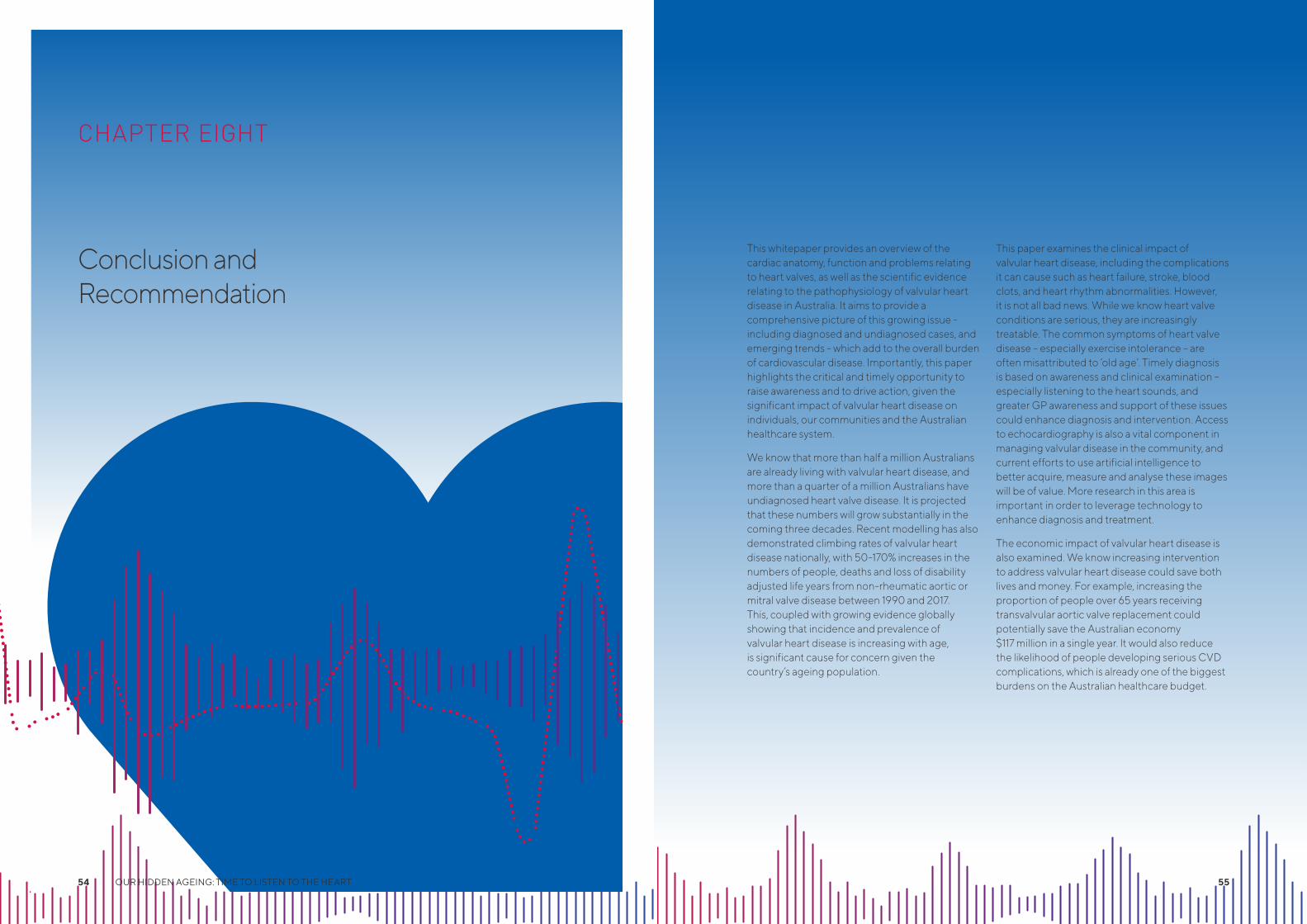
Conclusion and Recommendation
CHAPTER EIGHT
This whitepaper provides an overview of the cardiac anatomy, function and problems relating to heart valves, as well as the scientific evidence relating to the pathophysiology of valvular heart disease in Australia. It aims to provide a comprehensive picture of this growing issue - including diagnosed and undiagnosed cases, and emerging trends - which add to the overall burden of cardiovascular disease. Importantly, this paper highlights the critical and timely opportunity to raise awareness and to drive action, given the significant impact of valvular heart disease on individuals, our communities and the Australian healthcare system.
We know that more than half a million Australians are already living with valvular heart disease, and more than a quarter of a million Australians have undiagnosed heart valve disease. It is projected that these numbers will grow substantially in the coming three decades. Recent modelling has also demonstrated climbing rates of valvular heart disease nationally, with 50-170% increases in the numbers of people, deaths and loss of disability adjusted life years from non-rheumatic aortic or mitral valve disease between 1990 and 2017. This, coupled with growing evidence globally showing that incidence and prevalence of valvular heart disease is increasing with age, is significant cause for concern given the country’s ageing population.
This paper examines the clinical impact of valvular heart disease, including the complications it can cause such as heart failure, stroke, blood clots, and heart rhythm abnormalities. However, it is not all bad news. While we know heart valve conditions are serious, they are increasingly treatable. The common symptoms of heart valve disease – especially exercise intolerance – are often misattributed to ‘old age’. Timely diagnosis is based on awareness and clinical examination – especially listening to the heart sounds, and greater GP awareness and support of these issues could enhance diagnosis and intervention. Access to echocardiography is also a vital component in managing valvular disease in the community, and current efforts to use artificial intelligence to better acquire, measure and analyse these images will be of value. More research in this area is important in order to leverage technology to enhance diagnosis and treatment.
The economic impact of valvular heart disease is also examined. We know increasing intervention to address valvular heart disease could save both lives and money. For example, increasing the proportion of people over 65 years receiving transvalvular aortic valve replacement could potentially save the Australian economy $117 million in a single year. It would also reduce the likelihood of people developing serious CVD complications, which is already one of the biggest burdens on the Australian healthcare budget.
OUR HIDDEN AGEING: TIME TO LISTEN TO THE HEART 54 55
We know many people are worried about the health impacts of ageing but little is known in the wider community about vascular ageing. In many respects, vascular ageing is far more important than chronological age and is emerging as an important marker of cardiovascular disease risk. Vascular ageing refers to the deterioration in vascular structure and function over time, which
ultimately leads to end-organ damage in the heart, brain and kidney. Vascular ageing involves both arteriosclerosis and atherosclerosis. Arteriosclerosis is the thickening, hardening, and loss of elasticity of the arterial walls. While the process of atherosclerosis principally takes place in inner lining (intima), the ageing process affects the entire arterial wall. Given that a measure of
vascular age encompasses the cumulative effect of all cardiovascular risk factors on the arterial wall over the life course, compared to more classical risk factors, such as blood pressure which may vary with time, a measure of vascular age may help to identify those at elevated risk of cardiovascular disease.
The hidden toll of valvular heart disease and the potential role of vascular ageing in identifying cardiovascular disease are critical health issues that warrant more attention, exposure and action.
CALLS TO ACTION
Urgent attention to valvular heart disease is needed. The increasing burden of valvular heart disease should prompt consideration of a number of steps:
Individual and social marketing campaignsTo increase awareness of heart valve disease and other manifestations of cardiovascular ageing, particularly amongst GPs, healthcare and health advocacy groups. A benefit might be that more patients get heart murmurs checked.
Support for emerging technologies Development of translational research streams to more rapidly evaluate novel technologies for management of structural heart disease. Investment and clinical application of AI-supported and hand-held echo will require adjustments in current funding arrangements, which preclude these approaches from reimbursement.
Health service designThis includes improving access to echocardiography. These steps might encompass early detection and out-reach echocardiography programs in rural areas.
PolicyDedicated funding for service level interventions that improve access and equity to transcatheter valvular interventions (minimally-invasive interventions). This will require planning, training and resourcing, along with financial incentives to drive clinical change.
GuidelinesDevelopment of national heart valve disease guidelines to facilitate decision-making.
Strategies involving primary careThese might include educational updates and upskilling. People >65 years should have heart checks during GP visits for other problems. For people 65 and older who are not engaged with primary care, an extension of the current preventive cardiology item numbers (MBS 699 and 177) to include cardiac auscultation (listening to heart murmurs) should be considered.
OUR HIDDEN AGEING: TIME TO LISTEN TO THE HEART 5756
REFERENCES
1. Yadgir S, Johnson CO, Aboyans V, Adebayo OM, Adedoyin RA, Afarideh M, et al. Global, regional, and national burden of calcific aortic valve and degenerative mitral valve diseases, 1990–2017. Circulation. 2020;141(21):1670-80.
2. Bonow RO, Greenland P. Population-wide trends in aortic stenosis incidence and outcomes. Am Heart Assoc; 2015.
3. Statistics ABo. Causes of death, Australia. Canberra Australia: Australian Bureau of Statistics; 2020.
4. Statistics ABo. National Health Survey: First results. Canberra, Australia: Australian Bureau of Statistics; 2018.
5. Welfare AIoHa. Disease expenditure in Australia. Canberra Australia: Australian Institute of Health and Welfare; 2019.
6. Health AIo, Welfare. Australian Burden of Disease Study: impact and causes of illness and death in Australia 2015. Canberra: AIHW; 2019.
7. Welfare AIoHa. National Hospitals Data Collection. Canberra Australia: Australian Institute of Health and Welfare; 2020.
8. Abubakar I, Tillmann T, Banerjee A. Global, regional, and national age-sex specific all-cause and cause-specific mortality for 240 causes of death, 1990-2013: a systematic analysis for the Global Burden of Disease Study 2013. Lancet. 2015;385(9963):117-71.
9. Statistics ABo. National Aboriginal and Torres Strait Islander Health Survey. Canberra, Australia: Australian Bureau of Statistics; 2019.
10. Iung B, Delgado V, Rosenhek R, Price S, Prendergast B, Wendler O, et al. Contemporary presentation and management of valvular heart disease: the EURObservational Research Programme Valvular Heart Disease II Survey. Circulation. 2019;140(14):1156-69.
11. Mangieri A, Montalto C, Pagnesi M, Jabbour RJ, Rodés-Cabau J, Moat N, et al. Mechanism and implications of the tricuspid regurgitation: from the pathophysiology to the current and future therapeutic options. Circulation: Cardiovascular Interventions. 2017;10(7):e005043.
12. Nkomo VT, Gardin JM, Skelton TN, Gottdiener JS, Scott CG, Enriquez-Sarano M. Burden of valvular heart diseases: a population-based study. Lancet. 2006;368(9540):1005-11.
13. Singh JP, Evans JC, Levy D, Larson MG, Freed LA, Fuller DL, et al. Prevalence and clinical determinants of mitral, tricuspid, and aortic regurgitation (the Framingham Heart Study). The American journal of cardiology. 1999;83(6):897-902.
14. Kang K, Chau KW, Howell E, Anderson M, Smith S, Davis TJ, et al. The temporospatial epidemiology of rheumatic heart disease in Far North Queensland, tropical Australia 1997–2017; impact of socioeconomic status on disease burden, severity and access to care. PLOS Neglected Tropical Diseases. 2021;15(1):e0008990.
15. De Sciscio P, Brubert J, De Sciscio M, Serrani M, Stasiak J, Moggridge GD. Quantifying the shift toward transcatheter aortic valve replacement in low-risk patients: a meta-analysis. Circulation: Cardiovascular Quality and Outcomes. 2017;10(6):e003287.
16. Cardoso RN, Benjo AM, DiNicolantonio JJ, Garcia DC, Macedo FY, El-Hayek G, et al. Incidence of cardiovascular events and gastrointestinal bleeding in patients receiving clopidogrel with and without proton pump inhibitors: an updated meta-analysis. Open heart. 2015;2(1):e000248.
17. Coffey S, d'Arcy JL, Loudon MA, Mant D, Farmer AJ, Prendergast BD, et al. The OxVALVE population cohort study (OxVALVE-PCS)-population screening for undiagnosed valvular heart disease in the elderly: study design and objectives. Open Heart. 2014;1(1):e000043.
18. Danielsen R, Aspelund T, Harris TB, Gudnason V. The prevalence of aortic stenosis in the elderly in Iceland and predictions for the coming decades: the AGES-Reykjavik study. Int J Cardiol. 2014;176(3):916-22.
19. De Sciscio P, Brubert J, De Sciscio M, Serrani M, Stasiak J, Moggridge GD. Quantifying the Shift Toward Transcatheter Aortic Valve Replacement in Low-Risk Patients: A Meta-Analysis. Circ Cardiovasc Qual Outcomes. 2017;10(6).
20. von Kappelgaard L, Gislason G, Davidsen M, Zwisler A-D, Juel K. Temporal trends and socioeconomic differences in the incidence of left-sided valvular heart disease in Denmark. European Heart Journal-Quality of Care and Clinical Outcomes. 2020.
21. Czarnecki A, Qiu F, Koh M, Alter DA, Austin PC, Fremes SE, et al. Trends in the incidence and outcomes of patients with aortic stenosis hospitalization. American heart journal. 2018;199:144-9.
22. Beydoun HA, Beydoun MA, Liang H, Dore GA, Shaked D, Zonderman AB, et al. Sex, race, and socioeconomic disparities in patients with aortic stenosis (from a nationwide inpatient sample). The American journal of cardiology. 2016;118(6):860-5.
23. Martinsson A, Li X, Andersson C, Nilsson J, Smith JG, Sundquist K. Temporal trends in the incidence and prognosis of aortic stenosis: a nationwide study of the Swedish population. Circulation. 2015;131(11):988-94.
24. Ersboll M, Samad Z, Al Enezi F, Kisslo J, Schulte PJ, Shaw LK, et al. Temporal trends in disease severity and predicted surgical risk at the time of referral for echocardiography in patients diagnosed with aortic stenosis. Critical pathways in cardiology. 2015;14(3):103-9.
25. Leon MB, Smith CR, Mack M, Miller DC, Moses JW, Svensson LG, et al. Transcatheter aortic-valve implantation for aortic stenosis in patients who cannot undergo surgery. N Engl J Med Overseas. 2010;363(17):1597-607.
26. Messika-Zeitoun D, Candolfi P, Vahanian A, Chan V, Burwash IG, Philippon J-F, et al. Dismal Outcomes and High Societal Burden of Mitral Valve Regurgitation in France in the Recent Era: A Nationwide Perspective. Journal of the American Heart Association. 2020;9(15):e016086-e.
27. Hira R, Vemulapalli S, Li Z. Trends and outcomes of off-label use of transcatheter aortic valve replacement: insights from the NCDR STS/SCC TVT registry [published online June 21, 2017]. JAMA Cardiol.
28. Yadgir S, Johnson CO, Aboyans V, Adebayo OM, Adedoyin RA, Afarideh M, et al. Global, Regional, and National Burden of Calcific Aortic Valve and Degenerative Mitral Valve Diseases, 1990-2017. Circulation. 2020;141(21):1670-80.
29. Iung B, Delgado V, Rosenhek R, Price S, Prendergast B, Wendler O, et al. Contemporary Presentation and Management of Valvular Heart Disease. Circulation. 2019;140(14):1156-69.
30. von Kappelgaard L, Gislason G, Davidsen M, Zwisler A-D, Juel K. Temporal trends and socioeconomic differences in the incidence of left-sided valvular heart disease in Denmark. European Heart Journal - Quality of Care and Clinical Outcomes. 2020.
OUR HIDDEN AGEING: TIME TO LISTEN TO THE HEART 58 59

51. O’rourke MF, Hashimoto J. Mechanical factors in arterial aging: a clinical perspective. Journal of the American College of Cardiology. 2007;50(1):1-13.
52. Zeng Q, Dong S-Y, Wang M-L, Xiang H, Zhao X-L. Association of EZSCAN values with arterial stiffness in individuals without diabetes or cardiovascular disease. PloS one. 2014;9(3):e90854.
53. Yudkin JS, Eringa E, Stehouwer CD. “Vasocrine” signalling from perivascular fat: a mechanism linking insulin resistance to vascular disease. The Lancet. 2005;365(9473):1817-20.
54. Laurent S, Hayoz D, Trazzi S, Boutouyrie P, Waeber B, Omboni S, et al. Isobaric compliance of the radial artery is increased in patients with essential hypertension. Journal of hypertension. 1993;11(1):89-98.
55. Benetos A, Laurent S, Hoeks A, Boutouyrie P, Safar M. Arterial alterations with aging and high blood pressure. A noninvasive study of carotid and femoral arteries. Arteriosclerosis and thrombosis: a journal of vascular biology. 1993;13(1):90-7.
56. Laurent S, Girerd X, Mourad J-J, Lacolley P, Beck L, Boutouyrie P, et al. Elastic modulus of the radial artery wall material is not increased in patients with essential hypertension. Arteriosclerosis and thrombosis: a journal of vascular biology. 1994;14(7):1223-31.
57. Boutouyrie P, Bussy C, Lacolley P, Girerd X, Laloux B, Laurent S. Association between local pulse pressure, mean blood pressure, and large-artery remodeling. Circulation. 1999;100(13):1387-93.
58. Humphrey JD, Harrison DG, Figueroa CA, Lacolley P, Laurent S. Central artery stiffness in hypertension and aging: a problem with cause and consequence. Circulation research. 2016;118(3):379-81.
59. WW N, MF OR. McDonald's Blood Flow in Arteries: Theoretical, Experimental and Clinical Principles. London: Hodder Arnold Publishing; 2005.
60. McEniery CM, Yasmin, Hall IR, Qasem A, Wilkinson IB, Cockcroft JR. Normal vascular aging: differential effects on wave reflection and aortic pulse wave velocity: the Anglo-Cardiff Collaborative Trial (ACCT). Journal of the American College of Cardiology. 2005;46(9):1753-60.
61. Vermeersch S. Reference values for arterial stiffness’ collaboration. Determinants of pulse wave velocity in healthy people and in the presence of cardiovascular risk factors: Establishing normal and reference values. Eur Heart J. 2010;31(19):2338-50.
62. Franklin SS. Arterial stiffness and hypertension. Hypertension. 2005;45(3):349-51.
63. Stehouwer C, Henry R, Ferreira I. Arterial stiffness in diabetes and the metabolic syndrome: a pathway to cardiovascular disease. Diabetologia. 2008;51(4):527.
64. Climie RE, van Sloten TT, Bruno R-M, Taddei S, Empana J-P, Stehouwer CD, et al. Macrovasculature and Microvasculature at the Crossroads Between Type 2 Diabetes Mellitus and Hypertension. Hypertension. 2019;73(6):1138-49.
65. Henry RM, Kostense PJ, Spijkerman AM, Dekker JM, Nijpels G, Heine RJ, et al. Arterial stiffness increases with deteriorating glucose tolerance status: the Hoorn Study. Circulation. 2003;107(16):2089-95.
66. McEniery CM, Wilkinson IB, Johansen NB, Witte DR, Singh-Manoux A, Kivimaki M, et al. Nondiabetic glucometabolic status and progression of aortic stiffness: the Whitehall II study. Diabetes care. 2017;40(4):599-606.
67. Woolam G, Schnur P, Vallbona C, Hoff H. The pulse wave velocity as an early indicator of atherosclerosis in diabetic subjects. Circulation. 1962;25(3):533-9.
68. Chirinos JA, Segers P, Gillebert TC, De Buyzere ML, Khan ZA, Khawar U, et al. Central pulse pressure and its hemodynamic determinants in middle-aged adults with impaired fasting glucose and diabetes: the Asklepios study. Diabetes care. 2013;36(8):2359-65.
31. Singh JP, Evans JC, Levy D, Larson MG, Freed LA, Fuller DL, et al. Prevalence and clinical determinants of mitral, tricuspid, and aortic regurgitation (the Framingham Heart Study). Am J Cardiol. 1999;83(6):897-902.
32. Samad Z, Shaw LK, Phelan M, Glower DD, Ersboll M, Toptine JH, et al. Long-term outcomes of mitral regurgitation by type and severity. American heart journal. 2018;203:39-48.
33. Ramakrishna C, Khadar S, George R, Jayaprakash V, Sudhayakumar N, Jayaprakash K, et al. The age-specific clinical and anatomical profile of mitral stenosis. Singapore medical journal. 2009;50(7):680.
34. Kim JY, Kim S-H, Myong JP, Choi Y, Hwang YM, Kim T-S, et al. Ten-year trends in the incidence, treatment and outcomes of patients with mitral stenosis in Korea. Heart. 2020;106(10):746-50.
35. Lawrence JG, Carapetis JR, Griffiths K, Edwards K, Condon JR. Acute rheumatic fever and rheumatic heart disease: incidence and progression in the Northern Territory of Australia, 1997 to 2010. Circulation. 2013;128(5):492-501.
36. Russell EA, Walsh WF, Reid CM, Tran L, Brown A, Bennetts JS, et al. Outcomes after mitral valve surgery for rheumatic heart disease. Heart Asia. 2017;9(2).
37. Zhan Y, Debs D, Khan MA, Nguyen DT, Graviss EA, Khalaf S, et al. Natural history of functional tricuspid regurgitation quantified by cardiovascular magnetic resonance. Journal of the American College of Cardiology. 2020;76(11):1291-301.
38. Dhoble A, Zhao Y, Vejpongsa P, Loghin C, Smalling RW, Estrera A, et al. National 10-year trends and outcomes of isolated and concomitant tricuspid valve surgery. The Journal of cardiovascular surgery. 2018;60(1):119-27.
39. Gaede L, Sitges M, Neil J, Selvi E, Woan W, Derks R, et al. European heart health survey 2019. Clin Cardiol. 2020;43(12):1539-46.
40. Karavidas A, Lazaros G, Tsiachris D, Pyrgakis V. Aging and the cardiovascular system. Hellenic J Cardiol. 2010;51(5):421-7.
41. Olsen MH, Wachtell K, Bella JN, Gerdts E, Palmieri V, Nieminen MS, et al. Aortic valve sclerosis relates to cardiovascular events in patients with hypertension (a LIFE substudy). The American journal of cardiology. 2005;95(1):132-6.
42. Otto CM, Lind BK, Kitzman DW, Gersh BJ, Siscovick DS. Association of aortic-valve sclerosis with cardiovascular mortality and morbidity in the elderly. New England Journal of Medicine. 1999;341(3):142-7.
43. Aronow WS, Ahn C, Shirani J, Kronzon I. Comparison of frequency of new coronary events in older subjects with and without valvular aortic sclerosis. The American journal of cardiology. 1999;83(4):599-600, A8.
44. Nassimiha D, Aronow WS, Ahn C, Goldman ME. Association of coronary risk factors with progression of valvular aortic stenosis in older persons. American Journal of Cardiology. 2001;87(11):1313-4.
45. Jeon D-S, Atar S, Brasch AV, Luo H, Mirocha J, Naqvi TZ, et al. Association of mitral annulus calcification, aortic valve sclerosis and aortic root calcification with abnormal myocardial perfusion single photon emission tomography in subjects age=< 65 years old. Journal of the American College of Cardiology. 2001;38(7):1988-93.
46. Edwars MM OGP, Lilly LS. Valvular Heart Disease. In: LS L, editor. Pathophysiology of Heart Disease. Fourth ed. Baltimore, USA: Li[[incott Williams & Wilkins; 2007.
47. Nilsson PM. Early vascular aging (EVA): consequences and prevention. Vascular health and risk management. 2008;4(3):547.
48. Nilsson PM, Boutouyrie P, Laurent S. Vascular aging: a tale of EVA and ADAM in cardiovascular risk assessment and prevention. Hypertension. 2009;54(1):3-10.
49. Climie RE, Bruno RM, Hametner B, Mayer CC, Terentes-Printzios D. Vascular Age Is Not Only Atherosclerosis, it Is Also Arteriosclerosis. Journal of the American College of Cardiology. 2020;76(2):229-30.
50. Hansson GK, Robertson A-KL, Söderberg-Nauclér C. Inflammation and atherosclerosis. Annu Rev Pathol Mech Dis. 2006;1:297-329.
OUR HIDDEN AGEING: TIME TO LISTEN TO THE HEART 6160
86. Marijon E, Mocumbi A, Narayanan K, Jouven X, Celermajer DS. Persisting burden and challenges of rheumatic heart disease. Eur Heart J. 2021.
87. Otto CM, Nishimura RA, Bonow RO, Carabello BA, Erwin III JP, Gentile F, et al. 2020 ACC/AHA guideline for the management of patients with valvular heart disease: executive summary: a report of the American College of Cardiology/American Heart Association Joint Committee on Clinical Practice Guidelines. Journal of the American College of Cardiology. 2021;77(4):450-500.
88. Badiani S, van Zalen J, Treibel TA, Bhattacharyya S, Moon JC, Lloyd G. Aortic Stenosis, a Left Ventricular Disease: Insights from Advanced Imaging. Curr Cardiol Rep. 2016;18(8):80.
89. Lindman BR, Dweck MR, Lancellotti P, Généreux P, Piérard LA, O’Gara PT, et al. Management of asymptomatic severe aortic stenosis: evolving concepts in timing of valve replacement. Cardiovascular Imaging. 2020;13(2_Part_1):481-93.
90. Borregaard B, Møller JE, Dahl JS, Riber LPS, Berg SK, Ekholm O, et al. Early follow-up after open heart valve surgery reduces healthcare costs: a propensity matched study. Open heart. 2019;6(2):e001122.
91. Savage PD, Rengo JL, Menzies KE, Ades PA. Cardiac rehabilitation after heart valve surgery: comparison with coronary artery bypass grafting patients. Journal of cardiopulmonary rehabilitation and prevention. 2015;35(4):231.
92. Sibilitz KL, Berg SK, Rasmussen TB, Risom SS, Thygesen LC, Tang L, et al. Cardiac rehabilitation increases physical capacity but not mental health after heart valve surgery: a randomised clinical trial. Heart. 2016;102(24):1995-2003.
93. Berg SK, Zwisler A-D, Pedersen BD, Haase K, Sibilitz KL. Patient experiences of recovery after heart valve replacement: suffering weakness, struggling to resume normality. BMC nursing. 2013;12(1):1-8.
94. Habel-Verge C, Landry F, Desaulniers D, Dagenais GR, Moisan A, Cote L, et al. Physical fitness improves after mitral valve replacement. CMAJ: Canadian Medical Association journal= journal de l'Association medicale canadienne. 1987;136(2):142-7.
95. Meurin P, Iliou MC, Driss AB, Pierre B, Corone S, Cristofini P, et al. Early exercise training after mitral valve repair: a multicentric prospective French study. Chest. 2005;128(3):1638-44.
96. Sire S. Physical training and occupational rehabilitation after aortic valve replacement. European Heart Journal. 1987;8(11):1215-20.
97. Hansen TB, Zwisler AD, Berg SK, Sibilitz KL, Thygesen LC, Kjellberg J, et al. Cost–utility analysis of cardiac rehabilitation after conventional heart valve surgery versus usual care. European journal of preventive cardiology. 2017;24(7):698-707.
98. Karlsson A-K, Johansson M, Lidell E. Fragility—the price of renewed life. Patients experiences of open heart surgery. European Journal of Cardiovascular Nursing. 2005;4(4):290-7.
99. Lapum J, Angus JE, Peter E, Watt-Watson J. Patients' discharge experiences: returning home after open-heart surgery. heart & lung. 2011;40(3):226-35.
100. An Ageing Australia: Preparing for the Future. Canberra: Productivity Commission, Australian Government; 2013.
101. 2015 Intergenerational Report: Australia in 2055. Canberra: Commonwealth of Australia Department of Treasury; 2015.
102. Australia’s ageing population: understanding the fiscal impacts over the next decade. Canberra: Commonwealth of Australia, Parliamentary Budget Office. ; 2019.
103. Economic Implications of an Ageing Australia, Draft Research Report. Canberra: Productivity Commission; 2004.
104. Kochhar CAMBED-NYKK. Reducing and Redistributing Unpaid Work: Stronger Policies to Support Gender Equality. Washington D.C: International Monetary Fund; 2019.
69. Mitchell GF, Hwang S-J, Vasan RS, Larson MG, Pencina MJ, Hamburg NM, et al. Arterial stiffness and cardiovascular events: the Framingham Heart Study. Circulation. 2010;121(4):505.
70. Collaboration RVfAS. Determinants of pulse wave velocity in healthy people and in the presence of cardiovascular risk factors:‘establishing normal and reference values’. European heart journal. 2010;31(19):2338-50.
71. Conte M, Petraglia L, Campana P, Gerundo G, Caruso A, Grimaldi MG, et al. The role of inflammation and metabolic risk factors in the pathogenesis of calcific aortic valve stenosis. Aging Clinical and Experimental Research. 2020:1-6.
72. Perrot N, Boekholdt SM, Mathieu P, Wareham NJ, Khaw K-T, Arsenault BJ. Life's simple 7 and calcific aortic valve stenosis incidence in apparently healthy men and women. International journal of cardiology. 2018;269:226-8.
73. Im Cho K, Sakuma I, Sohn IS, Jo S-H, Koh KK. Inflammatory and metabolic mechanisms underlying the calcific aortic valve disease. Atherosclerosis. 2018;277:60-5.
74. Rahimi K, Mohseni H, Otto CM, Conrad N, Tran J, Nazarzadeh M, et al. Elevated blood pressure and risk of mitral regurgitation: A longitudinal cohort study of 5.5 million United Kingdom adults. PLoS medicine. 2017;14(10):e1002404.
75. Welfare AIoHa. Overweight and obesity. Canberra Australia: Australian Institute of Health and Welfare; 2021.
76. Khan SS, Ning H, Wilkins JT, Allen N, Carnethon M, Berry JD, et al. Association of body mass index with lifetime risk of cardiovascular disease and compression of morbidity. JAMA cardiology. 2018;3(4):280-7.
77. Larsson SC, Wolk A, Håkansson N, Bäck M. Overall and abdominal obesity and incident aortic valve stenosis: two prospective cohort studies. European heart journal. 2017;38(28):2192-7.
78. Salehi-Abargouei A, Maghsoudi Z, Shirani F, Azadbakht L. Effects of Dietary Approaches to Stop Hypertension (DASH)-style diet on fatal or nonfatal cardiovascular diseases—incidence: a systematic review and meta-analysis on observational prospective studies. Nutrition. 2013;29(4):611-8.
79. De Lorgeril M, Salen P. Mediterranean diet and n-3 fatty acids in the prevention and treatment of cardiovascular disease. Journal of Cardiovascular Medicine. 2007;8:S38-S41.
80. Sofi F, Abbate R, Gensini GF, Casini A. Accruing evidence on benefits of adherence to the Mediterranean diet on health: an updated systematic review and meta-analysis. The American journal of clinical nutrition. 2010;92(5):1189-96.
81. Markus MRP, Lieb W, Stritzke J, Siewert U, Troitzsch P, Koch M, et al. Light to moderate alcohol consumption is associated with lower risk of aortic valve sclerosis: The Study of Health in Pomerania (SHIP). Arteriosclerosis, thrombosis, and vascular biology. 2015;35(5):1265-70.
82. Banks E, Joshy G, Korda RJ, Stavreski B, Soga K, Egger S, et al. Tobacco smoking and risk of 36 cardiovascular disease subtypes: fatal and non-fatal outcomes in a large prospective Australian study. BMC medicine. 2019;17(1):1-18.
83. Myers J, Prakash M, Froelicher V, Do D, Partington S, Atwood JE. Exercise capacity and mortality among men referred for exercise testing. New England journal of medicine. 2002;346(11):793-801.
84. Mandsager K, Harb S, Cremer P, Phelan D, Nissen SE, Jaber W. Association of cardiorespiratory fitness with long-term mortality among adults undergoing exercise treadmill testing. JAMA network open. 2018;1(6):e183605-e.
85. Dayer MJ, Jones S, Prendergast B, Baddour LM, Lockhart PB, Thornhill MH. Incidence of infective endocarditis in England, 2000-13: a secular trend, interrupted time-series analysis. Lancet. 2015;385(9974):1219-28.
OUR HIDDEN AGEING: TIME TO LISTEN TO THE HEART 6362
randomised controlled trial of very early mobilisation after stroke (AVERT). BMJ Open. 2019;9(5):e026230.
124. Scuffham PA, Ball J, Horowitz JD, Wong C, Newton PJ, Macdonald P, et al. Standard vs. intensified management of heart failure to reduce healthcare costs: results of a multicentre, randomized controlled trial. Eur Heart J. 2017;38(30):2340-8.
125. Auensen A, Hussain AI, Garratt AM, Gullestad LL, Pettersen KI. Patient-reported outcomes after referral for possible valve replacement in patients with severe aortic stenosis. Eur J Cardiothorac Surg. 2018;53(1):129-35.
126. Reynolds MR, Magnuson EA, Wang K, Thourani VH, Williams M, Zajarias A, et al. Health-related quality of life after transcatheter or surgical aortic valve replacement in high-risk patients with severe aortic stenosis: results from the PARTNER (Placement of AoRTic TraNscathetER Valve) Trial (Cohort A). JACC CardioOncol. 2012;60(6):548-58.
127. Cumming TB, Churilov L, Collier J, Donnan G, Ellery F, Dewey H, et al. Early mobilization and quality of life after stroke: Findings from AVERT. Neurology. 2019;93(7):e717-e28.
128. Pharmaceutical Benefits Advisory Committee. Guidelines for preparing submissions to the Pharmaceutical Benefits Advisory Committee 2016 [Available from: https://pbac.pbs.gov.au/content/information/files/pbac-guidelines-version-5.pdf.
129. Raftery JP. Paying for costly pharmaceuticals: regulation of new drugs in Australia, England and New Zealand. Med J Aust. 2008;188(1):26-8.
130. Bin Sayeed MS, Joshy G, Paige E, Banks E, Korda R. Cardiovascular disease subtypes, physical disability and workforce participation: A cross-sectional study of 163,562 middle-aged Australians. PloS One. 2021;16(4):e0249738.
131. Nkomo VT, Gardin JM, Skelton TN, Gottdiener JS, Scott CG, Enriquez-Sarano M. Burden of valvular heart diseases: a population-based study. Lancet. 2006;368(9540):1005-11.
132. Australia Po. Age care: a quick guide 2019 [Available from: aph.gov.au.
133. Department of Social Services. Age Pension 2019 [Available from: https://www.dss.gov.au/seniors/benefits-payments/age-pension.
134. Lancellotti P, Magne J, Dulgheru R, Clavel M-A, Donal E, Vannan MA, et al. Outcomes of patients with asymptomatic aortic stenosis followed up in heart valve clinics. JAMA Cardiology. 2018;3(11):1060-8.
135. Moon I, Kim M, Choi J-W, Park J-B, Hwang H-Y, Kim H-K, et al. Early Surgery versus watchful waiting in patients with moderate aortic stenosis and left ventricular systolic dysfunction. Korean Circ J. 2020;50(9):791-800.
136. Taylor CJ, OrdóñezβMena JM, Jones NR, Roalfe AK, LayβFlurrie S, Marshall T, et al. National trends in heart failure mortality in men and women, United Kingdom, 2000–2017. Eur J Heart Fail. 2021;23(1):3-12.
137. Vánky FB, Håkanson E, Svedjeholm R. Long-term consequences of postoperative heart failure after surgery for aortic stenosis compared with coronary surgery. Ann Cardiothorac Surg. 2007;83(6):2036-43.
138. Kearney LG, Ord M, Buxton BF, Matalanis G, Patel SK, Burrell LM, et al. Progression of aortic stenosis in elderly patients over long-term follow up. International Journal of Cardiology. 2013;167(4):1226-31
139. Oterhals K, Haaverstad R, Nordrehaug JE, Eide GE, Norekvål TM. Self-reported health status, treatment decision and survival in asymptomatic and symptomatic patients with aortic stenosis in a Western Norway population undergoing conservative treatment: a cross-sectional study with 18 months follow-up. BMJ Open. 2017;7(8).
140. Deutsch M-A, Erlebach M, Burri M, Hapfelmeier A, Witt OG, Ziegelmueller JA, et al. Beyond the five-year horizon: long-term outcome of high-risk and inoperable patients undergoing TAVR with first-generation devices. EuroIntervention. 2018;14(1).
105. The value of informal care in 2020. Canberra: Deloitte; 2020.
106. Grandparents in their young grandchildren's lives. Melbourne: Australian Institute of Family Studies; 2015.
107. David de Vaus MG, David Stanton. Measuring the value of unpaid household, caring and voluntary work of older Australians. Melbourne: the Australian Institute of Family Studies; 2003.
108. Badran AA, Vohra HA, Livesey SA. Unoperated severe aortic stenosis: decision making in an adult UK-based population. Annals of the Royal College of Surgeons of England. 2012;94(6):416-21.
109. Strange G, Stewart S, Celermajer D, Prior D, Scalia GM, Marwick T, et al. Poor Long-Term Survival in Patients With Moderate Aortic Stenosis. Journal of the American College of Cardiology. 2019;74(15):1851-63.
110. van Geldorp MW, Heuvelman HJ, Kappetein AP, Busschbach JJ, Cohen DJ, Takkenberg JJ, et al. Quality of life among patients with severe aortic stenosis. Neth Heart J. 2013;21(1):21-7.
111. Bloom DE, Khoury A, Algur E, Sevilla JP. Valuing Productive Non-market Activities of Older Adults in Europe and the US. De Economist. 2020;168(2):153-81.
112. Sá MPB, Simonato M, Van den Eynde J, Cavalcanti LRP, Roever L, Bisleri G, et al. Asymptomatic severe aortic stenosis, bicuspid aortic valves and moderate aortic stenosis in heart failure: new indications for transcatheter aortic valve implantation. Trends in Cardiovascular Medicine. 2020.
113. Gialama F, Prezerakos P, Apostolopoulos V, Maniadakis N. Systematic review of the cost-effectiveness of transcatheter interventions for valvular heart disease. Eur Heart J Qual Care Clin Outcomes. 2018;4(2):81-90.
114. Zhou JY, Liew D, Duffy SJ, Walton A, Htun N, Stub D. Cost-Effectiveness of Transcatheter versus Surgical Aortic Valve Replacement in Low-Risk Patients With Severe Aortic Stenosis. Heart, Lung and Circulation. 2021;30(4):547-54.
115. Baumgartner H, Falk V, Bax JJ, De Bonis M, Hamm C, Holm PJ, et al. 2017 ESC/EACTS Guidelines for the management of valvular heart disease. Eur Heart J. 2017;38(36):2739-91.
116. Otto CM, Nishimura RA, Bonow RO, Carabello BA, Erwin III JP, Gentile F, et al. 2020 ACC/AHA guideline for the management of patients with valvular heart disease: executive summary: a report of the American College of Cardiology/American Heart Association Joint Committee on Clinical Practice Guidelines. JACC CardioOncol. 2021;77(4):450-500.
117. Gada H, Scuffham PA, Griffin B, Marwick TH. Quality-of-life implications of immediate surgery and watchful waiting in asymptomatic aortic stenosis: a decision-analytic model. Circ Cardiovasc Qual Outcomes. 2011;4(5):541-8.
118. Australian Institute of Health and Welfare. Health expenditure Australia 2017–18 2019 [Available from: https://www.aihw.gov.au/reports/health-welfare-expenditure/health-expenditure-australia-2017-18.
119. Ademi Z, Liew D, Chew D, Conner G, Shiel L, Nelson M, et al. Drug treatment and cost of cardiovascular disease in Australia. Cardiovascular Ther. 2009;27(3):164-72.
120. Independent Hospital Pricing Authority. National Hospital Cost Database Collection, Public Hospitals Cost Report, Round 22 (Financial Year2017-18) 2020 [Available from: National Hospital Cost Database Collection, Public Hospitals Cost Report, Round 22 (Financial Year2017-18).
121. Medicare Benefits Schedule. National Hospital Cost Database Collection, Public Hospitals Cost Report, Round 22 (Financial Year2017-18) 2017 [Available from: MBS online - MBS Listing for Transcatheter Aortic Valve Implantation (TAVI).
122. Hicks P, Huckson S, Fenney E, Leggett I, Pilcher D, Litton E. The financial cost of intensive care in Australia: a multicentre registry study. Med J Aust. 2019;211(7):324-5.
123. Gao L, Sheppard L, Wu O, Churilov L, Mohebbi M, Collier J, et al. Economic evaluation of a phase III international
OUR HIDDEN AGEING: TIME TO LISTEN TO THE HEART 6564
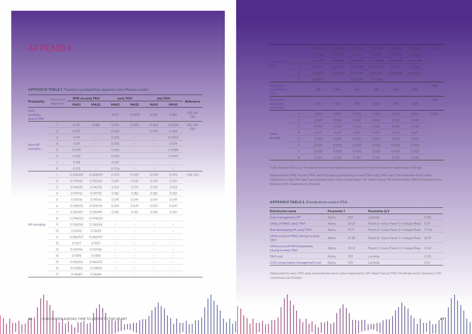
APPENDIX
APPENDIX TABLE 1. Transition probabilities applied in the Markov model
APPENDIX TABLE 2. Distributions used in PSA
*over 15 years’ follow up. The yearly transition probability was derived from the cumulative incidence accordingly.
Abbreviations: WW_no early TAVI: watchful waiting and having no early TAVI; early TAVI: early Transcatheter Aortic Valve Implantation; late TAVI: late Transcatheter Aortic Valve Implantation, HF: Heart Failure; RR: Relative Risk, MAS: Moderate Aortic Stenosis, SAS: Severe Aortic Stenosis.
Abbreviations: early TAVI: early transcatheter aortic valve implantation; HF: Heart Failure, MAS: Moderate Aortic Stenosis, CVD: Cardiovascular Disease
Probability Years since
diagnosis
WW_no early TAVI early TAVI late TAVIReference
MAS1 MAS2 MAS1 MAS2 MAS1 MAS2
CVD mortality due to TAVI
- - - 0.011 0.0335 0.021 0.186(25, 134,
135)
Non-HF mortality
1 0.101 0.188 0.016 0.053 0.264 0.0538 (25, 134, 135)
2 0.097 - 0.042 - 0.079 0.026
3 0.119 - 0.023 - - 0.0555
4 0.09 - 0.035 - - 0.074
5 0.099 - 0.062 - - 0.0588
6 0.242 - 0.034 - - 0.0499
7 0.138 - 0.031 - - -
8 0.215 - 0.006 - - -
HF mortality
1 0.258359 0.258359 0.093 0.093 0.093 0.093 (136, 137)
2 0.115002 0.115002 0.051 0.051 0.051 0.051
3 0.114035 0.114035 0.103 0.103 0.103 0.103
4 0.119732 0.119732 0.182 0.182 0.182 0.182
5 0.119016 0.119016 0.091 0.091 0.091 0.091
6 0.108076 0.108076 0.047 0.047 0.047 0.047
7 0.125499 0.125499 0.142 0.142 0.142 0.142
8 0.094023 0.094023 - - - -
9 0.136554 0.136554 - - - -
10 0.12652 0.12652 - - - -
11 0.086907 0.086907 - - - -
12 0.1507 0.1507 - - - -
13 0.130746 0.130746 - - - -
14 0.11818 0.11818 - - - -
15 0.146202 0.146202 - - - -
16 0.02854 0.02854 - - - -
17 0.14689 0.14689 - - - -
Develop HF (RR)
1 0.033014 0.118935 0.009574 0.009574 0.034491 0.034491 (138)
2 0.041828 0.104538 0.01213 0.01213 0.030316 0.030316
3 0.013339 0.018944 0.003868 0.003868 0.005494 0.005494
4 0.02273 0.049208 0.006592 0.006592 0.01427 0.01427
5 0.052877 0.083027 0.015334 0.015334 0.024078 0.024078
6 0.045331 - 0.013146 0.013146 - -
Progression from MAS to SAS*
74% 74% 74% 74% 74% 74%(138)
Proportion of patients with MAS1
35% 65% 35% 65% 35% 65%(139)
Valve durable
1 0.100 0.100 0.100 0.100 0.100 0.100 (140)
2 0.047 0.047 0.047 0.047 0.047 0.047
3 0.022 0.022 0.022 0.022 0.022 0.022
4 0.017 0.017 0.017 0.017 0.017 0.017
5 0.010 0.010 0.010 0.010 0.010 0.010
6 0.050 0.050 0.050 0.050 0.050 0.050
7 0.005 0.005 0.005 0.005 0.005 0.005
8 0.130 0.130 0.130 0.130 0.130 0.130
Distribution name Parameter 1 Parameter 2/3
Cost management HF Alpha 100 Lambda 0.00
Utility of MAS1, early TAVI Alpha 20.21 Param 2 = beta; Param 3 = Integer/Real; 5.37
Risk developing HF, early TAVI Alpha 70.71 Param 2 = beta; Param 3 = Integer/Real; 173.12
Utility score or MAS, having no early TAVI
Alpha 37.38 Param 2 = beta; Param 3 = Integer/Real; 22.91
Utility score of MAS at baseline, having no early TAVI
Alpha 30.31 Param 2 = beta; Param 3 = Integer/Real; 13.62
TAVI cost Alpha 100 Lambda 0.00
CVD complication management cost Alpha 100 Lambda 0.01
OUR HIDDEN AGEING: TIME TO LISTEN TO THE HEART 66 67
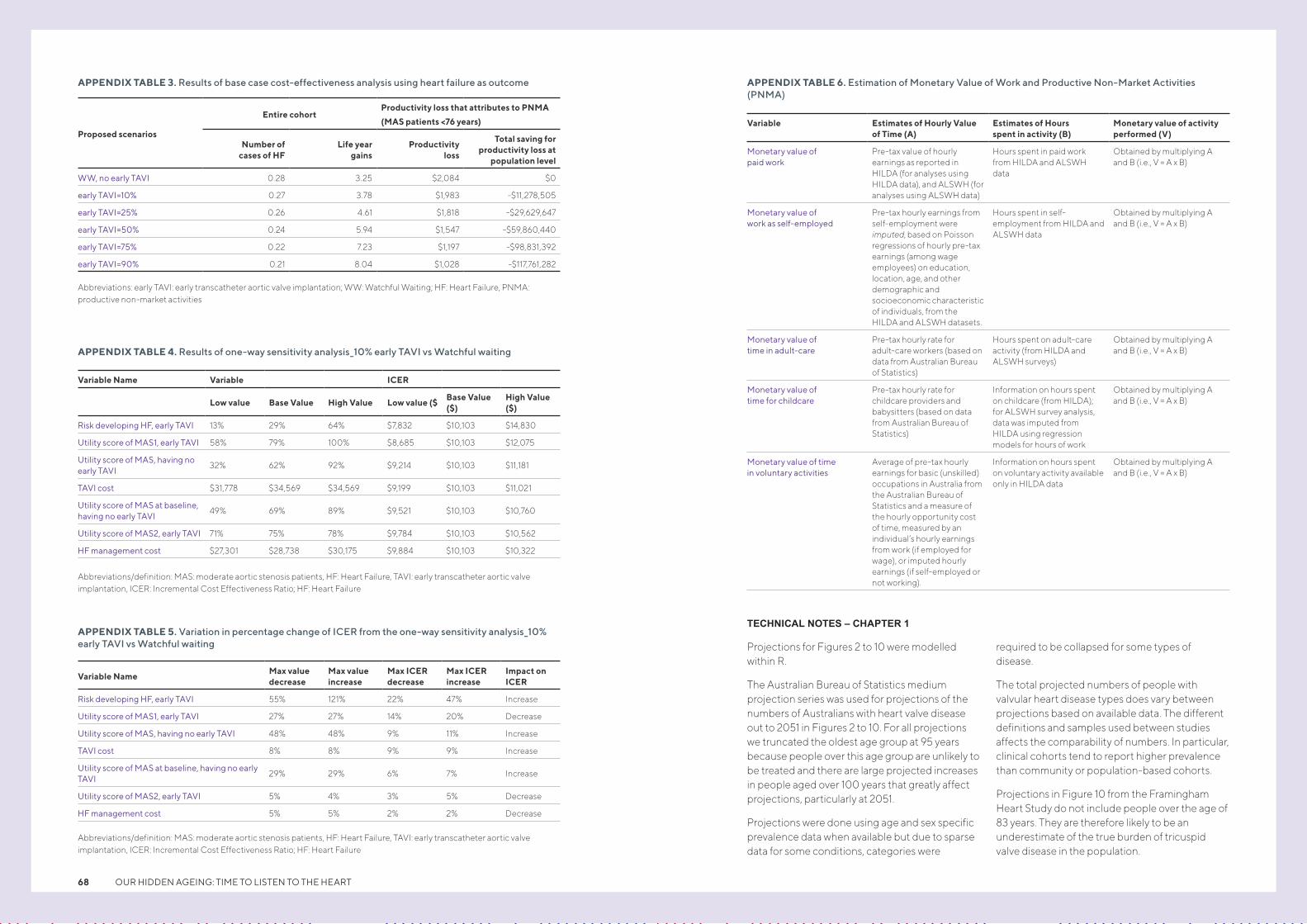
APPENDIX TABLE 3. Results of base case cost-effectiveness analysis using heart failure as outcome APPENDIX TABLE 6. Estimation of Monetary Value of Work and Productive Non-Market Activities (PNMA)
APPENDIX TABLE 4. Results of one-way sensitivity analysis_10% early TAVI vs Watchful waiting
APPENDIX TABLE 5. Variation in percentage change of ICER from the one-way sensitivity analysis_10% early TAVI vs Watchful waiting
Abbreviations: early TAVI: early transcatheter aortic valve implantation; WW: Watchful Waiting; HF: Heart Failure, PNMA: productive non-market activities
Abbreviations/definition: MAS: moderate aortic stenosis patients, HF: Heart Failure, TAVI: early transcatheter aortic valve implantation, ICER: Incremental Cost Effectiveness Ratio; HF: Heart Failure
Abbreviations/definition: MAS: moderate aortic stenosis patients, HF: Heart Failure, TAVI: early transcatheter aortic valve implantation, ICER: Incremental Cost Effectiveness Ratio; HF: Heart Failure
Proposed scenarios
Entire cohortProductivity loss that attributes to PNMA
(MAS patients <76 years)
Number of cases of HF
Life year gains
Productivity loss
Total saving for productivity loss at
population level
WW, no early TAVI 0.28 3.25 $2,084 $0
early TAVI=10% 0.27 3.78 $1,983 -$11,278,505
early TAVI=25% 0.26 4.61 $1,818 -$29,629,647
early TAVI=50% 0.24 5.94 $1,547 -$59,860,440
early TAVI=75% 0.22 7.23 $1,197 -$98,831,392
early TAVI=90% 0.21 8.04 $1,028 -$117,761,282
Variable Name Variable ICER
Low value Base Value High Value Low value ($Base Value ($)
High Value ($)
Risk developing HF, early TAVI 13% 29% 64% $7,832 $10,103 $14,830
Utility score of MAS1, early TAVI 58% 79% 100% $8,685 $10,103 $12,075
Utility score of MAS, having no early TAVI
32% 62% 92% $9,214 $10,103 $11,181
TAVI cost $31,778 $34,569 $34,569 $9,199 $10,103 $11,021
Utility score of MAS at baseline, having no early TAVI
49% 69% 89% $9,521 $10,103 $10,760
Utility score of MAS2, early TAVI 71% 75% 78% $9,784 $10,103 $10,562
HF management cost $27,301 $28,738 $30,175 $9,884 $10,103 $10,322
Variable NameMax value decrease
Max value increase
Max ICER decrease
Max ICER increase
Impact on ICER
Risk developing HF, early TAVI 55% 121% 22% 47% Increase
Utility score of MAS1, early TAVI 27% 27% 14% 20% Decrease
Utility score of MAS, having no early TAVI 48% 48% 9% 11% Increase
TAVI cost 8% 8% 9% 9% Increase
Utility score of MAS at baseline, having no early TAVI
29% 29% 6% 7% Increase
Utility score of MAS2, early TAVI 5% 4% 3% 5% Decrease
HF management cost 5% 5% 2% 2% Decrease
Variable Estimates of Hourly Value of Time (A)
Estimates of Hours spent in activity (B)
Monetary value of activity performed (V)
Monetary value of paid work
Pre-tax value of hourly earnings as reported in HILDA (for analyses using HILDA data), and ALSWH (for analyses using ALSWH data)
Hours spent in paid work from HILDA and ALSWH data
Obtained by multiplying A and B (i.e., V = A x B)
Monetary value of work as self-employed
Pre-tax hourly earnings from self-employment were imputed, based on Poisson regressions of hourly pre-tax earnings (among wage employees) on education, location, age, and other demographic and socioeconomic characteristic of individuals, from the HILDA and ALSWH datasets.
Hours spent in self-employment from HILDA and ALSWH data
Obtained by multiplying A and B (i.e., V = A x B)
Monetary value of time in adult-care
Pre-tax hourly rate for adult-care workers (based on data from Australian Bureau of Statistics)
Hours spent on adult-care activity (from HILDA and ALSWH surveys)
Obtained by multiplying A and B (i.e., V = A x B)
Monetary value of time for childcare
Pre-tax hourly rate for childcare providers and babysitters (based on data from Australian Bureau of Statistics)
Information on hours spent on childcare (from HILDA); for ALSWH survey analysis, data was imputed from HILDA using regression models for hours of work
Obtained by multiplying A and B (i.e., V = A x B)
Monetary value of time in voluntary activities
Average of pre-tax hourly earnings for basic (unskilled) occupations in Australia from the Australian Bureau of Statistics and a measure of the hourly opportunity cost of time, measured by an individual’s hourly earnings from work (if employed for wage), or imputed hourly earnings (if self-employed or not working).
Information on hours spent on voluntary activity available only in HILDA data
Obtained by multiplying A and B (i.e., V = A x B)
TECHNICAL NOTES – CHAPTER 1
Projections for Figures 2 to 10 were modelled within R.
The Australian Bureau of Statistics medium projection series was used for projections of the numbers of Australians with heart valve disease out to 2051 in Figures 2 to 10. For all projections we truncated the oldest age group at 95 years because people over this age group are unlikely to be treated and there are large projected increases in people aged over 100 years that greatly affect projections, particularly at 2051.
Projections were done using age and sex specific prevalence data when available but due to sparse data for some conditions, categories were
required to be collapsed for some types of disease.
The total projected numbers of people with valvular heart disease types does vary between projections based on available data. The different definitions and samples used between studies affects the comparability of numbers. In particular, clinical cohorts tend to report higher prevalence than community or population-based cohorts.
Projections in Figure 10 from the Framingham Heart Study do not include people over the age of 83 years. They are therefore likely to be an underestimate of the true burden of tricuspid valve disease in the population.
OUR HIDDEN AGEING: TIME TO LISTEN TO THE HEART 68

baker.edu.au
This whitepaper was made possible thanks to an unrestricted educational grant from Edwards Lifesciences.
ALICE SPRINGSBaker Institute Central Australia
W&E Rubuntja Research and Medical Education Building Alice Springs Hospital Campus Gap Road, Alice Springs NT 0870 Australia
T +61 8 8959 0111
F +61 8 8952 1557
PO Box 1294, Alice Springs NT 0871 Australia
MELBOURNE75 Commercial Road Melbourne VIC 3004 Australia
T +61 3 8532 1111
F +61 3 8532 1100
PO Box 6492, Melbourne VIC 3004 Australia