Embed Size (px)
Citation preview

Our Dermatology Online
© Our Dermatol Online 4.2018 407
How to cite this article: Siham M, Asmaa S, Dione JP, Zaitouna A, Karima S, Badr H, Nadia I. Pemphigus vulgaris and renal amyloidosis: a new association?. Our Dermatol Online. 2018;9(4):407-409.Submission: 29.01.2018; Acceptance: 22.03.2018DOI: 10.7241/ourd.20184.11
Pemphigus vulgaris and renal amyloidosis: a new Pemphigus vulgaris and renal amyloidosis: a new association?association?Mansouri Siham1, Sqalli Asmaa1, Jean Pierre Dione2, Alhamany Zaitouna3, Senouci Karima1, Hassam Badr1, Ismaili Nadia1
1Department of Dermatology, University Hospital Ibn Sina Rabat, Morocco, 2Department of Nephrology, University Hospital Ibn Sina Rabat, Morocco, 3Department of Pathology, University Hospital Ibn Sina Rabat, Morocco
Corresponding author: Dr Mansouri Siham, E-mail: [email protected]
INTRODUCTION
Pemphigus vulgaris (PV) is a bullous auto-immune disease affecting the skin and mucosa. It is characterised by acantholysis that results in the formation of intraepithelial bullous lesions [1]. There has been no description of the relationship between PV and kidney disease, even though the kidney is a frequent site of immune-mediated injury. Herein we report a case distinguished by its unusual clinical presentation.
CASE REPORT
A 61-year-old man came to our department with a 9 months history of two facial skin lesions treated with different topics but without improvement. Within 5 months, he presented painful oral ulceration. Within 07 month, he developed lower extremity edema.
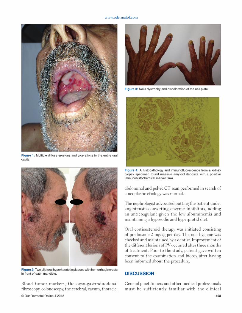
Physical examination revealed multiple diffuse erosions and ulcerations in the entire oral cavity (Fig. 1), mainly in the tongue, palate and cheek mucosa; two bilateral hyperkeratotic plaques with hemorrhagic crusts were present in front of each mandible (Fig. 2); nails dystrophy and discoloration of the nail plate (Fig. 3);
and edema of the lower extremities. In addition, Nikolsky’s sign was positive in perilesional at the facial level. The rest of the clinical examination was normal.
The mycological nail tests were negative. Histological examination of two biopsy sample taken from the erosion of skin and the nail bed of one finger, the direct and the indirect immunofluorescence confirmed the diagnosis of PV.
After confirming the nephrotic syndrome (positive proteinuria 15,75g/24h, hypoalbuminemia, positive urine protein electrophoresis test and normal renal function), histopathology and immunofluorescence from a kidney biopsy specimen was performed and confirmed glomerular and vascular amyloidosis AA. The biopsy containing 15 glomeruli without cell proliferation, found massive amyloid deposits in the glomerulus and vessels with an important intertitial fibrosis. Congo red coloration was positive. The immunohistochemical marker SAA was positive (Fig. 4). No detectable gammapathy was found.
No acute infection was detected (Hemoculture, hepatitis, syphilitic and HIV serology were negative).
ABSTRACT
Pemphigus vulgaris (PV) is a life- threatening chronic autoimmune disease characterized by the formation of intraepithelial blisters on the skin and mucous membranes. The etiology of PV is still unknown. It results from an autoimmune process. There has been virtually no description of the relationship between pemphigus vulgaris and kidney disease. The case we report illustrates a special situation in which PV was associated with renal amyloidosis.
Key words: Pemphigus vulgaris; Renal amyloidosis; Autoimmune
Case Report
www.odermatol.com
© Our Dermatol Online 4.2018 408
Blood tumor markers, the oeso-gastroduodenal fibroscopy, colonoscopy, the cerebral, cavum, thoracic,
abdominal and pelvic CT scan performed in search of a neoplastic etiology was normal.
The nephrologist advocated putting the patient under angiotensin-converting enzyme inhibitors, adding an anticoagulant given the low albuminemia and maintaining a hyposodic and hyperprotid diet.
Oral corticosteroid therapy was initiated consisting of prednisone 2 mg/kg per day. The oral hygiene was checked and maintained by a dentist. Improvement of the different lesions of PV occurred after three months of treatment. Prior to the study, patient gave written consent to the examination and biopsy after having been informed about the procedure.
DISCUSSION
General practitioners and other medical professionals must be sufficiently familiar with the clinical
Figure 1: Multiple diffuse erosions and ulcerations in the entire oral cavity.
Figure 2: Two bilateral hyperkeratotic plaques with hemorrhagic crusts in front of each mandible.
Figure 3: Nails dystrophy and discoloration of the nail plate.
Figure 4: A histopathology and immunofl uorescence from a kidney biopsy specimen found massive amyloid deposits with a positive immunohistochemical marker SAA.

www.odermatol.com
© Our Dermatol Online 4.2018 409
manifestations of PV to ensure early diagnosis and treatment. There has been no description of the relationship between PV and kidney disease, even though the kidney is a frequent site of immune-mediated injury. Our observation of renal amyloidosis and PV occurring simultaneously in a patient in the absence of offending agents or other clinically apparent disease processes represents a novel finding.
Glomerular involvement was also reported during acquired bullous dermatosis. The glomerular affections reported are varied but some associations appear: nephrosis with minimal glomerular lesions and pemphigus [2,3], extramembraneous glomerulonephritis and bullous pemphigoid [4], immunoglobulin A glomerulonephritis and dermatitis herpetiformis [5]. They are conceived in a context of autoimmunity, neoplasia, and even drug toxicity with D-penicillamide.
Amyloidosis, unlike dystrophic epidermolysis bullosa, is found only in an elderly patient with acquired epidermolysis bullosa [6].
One report describes an association of PV with renal disease, and, in this case, a clear offending agent (D-penicillamine) was identified [7].
Another observation of minimal change nephropathy and PV occurring simultaneously in a patient was reported [8].
Chronic inflammatory syndrome and recurrent skin infections are clearly the cause of amyloid glomerular complications.
The initial appearance of PV, followed by clinically apparent nephrotic syndrome, suggests the possibility of a causative effect of PV in the pathogenesis of renal amyloidosis. Otherwise, an unidentified process may have caused the simultaneous occurrence of the two disorders, although the absence of any other apparent disease makes this possibility speculative. Unfortunately, there are no data on which to address this issue: the small number of cases and the absence of a prospective study do not make it possible to retain
one of the factors more specifically responsible for such a glomerular entity.
CONCLUSION
This study adds to the limited number of cases of PV associated to kidney disease. An important question is raised as to whether patients with PV and autoimmune bullous diseases should be screened for proteinuria. We suggest that performed prospective studies could help resolve this issue.
CONSENT
The examination of the patient was conducted according to the Declaration of Helsinki principles.
REFERENCES
1. Aline Bicalho Matias, Ana Maria Ferreira Roselino: Pemphigus: a disease stamped in the skin. Our Dermatol Online. 2013;4(Suppl.3): 601-5.
2. Herron MD, Kohan DE, Hansen CD. Minimal change nephropathy associated with pemphigus vulgaris: a new relationship? J Am Acad Dermatol. 2004;10:645.
3. Karras A, De Montpreville V, Fakhouri F, Gru¨nfeld JP, Lesavre P. Renal and thymic patholygy in thymoma-associated nephropathy: report of 21 cases and review of the litterature. Nephrol Dial Transplant. 2005;20:1075-82.
4. Ross EA, Ahmed RA. Bullous pemphigoid-associated nephropathy: report of two cases and review of the literature. Am J Kidney Dis. 1989;14:225-9.
5. Julian BA, Czerlinsky C, Russell MW, Galla JH, Koopman WJ, Mestecky J, et al. Striking elevation of serum IgA, IgA-containing immune complexes, and IgA rheumatoid factor in clinically silent dermatitis herpetiformis. Am J Kidney Dis. 1987;10:378-84.
6. Trump DL, Allen H, Olson J, Wright J, Humphrey RL. Epidermolysis bullosa acquisita; association with amyloidosis and multiple myeloma. JAMA. 1980;243:1461-2.
7. Shapiro M, Jimenez S, Werth VP. Pemphigus vulgaris induced by D-penicillamine therapy in a patient with systemic sclerosis. J Am Acad Dermatol. 2000;42:297-9.
8. Herron MD, Kohan DE, Hansen CD. Minimal change nephropathy associated with pemphigus vulgaris: A new relationship? J Am Acad Dermatol. 2004;50:645.
Copyright by Mansouri Siham, et al. This is an open-access article distributed under the terms of the Creative Commons Attribution License, which permits unrestricted use, distribution, and reproduction in any medium, provided the original author and source are credited.Source of Support: Nil, Confl ict of Interest: None declared.


![Case Report AAtypical presentation of pemphigus vulgaris - A … · 2018-12-03 · involvement and pemphigus vulgaris presents as oral lesions in 50 to 70% patients [1-3]. These may](https://img.pdfslide.us/doc/110x75/5ccfc74d88c993cc718c625a/case-report-aatypical-presentation-of-pemphigus-vulgaris-a-2018-12-03.jpg)