Embed Size (px)
Citation preview
Pemphigus in Korea: A retrospective analysis of 199 patients over a 16-year period
Mi Ri Kim
Department of Medicine
The Graduate School, Yonsei University
Pemphigus in Korea: A retrospective analysis of 199 patients over a 16-year period
Directed by Professor Soo-Chan Kim
The Master’s Thesis
Submitted to the Department of Medicine
and the Graduate School of Yonsei University
in partial fulfillment of the requirements for the
degree of Master of Medical science
Mi Ri Kim
June 2010
3
This certifies that the Master's Thesis of Mi Ri Kim is approved.
------------------------------------------------------------------ Thesis Supervisor : Soo-Chan Kim ------------------------------------------------------------------ Thesis Committee Member : Seung Hun Lee ------------------------------------------------------------------ Thesis Committee Member: Hyeon Chang Kim
The Graduate School Yonsei University
June 2010
Acknowledgements
I appreciate my thesis supervisor, Prof. Soo-Chan Kim for
giving me great advice and guidance that has been helpful for
taking a degree. I thank him for his supervision and
encouragement to study this subject.
I also appreciate professors Seung Hun Lee and Hyeon
Chang Kim who gave me expert advice and warm support.
And thanks to all members of my department.
Finally, I am especially grateful to my hudsband who
always cheer me up.
1
Table of Contents I. INTRODUCTION………………………….………………..………3 II. MATERIALS AND METHODS……………………………………5
1. Subjects ……………………...…………………………….……5
2. Statistical analysis……………………………………………….5
3. Assessment methods ….………………..……………………….6
A. Disease severity score.……………………………...………6
B. Late end points of disease activity.………………..………..8
4. Treatment protocol………………………………………………8
III. RESULTS…………………..…………………...……….………..11 1. Clinical characteristics................................................................11
2. Treatment regimens……………………………………….……12
3. Treatment outcome…………………………………….………14
A. Both complete and overall remission rate were higher in PF
patients than PV patients…………………………………….14
B. Prednisolone plus adjuvant immunosuppressive therapy
compared with prednisolone alone in patients with
pemphigus…………………………………………………...16
C. Conventional therapy plus rituximab compared with
conventional therapy alone in patients with pemphigus…….17
D. Mortality………………………………………………….…21
IV. DISCUSSION.…………….................................…........................22 V. CONCLUSION..………………………....……….……..................26 REFERENCES.……………………. ………….……………………..27 ABSTRACT(IN KOREAN) .………………………….……..............29
2
LIST OF FIGURES
Figure 1. Treatment protocol fore pemphigus………………..7
Figure 2. Disease severity at initial visit…………………….11
Figure 3. Remission rate of pemphigus vulgaris during follow-
up period…………………………………………14
Figure 4. Remission rate of pemphigus foliaceus during follow-
up period…………………………………15
Figure 5. Comparision of pemphigus severity score before and
after rituximab treatment………………………...17
Figure 6. Clinical response to rituximab in recalcitrant
pemphigus vulgaris………………………………18
3
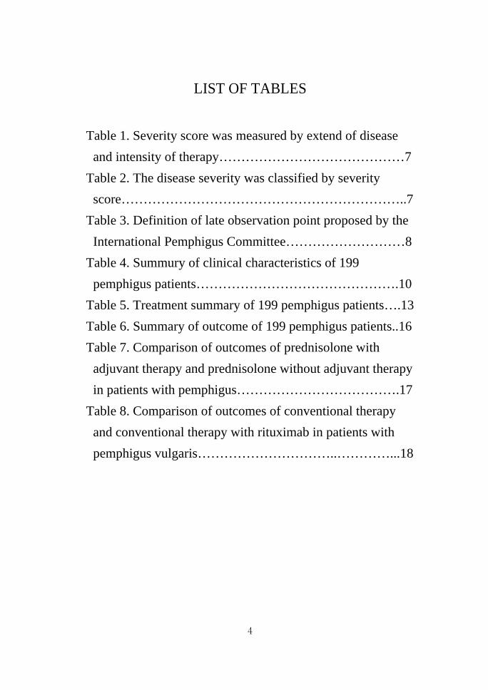
LIST OF TABLES
Table 1. Severity score was measured by extend of disease
and intensity of therapy……………………………………7
Table 2. The disease severity was classified by severity
score………………………………………………………..7
Table 3. Definition of late observation point proposed by the
International Pemphigus Committee………………………8
Table 4. Summury of clinical characteristics of 199
pemphigus patients……………………………………….10
Table 5. Treatment summary of 199 pemphigus patients….13
Table 6. Summary of outcome of 199 pemphigus patients..16
Table 7. Comparison of outcomes of prednisolone with
adjuvant therapy and prednisolone without adjuvant therapy
in patients with pemphigus……………………………….17
Table 8. Comparison of outcomes of conventional therapy
and conventional therapy with rituximab in patients with
pemphigus vulgaris…………………………..…………...18
4
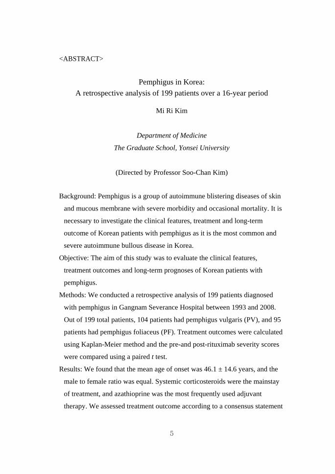
<ABSTRACT>
Pemphigus in Korea:
A retrospective analysis of 199 patients over a 16-year period
Mi Ri Kim
Department of Medicine
The Graduate School, Yonsei University
(Directed by Professor Soo-Chan Kim)
Background: Pemphigus is a group of autoimmune blistering diseases of skin
and mucous membrane with severe morbidity and occasional mortality. It is
necessary to investigate the clinical features, treatment and long-term
outcome of Korean patients with pemphigus as it is the most common and
severe autoimmune bullous disease in Korea.
Objective: The aim of this study was to evaluate the clinical features,
treatment outcomes and long-term prognoses of Korean patients with
pemphigus.
Methods: We conducted a retrospective analysis of 199 patients diagnosed
with pemphigus in Gangnam Severance Hospital between 1993 and 2008.
Out of 199 total patients, 104 patients had pemphigus vulgaris (PV), and 95
patients had pemphigus foliaceus (PF). Treatment outcomes were calculated
using Kaplan-Meier method and the pre-and post-rituximab severity scores
were compared using a paired t test.
Results: We found that the mean age of onset was 46.1 ± 14.6 years, and the
male to female ratio was equal. Systemic corticosteroids were the mainstay
of treatment, and azathioprine was the most frequently used adjuvant
therapy. We assessed treatment outcome according to a consensus statement
5
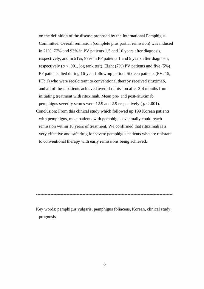
on the definition of the disease proposed by the International Pemphigus
Committee. Overall remission (complete plus partial remission) was induced
in 21%, 77% and 93% in PV patients 1,5 and 10 years after diagnosis,
respectively, and in 51%, 87% in PF patients 1 and 5 years after diagnosis,
respectively (p < .001, log rank test). Eight (7%) PV patients and five (5%)
PF patients died during 16-year follow-up period. Sixteen patients (PV: 15,
PF: 1) who were recalcitrant to conventional therapy received rituximab,
and all of these patients achieved overall remission after 3-4 months from
initiating treatment with rituximab. Mean pre- and post-rituximab
pemphigus severity scores were 12.9 and 2.9 respectively ( p < .001).
Conclusion: From this clinical study which followed up 199 Korean patients
with pemphigus, most patients with pemphigus eventually could reach
remission within 10 years of treatment. We confirmed that rituximab is a
very effective and safe drug for severe pemphigus patients who are resistant
to conventional therapy with early remissions being achieved.
----------------------------------------------------------------------------------------------
Key words: pemphigus vulgaris, pemphigus foliaceus, Korean, clinical study,
prognosis
6
Pemphigus in Korea:
A retrospective analysis of 199 patients over a 16-year period
Mi Ri Kim
Department of Medicine The Graduate School, Yonsei University
(Directed by Professor Soo Chan Kim)
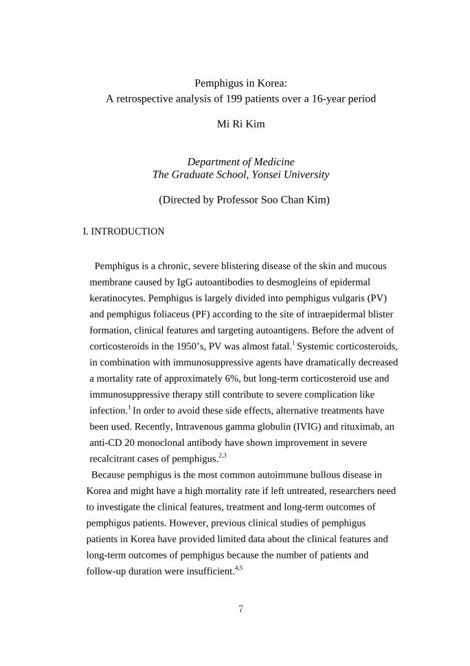
I. INTRODUCTION
Pemphigus is a chronic, severe blistering disease of the skin and mucous
membrane caused by IgG autoantibodies to desmogleins of epidermal
keratinocytes. Pemphigus is largely divided into pemphigus vulgaris (PV)
and pemphigus foliaceus (PF) according to the site of intraepidermal blister
formation, clinical features and targeting autoantigens. Before the advent of
corticosteroids in the 1950’s, PV was almost fatal.1 Systemic corticosteroids,
in combination with immunosuppressive agents have dramatically decreased
a mortality rate of approximately 6%, but long-term corticosteroid use and
immunosuppressive therapy still contribute to severe complication like
infection.1 In order to avoid these side effects, alternative treatments have
been used. Recently, Intravenous gamma globulin (IVIG) and rituximab, an
anti-CD 20 monoclonal antibody have shown improvement in severe
recalcitrant cases of pemphigus.2,3
Because pemphigus is the most common autoimmune bullous disease in
Korea and might have a high mortality rate if left untreated, researchers need
to investigate the clinical features, treatment and long-term outcomes of
pemphigus patients. However, previous clinical studies of pemphigus
patients in Korea have provided limited data about the clinical features and
long-term outcomes of pemphigus because the number of patients and
follow-up duration were insufficient.4,5
7
A previous clinical study conducted by Herbst and Bystryn6 revealed that
25%, 50%, and 75% of 40 PV patients eventually achieved complete and
durable remission 2, 5, and 10 years after diagnosis during a 7.7-year
follow-up period. However, it is obscure at present how many Korean
patients with pemphigus can reach long-term remission.
Recently, the International Pemphigus Committee (IPC) proposed a
consensus statement to accurately assess disease activity, severity and
therapeutic response, because common terms and end point of pemphigus
are needed for comparing disease severity and therapeutic outcomes
between pemphigus treatment centers.
To our knowledge, this is the first clinical study for Korean pemphigus
patients that enrolled a large number of patients and conducted long-term
follow-up with the application of common terms from the IPC consensus
statement to estimate treatment outcomes.
8
II. MATERIALS AND METHODS
1. Subjects
To evaluate the clinical features, treatment and prognoses of patients
with pemphigus, the medical records of all patients with pemphigus
diagnosed at Gangnam Severance Hospital in Seoul, Korea from 1993
to 2008 were reviewed retrospectively. The following data were
recorded and analyzed; gender, age at onset, disease severity, treatment
modalities, treatment outcome and time to remission.
Diagnosis was made on the basis of typical clinical features and
confirmed by histopathology and immunofluorescence examinations.7
Enzyme-linked immunosorbent assay (ELISA) for recombinant
desmoglein (Dsg) 1 and Dsg3 were also performed in some of the
patients. The patients who have more than 6 months follow-up data
were included in this study to avoid a bias.
2. Statistical analysis
We compared demographic and clinical characteristics between PV
and PF patients using independent two-sample t test for continuous
variables and chi-square test for categorical variables. The cumulative
probabilities of complete remission and overall remission ( complete
plus partial remission) were calculated using Kaplan-Meier method,
and the curves were compared between PV and PF groups using the
log rank test. We censored patients who did not achieve remission and
those who were withdrawn during follow-up period. The pre-rituximab
and post-rituximab pemphigus severity scores were compared using a
paired t test. Two-sided p < .05 were considered significant. We used
9
SPSS for windows, version 17.0 (SPSS, Chicago, Illinois), for all
statistical analyses.
3. Assessment methods
A. Disease severity score
Scores for disease severity were assessed using our revised criteria
modified from those designed by Herbst and Bystryn6, who minutely
divided the body areas involved, plus an additional oral mucosa score.
The following grading system was devised to provide objective scores
for disease severity and treatment outcome (Table 1).
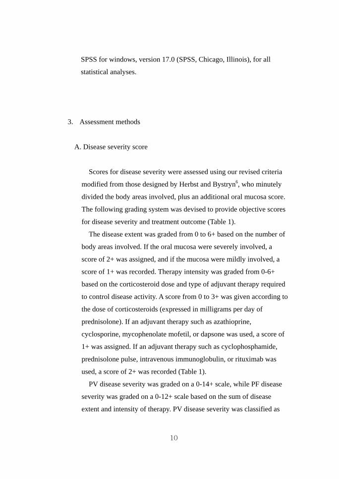
The disease extent was graded from 0 to 6+ based on the number of
body areas involved. If the oral mucosa were severely involved, a
score of 2+ was assigned, and if the mucosa were mildly involved, a
score of 1+ was recorded. Therapy intensity was graded from 0-6+
based on the corticosteroid dose and type of adjuvant therapy required
to control disease activity. A score from 0 to 3+ was given according to
the dose of corticosteroids (expressed in milligrams per day of
prednisolone). If an adjuvant therapy such as azathioprine,
cyclosporine, mycophenolate mofetil, or dapsone was used, a score of
1+ was assigned. If an adjuvant therapy such as cyclophosphamide,
prednisolone pulse, intravenous immunoglobulin, or rituximab was
used, a score of 2+ was recorded (Table 1).
PV disease severity was graded on a 0-14+ scale, while PF disease
severity was graded on a 0-12+ scale based on the sum of disease
extent and intensity of therapy. PV disease severity was classified as
10
mild, moderate or severe based on a severity score of 0-4, 5-8 or 9-14,
respectively, and PF disease severity was classified as mild, moderate
or severe based on a severity score of 0-3, 4-7 or 8-12, respectively
(Table 2). Scores were recorded at the initial visit and during all
follow-up visits.
Table 1. Severity score was measured by extend of disease and
intensity of therapy.
Extend of disease Score Intensity of therapy Score
scalp, face/ neck, chest/ abdomen/ back/ arm/ leg
1 in each body area involved
Pd 10mg >
10~30mg > 30mg <
1 2 3
oral mucosa ( only for PV) Adjuvant therapy
mild 1 AZA, MMF, CsA, Dapsone 1
severe 2 CTX, Pd pulse, IVIG, Rituximab 2
Pd: prednisolone, AZA: azathioprine, MMF: mycophenolate mofetil, CsA: cyclosporine A, CTX: cyclophosphamide, Pd pulse: prednisolone pulse, IVIG: intravenous immunoglobulins
Table 2. The disease severity was classified by severity score.
Severity PV severity score
(total 14) PF severity score
(total 12)
Mild 0~4 0~3
Moderate 5~8 4~7
Severe 9~14 8~12
11
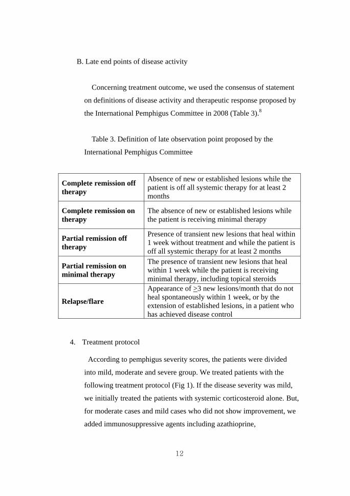
B. Late end points of disease activity
Concerning treatment outcome, we used the consensus of statement
on definitions of disease activity and therapeutic response proposed by
the International Pemphigus Committee in 2008 (Table 3).8
Table 3. Definition of late observation point proposed by the
International Pemphigus Committee
Complete remission off therapy
Absence of new or established lesions while the patient is off all systemic therapy for at least 2 months
Complete remission on therapy
The absence of new or established lesions while the patient is receiving minimal therapy
Partial remission off therapy
Presence of transient new lesions that heal within 1 week without treatment and while the patient is off all systemic therapy for at least 2 months
Partial remission on minimal therapy
The presence of transient new lesions that heal within 1 week while the patient is receiving minimal therapy, including topical steroids
Relapse/flare
Appearance of >3 new lesions/month that do not heal spontaneously within 1 week, or by the extension of established lesions, in a patient who has achieved disease control
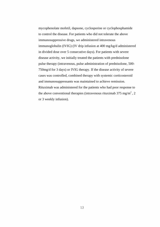
4. Treatment protocol
According to pemphigus severity scores, the patients were divided
into mild, moderate and severe group. We treated patients with the
following treatment protocol (Fig 1). If the disease severity was mild,
we initially treated the patients with systemic corticosteroid alone. But,
for moderate cases and mild cases who did not show improvement, we
added immunosuppressive agents including azathioprine,
12
mycophenolate mofetil, dapsone, cyclosporine or cyclophosphamide
to control the disease. For patients who did not tolerate the above
immunosuppressive drugs, we administered intravenous
immunoglobulin (IVIG) (IV drip infusion at 400 mg/kg/d administered
in divided dose over 5 consecutive days). For patients with severe
disease activity, we initially treated the patients with prednisolone
pulse therapy (intravenous, pulse administration of prednisolone, 500-
750mg/d for 3 days) or IVIG therapy. If the disease activity of severe
cases was controlled, combined therapy with systemic corticosteroid
and immunosuppressants was maintained to achieve remission.
Rituximab was administered for the patients who had poor response to
the above conventional therapies (intravenous rituximab 375 mg/m2 , 2
or 3 weekly infusion).
13
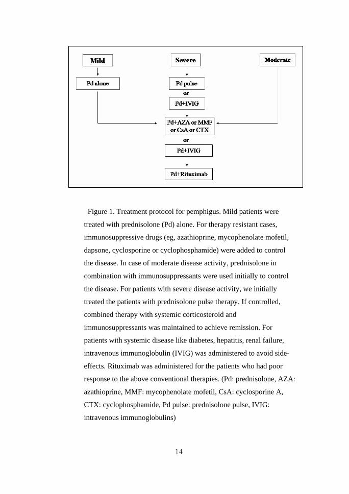
Figure 1. Treatment protocol for pemphigus. Mild patients were
treated with prednisolone (Pd) alone. For therapy resistant cases,
immunosuppressive drugs (eg, azathioprine, mycophenolate mofetil,
dapsone, cyclosporine or cyclophosphamide) were added to control
the disease. In case of moderate disease activity, prednisolone in
combination with immunosuppressants were used initially to control
the disease. For patients with severe disease activity, we initially
treated the patients with prednisolone pulse therapy. If controlled,
combined therapy with systemic corticosteroid and
immunosuppressants was maintained to achieve remission. For
patients with systemic disease like diabetes, hepatitis, renal failure,
intravenous immunoglobulin (IVIG) was administered to avoid side-
effects. Rituximab was administered for the patients who had poor
response to the above conventional therapies. (Pd: prednisolone, AZA:
azathioprine, MMF: mycophenolate mofetil, CsA: cyclosporine A,
CTX: cyclophosphamide, Pd pulse: prednisolone pulse, IVIG:
intravenous immunoglobulins)
14
III. RESULTS
1. Clinical characteristics of pemphigus patients
Out of 199 pemphigus patients, 104 patients were diagnosed with PV and
95 patients were diagnosed with PF. Therefore the prevalence rate of PV
to PF is 1.1:1.0. For the 104 PV patients, the mean age of onset was 47.0
years (range, 19-75 years), the male-to-female ratio was 1.0:1.0 (men 51,
women 53), and the mean follow-up duration was 48.6 months (range, 6-
172 months). For the 95 PF patients, the mean age of onset was 45.0 years
(range, 24-83 years), the male-to-female ratio was 1.0:1.1 (men 46, women
49), and the mean follow-up duration was 45.9 months (range, 6-181
months). We didn’t observe any significant differences between PV and
PF patients in terms of gender and age. The clinical characteristics for our
patients with PV and PF are summarized in Table 4.
With regard to the disease severity at initial visit, eighty-six patients with
PV (83%) had moderate to severe disease, whereas 82 PF patients (86%)
had mild to moderate disease (figure 2).
Table 4. Summary of clinical characteristics of 199 pemphigus patients
PV PF P value
Number of patients 104 95 Mean age at onset (years)
47.0+13.7 45.0+15.5 0.408
Sex ratio (M:F) 51:53(1:1.04) 46:49(1:1.07) 0.931 Follow up duration (months)
48.6+36.9 45.9+39.6 0.587
Skin involvement* 99(95.2%) 95(100%) 0.061 Oral mucosa involvement*
102(98.1%) 0(0%) .
# Results are presented as number (%) or mean + SD (standard
deviation).
*at initial visit
15
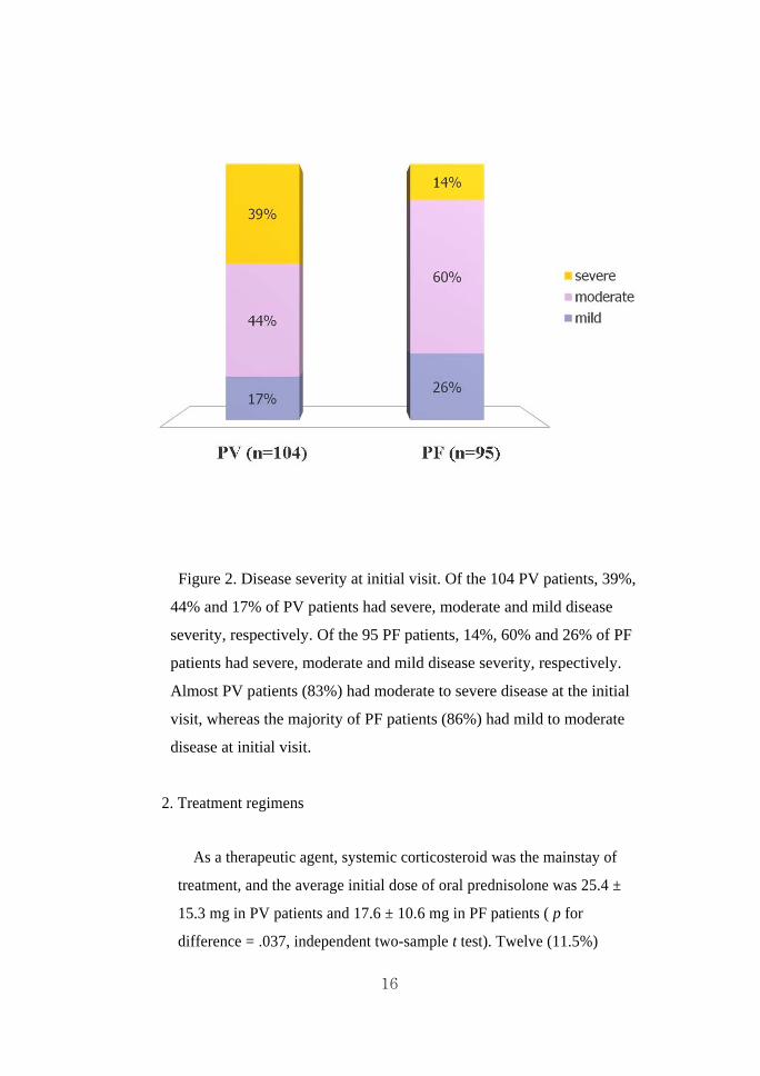
Figure 2. Disease severity at initial visit. Of the 104 PV patients, 39%,
44% and 17% of PV patients had severe, moderate and mild disease
severity, respectively. Of the 95 PF patients, 14%, 60% and 26% of PF
patients had severe, moderate and mild disease severity, respectively.
Almost PV patients (83%) had moderate to severe disease at the initial
visit, whereas the majority of PF patients (86%) had mild to moderate
disease at initial visit.
2. Treatment regimens
As a therapeutic agent, systemic corticosteroid was the mainstay of
treatment, and the average initial dose of oral prednisolone was 25.4 ±
15.3 mg in PV patients and 17.6 ± 10.6 mg in PF patients ( p for
difference = .037, independent two-sample t test). Twelve (11.5%)
16
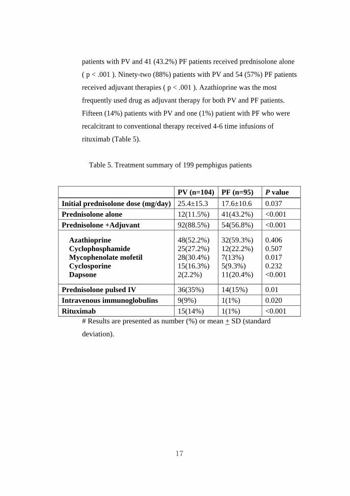
patients with PV and 41 (43.2%) PF patients received prednisolone alone
( p < .001 ). Ninety-two (88%) patients with PV and 54 (57%) PF patients
received adjuvant therapies ( p < .001 ). Azathioprine was the most
frequently used drug as adjuvant therapy for both PV and PF patients.
Fifteen (14%) patients with PV and one (1%) patient with PF who were
recalcitrant to conventional therapy received 4-6 time infusions of
rituximab (Table 5).
Table 5. Treatment summary of 199 pemphigus patients
PV (n=104) PF (n=95) P value
Initial prednisolone dose (mg/day) 25.4±15.3 17.6±10.6 0.037
Prednisolone alone 12(11.5%) 41(43.2%) <0.001
Prednisolone +Adjuvant 92(88.5%) 54(56.8%) <0.001
Azathioprine Cyclophosphamide Mycophenolate mofetil Cyclosporine Dapsone
48(52.2%) 25(27.2%) 28(30.4%) 15(16.3%) 2(2.2%)
32(59.3%) 12(22.2%) 7(13%) 5(9.3%) 11(20.4%)
0.406 0.507 0.017 0.232 <0.001
Prednisolone pulsed IV 36(35%) 14(15%) 0.01
Intravenous immunoglobulins 9(9%) 1(1%) 0.020
Rituximab 15(14%) 1(1%) <0.001 # Results are presented as number (%) or mean + SD (standard
deviation).
17
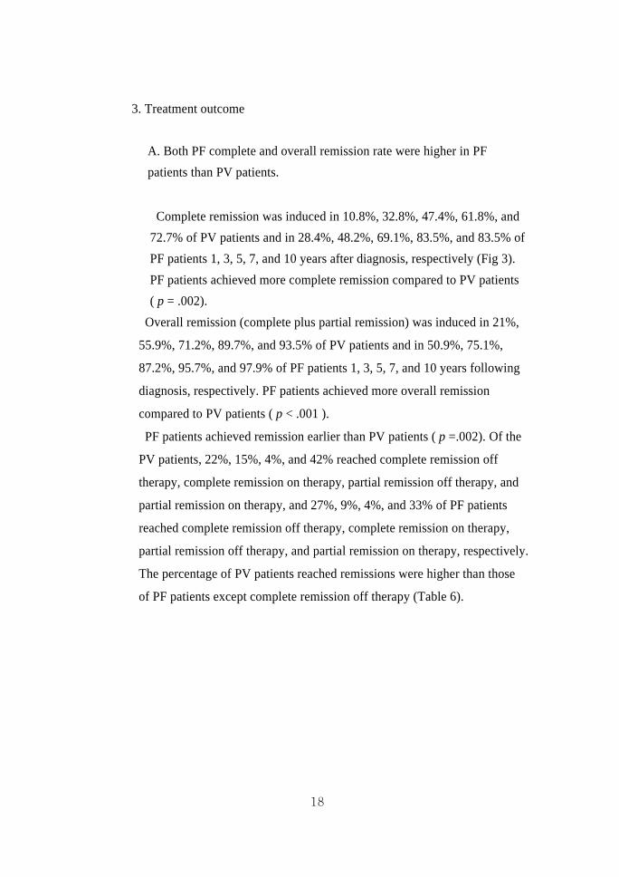
3. Treatment outcome
A. Both PF complete and overall remission rate were higher in PF
patients than PV patients.
Complete remission was induced in 10.8%, 32.8%, 47.4%, 61.8%, and
72.7% of PV patients and in 28.4%, 48.2%, 69.1%, 83.5%, and 83.5% of
PF patients 1, 3, 5, 7, and 10 years after diagnosis, respectively (Fig 3).
PF patients achieved more complete remission compared to PV patients
( p = .002).
Overall remission (complete plus partial remission) was induced in 21%,
55.9%, 71.2%, 89.7%, and 93.5% of PV patients and in 50.9%, 75.1%,
87.2%, 95.7%, and 97.9% of PF patients 1, 3, 5, 7, and 10 years following
diagnosis, respectively. PF patients achieved more overall remission
compared to PV patients ( p < .001 ).
PF patients achieved remission earlier than PV patients ( p =.002). Of the
PV patients, 22%, 15%, 4%, and 42% reached complete remission off
therapy, complete remission on therapy, partial remission off therapy, and
partial remission on therapy, and 27%, 9%, 4%, and 33% of PF patients
reached complete remission off therapy, complete remission on therapy,
partial remission off therapy, and partial remission on therapy, respectively.
The percentage of PV patients reached remissions were higher than those
of PF patients except complete remission off therapy (Table 6).
18
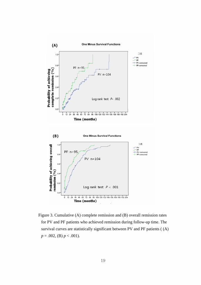
Figure 3. Cumulative (A) complete remission and (B) overall remission rates
for PV and PF patients who achieved remission during follow-up time. The
survival curves are statistically significant between PV and PF patients ( (A)
p = .002, (B) p < .001).
19
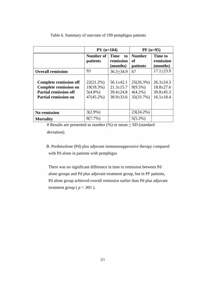
Table 6. Summary of outcome of 199 pemphigus patients
PV (n=104) PF (n=95)
Number ofpatients
Time to remission(months)
Number of patients
Time to remission (months)
Overall remission 93 36.5+34.9 67 17.1+23.9
Complete remission off Complete remission on Partial remission off Partial remission on
22(21.2%) 19(18.3%) 5(4.8%) 47(45.2%)
56.1±42.121.3±15.739.4±24.838.9±33.6
25(26.3%) 9(9.5%) 4(4.2%) 32(33.7%)
26.3±24.3 18.8±27.6 39.8±45.3 16.5±18.4
No remission 3(2.9%) 23(24.2%)
Mortality 8(7.7%) 5(5.3%)
# Results are presented as number (%) or mean + SD (standard
deviation).
B. Prednisolone (Pd) plus adjuvant immunosuppressive therapy compared
with Pd alone in patients with pemphigus
There was no significant difference in time to remission between Pd
alone groups and Pd plus adjuvant treatment group, but in PF patients,
Pd alone group achieved overall remission earlier than Pd plus adjuvant
treatment group ( p < .001 ).
20
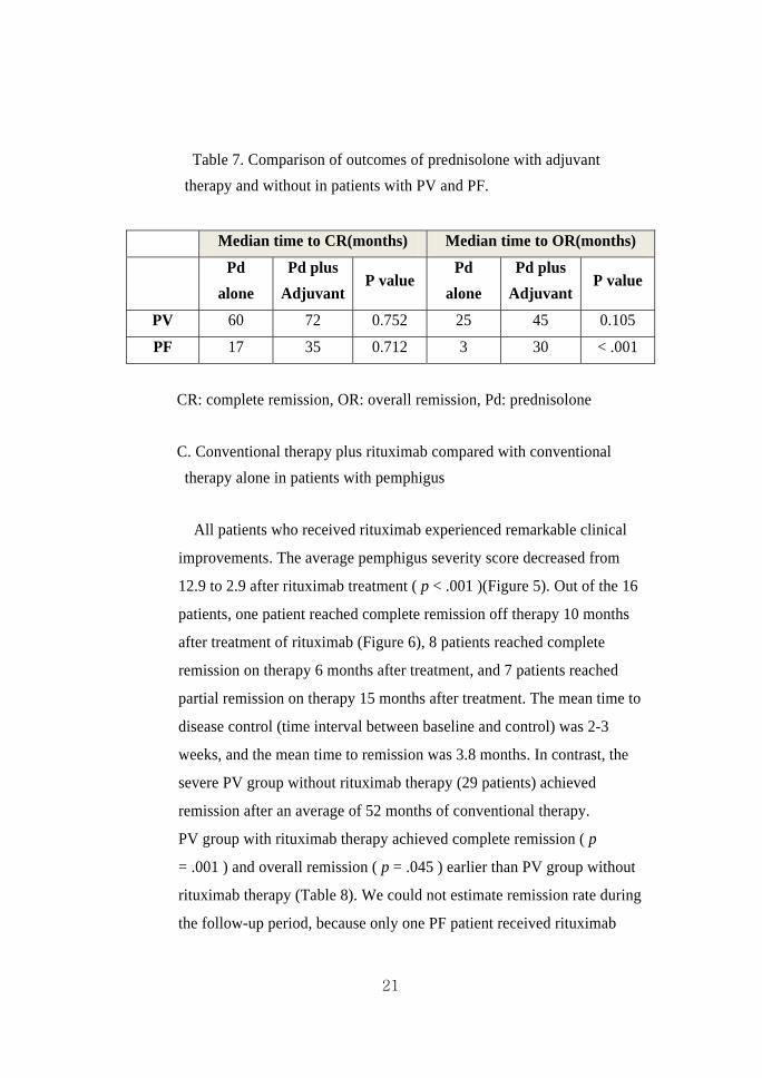
Table 7. Comparison of outcomes of prednisolone with adjuvant
therapy and without in patients with PV and PF.
Median time to CR(months) Median time to OR(months)
Pd
alone
Pd plus
Adjuvant P value
Pd
alone
Pd plus
Adjuvant P value
PV 60 72 0.752 25 45 0.105
PF 17 35 0.712 3 30 < .001
CR: complete remission, OR: overall remission, Pd: prednisolone
C. Conventional therapy plus rituximab compared with conventional
therapy alone in patients with pemphigus
All patients who received rituximab experienced remarkable clinical
improvements. The average pemphigus severity score decreased from
12.9 to 2.9 after rituximab treatment ( p < .001 )(Figure 5). Out of the 16
patients, one patient reached complete remission off therapy 10 months
after treatment of rituximab (Figure 6), 8 patients reached complete
remission on therapy 6 months after treatment, and 7 patients reached
partial remission on therapy 15 months after treatment. The mean time to
disease control (time interval between baseline and control) was 2-3
weeks, and the mean time to remission was 3.8 months. In contrast, the
severe PV group without rituximab therapy (29 patients) achieved
remission after an average of 52 months of conventional therapy.
PV group with rituximab therapy achieved complete remission ( p
= .001 ) and overall remission ( p = .045 ) earlier than PV group without
rituximab therapy (Table 8). We could not estimate remission rate during
the follow-up period, because only one PF patient received rituximab
21
therapy.
Out of the 16 patients who received rituximab, 5 patients relapsed after a
mean follow up period of 18.8 months, and these patients reached
remission again after two more infusions of rituximab. The time to
remission after the second rituximab infusion was 2 months. No
significant adverse effects were observed except one patient who
developed transient pruritic rash that occurred 30 minutes after infusion.
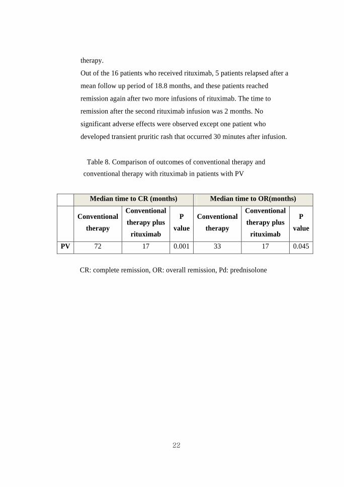
Table 8. Comparison of outcomes of conventional therapy and
conventional therapy with rituximab in patients with PV
Median time to CR (months) Median time to OR(months)
Conventional
therapy
Conventional
therapy plus
rituximab
P
value
Conventional
therapy
Conventional
therapy plus
rituximab
P
value
PV 72 17 0.001 33 17 0.045
CR: complete remission, OR: overall remission, Pd: prednisolone
22
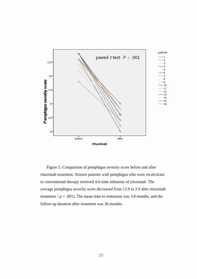
Figure 5. Comparison of pemphigus severity score before and after
rituximab treatment. Sixteen patients with pemphigus who were recalcitrant
to conventional therapy received 4-6 time infusions of rituximab. The
average pemphigus severity score decreased from 12.9 to 2.9 after rituximab
treatment ( p < .001). The mean time to remission was 3.8 months, and the
follow-up duration after treatment was 36 months.
23
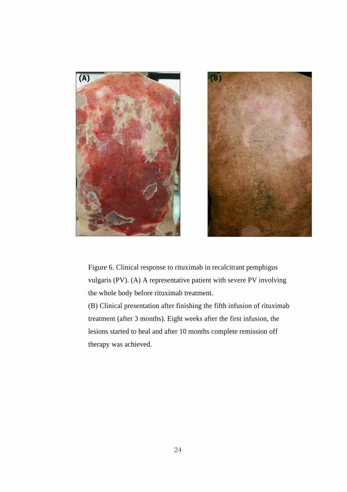
Figure 6. Clinical response to rituximab in recalcitrant pemphigus
vulgaris (PV). (A) A representative patient with severe PV involving
the whole body before rituximab treatment.
(B) Clinical presentation after finishing the fifth infusion of rituximab
treatment (after 3 months). Eight weeks after the first infusion, the
lesions started to heal and after 10 months complete remission off
therapy was achieved.
24
D. Mortality
During the 16-year follow-up period, eight (7%) PV patients and five
(5%) PF patients died (Table 6). Out of the PV deaths, sepsis was the
cause of death in three cases, and hepatic failure, lung cancer,
esophageal cancer, gastric perforation, and suicide was the cause of
death in each patient. Out of the PF deaths, four patients died due to
sepsis and one patient died due to cardiorespiratory failure.
25
IV. DISCUSSION
Pemphigus is a rare autoimmune blistering disorder varying in incidence
from 0.5 to 3.2 cases per 100,000 people per year.9 Lee10 reported that
pemphigus is the most common immunobullous disease in Korea, varying
in incidence from 0.1-0.5 cases per 100,000 people per year. We also
confirmed that pemphigus is the most common immunobullous disease in
Korea and bullous pemphigoid is the second common disease (unpublished
data).
The incidence of pemphigus subtype is dependent on the ethnic
background. In New York, Los Angeles, and Croatia where the Jewish,
Middle Eastern, and Mediterranean population predominates, the ratio of
PV to PF cases is approximately 5:1 whereas in Finland, it is only 0.5:1.0.11
Previous Korean studies revealed that PV was the predominant clinical
subtype, however, this study demonstrated that the prevalence of PV and
PF is almost same.4,5 A previous study from Korea revealed a female-to-
male ratio was 1.3:1, and Japanese study revealed female-to-male ratio was
2:1. 5,12 A study from Israel including 155 patients revealed the female-to-
male ratio was 1.5:1.13 However, in our study the female-to-male ratio was
approximately 1:1 for both PV and PF. This result is similar to the previous
studies from Malaysia and Finland.14,15
Pemphigus primarily affects middle-aged patients, and in our study the
mean age of onset was 46.0 years (PV, 47.0 years; PF, 45.0 years).
However, eight PV patients (7.7%) and 15 PF patients (15.8%) first
developed pemphigus lesions in their later teens or twenties. Therefore if
young patients present with blisters on their skin or in their oral cavity,
physicians should consider pemphigus as a differential diagnosis and
appropriately evaluate further.
26
Similar to other studies, we found that prednisolone and
immunosuppressive agents were the mainstays of therapy for our patients.2
We determined the treatment regimen based on pemphigus subtype, disease
severity and other associated diseases. In general, mild cases were treated
with systemic corticosteroid alone, and moderate and severe cases were
treated with a combination therapy of systemic corticosteroids and
immunosuppressants including azathioprine, mycophenolate mofetil,
cyclosporine and cyclophosphamide. The average initial dose of oral
prednisolone in PV patients was higher than that for PF patients ( p = .037 ),
and more PV patients received adjuvant therapies than PF patients ( p =
< .001 ). This is closely related to the fact that most PV patients had
moderate to severe disease, while most PF patients had mild to moderate
disease at the initial visit.
We used relatively small initial dose of prednisolone (0.3-0.5mg/kg/day)
to control the disease, while other institutes usually treated patients with
high initial dose of prednisolone (1mg/kg/day). This is because we favored
low dose of systemic corticosteroid in order to avoid long-term side effects.
In addition, patients were not likely to take high dose of corticosteroid,
because many of them had negative perception of systemic corticosteroid.
We found that in PV patients, there were no difference in remission
between prednisolone alone group and prednisolone with adjuvant therapy
group, but in PF patients, prednisolone alone group had more favorable
prognosis than prednisolone with adjuvant therapy group. This is because
PF group composed much more mild patients than PV. Therefore, we
consider that disease severity is a more important prognostic factor than
adjuvant therapy.
Although systemic corticosteroids, in combination with
immunosuppressive agents have improved prognosis of pemphigus
remarkably, treatment of pemphigus is still challenging. Long-term use of
27
immunosuppressive therapy also increase the chance of adverse effect or
complications. Furthermore some patients are unresponsive to conventional
immunosuppressive treatments, so novel effective therapy is required.
Rituximab is a chimeric monoclonal antibody that binds to the CD20
antigen on the surface of B cells and has been proved to be effective in
recalcitrant pemphigus.3,16 Joly16 et al. reported that 18 of 21 (86%) patients
with severe PV or PF had complete remissions three months after a single
cycle of four weekly infusions of rituximab. In the case reported by
Cianchini17 et al, 10 PV patients and 2 PF patients achieved prolonged
clinical remission after a single course of rituximab treatment. In this study,
all 16 patients who received rituximab showed successful controlling of the
disease, producing complete remission in 9 patients and partial remission in
7 patients during 16 month follow-up period. Also, PV group with
rituximab therapy achieved both complete remission and overall remission
significantly earlier than PV group without rituximab therapy. We confirmed
that rituximab is an effective and safe treatment option for patients
unresponsive to conventional therapies.
With regard to the treatment outcomes and prognoses, overall remission
(complete plus partial remission) was induced in 21%, 53%, 71%, 87%,
and 89% of PV patients 1, 3, 5, 10, and 15 years after diagnosis,
respectively. Herbst and Bystryn6 reported that complete and long-lasting
remission (no evidence of disease and no systemic therapy for at least six
months) was induced in 25%, 50%, and 75% of 40 PV patients 2, 5, and 10
years after diagnosis, respectively. In this study, we achieved more
remission rate than previous study.6 This difference is partially because our
definition of remission is more generous than those of previous studies and
we could introduce more effective therapy like rituximab in some patients.
This study confirmed that PF patients achieved higher rates of complete
and overall remission than PV patients11 ( P = .002, P < .001, respectively).
28
The overall mortality rate in pemphigus has been reported to be 5-9%18,
and the most common cause of death is attributed to the side effects of
treatment.1,19,20 In our study, the overall pemphigus mortality rate was
around 7% during the 16-year follow-up period and the most common
cause of death is sepsis which was comparable with the results of a
previous study. One of our patients who is a 49-year-old female with
severe PV committed suicide during treatment. As pemphigus is difficult to
control and is a relapsing disorder which destroys the patient's appearance
and requires long-term period of treatment, the disease creates significant
psychological problems for patients and may lead to depression and even
suicide.21 Therefore, we propose that doctors should carefully evaluate
pemphigus patients’ psychological distress and treat the patients
appropriately.
29
V. CONCLUSION
The aim of this study is to evaluate the clinical features, treatment outcomes
and long-term prognoses of Korean patients with pemphigus. The summary of
the results are described below.
1. The mean age of onset was 46.1 ± 14.6 years, and the male to female
ratio was equal.
2. Most patients with pemphigus reached complete plus partial
remission during follow-up period.
3. Rituximab is an effective and safe drug for severe pemphigus patients
who are resistant to conventional therapy with early remissions being
achieved.
In conclusion, this study is the first large scale follow-up study for
pemphigus patients of Korea. We conducted long-term follow-up
observation with the application of common terms from the International
Pemphigus Committee consensus statement to estimate treatment
outcomes. This study should enhance our understanding of clinical
characteristics and long-term prognosis in pemphigus patients.
30
REFERENCES
1 Bystryn JC, Steinman NM. The adjuvant therapy of pemphigus.
An update. Arch Dermatol 1996; 132: 203-12.
2 Ljubojevic S, Lipozencic J, Brenner S et al. Pemphigus vulgaris:
a review of treatment over a 19-year period. J Eur Acad Dermatol Venereol 2002; 16: 599-603.
3 Ahmed AR, Spigelman Z, Cavacini LA et al. Treatment of
pemphigus vulgaris with rituximab and intravenous immune
globulin. N Engl J Med 2006; 355: 1772-9.
4 Park BS, Chung JH. Treatment of pemphigus. Korean J Dermatol 1997: 465-74.
5 Seo PG, Choi WW, Chung JH. Pemphigus in Korea: clinical
manifestations and treatment protocol. J Dermatol 2003; 30:
782-8.
6 Herbst A, Bystryn JC. Patterns of remission in pemphigus
vulgaris. J Am Acad Dermatol 2000; 42: 422-7.
7 Ishii N, Maeyama Y, Karashima T et al. Immunoserological
analyses of 55 patients with pemphigus at the Dermatological
Department of Kurume University Hospital: an 11-year
retrospective study (1996-2006). Int J Dermatol 2008; 47:
1321-2.
8 Murrell DF, Dick S, Ahmed AR et al. Consensus statement on
definitions of disease, end points, and therapeutic response for
pemphigus. J Am Acad Dermatol 2008; 58: 1043-6.
9 Korman NJ. Pemphigus. Dermatol Clin 1990; 8: 689-700.
10 Lee CW. Autoimmune diseases of the skin. Seoul: Academia
2002: 9-42.
11 Stanley JR. Pemphigus. Fitzpatrick's dermatology in general medicine 2007.
12 Ishii N, Maeyama Y, Karashima T et al. A clinical study of
patients with pemphigus vulgaris and pemphigus foliaceous: an
11-year retrospective study (1996-2006). Clin Exp Dermatol 2008; 33: 641-3.
13 Mimouni D, Bar H, Gdalevich M et al. Pemphigus--analysis of
epidemiological factors in 155 patients. J Eur Acad Dermatol Venereol 2008; 22: 1232-5.
14 Adam. B. Bullous disease in Malaysia: Epidemiology and natural
history. Int J Dermatol 1992; 1: 42-5.
15 Hietanen J, Salo OP. Pemphigus: an epidemiological study of
patients treated in Finnish hospitals between 1969 and 1978.
31
Acta Derm Venereol 1982; 62: 491-6.
16 Joly P, Mouquet H, Roujeau JC et al. A single cycle of rituximab
for the treatment of severe pemphigus. N Engl J Med 2007; 357:
545-52.
17 Cianchini G, Corona R, Frezzolini A et al. Treatment of severe
pemphigus with rituximab: report of 12 cases and a review of
the literature. Arch Dermatol 2007; 143: 1033-8.
18 Korman N. Pemphigus. J Am Acad Dermatol 1988; 18: 1219-38.
19 Huilgol SC, Black MM. Management of the immunobullous
disorders. II. Pemphigus. Clin Exp Dermatol 1995; 20: 283-93.
20 Carson PJ, Hameed A, Ahmed AR. Influence of treatment on the
clinical course of pemphigus vulgaris. J Am Acad Dermatol 1996; 34: 645-52.
21 Namazi MR. Prescribing antidepressant drugs for pemphigus
patients: An important point to keep in mind. Dermatol Online J 2004; 10: 22.
32
< ABSTRACT(IN KOREAN)>
한국인 천포창:
16년 동안 199명의 환자를 대상으로 한
후향적 연구
<지도교수 김수찬>
연세대학교 대학원 의학과
김미리
배 경: 천포창(pemphigus)은 피부와 점막에 수포를 형성하
는 만성 수포성 질환으로서 심할 경우 사망까지 할 수 있
는 심각한 질환이다. 한국에서 천포창은 자가면역수포성 질
환 가장 빈도가 높으며, 치료하지 않을 경우 사망률이 높은
위중한 피부병이기 때문에 치료와 예후에 대한 분석이 매
우 중요하다.
목 적: 본 연구의 목적은 한국인 천포창 환자의 임상적 특
징, 치료 결과, 장기 예후 등을 분석하여 규명하는데 있다.
방 법: 본 연구자들은 지난 16년간 강남세브란스병원 피부
과 수포성질환 클리닉에서 진단 및 치료 한 199명의 천포
창 환자의 임상적 특징과 치료 효과 및 예후를 분석하고자
차트 리뷰를 통한 후향적 연구를 시행하였다.
결 과: 한국인 천포창 환자를 대상으로 한 이번 연구에서
는 심상성 천포창 (pemphigus vulgaris)과 낙엽상 천포창
33
(pemphigus foliaceus)이 같은 비율로 발생하였으며, 심상성
천포창과 낙엽상 천포창 모두에서 남성과 여성이 같은 발
생비율을 보였다. 평균적으로 중년에 질병이 발생하였으며,
평균 초발연령은 46세였다. 치료로는 전신적 스테로이드를
기본적으로 사용하였으며, 면역억제제 중에서는 azathioprine
이 가장 많이 사용되었다. 본 연구에서는 2008년 국제 천포
창 연합 (International Pemphigus Committee)에서 제시한 천포
창의 관해에 관한 정의에 관한 합의내용을 적용하여, 한국
인 천포창 환자의 치료후 결과에 대해 분석 하였다. 유병기
간에 따라 관해의 비율을 분석해 본 결과 심상성 천포창
환자는 1년, 5년, 10년 후에 각각 21%, 71%, 87%의 환자가
부분관해 이상에 도달하였으며, 낙엽상 천포창 환자는 각각
41%, 68%, 74%의 환자가 부분관해 이상에 도달하였다. 16년
간의 연구 기간 동안 총 8명의 심상성 천포창 환자와 5명
의 낙엽상 천포창 환자가 사망하였다. 기존 치료법에 잘 반
응하지 않는 난치성 천포창 환자 16명 (심상성 천포창: 15
명, 낙엽상 천포창: 1명)에게 rituximab을 투여하였고, 모든
환자가 평균적으로 rituximab 치료시작 3~4개월 후에 완전
관해나 부분관해에 도달하였다.
결 론: 199명의 한국인 천포창 환자를 대상으로 한 본 연
구에서 심상성 천포창과 낙엽상 천포창의 발병률의 차이는
없었으며, 성별에 따른 유병률도 동일하였다. 질병은 주로
중년에 시작하였으며, 사망률은 7%로, 이는 과거의 외국
및 국내 보고와 큰 차이가 없었다. 거의 대부분의 천포창
34
35
환자는 유병기간이 10년이 지나면 결국에는 관해에 도달하
였다. 또한 본 연구에서는 rituximab이 기존 치료에는 효과
가 없는 난치성 천포창 환자에게 사용할 수 있는 안전하고
효과적이며 빨리 관해에 도달하게 하는 유용한 약제라는
것을 확인하였다.
핵심되는 말: 심상성 천포창, 낙엽상 천포창, 한국인, 임상적
연구, 예후


![Oral Manifestations of Pemphigus Vulgaris: Clinical ... · bullous pemphigus, and paraneoplastic pemphigus [4]. The differential diagnosis includes other dermatological diseases with](https://img.pdfslide.us/doc/110x75/5cbb138688c9930c5f8bb27d/oral-manifestations-of-pemphigus-vulgaris-clinical-bullous-pemphigus-and.jpg)