Embed Size (px)
Citation preview
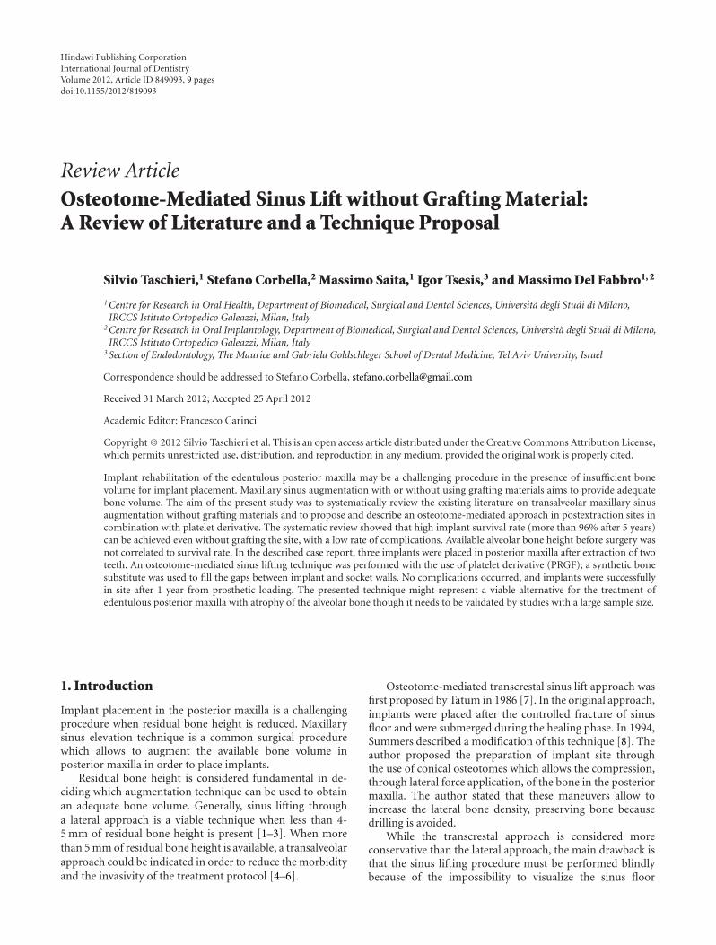
Hindawi Publishing CorporationInternational Journal of DentistryVolume 2012, Article ID 849093, 9 pagesdoi:10.1155/2012/849093
Review Article
Osteotome-Mediated Sinus Lift without Grafting Material:A Review of Literature and a Technique Proposal
Silvio Taschieri,1 Stefano Corbella,2 Massimo Saita,1 Igor Tsesis,3 and Massimo Del Fabbro1, 2
1 Centre for Research in Oral Health, Department of Biomedical, Surgical and Dental Sciences, Universita degli Studi di Milano,IRCCS Istituto Ortopedico Galeazzi, Milan, Italy
2 Centre for Research in Oral Implantology, Department of Biomedical, Surgical and Dental Sciences, Universita degli Studi di Milano,IRCCS Istituto Ortopedico Galeazzi, Milan, Italy
3 Section of Endodontology, The Maurice and Gabriela Goldschleger School of Dental Medicine, Tel Aviv University, Israel
Correspondence should be addressed to Stefano Corbella, [email protected]
Received 31 March 2012; Accepted 25 April 2012
Academic Editor: Francesco Carinci
Copyright © 2012 Silvio Taschieri et al. This is an open access article distributed under the Creative Commons Attribution License,which permits unrestricted use, distribution, and reproduction in any medium, provided the original work is properly cited.
Implant rehabilitation of the edentulous posterior maxilla may be a challenging procedure in the presence of insufficient bonevolume for implant placement. Maxillary sinus augmentation with or without using grafting materials aims to provide adequatebone volume. The aim of the present study was to systematically review the existing literature on transalveolar maxillary sinusaugmentation without grafting materials and to propose and describe an osteotome-mediated approach in postextraction sites incombination with platelet derivative. The systematic review showed that high implant survival rate (more than 96% after 5 years)can be achieved even without grafting the site, with a low rate of complications. Available alveolar bone height before surgery wasnot correlated to survival rate. In the described case report, three implants were placed in posterior maxilla after extraction of twoteeth. An osteotome-mediated sinus lifting technique was performed with the use of platelet derivative (PRGF); a synthetic bonesubstitute was used to fill the gaps between implant and socket walls. No complications occurred, and implants were successfullyin site after 1 year from prosthetic loading. The presented technique might represent a viable alternative for the treatment ofedentulous posterior maxilla with atrophy of the alveolar bone though it needs to be validated by studies with a large sample size.
1. Introduction
Implant placement in the posterior maxilla is a challengingprocedure when residual bone height is reduced. Maxillarysinus elevation technique is a common surgical procedurewhich allows to augment the available bone volume inposterior maxilla in order to place implants.
Residual bone height is considered fundamental in de-ciding which augmentation technique can be used to obtainan adequate bone volume. Generally, sinus lifting througha lateral approach is a viable technique when less than 4-5 mm of residual bone height is present [1–3]. When morethan 5 mm of residual bone height is available, a transalveolarapproach could be indicated in order to reduce the morbidityand the invasivity of the treatment protocol [4–6].
Osteotome-mediated transcrestal sinus lift approach wasfirst proposed by Tatum in 1986 [7]. In the original approach,implants were placed after the controlled fracture of sinusfloor and were submerged during the healing phase. In 1994,Summers described a modification of this technique [8]. Theauthor proposed the preparation of implant site throughthe use of conical osteotomes which allows the compression,through lateral force application, of the bone in the posteriormaxilla. The author stated that these maneuvers allow toincrease the lateral bone density, preserving bone becausedrilling is avoided.
While the transcrestal approach is considered moreconservative than the lateral approach, the main drawback isthat the sinus lifting procedure must be performed blindlybecause of the impossibility to visualize the sinus floor
2 International Journal of Dentistry
[5, 6]. In spite of this limitation, membrane perforationwas reported to be less frequent in the osteotome-mediatedprocedure [6] than in the lateral approach, for which suchcomplication was described in 25–44% of cases [9–11].
Transcrestal, osteotome-mediated sinus lift surgery maybe performed with or without the use of bone graftingmaterial as allograft, autogenous bone, or heterologous bonematerial [6]. No significant differences in terms of implantsurvival and success rates were observed comparing the twomethods [6]. Also, the use of platelet derivatives without anybone substitute is described in literature [12, 13] with theaim of allowing a better control of forces during sinus floorelevation and reducing the incidence of complications.
The aim of this study was to perform a literature reviewregarding osteotome-mediated sinus lifting without bonegrafting material and to present a technique to perform theprocedure with the use of plasma rich in growth factors(PRGFs).
2. Literature Review
2.1. Materials and Methods. An electronic search was con-ducted via MEDLINE (PubMed) in the dental literatureto select human clinical trials published from 1986 toJanuary 2012. The search terms used were “sinus lift,” “sinusaugmentation,” “sinus grafting,” “sinus elevation” alone or incombination with “osteotome,” “dental implants,” “crestal,”and “transalveolar” using boolean operator “AND” and werechosen accordingly with previously published reviews [1, 5,6]. Bibliographies of the selected articles were also manuallysearched.
Inclusion criteria for the studies were
(i) studies concerning osteotome-mediated sinus liftingprocedure without using grafting materials;
(ii) a minimum of 1-year followup after prosthetic re-habilitation;
(iii) at least 20 patients treated;
(iv) data on implant survival (SR) were reported.
Two authors (SC and MDF) independently screenedabstracts and fulltext of the eligible articles for possibleinclusion. In case of disagreement, a joint decision was takenby discussion.
Data from selected studies were extracted and recordedin a previously designed electronic form.
The fulltext of each included study was reviewed, and thefollowing parameters were extracted:
(i) demographics of treated patients (age, gender, samplesize);
(ii) bone height (distance between bone crest and floor ofthe sinus);
(iii) implant length;
(iv) Implant survival rate;
(v) surgical or postsurgical complications;
(vi) causes and occurrence of implant failure.
80
70
60
50
40
30
20
10
66.04%
16.98%
9.43% 7.55%
0%
0
3–5
year
s
Mor
e th
an5
year
s
0-1
year
1-2
year
s
2-3
year
s
(%)
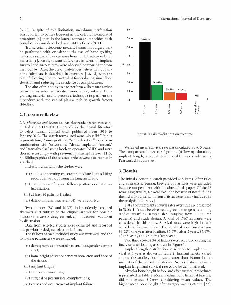
Figure 1: Failures distribution over time.
Weighted mean survival rate was calculated up to 5 years.The comparison between subgroups (follow-up duration,implant length, residual bone height) was made usingPearson’s chi square test.
3. Results
The initial electronic search provided 438 items. After titlesand abstracts screening, they are 361 articles were excludedbecause not pertinent with the aims of this paper. Of the 77remaining articles, 62 were excluded because of not fulfillingthe inclusion criteria. Fifteen articles were finally included inthe analysis [12, 14–27].
Data about implant survival rates over time are presentedin Table 1. It can be observed a great heterogeneity amongstudies regarding sample size (ranging from 20 to 983patients) and study design. A total of 1767 implants wereconsidered in this study. Survival rates were high in eachconsidered follow-up time. The weighted mean survival was98.02% one year after loading, 97.37% after 2 years, 97.47%after 3 years, and 96.77% after 5 years.
Two thirds (66.04%) of failures were recorded during thefirst year after loading as shown in Figure 1.
Implant length distribution in relation to implant sur-vival at 1 year is shown in Table 2. Implant length variedamong the studies, but it was greater than 10 mm in themajority of the considered studies. No correlation betweenimplant length and survival rate could be demonstrated.
Alveolar bone height before and after surgical proceduresis presented in Table 2. Mean residual bone height at baselinedid not exceed 8.2 mm considering mean values. Thehigher mean bone height after surgery was 13.28 mm [27].

International Journal of Dentistry 3
Table 1: Cumulative implant survival rates.
Study N n 1 y n 2 y n 3 y n 5 y
Fermergard and Astrand [14] 53 53 96,23 50 94,30
Tetsch et al. [15] 983 983 98,88 887 97,88 805 98,39 529 97,83
Bruschi et al. [16] 66 66 95,45 63 95,45 63 95,45 63 95,45
Gabbert et al. [17] 92 92 95,65 83 95,65 83 95,65
Jurisic et al. [18] 40 40 100,00 40 100,00 40 100,00
Nedir et al. [19] 25 25 100,00 25 100,00 25 100,00
Nedir et al. [20] 54 54 100,00
Cavicchia et al. [21] 97 97 89,69 87 89,69 87 89,69 86 88,65
Diss et al. [12] 35 35 97,14
Schmidlin et al. [22] 24 24 100,00 24 100,00
Leblebicioglu et al. [23] 75 75 97,33 73 97,33
Fugazzotto [24] 114 114 98,25 83 98,25 40 98,25
Volpe et al. [25] 20 20 100,00
Bruschi et al. [27] 68 68 100,00 68 100,00 68 100,00 68 100,00
Fornell et al. [26] 21 21 100,00
n Total 1767 1767 98,02 1433 97,37 1261 97,47 746 96,77
Table 2: Bone height before and after surgery.
Study Mean implant length Mean ± SD (range) before surgery Mean ± SD after surgery
Fermergard and Astrand [14] 10,89 6,3 ± 0,3 10,7 ± 0,3
Tetsch et al. [15] 11,50 8,2 3,3
Bruschi et al. [16] 13,57 1–3 13,28 ± 1,23
Gabbert et al. [17] 10,29 NE NE
Jurisic et al. [18] 10,72 NE NE
Nedir et al. [19] 9,60 5,4 ± 2,3 10,3 ± 2,2
Nedir et al. [20] 8,37 2,5 ± 1,7 6,3 ± 1,5
Cavicchia et al. [21] 12,30 NE NE
Diss et al. [12] 10,51 6,5 ± 1,7 9,8 ± 1,5
Schmidlin et al. [22] 8,60 5,0 ± 1,5 8,6 ± 1,3
Leblebicioglu et al. [23] >11 mm 7 ± 1,3 10,9 ± 1,7
Fugazzotto [24] 9,16 NE NE
Volpe et al. [25] NR 7.2 ± 1.5 10.0 ± 1.0
Bruschi et al. [27] 13,50 6.02 ± 0,75 7.99 ± 1.16
Fornell et al. [26] 10,00 5.6 ± 2.1 8.6 ± 2.1
No correlation could be found between bone height andimplant survival rate.
4. Technique Description and Case Report
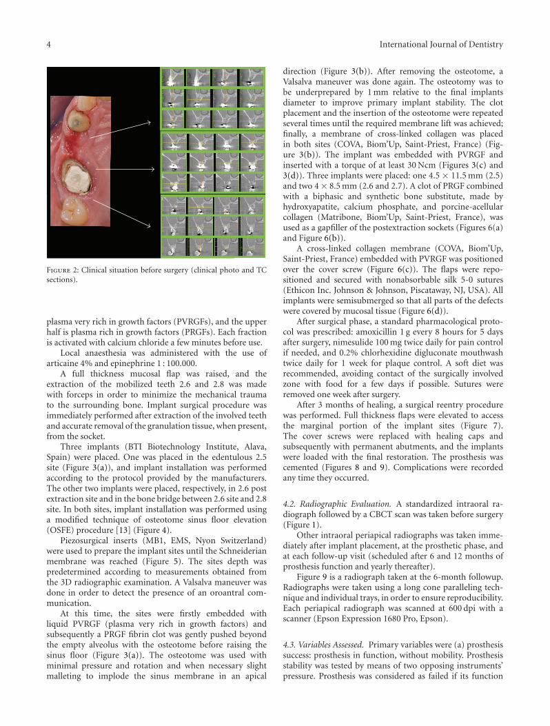
A 45-year-old male patient, in general good health (ASA1), nonsmoker, presented with a first left maxillary molar(2.6) exhibiting a destructive caries and referring vague,nonspecific symptoms. Radiographic examination revealedthe presence of periradicular lesion of strictly endodonticorigin, and a suitable restoration was considered unfeasible.In the same quadrant, the maxillary second premolar andsecond molar (2.5 and 2.7) were missing. Moreover, a tiltedwisdom teeth (2.8) showed a lateral and vertical mobility
associated with a pathological periodontal status (Figure 2).An experienced surgeon (ST) performed the entire sur-
gical procedure.
4.1. Surgical Procedure. Preoperatively all patients rinsedwith a 0.2% chlorhexidine solution for a minute as an an-tiseptic treatment in order to reduce the contamination ofthe surgical field.
Patients’ peripheral blood was collected using citratedtubes in order to prepare the platelet concentrate [28–30]. Briefly, the platelet concentrate is obtained by one-step centrifugation process (580 g for 8 minutes). Thesupernatant is then separated into two fractions paying carenot to collect the leukocyte-rich layer: the deeper half is
4 International Journal of Dentistry
Figure 2: Clinical situation before surgery (clinical photo and TCsections).
plasma very rich in growth factors (PVRGFs), and the upperhalf is plasma rich in growth factors (PRGFs). Each fractionis activated with calcium chloride a few minutes before use.
Local anaesthesia was administered with the use ofarticaine 4% and epinephrine 1 : 100.000.
A full thickness mucosal flap was raised, and theextraction of the mobilized teeth 2.6 and 2.8 was madewith forceps in order to minimize the mechanical traumato the surrounding bone. Implant surgical procedure wasimmediately performed after extraction of the involved teethand accurate removal of the granulation tissue, when present,from the socket.
Three implants (BTI Biotechnology Institute, Alava,Spain) were placed. One was placed in the edentulous 2.5site (Figure 3(a)), and implant installation was performedaccording to the protocol provided by the manufacturers.The other two implants were placed, respectively, in 2.6 postextraction site and in the bone bridge between 2.6 site and 2.8site. In both sites, implant installation was performed usinga modified technique of osteotome sinus floor elevation(OSFE) procedure [13] (Figure 4).
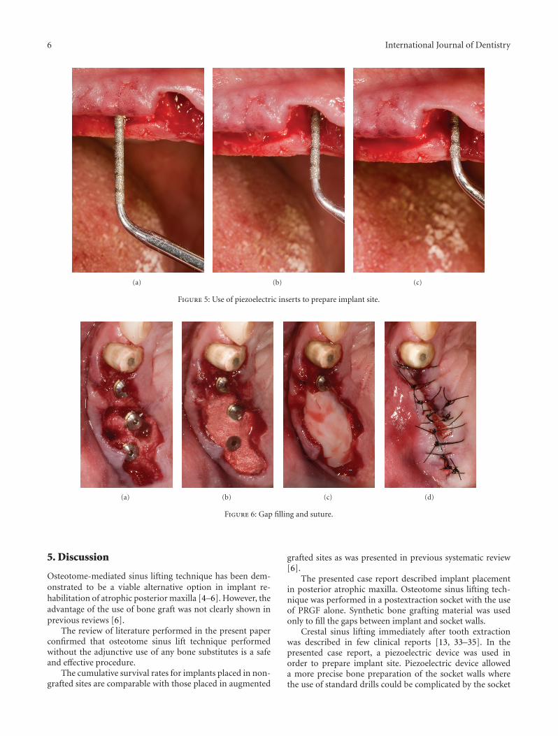
Piezosurgical inserts (MB1, EMS, Nyon Switzerland)were used to prepare the implant sites until the Schneiderianmembrane was reached (Figure 5). The sites depth waspredetermined according to measurements obtained fromthe 3D radiographic examination. A Valsalva maneuver wasdone in order to detect the presence of an oroantral com-munication.
At this time, the sites were firstly embedded withliquid PVRGF (plasma very rich in growth factors) andsubsequently a PRGF fibrin clot was gently pushed beyondthe empty alveolus with the osteotome before raising thesinus floor (Figure 3(a)). The osteotome was used withminimal pressure and rotation and when necessary slightmalleting to implode the sinus membrane in an apical
direction (Figure 3(b)). After removing the osteotome, aValsalva maneuver was done again. The osteotomy was tobe underprepared by 1 mm relative to the final implantsdiameter to improve primary implant stability. The clotplacement and the insertion of the osteotome were repeatedseveral times until the required membrane lift was achieved;finally, a membrane of cross-linked collagen was placedin both sites (COVA, Biom’Up, Saint-Priest, France) (Fig-ure 3(b)). The implant was embedded with PVRGF andinserted with a torque of at least 30 Ncm (Figures 3(c) and3(d)). Three implants were placed: one 4.5 × 11.5 mm (2.5)and two 4 × 8.5 mm (2.6 and 2.7). A clot of PRGF combinedwith a biphasic and synthetic bone substitute, made byhydroxyapatite, calcium phosphate, and porcine-acellularcollagen (Matribone, Biom’Up, Saint-Priest, France), wasused as a gapfiller of the postextraction sockets (Figures 6(a)and Figure 6(b)).
A cross-linked collagen membrane (COVA, Biom’Up,Saint-Priest, France) embedded with PVRGF was positionedover the cover screw (Figure 6(c)). The flaps were repo-sitioned and secured with nonabsorbable silk 5-0 sutures(Ethicon Inc. Johnson & Johnson, Piscataway, NJ, USA). Allimplants were semisubmerged so that all parts of the defectswere covered by mucosal tissue (Figure 6(d)).
After surgical phase, a standard pharmacological proto-col was prescribed: amoxicillin 1 g every 8 hours for 5 daysafter surgery, nimesulide 100 mg twice daily for pain controlif needed, and 0.2% chlorhexidine digluconate mouthwashtwice daily for 1 week for plaque control. A soft diet wasrecommended, avoiding contact of the surgically involvedzone with food for a few days if possible. Sutures wereremoved one week after surgery.
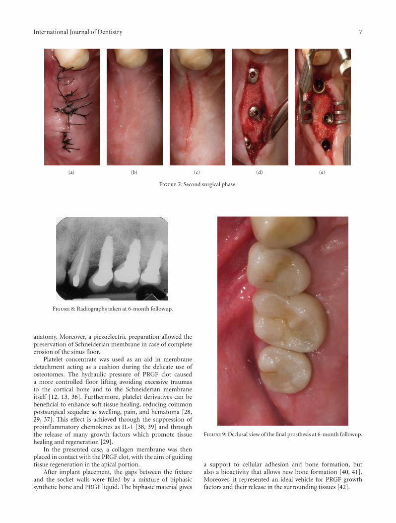
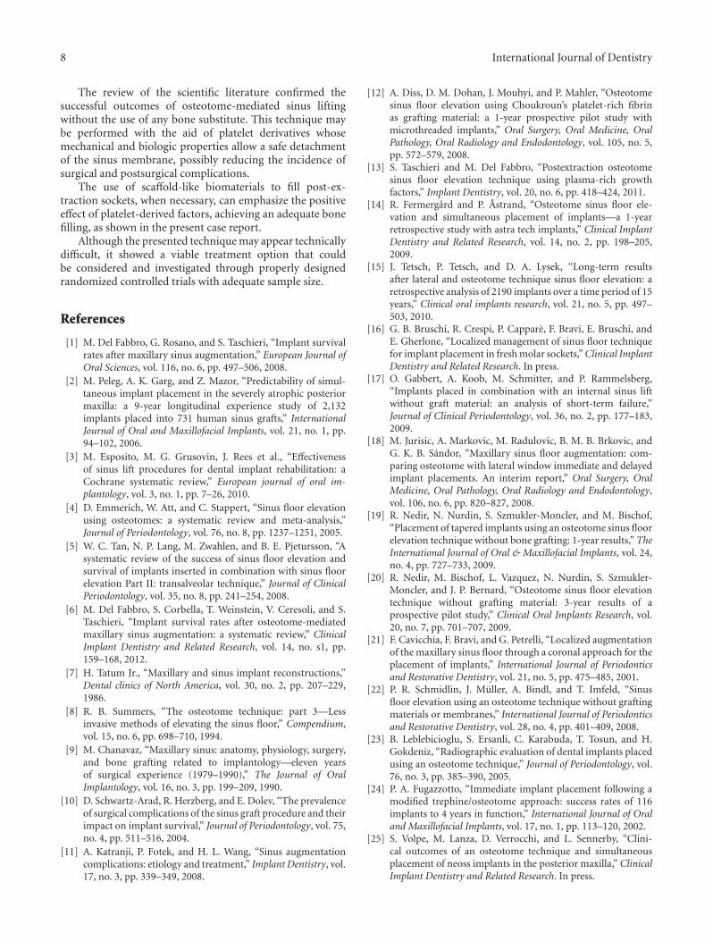
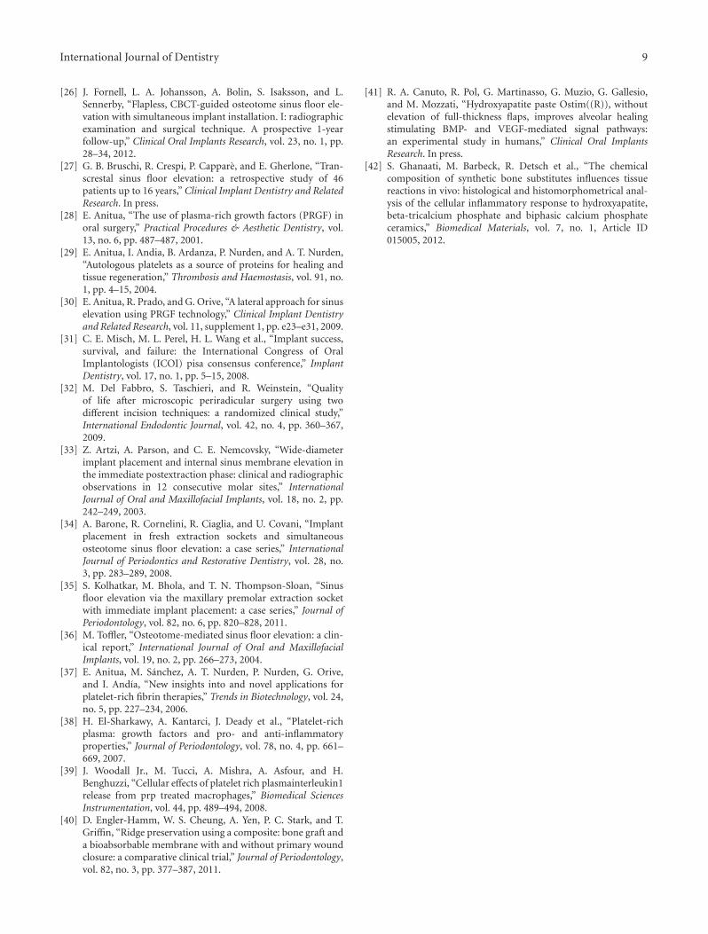
After 3 months of healing, a surgical reentry procedurewas performed. Full thickness flaps were elevated to accessthe marginal portion of the implant sites (Figure 7).The cover screws were replaced with healing caps andsubsequently with permanent abutments, and the implantswere loaded with the final restoration. The prosthesis wascemented (Figures 8 and 9). Complications were recordedany time they occurred.
4.2. Radiographic Evaluation. A standardized intraoral ra-diograph followed by a CBCT scan was taken before surgery(Figure 1).
Other intraoral periapical radiographs was taken imme-diately after implant placement, at the prosthetic phase, andat each follow-up visit (scheduled after 6 and 12 months ofprosthesis function and yearly thereafter).
Figure 9 is a radiograph taken at the 6-month followup.Radiographs were taken using a long cone paralleling tech-nique and individual trays, in order to ensure reproducibility.Each periapical radiograph was scanned at 600 dpi with ascanner (Epson Expression 1680 Pro, Epson).
4.3. Variables Assessed. Primary variables were (a) prosthesissuccess: prosthesis in function, without mobility. Prosthesisstability was tested by means of two opposing instruments’pressure. Prosthesis was considered as failed if its function
International Journal of Dentistry 5
(a) (b) (c) (d)
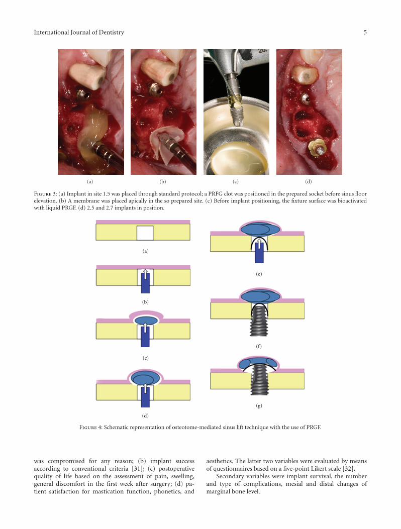
Figure 3: (a) Implant in site 1.5 was placed through standard protocol; a PRFG clot was positioned in the prepared socket before sinus floorelevation. (b) A membrane was placed apically in the so prepared site. (c) Before implant positioning, the fixture surface was bioactivatedwith liquid PRGF. (d) 2.5 and 2.7 implants in position.
(a)
(b)
(c)
(d)
(e)
(f)
(g)
Figure 4: Schematic representation of osteotome-mediated sinus lift technique with the use of PRGF.
was compromised for any reason; (b) implant successaccording to conventional criteria [31]; (c) postoperativequality of life based on the assessment of pain, swelling,general discomfort in the first week after surgery; (d) pa-tient satisfaction for mastication function, phonetics, and
aesthetics. The latter two variables were evaluated by meansof questionnaires based on a five-point Likert scale [32].
Secondary variables were implant survival, the numberand type of complications, mesial and distal changes ofmarginal bone level.
6 International Journal of Dentistry
(a) (b) (c)
Figure 5: Use of piezoelectric inserts to prepare implant site.
(a) (b) (c) (d)
Figure 6: Gap filling and suture.
5. Discussion
Osteotome-mediated sinus lifting technique has been dem-onstrated to be a viable alternative option in implant re-habilitation of atrophic posterior maxilla [4–6]. However, theadvantage of the use of bone graft was not clearly shown inprevious reviews [6].
The review of literature performed in the present paperconfirmed that osteotome sinus lift technique performedwithout the adjunctive use of any bone substitutes is a safeand effective procedure.
The cumulative survival rates for implants placed in non-grafted sites are comparable with those placed in augmented
grafted sites as was presented in previous systematic review[6].
The presented case report described implant placementin posterior atrophic maxilla. Osteotome sinus lifting tech-nique was performed in a postextraction socket with the useof PRGF alone. Synthetic bone grafting material was usedonly to fill the gaps between implant and socket walls.
Crestal sinus lifting immediately after tooth extractionwas described in few clinical reports [13, 33–35]. In thepresented case report, a piezoelectric device was used inorder to prepare implant site. Piezoelectric device alloweda more precise bone preparation of the socket walls wherethe use of standard drills could be complicated by the socket
International Journal of Dentistry 7
(a) (b) (c) (d) (e)
Figure 7: Second surgical phase.
Figure 8: Radiographs taken at 6-month followup.
anatomy. Moreover, a piezoelectric preparation allowed thepreservation of Schneiderian membrane in case of completeerosion of the sinus floor.
Platelet concentrate was used as an aid in membranedetachment acting as a cushion during the delicate use ofosteotomes. The hydraulic pressure of PRGF clot causeda more controlled floor lifting avoiding excessive traumasto the cortical bone and to the Schneiderian membraneitself [12, 13, 36]. Furthermore, platelet derivatives can bebeneficial to enhance soft tissue healing, reducing commonpostsurgical sequelae as swelling, pain, and hematoma [28,29, 37]. This effect is achieved through the suppression ofproinflammatory chemokines as IL-1 [38, 39] and throughthe release of many growth factors which promote tissuehealing and regeneration [29].
In the presented case, a collagen membrane was thenplaced in contact with the PRGF clot, with the aim of guidingtissue regeneration in the apical portion.
After implant placement, the gaps between the fixtureand the socket walls were filled by a mixture of biphasicsynthetic bone and PRGF liquid. The biphasic material gives
Figure 9: Occlusal view of the final prosthesis at 6-month followup.
a support to cellular adhesion and bone formation, butalso a bioactivity that allows new bone formation [40, 41].Moreover, it represented an ideal vehicle for PRGF growthfactors and their release in the surrounding tissues [42].
8 International Journal of Dentistry
The review of the scientific literature confirmed thesuccessful outcomes of osteotome-mediated sinus liftingwithout the use of any bone substitute. This technique maybe performed with the aid of platelet derivatives whosemechanical and biologic properties allow a safe detachmentof the sinus membrane, possibly reducing the incidence ofsurgical and postsurgical complications.
The use of scaffold-like biomaterials to fill post-ex-traction sockets, when necessary, can emphasize the positiveeffect of platelet-derived factors, achieving an adequate bonefilling, as shown in the present case report.
Although the presented technique may appear technicallydifficult, it showed a viable treatment option that couldbe considered and investigated through properly designedrandomized controlled trials with adequate sample size.
References
[1] M. Del Fabbro, G. Rosano, and S. Taschieri, “Implant survivalrates after maxillary sinus augmentation,” European Journal ofOral Sciences, vol. 116, no. 6, pp. 497–506, 2008.
[2] M. Peleg, A. K. Garg, and Z. Mazor, “Predictability of simul-taneous implant placement in the severely atrophic posteriormaxilla: a 9-year longitudinal experience study of 2,132implants placed into 731 human sinus grafts,” InternationalJournal of Oral and Maxillofacial Implants, vol. 21, no. 1, pp.94–102, 2006.
[3] M. Esposito, M. G. Grusovin, J. Rees et al., “Effectivenessof sinus lift procedures for dental implant rehabilitation: aCochrane systematic review,” European journal of oral im-plantology, vol. 3, no. 1, pp. 7–26, 2010.
[4] D. Emmerich, W. Att, and C. Stappert, “Sinus floor elevationusing osteotomes: a systematic review and meta-analysis,”Journal of Periodontology, vol. 76, no. 8, pp. 1237–1251, 2005.
[5] W. C. Tan, N. P. Lang, M. Zwahlen, and B. E. Pjetursson, “Asystematic review of the success of sinus floor elevation andsurvival of implants inserted in combination with sinus floorelevation Part II: transalveolar technique,” Journal of ClinicalPeriodontology, vol. 35, no. 8, pp. 241–254, 2008.
[6] M. Del Fabbro, S. Corbella, T. Weinstein, V. Ceresoli, and S.Taschieri, “Implant survival rates after osteotome-mediatedmaxillary sinus augmentation: a systematic review,” ClinicalImplant Dentistry and Related Research, vol. 14, no. s1, pp.159–168, 2012.
[7] H. Tatum Jr., “Maxillary and sinus implant reconstructions,”Dental clinics of North America, vol. 30, no. 2, pp. 207–229,1986.
[8] R. B. Summers, “The osteotome technique: part 3—Lessinvasive methods of elevating the sinus floor,” Compendium,vol. 15, no. 6, pp. 698–710, 1994.
[9] M. Chanavaz, “Maxillary sinus: anatomy, physiology, surgery,and bone grafting related to implantology—eleven yearsof surgical experience (1979–1990),” The Journal of OralImplantology, vol. 16, no. 3, pp. 199–209, 1990.
[10] D. Schwartz-Arad, R. Herzberg, and E. Dolev, “The prevalenceof surgical complications of the sinus graft procedure and theirimpact on implant survival,” Journal of Periodontology, vol. 75,no. 4, pp. 511–516, 2004.
[11] A. Katranji, P. Fotek, and H. L. Wang, “Sinus augmentationcomplications: etiology and treatment,” Implant Dentistry, vol.17, no. 3, pp. 339–349, 2008.
[12] A. Diss, D. M. Dohan, J. Mouhyi, and P. Mahler, “Osteotomesinus floor elevation using Choukroun’s platelet-rich fibrinas grafting material: a 1-year prospective pilot study withmicrothreaded implants,” Oral Surgery, Oral Medicine, OralPathology, Oral Radiology and Endodontology, vol. 105, no. 5,pp. 572–579, 2008.
[13] S. Taschieri and M. Del Fabbro, “Postextraction osteotomesinus floor elevation technique using plasma-rich growthfactors,” Implant Dentistry, vol. 20, no. 6, pp. 418–424, 2011.
[14] R. Fermergard and P. Astrand, “Osteotome sinus floor ele-vation and simultaneous placement of implants—a 1-yearretrospective study with astra tech implants,” Clinical ImplantDentistry and Related Research, vol. 14, no. 2, pp. 198–205,2009.
[15] J. Tetsch, P. Tetsch, and D. A. Lysek, “Long-term resultsafter lateral and osteotome technique sinus floor elevation: aretrospective analysis of 2190 implants over a time period of 15years,” Clinical oral implants research, vol. 21, no. 5, pp. 497–503, 2010.
[16] G. B. Bruschi, R. Crespi, P. Cappare, F. Bravi, E. Bruschi, andE. Gherlone, “Localized management of sinus floor techniquefor implant placement in fresh molar sockets,” Clinical ImplantDentistry and Related Research. In press.
[17] O. Gabbert, A. Koob, M. Schmitter, and P. Rammelsberg,“Implants placed in combination with an internal sinus liftwithout graft material: an analysis of short-term failure,”Journal of Clinical Periodontology, vol. 36, no. 2, pp. 177–183,2009.
[18] M. Jurisic, A. Markovic, M. Radulovic, B. M. B. Brkovic, andG. K. B. Sandor, “Maxillary sinus floor augmentation: com-paring osteotome with lateral window immediate and delayedimplant placements. An interim report,” Oral Surgery, OralMedicine, Oral Pathology, Oral Radiology and Endodontology,vol. 106, no. 6, pp. 820–827, 2008.
[19] R. Nedir, N. Nurdin, S. Szmukler-Moncler, and M. Bischof,“Placement of tapered implants using an osteotome sinus floorelevation technique without bone grafting: 1-year results,” TheInternational Journal of Oral & Maxillofacial Implants, vol. 24,no. 4, pp. 727–733, 2009.
[20] R. Nedir, M. Bischof, L. Vazquez, N. Nurdin, S. Szmukler-Moncler, and J. P. Bernard, “Osteotome sinus floor elevationtechnique without grafting material: 3-year results of aprospective pilot study,” Clinical Oral Implants Research, vol.20, no. 7, pp. 701–707, 2009.
[21] F. Cavicchia, F. Bravi, and G. Petrelli, “Localized augmentationof the maxillary sinus floor through a coronal approach for theplacement of implants,” International Journal of Periodonticsand Restorative Dentistry, vol. 21, no. 5, pp. 475–485, 2001.
[22] P. R. Schmidlin, J. Muller, A. Bindl, and T. Imfeld, “Sinusfloor elevation using an osteotome technique without graftingmaterials or membranes,” International Journal of Periodonticsand Restorative Dentistry, vol. 28, no. 4, pp. 401–409, 2008.
[23] B. Leblebicioglu, S. Ersanli, C. Karabuda, T. Tosun, and H.Gokdeniz, “Radiographic evaluation of dental implants placedusing an osteotome technique,” Journal of Periodontology, vol.76, no. 3, pp. 385–390, 2005.
[24] P. A. Fugazzotto, “Immediate implant placement following amodified trephine/osteotome approach: success rates of 116implants to 4 years in function,” International Journal of Oraland Maxillofacial Implants, vol. 17, no. 1, pp. 113–120, 2002.
[25] S. Volpe, M. Lanza, D. Verrocchi, and L. Sennerby, “Clini-cal outcomes of an osteotome technique and simultaneousplacement of neoss implants in the posterior maxilla,” ClinicalImplant Dentistry and Related Research. In press.
International Journal of Dentistry 9
[26] J. Fornell, L. A. Johansson, A. Bolin, S. Isaksson, and L.Sennerby, “Flapless, CBCT-guided osteotome sinus floor ele-vation with simultaneous implant installation. I: radiographicexamination and surgical technique. A prospective 1-yearfollow-up,” Clinical Oral Implants Research, vol. 23, no. 1, pp.28–34, 2012.
[27] G. B. Bruschi, R. Crespi, P. Cappare, and E. Gherlone, “Tran-screstal sinus floor elevation: a retrospective study of 46patients up to 16 years,” Clinical Implant Dentistry and RelatedResearch. In press.
[28] E. Anitua, “The use of plasma-rich growth factors (PRGF) inoral surgery,” Practical Procedures & Aesthetic Dentistry, vol.13, no. 6, pp. 487–487, 2001.
[29] E. Anitua, I. Andia, B. Ardanza, P. Nurden, and A. T. Nurden,“Autologous platelets as a source of proteins for healing andtissue regeneration,” Thrombosis and Haemostasis, vol. 91, no.1, pp. 4–15, 2004.
[30] E. Anitua, R. Prado, and G. Orive, “A lateral approach for sinuselevation using PRGF technology,” Clinical Implant Dentistryand Related Research, vol. 11, supplement 1, pp. e23–e31, 2009.
[31] C. E. Misch, M. L. Perel, H. L. Wang et al., “Implant success,survival, and failure: the International Congress of OralImplantologists (ICOI) pisa consensus conference,” ImplantDentistry, vol. 17, no. 1, pp. 5–15, 2008.
[32] M. Del Fabbro, S. Taschieri, and R. Weinstein, “Qualityof life after microscopic periradicular surgery using twodifferent incision techniques: a randomized clinical study,”International Endodontic Journal, vol. 42, no. 4, pp. 360–367,2009.
[33] Z. Artzi, A. Parson, and C. E. Nemcovsky, “Wide-diameterimplant placement and internal sinus membrane elevation inthe immediate postextraction phase: clinical and radiographicobservations in 12 consecutive molar sites,” InternationalJournal of Oral and Maxillofacial Implants, vol. 18, no. 2, pp.242–249, 2003.
[34] A. Barone, R. Cornelini, R. Ciaglia, and U. Covani, “Implantplacement in fresh extraction sockets and simultaneousosteotome sinus floor elevation: a case series,” InternationalJournal of Periodontics and Restorative Dentistry, vol. 28, no.3, pp. 283–289, 2008.
[35] S. Kolhatkar, M. Bhola, and T. N. Thompson-Sloan, “Sinusfloor elevation via the maxillary premolar extraction socketwith immediate implant placement: a case series,” Journal ofPeriodontology, vol. 82, no. 6, pp. 820–828, 2011.
[36] M. Toffler, “Osteotome-mediated sinus floor elevation: a clin-ical report,” International Journal of Oral and MaxillofacialImplants, vol. 19, no. 2, pp. 266–273, 2004.
[37] E. Anitua, M. Sanchez, A. T. Nurden, P. Nurden, G. Orive,and I. Andıa, “New insights into and novel applications forplatelet-rich fibrin therapies,” Trends in Biotechnology, vol. 24,no. 5, pp. 227–234, 2006.
[38] H. El-Sharkawy, A. Kantarci, J. Deady et al., “Platelet-richplasma: growth factors and pro- and anti-inflammatoryproperties,” Journal of Periodontology, vol. 78, no. 4, pp. 661–669, 2007.
[39] J. Woodall Jr., M. Tucci, A. Mishra, A. Asfour, and H.Benghuzzi, “Cellular effects of platelet rich plasmainterleukin1release from prp treated macrophages,” Biomedical SciencesInstrumentation, vol. 44, pp. 489–494, 2008.
[40] D. Engler-Hamm, W. S. Cheung, A. Yen, P. C. Stark, and T.Griffin, “Ridge preservation using a composite: bone graft anda bioabsorbable membrane with and without primary woundclosure: a comparative clinical trial,” Journal of Periodontology,vol. 82, no. 3, pp. 377–387, 2011.
[41] R. A. Canuto, R. Pol, G. Martinasso, G. Muzio, G. Gallesio,and M. Mozzati, “Hydroxyapatite paste Ostim((R)), withoutelevation of full-thickness flaps, improves alveolar healingstimulating BMP- and VEGF-mediated signal pathways:an experimental study in humans,” Clinical Oral ImplantsResearch. In press.
[42] S. Ghanaati, M. Barbeck, R. Detsch et al., “The chemicalcomposition of synthetic bone substitutes influences tissuereactions in vivo: histological and histomorphometrical anal-ysis of the cellular inflammatory response to hydroxyapatite,beta-tricalcium phosphate and biphasic calcium phosphateceramics,” Biomedical Materials, vol. 7, no. 1, Article ID015005, 2012.

Submit your manuscripts athttp://www.hindawi.com
Hindawi Publishing Corporationhttp://www.hindawi.com Volume 2014
Oral OncologyJournal of
DentistryInternational Journal of
Hindawi Publishing Corporationhttp://www.hindawi.com Volume 2014
Hindawi Publishing Corporationhttp://www.hindawi.com Volume 2014
International Journal of
Biomaterials
Hindawi Publishing Corporationhttp://www.hindawi.com Volume 2014
BioMed Research International
Hindawi Publishing Corporationhttp://www.hindawi.com Volume 2014
Case Reports in Dentistry
Hindawi Publishing Corporationhttp://www.hindawi.com Volume 2014
Oral ImplantsJournal of
Hindawi Publishing Corporationhttp://www.hindawi.com Volume 2014
Anesthesiology Research and Practice
Hindawi Publishing Corporationhttp://www.hindawi.com Volume 2014
Radiology Research and Practice
Environmental and Public Health
Journal of
Hindawi Publishing Corporationhttp://www.hindawi.com Volume 2014
The Scientific World JournalHindawi Publishing Corporation http://www.hindawi.com Volume 2014
Hindawi Publishing Corporationhttp://www.hindawi.com Volume 2014
Dental SurgeryJournal of
Drug DeliveryJournal of
Hindawi Publishing Corporationhttp://www.hindawi.com Volume 2014
Hindawi Publishing Corporationhttp://www.hindawi.com Volume 2014
Oral DiseasesJournal of
Hindawi Publishing Corporationhttp://www.hindawi.com Volume 2014
Computational and Mathematical Methods in Medicine
ScientificaHindawi Publishing Corporationhttp://www.hindawi.com Volume 2014
PainResearch and TreatmentHindawi Publishing Corporationhttp://www.hindawi.com Volume 2014
Preventive MedicineAdvances in
Hindawi Publishing Corporationhttp://www.hindawi.com Volume 2014
EndocrinologyInternational Journal of
Hindawi Publishing Corporationhttp://www.hindawi.com Volume 2014
Hindawi Publishing Corporationhttp://www.hindawi.com Volume 2014
OrthopedicsAdvances in