Embed Size (px)
Citation preview

Osteomyelitis
Pediatric Surgery department
Andreev D.A.


How Common Is Osteomyelitis?
• Chronic osteomyelitis occurs in about 2 in 10,000 adults. Children have the acute form of the disease more often than adults do, at a rate of about 1 in 5,000. People who have diabetes, who have had a traumatic injury recently, or who use intravenous* drugs are at greatest risk for chronic infection.

Mortality/Morbidity
• Mortality from osteomyelitis was 5-25% in the preantibiotic era. Presently, the mortality rate is approaching 0%.
• Complications of osteomyelitis include • (1) septic arthritis,• (2) destruction of the adjacent soft tissues,• (3) malignant transformation (eg, Marjolin
ulcer [squamous cell carcinoma], epidermoid carcinoma of the sinus tract),
• (4) secondary amyloidoses, and• (5) pathologic fractures.
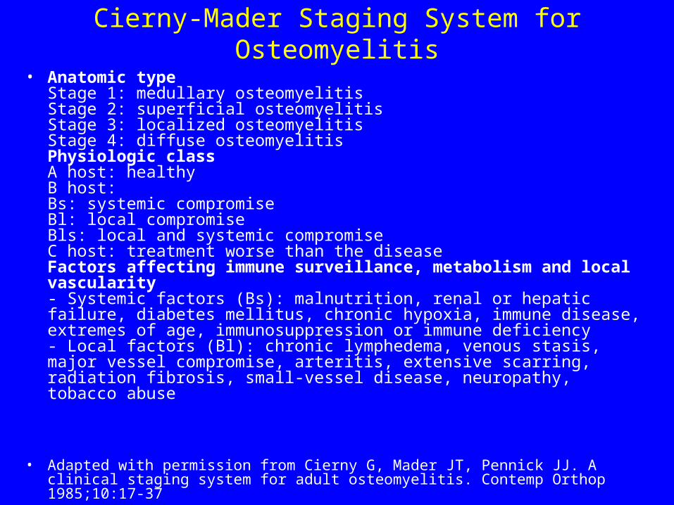
Cierny-Mader Staging System for Osteomyelitis
• Anatomic type Stage 1: medullary osteomyelitis Stage 2: superficial osteomyelitis Stage 3: localized osteomyelitis Stage 4: diffuse osteomyelitis Physiologic class A host: healthy B host: Bs: systemic compromise Bl: local compromise Bls: local and systemic compromise C host: treatment worse than the disease Factors affecting immune surveillance, metabolism and local vascularity - Systemic factors (Bs): malnutrition, renal or hepatic failure, diabetes mellitus, chronic hypoxia, immune disease, extremes of age, immunosuppression or immune deficiency - Local factors (Bl): chronic lymphedema, venous stasis, major vessel compromise, arteritis, extensive scarring, radiation fibrosis, small-vessel disease, neuropathy, tobacco abuse
• Adapted with permission from Cierny G, Mader JT, Pennick JJ. A clinical staging system for adult osteomyelitis. Contemp Orthop 1985;10:17-37

Organisms Commonly Isolated in Osteomyelitis Based on Patient Age
• Infants (<1 year) Group B streptococci Staphylococcus aureus Escherichia coli Children (1 to 16 years) S. aureus Streptococcus pyogenes Haemophilus influenzae Adults (>16 years) Staphylococcus epidermidis S. aureus Pseudomonas aeruginosa Serratia marcescens E. coli
• Adapted with permission from Dirschl DR, Almekinders LC. Osteomyelitis. Common causes and treatment recommendations. Drugs 1993;45:29-43.


• The body is infected and the bacteria invade the blood through injured skin and mucous membranes, and the lymphoid throat ring.
• Pyoderma of the skin, inflammation of the nasopharynx, and latent infections are of definite importance.
• The umbilical wound is a frequent infection atrium in infants.

• The anatomical age features of the structure and blood supply of the bones play a significant role in the development of osteomyelitis in children:
- the richly developed network of blood vessels;- the autonomous supply of blood to the epiphysis,
metaphysis, and diaphysis; - the presence of a great number of small vascular
branchings stretching radially through the epiphyseal cartilage to the ossification nucleus.
- The epiphyseal system of blood supply prevails in children under the age of 2 years, the metaphyseal system begins developing after this age. The epiphyseal and metaphyseal systems are isolated but there are anastomoses between them. The common vascular network forms only after ossification of the epiphysis.

• Affection of the epiphyseal zone is characteristic of children under the age of 2-3 years.
• With age, when the
system of blood supply to the metaphysis begins developing intensively, it is the metaphysis that predominantly becomes affected.

Localization


Pain
• which is a consequence of hypertension in the marrow cavity, is indirect proof of this interpretation of the circulatory disorders in the bone. Intraosseous pressure in acute osteomyelitis reaches 300-500 mm water (normal value in healthy children, 60-100 mm water).

If the osteomyelitic process is not recognized
• when it is still in the stage of inflammation within the boundaries of the bone-marrow cavity, then beginning from the 4th or 5th day of the disease the pus spreads along the bony haversian and Volkmann's canals under the periosteum and gradually separates it.
• Later (the 8th to 10th day and later) pus and the products of disintegration continue separating the periosteum, then the pus breaks through into the soft tissues and forms intermuscular and subcutaneous phlegmons.

Clinical picture • The toxic (adynamic) form follows an extremely violent
course with signs of endotoxic shock. A state of collapse is observed as a rule, with loss of consciousness, delirium, high body temperature (up to 40-41 °C), and sometimes with convulsions and vomiting.
• Dyspnoea is found but without any clear clinical picture of pneumonia.
• The cardiovascular abnormalities include disorders of central and peripheral circulation, reduced arterial pressure, with the development within a short time of cardiac insufficiency and signs of myocarditis.
• Punctate extravasations are often seen on the skin. • The tongue is dry and with a brownish coating. The
abdomen is usually distended and tender in the upper parts. The liver is enlarged.

Septicopyaemic form The onset of the disease is also acute: • body temperature rises to a high level (39-40°C),• signs of toxicosis increase, and the activity of
vital organs and systems is disturbed.• Confused consciousness, delirium, and euphoria
are sometimes encountered.• Pain is experienced in the affected limb from the
first days of the disease and becomes very intense due to the development of intraosseous hypertension.
• Septic complications caused by the spread of the purulent foci to various organs (the lungs, heart, and kidneys, as well as to the other bones) often occur.

The localized form
• characterized by the predominance of local signs of purulent inflammation over the general clinical manifestations of the disease


The main constant local signs of osteomyelitis
• sharp local tenderness to palpation and particularly to percussion over the site of the lesion.
• Oedema and tenderness extend also to the adjoining areas.
• Such signs as hyperaemia of the skin and fluctuation in the region of the lesion are very late signs and are evidence of neglected osteomyelitis
The main constant local signs of osteomyelitis
• Considerable diagnostic difficulties arise in osteomyelitis of bones forming the hip joint. The local signs are indistinct on the first days of the disease due to the powerful muscular casing in this region.
• On careful inspection it can be seen that the lower limb is slightly flexed at the hip joint; abduction and mild external rotation.
• Movements at the hip joint are painful. The joint itself and the overlying skin are oedematous .

Findings in infants include the following:
• Failure to thrive
• Drowsiness but irritability
• Minimal constitutional symptoms
• Effusions into neighboring joints (60%)

Findings in older children include the following:
• History of preceding minor trauma to the involved limb and/or recent infection, eg, upper respiratory tract or skin infection
• Bone pain • Malaise, irritability, and anorexia • Fever • Reluctance to use the limb • Localized swelling, redness, and warmth • Tenderness to finger pressure at a particular point • Pain on moving an adjacent joint • Regional lymphadenopathy
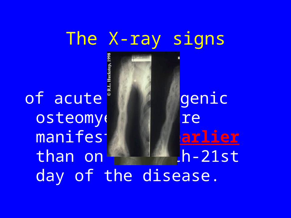
The X-ray signs
of acute haematogenic osteomyelitis are manifested no earlier than on the 14th-21st day of the disease.


The X-ray signs• Reduced density of the
bone shadow and blurring of its contours are usually found, osteoporosis in the region corresponding to the zone of the inflammation can also be detected. The spongy substance of the bone produces a macromacular pattern due to resorption of the bony trabeculae and merging of the intertrabecular spaces as the result of intensified resorption.


Nuclear medicine
• Nuclear medicine bone scans are a highly sensitive (>90%) modality in the diagnosis of osteomyelitis. This procedure is done in 3 parts. Technetium Tc 99m is used to create images to determine areas of infection and bone remodeling dependent on local blood flow. The sensitivity of bone scans is often helpful when the exact site and extent of the infection is not known.
MRI
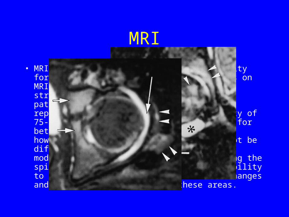
• MRI if available is another useful modality for imaging acute osteomyelitis. Findings on MRI accurately illustrate the extent and structure of the area involved in the pathologic process. Sensitivity has been reported to be 88-100%, with a specificity of 75-100%. Fat-suppression sequences allow for better detection of bone marrow edema; however, infection and inflammation cannot be differentiated. MRI may be the imaging modality of choice in infections involving the spine, pelvis, or limbs because of its ability to provide fine details of the osseous changes and soft-tissue extension in these areas.

Limitations of Techniques:
• MRI has limited availability and is relatively expensive. MRI is also contraindicated in patients with certain implant devices and metallic clips, and it is not tolerated by all patients because of claustrophobia or morbid obesity. In addition, young children may requiring sedation, Good MRI require patient cooperation because patient motion can degrade the images.
• CT is quick and inexpensive, but exposes the patient to ionizing radiation. The risk of a reaction to radio-iodinated contrast material is low, though the detection of bone destruction or a paraspinal mass does not require the use of contrast material.
• Although radionuclide studies are sensitive, they can be time-consuming, and they have lower spatial resolution. The incidence of false-negative scans is low in neonates and in elderly patients with osteomyelitis.

Diagnosis of osteomyelitis
• Diagnostic puncture of the bone with subsequent cytological examination of the aspirated material should be carried out more extensively in questionable cases.
• Measurement of intraosseous pressure is very important in establishing the early diagnosis of acute haematogenic osteomyelitis. The discovery of intraosseous hypertension confirms the diagnosis even in the absence of pus under the periosteum and in the marrow cavity.


Diagnosis of osteomyelitis• Blood tests show leukocytosis (up to 30 000-40 000 per
mm3) with a shift of the differential count to the left and toxic neutrophil granulation. The ESR is markedly increased (up to 60 mm/hour) and remains high for a long time.
• Marked changes are found in the blood serum protein spectrum. These are dysproteinaemia, an increase in the globulin fractions, and the development of hypoalbuminaemia. Anaemia caused by bone marrow inhibition by the prolonged effect of toxins develops in a persisting and severe disease.
• Disorders of the blood coagulation system are also found (the fibrinogen concentration and the fibrinolytic activity increase, the recalcification time and the coagulation time become shorter, the prothrombin index increases).

differential diagnosis
• articular form of rheumatism,
• phlegmon,
• tuberculosis of the bones,
• and injury.
• Rheumatism is characterized by shifting pains in the joints and typical changes in the heart confirmed by electrocardiography. Careful inspection and palpation of the involved region reveals that in rheumatism, in contrast to osteomyelitis, tenderness and swelling are mainly localized over the joint and not over the bone. Improvement of the local process with the prescription of salicylates is an important factor

Tuberculosis of the bones• Though experiencing pain in the limb, the
child still uses it.• Alexandrov's sign (thickening of the skin
fold on the involved limb) and muscle atrophy are found.
The radiograph demonstrates osteoporosis (the "melting sugar" symptom,) and an indistinct periosteaLreaction. This reaction, however, maybe clearly pronounced in mixed infection and in accompanying ordinary flora. The so-called acute forms of osteoarticular tuberculosis are actually cases of delayed diagnosis made when pus has already penetrated the joint. In addition to the X-ray picture, identification of the specific causative agent in material aspirated from the joint helps in establishing the correct diagnosis.
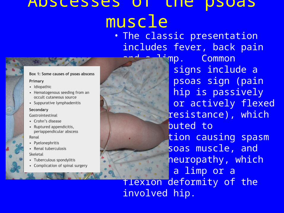
Abscesses of the psoas muscle • The classic presentation includes
fever, back pain and a limp. Common clinical signs include a positive psoas sign (pain when the hip is passively extended or actively flexed against resistance), which is attributed to inflammation causing spasm of the psoas muscle, and femoral neuropathy, which includes a limp or a flexion deformity of the involved hip.

Abscesses of the psoas muscle
• CT scanning is an accurate, rapid and noninvasive method for diagnosing psoas abscess and delineating its cause.
• Extraperitoneal surgical drainage has been the standard method of treatment; however, image-guided percutaneous drainage has become an effective alternative.


Treatment • In view of the fact that most severe
forms of osteomyelitis are consequent upon intraosseous hypertension, early surgical intervention, osteoperfora-tion, acquires primary importance. An incision, no less than 10-15 cm in length, is made in the soft tissues overlying the lesion and the periosteum is cut longitudinally. Two or three perforating openings 3-5 mm in diameter are made at the junction with the healthy bone. Pus is usually discharged under pressure in such cases, while in a disease of a long duration the contents of the marrow cavity may be seropurulent for two or three days. The marrow cavity is irrigated with 1 : 5000 nifrofurazone solu tion and antibiotics through the perforation in the bone.
Metaepiphyseal osteomyelitis
• is mostly encountered among in fants, predominantly among the newborn. By the haematogenic route the infection (usually staphylococcus) enters the bone metaphysis and the inflammatory process develops here. Due to the peculiar blood supply of the metaepiphyseal junction in very young children, however, the inflammation spreads to the growth zone and epiphysis located in the joint. As a result, the main clinical symptoms are caused by the developing acute arthritis.


Clinical picture
• Metaepiphyseal osteomyelitis sets in acutely as a rule with a rise of body temperature, debility, refusal of food, reluctance to move the involved limb which the child holds in a forced position.
• Examination reveals swelling over the zone of affection, deformity of the'adjoining joint, increase of local temperature. Hyperaemia appears later. Palpation and passive movement of the limb cause sharp pain. The "pseudoparesis" symptom (the hand or foot of the involved limb hangs and movements in it are sharply limited). The local form of osteomyelitis may be complicated by phlegmon of the soft tissues around the joint.

The X-ray signs
• are demonstrated earlier in metaepiphyseal osteomyelitis than in the other forms. Some characteristic signs can be detected as early as the 8th-10th day: thickening of soft tissues on the affected side, widening of the X-ray joint space, a fine periosteal reaction . Foci of destruction in the metaphysis are demonstrated on the radiographs only on the 3rd week after the onset of the disease, whereas the degree of destruction of the bone epiphysis

X-ray signs


• Immobilization plays an important role: Schede's traction is applied to the lower limb and Desault's bandage to the upper limb.

• In location of the process in the proximal femoral epiphysis, spreader-bandages are applied after the acute inflammation abates to prevent pathological dislocation of the hip. After recovery from acute haematogenic osteomyelitis the child must be kept under regu lar observation of an orthopaedist or surgeon.

Complications

Chronic Osteomyelitis
• If the process fails to abate completely in 4 to 6 months, regular exacerbations occur, fistulae remain, and the discharge of pus continues, then it is considered that osteomyelitis has taken the chronic stage.
• This outcome depends on the severity and rate of the occurring alternative changes in the bone tissue and how early and properly is the treatment applied. A change to the chronic stage may be encountred in 10 to 30 per cent of cases.

Anatomy:• Sharp loops of nonanastomosing are present at the
capillary ends of nutrient artery and enter into large venous sinusoids. This anatomy results in slowing of circulation and reduced oxygen tension. The capillaries do not communicate because columns of calcified cartilage separate them from each other.
• Children younger than 2 years of have transphyseal vessels, which cross from metaphysis to epiphysis. This causes the spread of infection into the joint. In children older than 2 years, the transphyseal vessels are absent, and hence the epiphyseal plate acts as a barrier to the spread of infection into the joint.
• Cierny and Mader proposed an anatomic classification of chronic osteomyelitis:
• Type 1 - Endosteal or medullary lesion • Type 2 - Superficial osteomyelitis limited to the surface • Type 3 - Localized, well-marked legion with sequestration
and cavity formation • Type 4 - Diffuse osteomyelitis lesions

Chronic osteomyelitis
• Chronic osteomyelitis is marked by a prolonged course with remissions and periods of deterioration.
• Typical forms are characterized pathomorphologically by pieces of necrotic bone (sequestra), a sequestral cavity, and sequestral capsule (involucrum). Granulations and pus are usually present between the involucrum and the sequestrum.


• After the sequestrum forms the inflammatory process continues. Pus collecting in the focus is discharged through the fistulae from time to time. Small sequestra are sometimes discharged, especially in a disease of a long duration. In such cases large sequestra may break into small ones. Sharp eburnation of bone (sclerosis and hardening) occurs around the focus of chronic inflammation. The soft tissues are also sclerosed, nutrition is disturbed, and the muscles atrophied. In a severe and extensive process the periosteum may be destroyed. Bone regeneration is greatly delayed in such cases and the involucrum fails to form or is deficient as a result of which pathological fracture or pseudoarthrosis often forms

Clinical picture• Chronic osteomyelitis is characterized by a protracted course with
remissions and exacerbations. • The fistulae may close during a remission.• In exacerbation, body temperature increases, tenderness and
toxicosis intensify. • Pus is again discharged from the fistulae, sometimes in abundance.• Examination of the patient reveals oedema of the soft tissues and
sometimes a swelling of the limb on the level of the lesion.• Fistulae and scars in places of previously existing fistulae are
typical of chronic osteomyelitis.• Palpation of the limb usually causes only mild tenderness and
often reveals atrophy of the soft tissues and thickening of the bone.
• Pallor of the skin and signs of malnutrition are also found.• Body temperature is subfebrile, particularly in the evening, but
sometimes reaches high levels during exacerbation

The X-ray diagnosis
• The X-ray diagnosis in typical cases with chronic osteomyelitis is quite easy. Radiographs show areas of osteoporosis and those of pronounced osteosclerosis. The involucrum containing sequestra, usually clearly outlined, is seen

Degree of Confidence:
• Plain radiographs are inexpensive and universally available.
• For the detection of acute osteomyelitis, the sensitivity is less than 5% at presentation and about 33% at 1 week; however, the sensitivity is 90% 3-4 weeks after presentation.
• For the detection of chronic osteomyelitis, the sensitivity of plain radiography is high, though the specificity is low.

CT scan• Findings: CT is of definite value for studying the entire
articular surface of bone and periarticular soft tissues; for delineating the extent of medullary and soft-tissue involvement; and for demonstrating cavities, serpiginous tracts, sequestra, or cloacae in osteomyelitis.
• CT scans sometimes show soft-tissue edema or bone destruction not seen on plain images, particularly in the setting of acute osteomyelitis. Sclerosis, demineralization, and periosteal reactions are usually well depicted in chronic osteomyelitis.
• CT scanning also helps in evaluating the need for surgery, and it provides vital information about the extent of disease. This data helps in planning appropriate surgery. CT is also an important modality in image-guided biopsy.

MRI
• MRI findings in osteomyelitis are usually secondary to the replacement of marrow fat with water secondary to edema, exudate, hyperemia, and bone ischemia. Findings include the following: decreased signal intensity in the involved bone on T1-weighted images, increased signal intensity in the involved bone on T2-weighted image, and increased signal intensity in the involved bone on short-tau inversion recovery (STIR) images.

Degree of Confidence:• MRI has sensitivity and specificity higher than those of
plain radiography and CT, and it is particularly good at depicting bone marrow abnormalities. On MRI, marrow signal abnormality is more sensitive than lytic changes on plain images, and findings become positive earlier with MRI than with radiography. Intramedullary bone pathology can be directly visualized with MRI, and in osteomyelitis marrow, these findings may precede bone changes.
• However, MRI findings of osteomyelitis are nonspecific, and similar changes can occur as a result of tumors, fractures, and a variety of other intramedullary or juxtamedullary processes that may cause bone marrow edema.
• The sensitivity and specificity has been reported as 92-100% and 89-100%, respectively. Prior fracture changes due to surgery or the fracture itself are difficult to differentiate from infection.
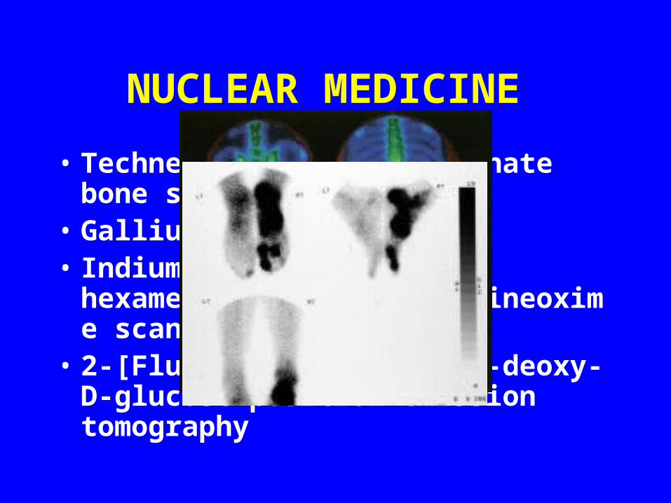
NUCLEAR MEDICINE
• Technetium-99m diphosphonate bone scanning
• Gallium-67 scanning • Indium-111 WBC and 99mTc
hexamethylenepropyleneamineoxime scanning
• 2-[Fluorine 18]-fluoro-2-deoxy-D-glucose positron emission tomography
DIFFERENTIALS • Chronic osteomyelitis has to be differentiated
from other diseases in some cases, namely, from tuberculosis and sarcoma.
• In contrast to osteomyelitis, tuberculosis sets in gradually, with no high temperature. Atrophy and contracture of the joint occur early. The fistulae are usually connected with the joint and have flacid and glass-like granulations. Processes of osteoporosis prevail on the radiograph and there are neither large sequestra (the sequestra seen usually re semble melting sugar) nor pronounced periostitis. Restoration of bone trabeculae (which at first are tangled) imperceptibly continuous with the normal tissue and diminution of osteoporosis are seen in the stage of reparation.

• Ewing's tumour (sarcoma) follows a wave-like course. Body temperature rises and pain increases during an attack. The diaphy-ses of the long tubular bones are involved in the process most often.
The X-ray picture of this tumour is characterized by a bulbous contour on a localized area of the diaphysis, scattered macular osteo-porosis, cortical osteolysis without sequestration, and narrowing of the marrow cavity. Osteogenic sarcoma is marked by the absence of a zone of sclerosis around the focus, by separation of the cortex and periosteum in the form of a peak, and by "spicles" (spicular periostitis).
• It is often very difficult to differentiate osteoid osteoma from• osteomyelitis. This tumour is characterized by a clearly
demonstrated band of perifocal thickening of trabeculae around the focus of diminished density and extensive periosteal deposits in the absence of marked destruction. Severe night pain in the involved bone is typical of osteoid osteoma. In some cases the diagnosis is established only with the aid of biopsy.

Findings in syphilis include the following:
• Pain, refusal to move the affected limb • Restriction of movement in an adjacent joint • Pain in the bone • Local swelling, redness, and warmth • Fever • Nausea • General discomfort, uneasiness, or ill feeling (malaise) • Drainage of pus through the skin (in chronic osteomyelitis)

Treatment
• Treatment in chronic osteomyelitis
• comprises trephination of the bone,
• removal of the sequestrum (sequestrectomy),
• curettage of the purulent granulations.

"Trough" resection
• "Trough" resection of the bone is therefore advisable in an extensive lesion. With this type of resection the possibility of sequestration of the overhanging bone edges to less, whereas the soft tissues adjoining closely the surface of the bone improve its nutrition

• Sir Benjamin Collins Brodie (1783-1862) Sir Benjamin Brodie was one of the most recognized surgeons at St. George’s Hospital in London during the nineteenth century. His early education began at home, being taught by his father, Reverend Peter Brodie. In 1801, he went to London to study medicine, attending anatomy lectures at St. Bartholomew’s Hospital. In 1802, he attended the Windmill Street School of Anatomy. By May 1805, Brodie’s work earned him the position of Assistant Surgeon at St. George’s Hospital. A few months later, he was admitted as a member of the prestigious and influential Royal College of Surgeons. Acknowledged as an outstanding physician and statesman, he served as personal surgeon to King George IV.
• Brodie was a skilled surgeon and successful writer, and his influence remains. In 1819 he published, On the Disease of Joints which served as a manual in understanding and classifying clinical aspects and pathology of joint disease. He first described a chronic abscess of the tibia in 1832 that has since been named Brodie’s abscess.

Atypical Forms of Osteomyelitis• Brodie's abscess is marked by a protracted course,
mild aching pains in the region of the lesion, and moderate increase of tempera ture. The proximal tibial, distal femoral, and proximal humeral metaphyses are the favoured sites. It can be seen on examination that the limb is moderately swollen and mildly tender to intense palpation.
• X-ray shows a round zone of destruction with pronounced perifocal sclerosis. Sequestra and fistulae do not usually form. Aband of diminished density, a "strip" connecting the focus with the growth zone, can often be seen

Ollier's albuminous osteomyelitis.
• This is a very rare disease. The clinical manifestations are similar'to those of other forms of atypical osteomyelitis though in some cases they are more pronounced. The bone is sclerosed and the marrow canal, which contains White or yellow fluid, is narrowed.
• Treatment consists in trephination of the bone with removal of albuminous fluid and tight filling of the cavity with antibiotics.
• Sclerosing osteomyelitis of Garré most commonly affects the mandible and appears with a focal sclerosing periosteal reaction on radiologic studies.
• Chronic recurrent osteomyelitis is benign self-limiting condition that primarily affects long bones in children and adolescents. The metaphysis of long bones are usually affected, and changes may be symmetrical. The appearances are those of confluent areas of bone lysis.

• In sickle cell anaemia, approximately 50% of all cases of infection are caused by a salmonella bacteraemia spreading from the intestinal tract. In sickle cell anaemia, however, considerable sterile bone destruction can occur without an associated infection. This is due to the multiple bone infarcts associated with cutting off of the cortical blood supply to the bone. Massive thrombosis to the arterioles supplying the bone occurs. If, at the same time the child has a bacteraemia, infection of the bone affected is likely. In the X-ray illustrated, the baby had sickle cell anaemia. She had no fewer than 9 bones infected at one time by a salmonella typhimurium. Note the multiple pathological fractures and osteomyelitis affecting both radius and ulna.

Chronic recurrent multifocal osteomyelitis (CRMO) • . Diagnostic criteria for CRMO have been proposed to
include all of the following: • (a) the presence of two or more radiographically confirmed
bone lesions,• (b) a prolonged course of at least 6 months with
characteristic exacerbation and remission, • (c) radiographic and nuclear scintigraphic evidence of
osteomyelitis,• (d) a lack of response to antimicrobial therapy of at least 1
month’s duration, and • (e) the lack of an identifiable etiology . • A definitive role for steroids or long term antibiotics has
not been established. Supportive management with anti-inflammatory medication is recommended, as the typical course of CRMO is self-limited.

The end


![Andreev Reflections and transport phenomena in ...€¦ · Andreev re°ections [S2,S8]. The point contact Andreev re°ection spectroscopy carried out on RuSr2GdCu2O8 has evidenced](https://img.pdfslide.us/doc/110x75/6060572c65c18a52267c888c/andreev-reflections-and-transport-phenomena-in-andreev-reections-s2s8.jpg)