Embed Size (px)
Citation preview
1
Orthopedic Manual Therapy of the Sacroiliac Joint and Pelvis:
An Evidence Based Approach to Success
!Dr. Eric (Rick) Douglass, !
PT, DPT, OCS, KTCC, FAAOMPT
2
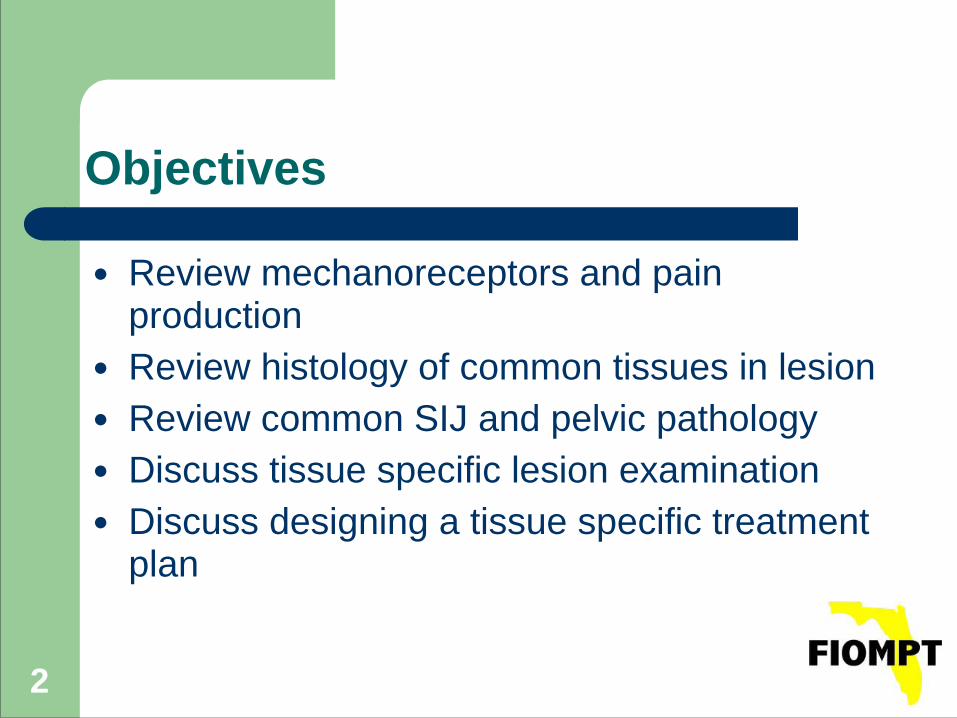
Objectives
● Review mechanoreceptors and pain production
● Review histology of common tissues in lesion ● Review common SIJ and pelvic pathology ● Discuss tissue specific lesion examination ● Discuss designing a tissue specific treatment
plan
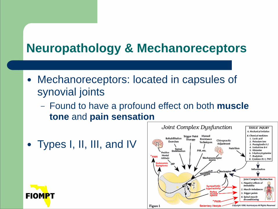
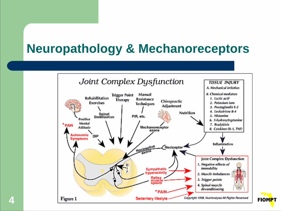
Neuropathology & Mechanoreceptors
● Mechanoreceptors: located in capsules of synovial joints – Found to have a profound effect on both muscle
tone and pain sensation !
● Types I, II, III, and IV
4
Neuropathology & Mechanoreceptors
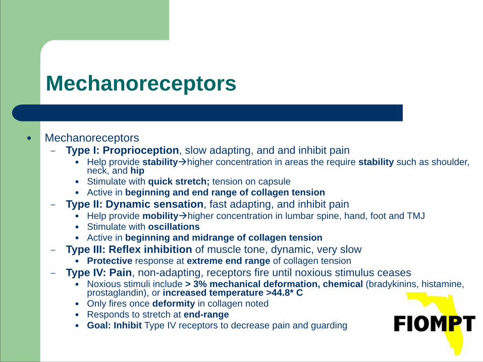
Mechanoreceptors!!!
● Mechanoreceptors – Type I: Proprioception, slow adapting, and and inhibit pain
● Help provide stabilityhigher concentration in areas the require stability such as shoulder, neck, and hip
● Stimulate with quick stretch; tension on capsule ● Active in beginning and end range of collagen tension
– Type II: Dynamic sensation, fast adapting, and inhibit pain ● Help provide mobilityhigher concentration in lumbar spine, hand, foot and TMJ ● Stimulate with oscillations ● Active in beginning and midrange of collagen tension
– Type III: Reflex inhibition of muscle tone, dynamic, very slow ● Protective response at extreme end range of collagen tension
– Type IV: Pain, non-adapting, receptors fire until noxious stimulus ceases ● Noxious stimuli include > 3% mechanical deformation, chemical (bradykinins, histamine,
prostaglandin), or increased temperature >44.8* C ● Only fires once deformity in collagen noted ● Responds to stretch at end-range ● Goal: Inhibit Type IV receptors to decrease pain and guarding
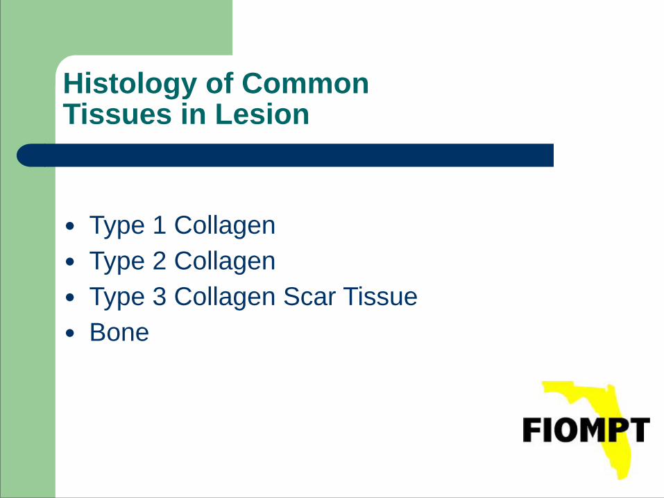
Histology of Common Tissues in Lesion
!● Type 1 Collagen ● Type 2 Collagen ● Type 3 Collagen Scar Tissue ● Bone
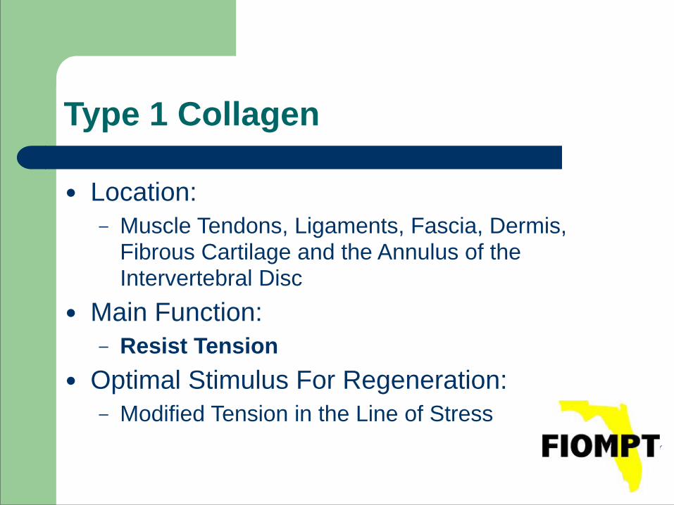
Type 1 Collagen
● Location: – Muscle Tendons, Ligaments, Fascia, Dermis,
Fibrous Cartilage and the Annulus of the Intervertebral Disc
● Main Function: – Resist Tension
● Optimal Stimulus For Regeneration: – Modified Tension in the Line of Stress
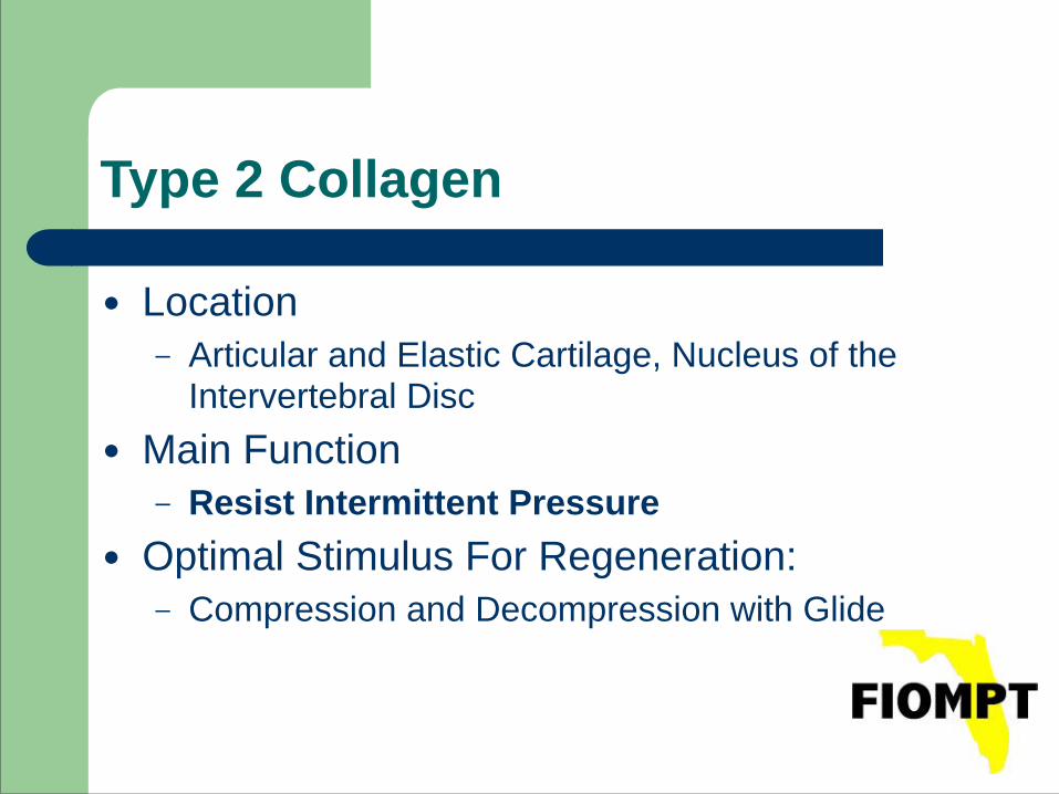
Type 2 Collagen
● Location – Articular and Elastic Cartilage, Nucleus of the
Intervertebral Disc ● Main Function
– Resist Intermittent Pressure ● Optimal Stimulus For Regeneration:
– Compression and Decompression with Glide
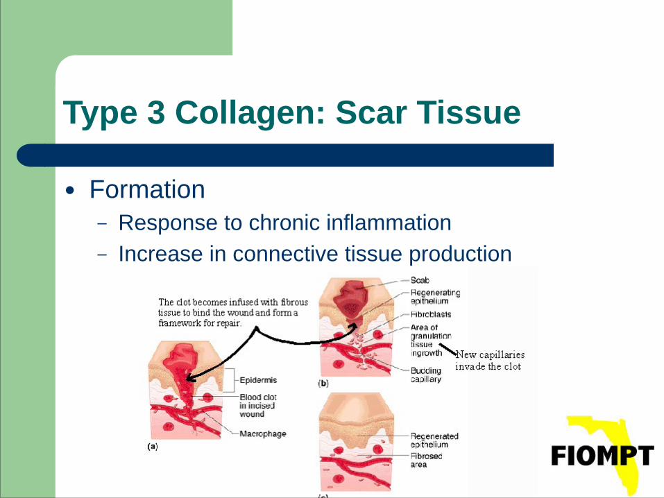
Type 3 Collagen: Scar Tissue
● Formation – Response to chronic inflammation – Increase in connective tissue production
Scar Tissue
● Result – Leads to Tight and Short tissue – Decreased ROM
● Inhibit Formation – Inhibit myofibroblastic activity – Goal: make scar as Small as possible and increase
the capacity of the scar to tolerate tension. Achieved by the providing optimal stimulus for regeneration for the tissue in lesion
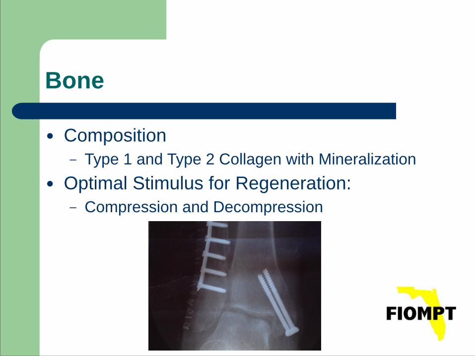
Bone
● Composition – Type 1 and Type 2 Collagen with Mineralization
● Optimal Stimulus for Regeneration: – Compression and Decompression
The Manual Therapy Lesion
1. Collagen/Tissue Trauma: Tear in capsule, ligament, tendon, muscle etc. May be acute or slow progression
2. Receptor Damage: Structural damage in capsules. Type I tonic and stability receptors most easily damaged due to its superficial placement. Type II also damaged
3. Reduced Muscle Fiber Recruitment: Type I tonic muscles reduced ability to recruit muscle fibers secondary to trauma
4. Tonic Fiber Atrophy: Decreased recruitment and trauma leads to atrophy of the muscle. Type I tonic more easily affected. Type II phasic
The Manual Therapy Lesion
5. Reduced Anti-Gravity Stability: Atrophy and reduced ability to recruit desired musculature leads to decreased stability and support in the joint
6. Motion Around Nonphysiological Axis: The stabilizing muscles are no longer able to support the joint around a physiologic axis and creates alteration in motion. See compensatory motion
7. Trauma/Acute Locking/Degeneration: The movement around a nonphysiologic axis increases trauma and may lead to locking or degeneration of the joint
8. Pain/Guarding: The deformity leads to firing of type IV mechanoreceptors, thus causing pain and guarding
Tissue Specific Evaluation
● PURPOSE – To identify:
1. Load sensitivity of key ligamentous and fascial structures
2. Abnormal function at articulations between the ilia, the sacrum and the pubis
SIJD: What is the “Tissue in Lesion”
● Levangie, PK (Phys Ther 1999; 79: 1043-1057) presented two hypotheses:
1. Asymmetry of the pelvis causes a nociceptive mechanical stress on the structures attached to the innominates or within the SIJ
2. SIJ hypomobility, with or without positional abnormalities, places painful mechanical stress on surrounding tissues
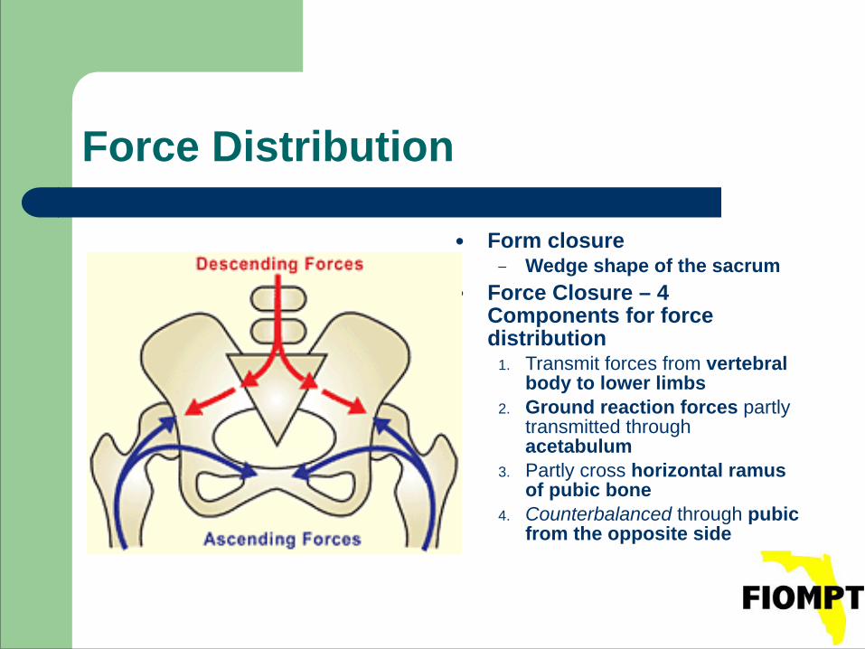
Force Distribution
● Form closure – Wedge shape of the sacrum
● Force Closure – 4 Components for force distribution
1. Transmit forces from vertebral body to lower limbs
2. Ground reaction forces partly transmitted through acetabulum
3. Partly cross horizontal ramus of pubic bone
4. Counterbalanced through pubic from the opposite side
17
SIJD: What is the “Tissue in Lesion”
● Peter O'Sullivan ● https://www.youtube.com/watch?
feature=youtu.be&v=RbSF3-_b7bI&app=desktop
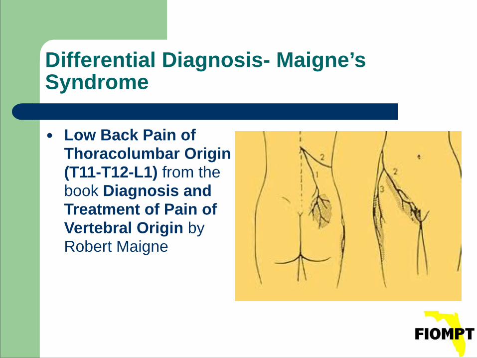
Differential Diagnosis- Maigne’s Syndrome
● Low Back Pain of Thoracolumbar Origin (T11-T12-L1) from the book Diagnosis and Treatment of Pain of Vertebral Origin by Robert Maigne
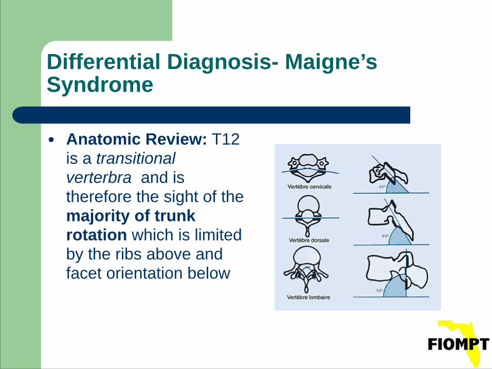
Differential Diagnosis- Maigne’s Syndrome
● Anatomic Review: T12 is a transitional verterbra and is therefore the sight of the majority of trunk rotation which is limited by the ribs above and facet orientation below
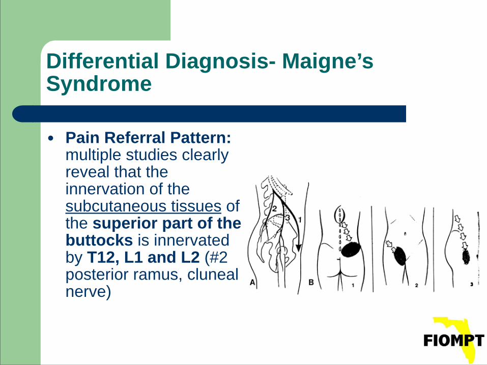
Differential Diagnosis- Maigne’s Syndrome
● Pain Referral Pattern: multiple studies clearly reveal that the innervation of the subcutaneous tissues of the superior part of the buttocks is innervated by T12, L1 and L2 (#2 posterior ramus, cluneal nerve)
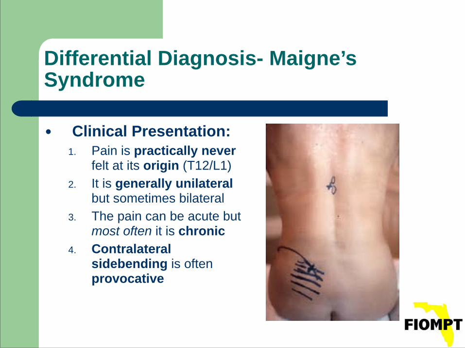
Differential Diagnosis- Maigne’s Syndrome
● Clinical Presentation: 1. Pain is practically never
felt at its origin (T12/L1) 2. It is generally unilateral
but sometimes bilateral 3. The pain can be acute but
most often it is chronic 4. Contralateral
sidebending is often provocative
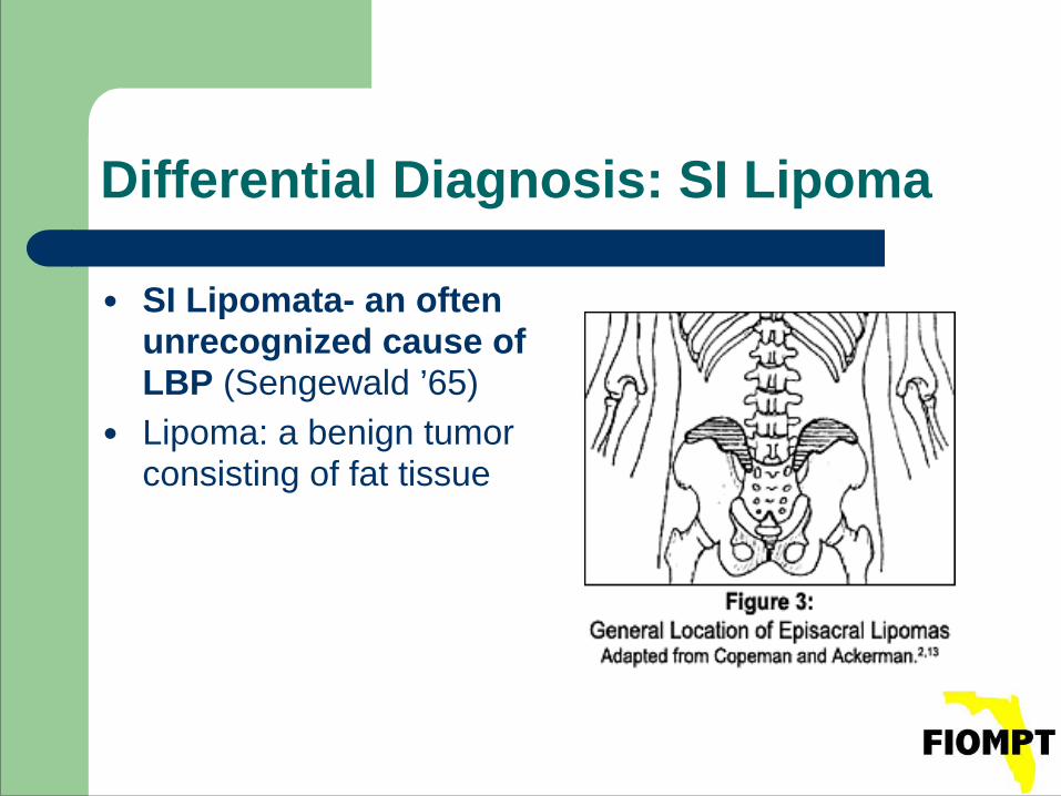
Differential Diagnosis: SI Lipoma
● SI Lipomata- an often unrecognized cause of LBP (Sengewald ’65)
● Lipoma: a benign tumor consisting of fat tissue
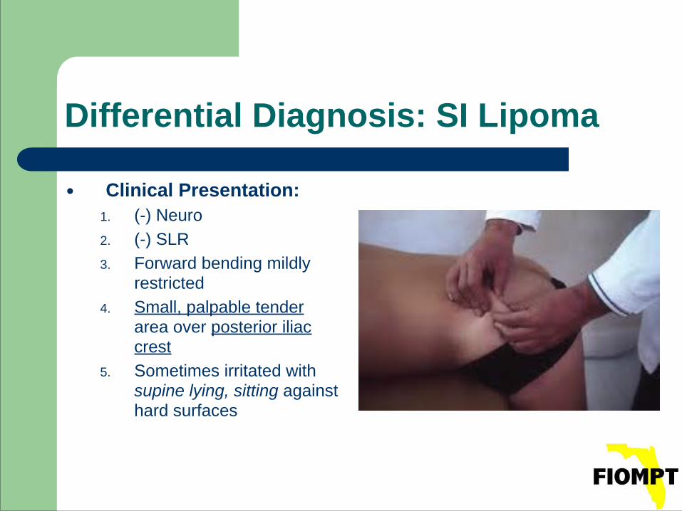
Differential Diagnosis: SI Lipoma
● Clinical Presentation: 1. (-) Neuro 2. (-) SLR 3. Forward bending mildly
restricted 4. Small, palpable tender
area over posterior iliac crest
5. Sometimes irritated with supine lying, sitting against hard surfaces
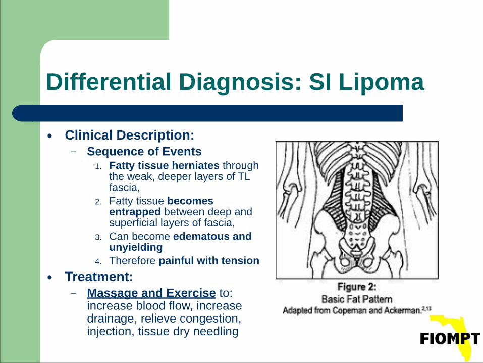
Differential Diagnosis: SI Lipoma
● Clinical Description: – Sequence of Events
1. Fatty tissue herniates through the weak, deeper layers of TL fascia,
2. Fatty tissue becomes entrapped between deep and superficial layers of fascia,
3. Can become edematous and unyielding
4. Therefore painful with tension ● Treatment:
– Massage and Exercise to: increase blood flow, increase drainage, relieve congestion, injection, tissue dry needling
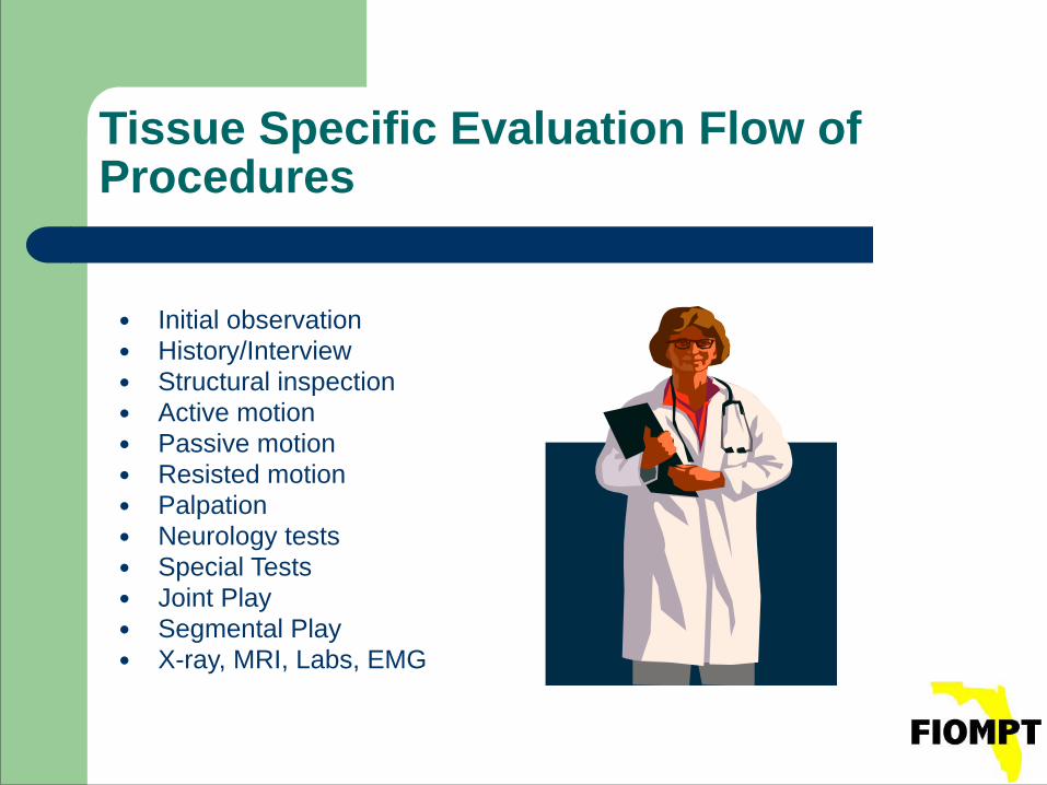
Tissue Specific Evaluation Flow of Procedures
!!
● Initial observation ● History/Interview ● Structural inspection ● Active motion ● Passive motion ● Resisted motion ● Palpation ● Neurology tests ● Special Tests ● Joint Play ● Segmental Play ● X-ray, MRI, Labs, EMG
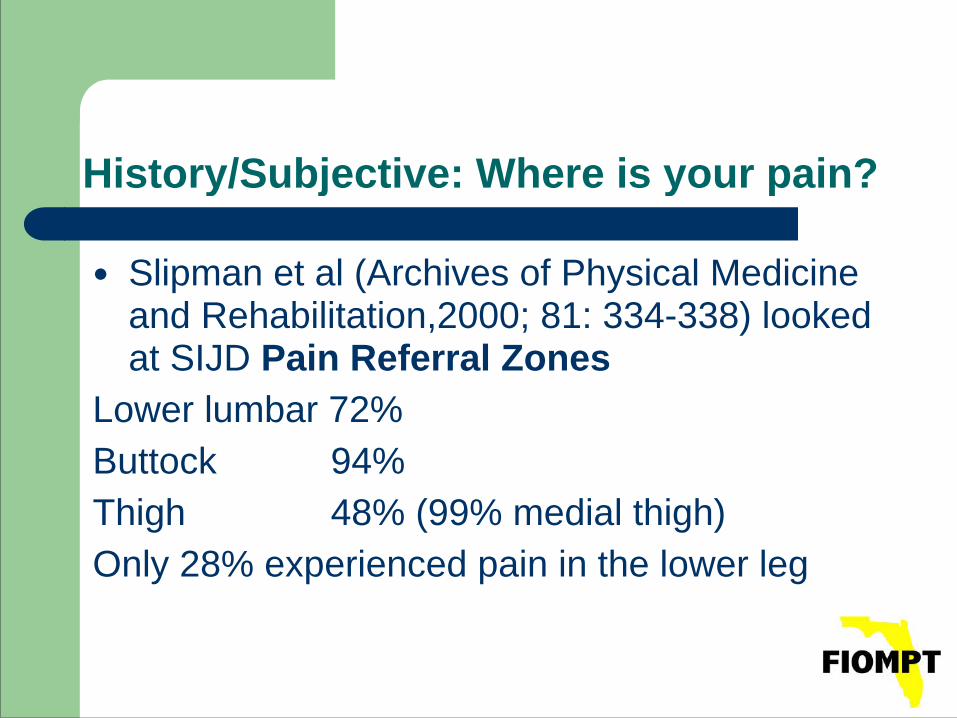
History/Subjective: Where is your pain?
● Slipman et al (Archives of Physical Medicine and Rehabilitation,2000; 81: 334-338) looked at SIJD Pain Referral Zones
Lower lumbar 72% Buttock 94% Thigh 48% (99% medial thigh) Only 28% experienced pain in the lower leg
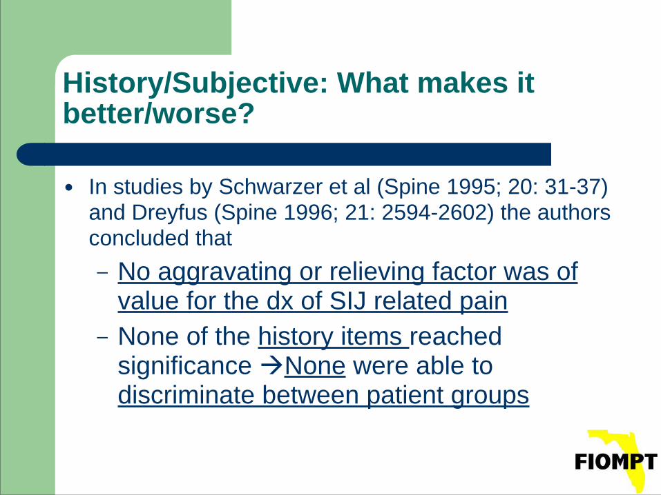
History/Subjective: What makes it better/worse?
● In studies by Schwarzer et al (Spine 1995; 20: 31-37) and Dreyfus (Spine 1996; 21: 2594-2602) the authors concluded that – No aggravating or relieving factor was of
value for the dx of SIJ related pain – None of the history items reached
significance None were able to discriminate between patient groups
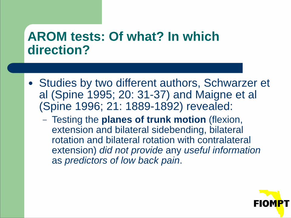
AROM tests: Of what? In which direction?
● Studies by two different authors, Schwarzer et al (Spine 1995; 20: 31-37) and Maigne et al (Spine 1996; 21: 1889-1892) revealed: – Testing the planes of trunk motion (flexion,
extension and bilateral sidebending, bilateral rotation and bilateral rotation with contralateral extension) did not provide any useful information as predictors of low back pain.
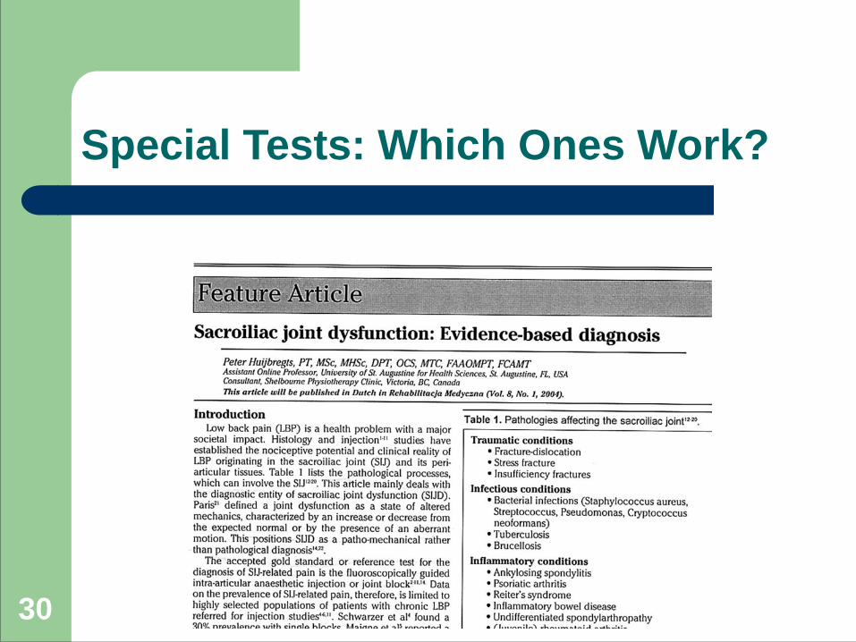
Special Tests: Which Ones Work?
● “Sacroiliac joint dysfunction: Evidence-based diagnosis”- Peter Huijbregts, PT, MSc, MHSc, DPT, OCS, MTC, FAAOMPT, FCAMT, Orthopedic Division Review, May/June 2004 !– GOAL: discuss reliability and validity of history
items and physical tests thought relevant for making a diagnosis of SIJD
30
Special Tests: Which Ones Work?
“Sacroiliac joint dysfunction: Evidence-based diagnosis”- Huijbregts
● Accepted Gold Standard – flouroscopically guided intra-articular anaesthetic injection or joint block
● Therefore: the only TRUE SI related LBP patients are that small subset who were referred for and got better with injection
● Bottom Line: knowing the clinically applicable special tests that worked well on the true SI population would be great information BUT is a very small data base
“Sacroiliac joint dysfunction: Evidence-based diagnosis”- Huijbregts
● Three categories of Special Tests for SIJ dysfunction
1. Positional palpation tests 2. Motion palpation tests 3. Provocation tests
● For these tests to be clinically useful, the data they yield needs to be reliable, valid and responsive to clinically relevant change.
“Sacroiliac joint dysfunction: Evidence-based diagnosis”- Huijbregts
● Looking for: 1. Test- re-test reliability: consistency of measures repeated
over time 2. Intra-rater reliability: stability of measurements taken by
one rater across two or more trials 3. Inter-rater reliability: the level of agreement between
findings of two or more raters measuring the same group of subjects
4. Validity: the degree to which a meaningful interpretation can be inferred from this measurement
“Sacroiliac joint dysfunction: Evidence-based diagnosis”- Huijbregts
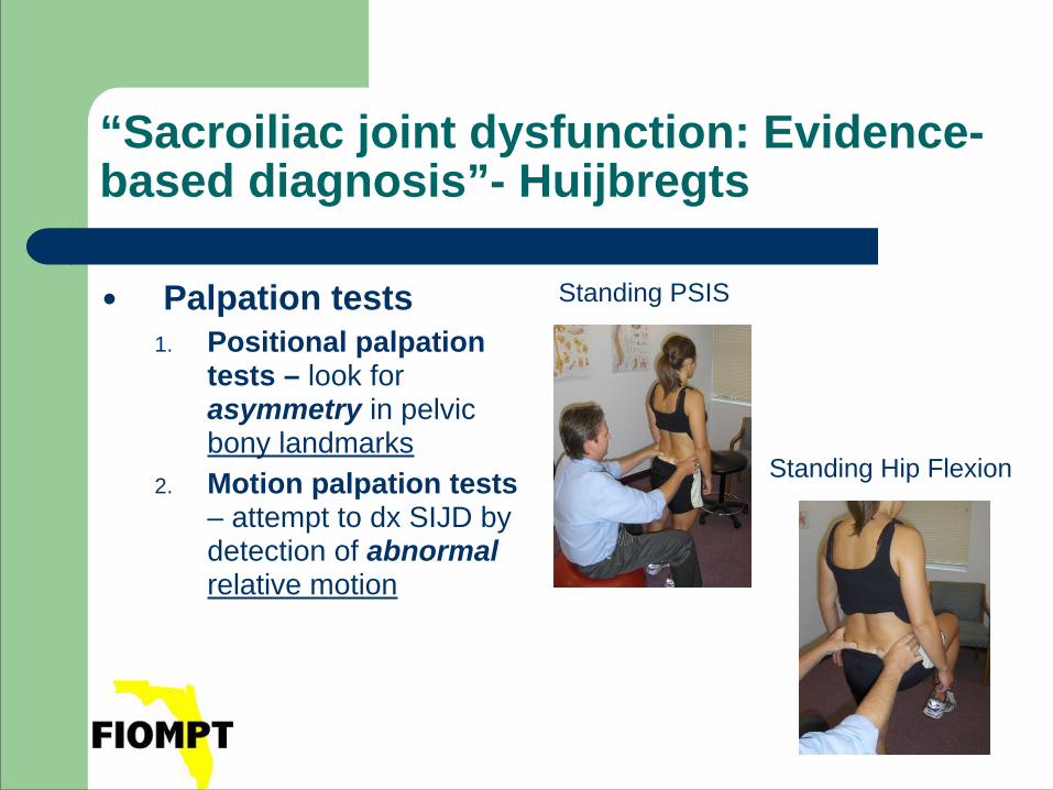
● Palpation tests 1. Positional palpation
tests – look for asymmetry in pelvic bony landmarks
2. Motion palpation tests – attempt to dx SIJD by detection of abnormal relative motion
Standing PSIS
Standing Hip Flexion
“Sacroiliac joint dysfunction: Evidence-based diagnosis”- Huijbregts
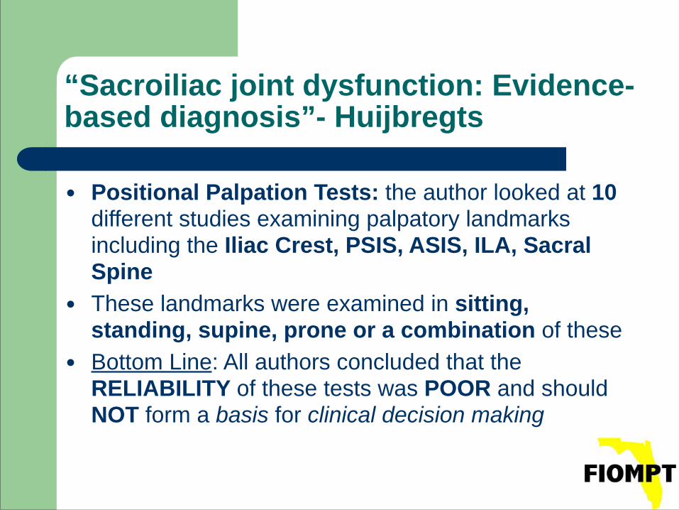
● Positional Palpation Tests: the author looked at 10 different studies examining palpatory landmarks including the Iliac Crest, PSIS, ASIS, ILA, Sacral Spine
● These landmarks were examined in sitting, standing, supine, prone or a combination of these
● Bottom Line: All authors concluded that the RELIABILITY of these tests was POOR and should NOT form a basis for clinical decision making
“Sacroiliac joint dysfunction: Evidence-based diagnosis”- Huijbregts
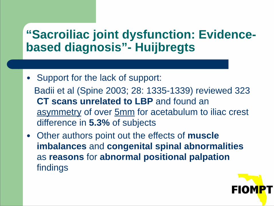
● Support for the lack of support: Badii et al (Spine 2003; 28: 1335-1339) reviewed 323
CT scans unrelated to LBP and found an asymmetry of over 5mm for acetabulum to iliac crest difference in 5.3% of subjects
● Other authors point out the effects of muscle imbalances and congenital spinal abnormalities as reasons for abnormal positional palpation findings
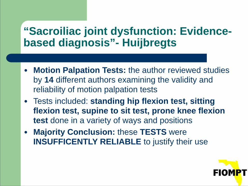
“Sacroiliac joint dysfunction: Evidence-based diagnosis”- Huijbregts
● Motion Palpation Tests: the author reviewed studies by 14 different authors examining the validity and reliability of motion palpation tests
● Tests included: standing hip flexion test, sitting flexion test, supine to sit test, prone knee flexion test done in a variety of ways and positions
● Majority Conclusion: these TESTS were INSUFFICENTLY RELIABLE to justify their use
“Sacroiliac joint dysfunction: Evidence-based diagnosis”- Huijbregts
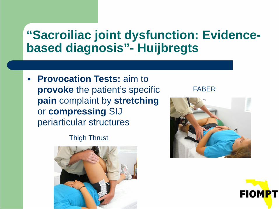
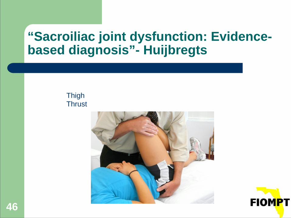
● Provocation Tests: aim to provoke the patient’s specific pain complaint by stretching or compressing SIJ periarticular structures
FABER
Thigh Thrust
“Sacroiliac joint dysfunction: Evidence-based diagnosis”- Huijbregts
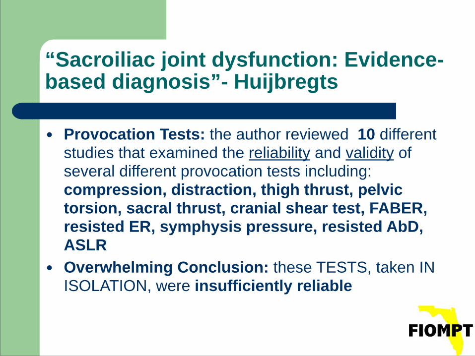
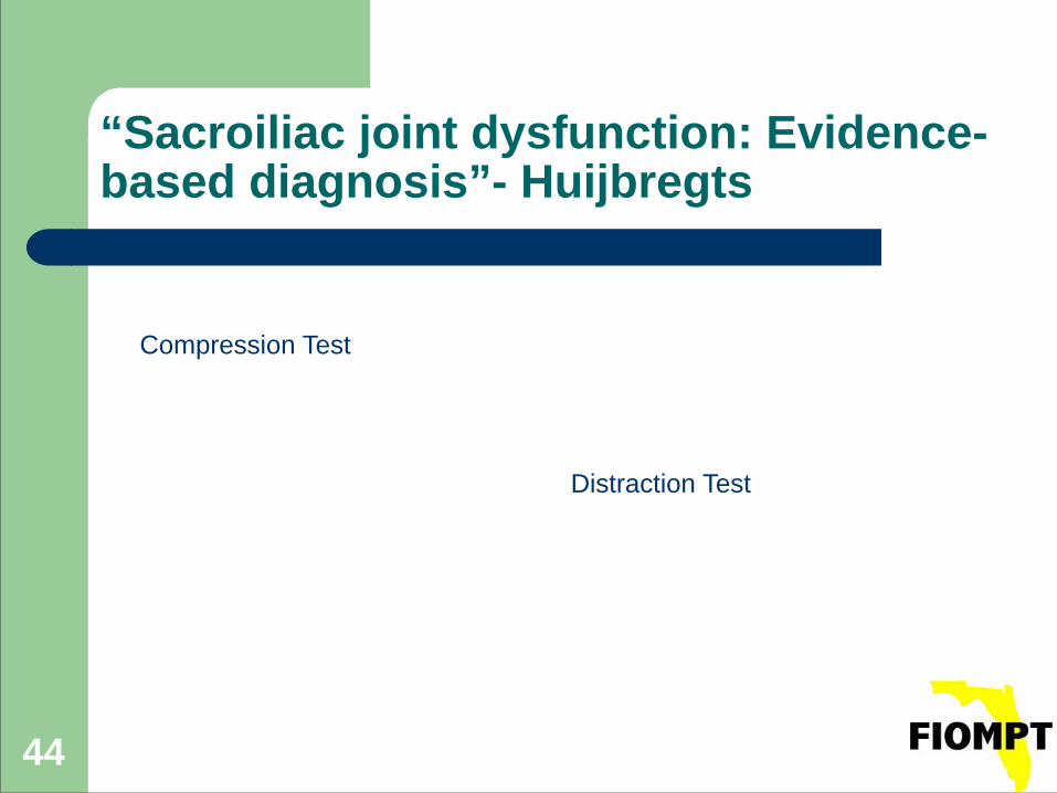
● Provocation Tests: the author reviewed 10 different studies that examined the reliability and validity of several different provocation tests including: compression, distraction, thigh thrust, pelvic torsion, sacral thrust, cranial shear test, FABER, resisted ER, symphysis pressure, resisted AbD, ASLR
● Overwhelming Conclusion: these TESTS, taken IN ISOLATION, were insufficiently reliable
“Sacroiliac joint dysfunction: Evidence-based diagnosis”- Huijbregts
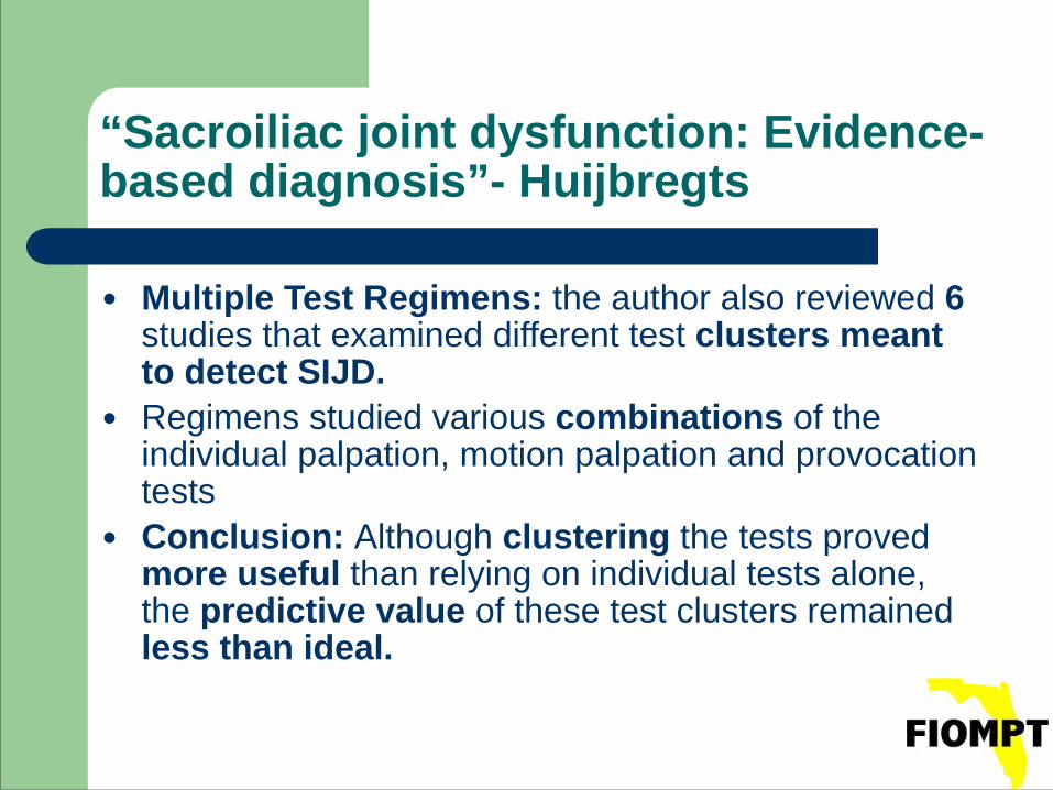
● Multiple Test Regimens: the author also reviewed 6 studies that examined different test clusters meant to detect SIJD.
● Regimens studied various combinations of the individual palpation, motion palpation and provocation tests
● Conclusion: Although clustering the tests proved more useful than relying on individual tests alone, the predictive value of these test clusters remained less than ideal.
“Sacroiliac joint dysfunction: Evidence-based diagnosis”- Huijbregts
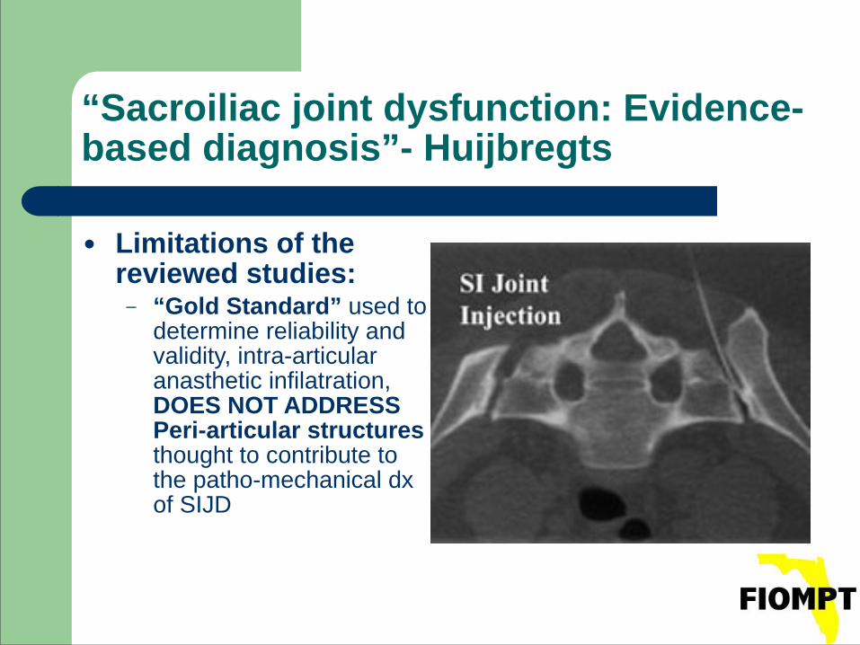
● Limitations of the reviewed studies:
– “Gold Standard” used to determine reliability and validity, intra-articular anasthetic infilatration, DOES NOT ADDRESS Peri-articular structures thought to contribute to the patho-mechanical dx of SIJD
● Conclusions: 1. Palpation in standing, prone or supine of the iliac
crests, PSIS and ASIS and of the ILA in supine is NOT a valid descriptor of SIJD
2. Standing hip flexion, sitting flexion and standing flexion tests have a false positive rate of near 20%
3. Positive ASLR test is associated with ipsilateral increased SIJ mobility
“Sacroiliac joint dysfunction: Evidence-based diagnosis”- Huijbregts
“Sacroiliac joint dysfunction: Evidence-based diagnosis”- Huijbregts
● Conclusions, cont.: 1. ASLR and thigh thrust tests have good predictive validity
for identifying patients with post partum pelvic pain associated with SIJ laxity.
2. Cluster of 5 Tests: Compression, Distraction, Pelvic Torsion, Sacral Thrust and Thigh Thrust tests ● When 3/5 POSITIVE = SUBSTANTIAL Inter-Rater Agreement
3. Further research on the level of patient outcomes with manual medicine is needed to validate claims of manual medicine’s diagnostic and therapeutic efficacy
44
“Sacroiliac joint dysfunction: Evidence-based diagnosis”- Huijbregts
Compression Test
Distraction Test
45
“Sacroiliac joint dysfunction: Evidence-based diagnosis”- Huijbregts
Pelvic Torsion
Sacral Thrust
46
“Sacroiliac joint dysfunction: Evidence-based diagnosis”- Huijbregts
Thigh Thrust
47
SIJ: Dosed Exercise Concepts: Ligamentous Influences
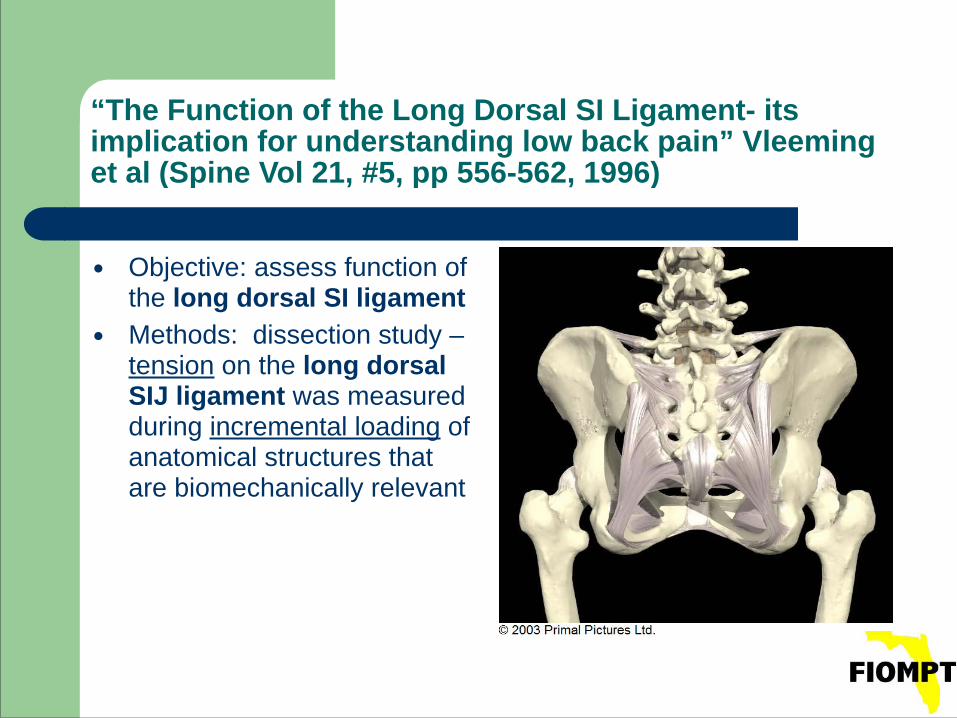
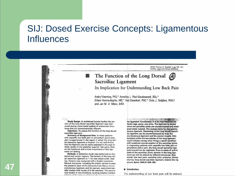
“The Function of the Long Dorsal SI Ligament- its implication for understanding low back pain” Vleeming et al (Spine Vol 21, #5, pp 556-562, 1996)
● Objective: assess function of the long dorsal SI ligament
● Methods: dissection study – tension on the long dorsal SIJ ligament was measured during incremental loading of anatomical structures that are biomechanically relevant
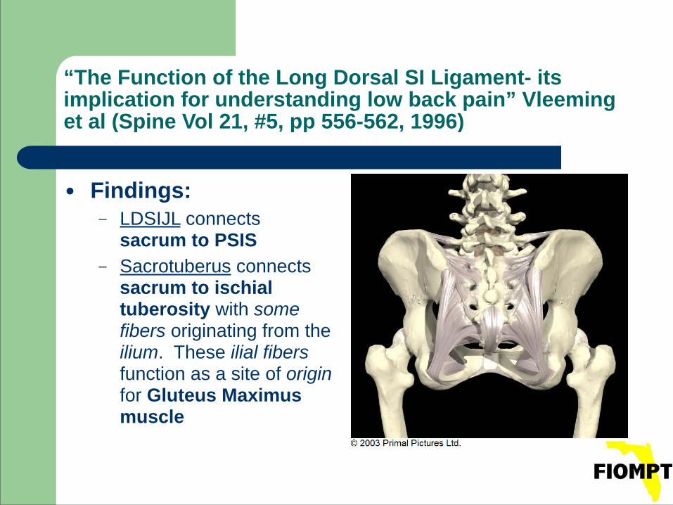
“The Function of the Long Dorsal SI Ligament- its implication for understanding low back pain” Vleeming et al (Spine Vol 21, #5, pp 556-562, 1996)
● Findings: – LDSIJL connects
sacrum to PSIS – Sacrotuberus connects
sacrum to ischial tuberosity with some fibers originating from the ilium. These ilial fibers function as a site of origin for Gluteus Maximus muscle
“The Function of the Long Dorsal SI Ligament- its implication for understanding low back pain” Vleeming et al (Spine Vol 21, #5, pp 556-562, 1996)
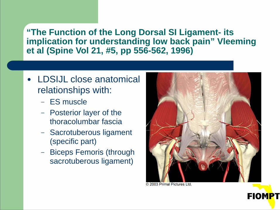
● LDSIJL close anatomical relationships with:
– ES muscle – Posterior layer of the
thoracolumbar fascia – Sacrotuberous ligament
(specific part) – Biceps Femoris (through
sacrotuberous ligament)
“The Function of the Long Dorsal SI Ligament- its implication for understanding low back pain” Vleeming et al (Spine Vol 21, #5, pp 556-562, 1996)
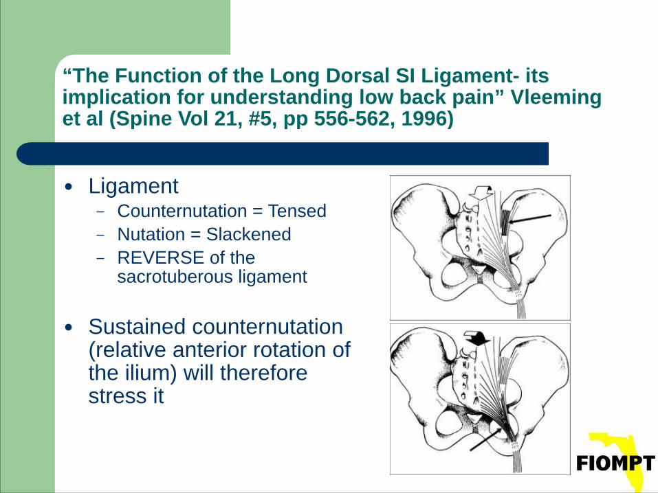
● Ligament – Counternutation = Tensed – Nutation = Slackened – REVERSE of the
sacrotuberous ligament !
● Sustained counternutation (relative anterior rotation of the ilium) will therefore stress it
“The Function of the Long Dorsal SI Ligament- its implication for understanding low back pain” Vleeming et al (Spine Vol 21, #5, pp 556-562, 1996)
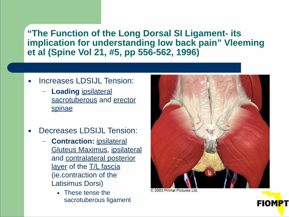
● Increases LDSIJL Tension: – Loading ipsilateral
sacrotuberous and erector spinae
!● Decreases LDSIJL Tension:
– Contraction: ipsilateral Gluteus Maximus, ipsilateral and contralateral posterior layer of the T/L fascia (ie.contraction of the Latisimus Dorsi) ● These tense the
sacrotuberous ligament
“The Function of the Long Dorsal SI Ligament- its implication for understanding low back pain” Vleeming et al (Spine Vol 21, #5, pp 556-562, 1996)
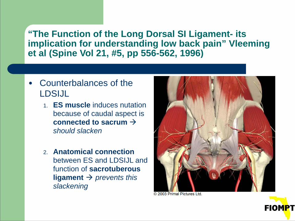
● Counterbalances of the LDSIJL
1. ES muscle induces nutation because of caudal aspect is connected to sacrum should slacken !
2. Anatomical connection between ES and LDSIJL and function of sacrotuberous ligament prevents this slackening
“The Function of the Long Dorsal SI Ligament- its implication for understanding low back pain” Vleeming et al (Spine Vol 21, #5, pp 556-562, 1996)
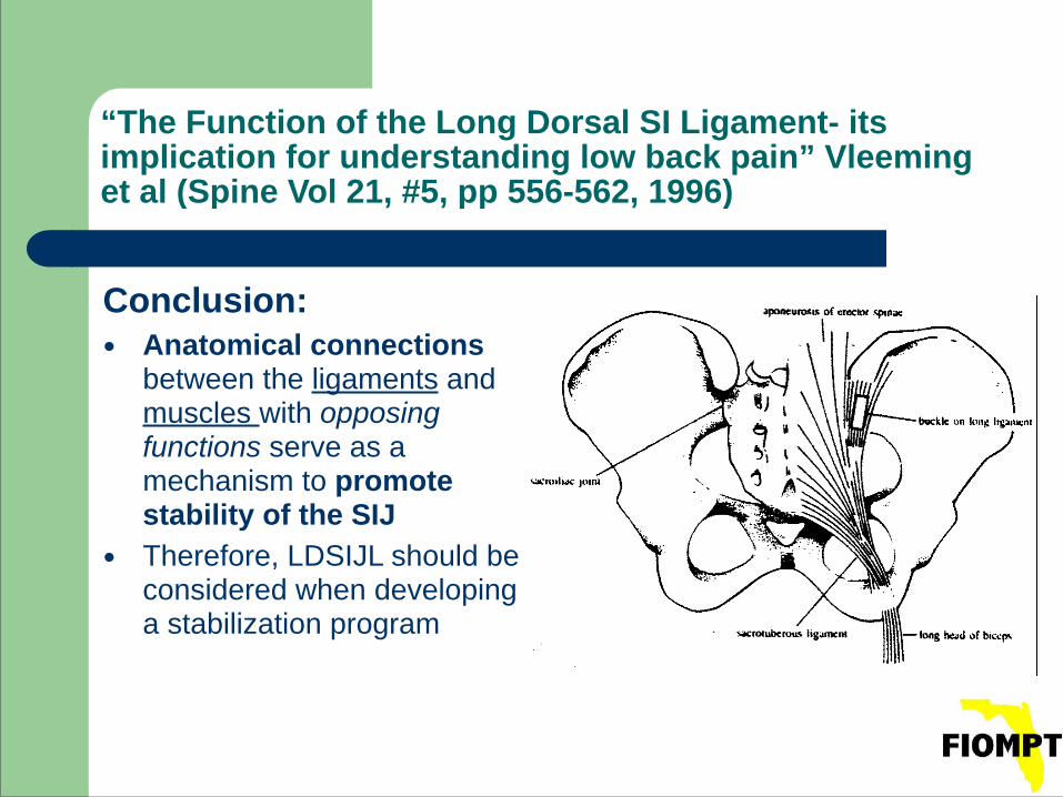
Conclusion: ● Anatomical connections
between the ligaments and muscles with opposing functions serve as a mechanism to promote stability of the SIJ
● Therefore, LDSIJL should be considered when developing a stabilization program
55
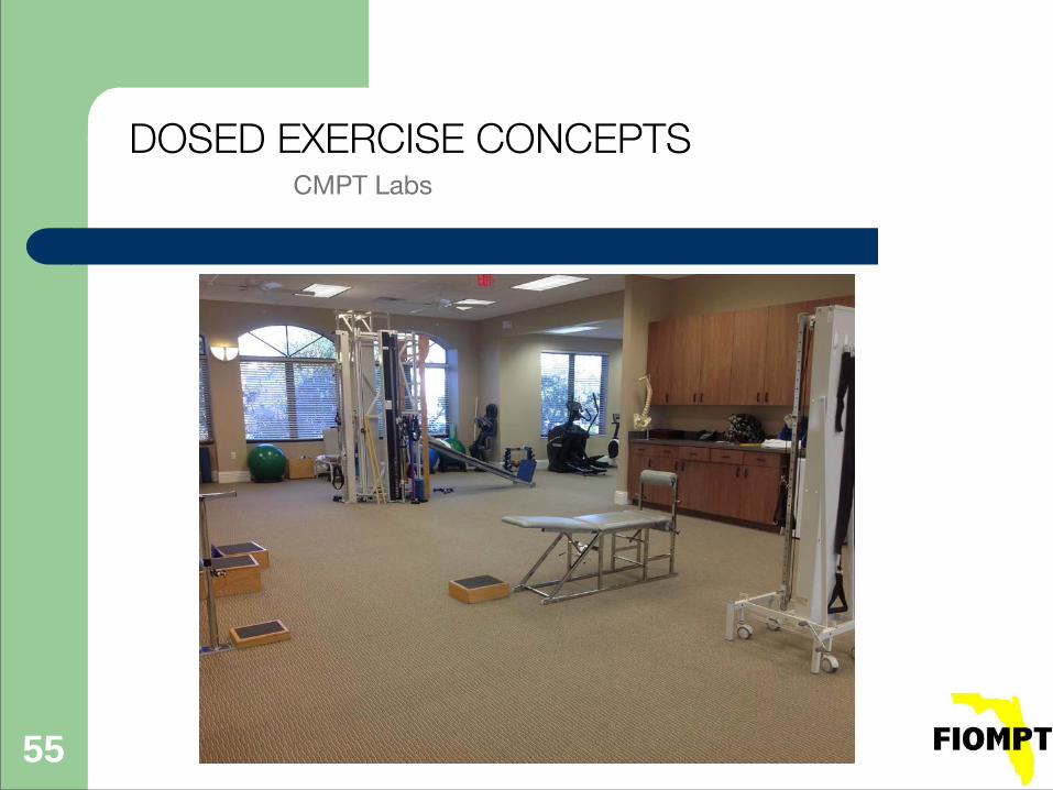
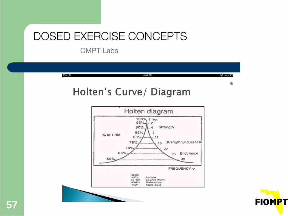
DOSED EXERCISE CONCEPTSCMPT Labs
56
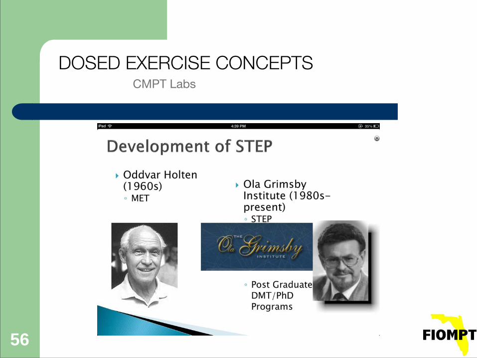
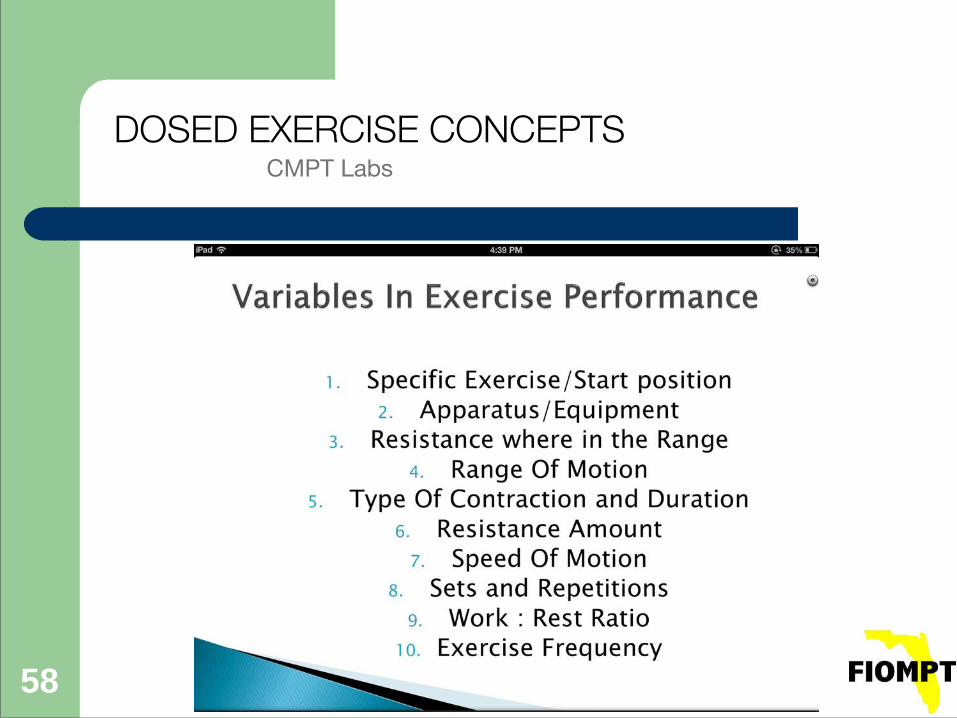
DOSED EXERCISE CONCEPTSCMPT Labs
57
CMPT LabsDOSED EXERCISE CONCEPTS
58
CMPT LabsDOSED EXERCISE CONCEPTS
59
Mobilizing Exercises
• Focus on arthrokinematic motions of distraction/translation
• Exercise performed for viscoelastic changes in joint capsules
• Sustained mobilizing force of greater than 10 seconds for plasticity
• < 50% of 1 RM
60
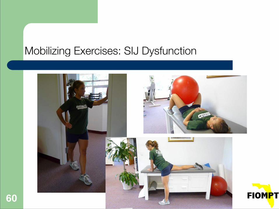
Mobilizing Exercises: SIJ Dysfunction
61
• Signs and symptoms
• Stages of healing of specific tissues/tolerance to stress
• Selective tissue training: bone, collagen, cartilage, disc, muscle, nerve
• The functional quality you are trying to achieve
• Functional progressions
Stage 1Dosage based on
62
Stage 1 Potential Tissue States
• Reduced arthrokinematic motion
• Decrease in active and passive range
• Joint pain
• Pain w weight bearing
• Edema/increased temp
• Muscle guarding
• Poor coordination
63
Dosage outline Stage 1
• 3-5 exercises
• Many repetitions, minimal resistance
• < 60% 1 RM for vascularity, < 40% 1RM tonic atrophy
• Low speed
• Hypomobile: train in outer range
• Hypermobile: train in mid to inner range
• Start contraction from length/tension position
64
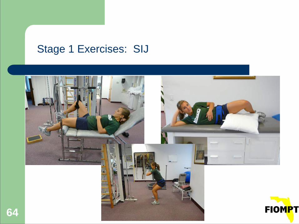
Stage 1 Exercises: SIJ
65
Stage 2 Training
• Increase repetitions with additional exercises (5-10)
• Increase repetitions with additional sets
• Increase speed – not weight
• Combine concentric and eccentric work for further tissue tension accommodation
• Remove external supports, progress to dependent positions
• Progress to full range of motion
66
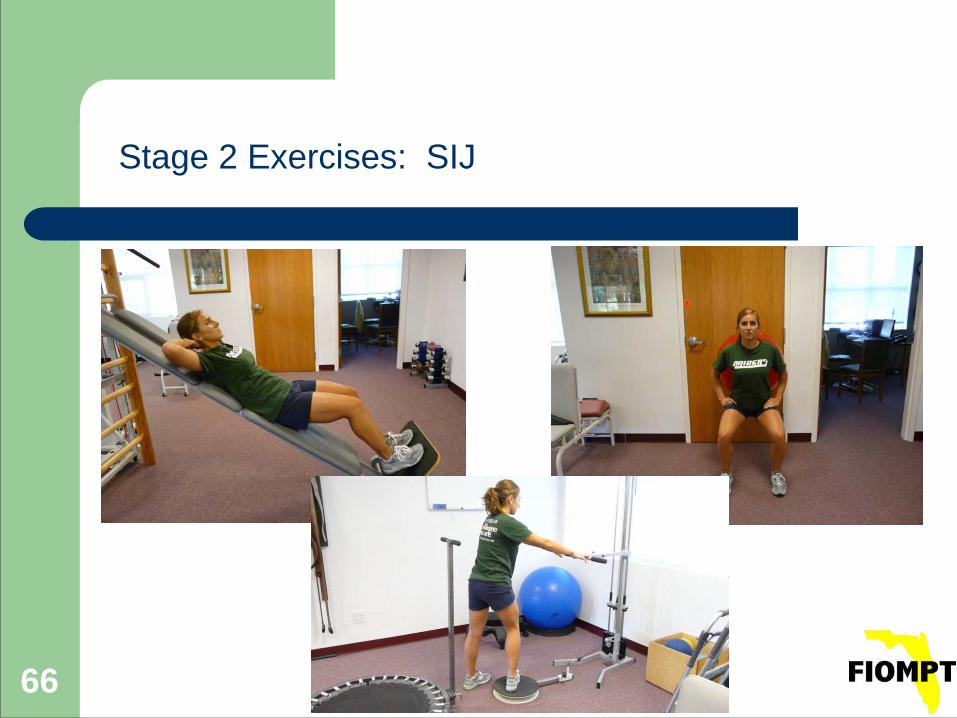
Stage 2 Exercises: SIJ
67
Stage 3 Training
• Increase weight (60 to 80% of 1RM) therefore decreased repetitions
• Eccentric emphasis to stabilize new range
• Diagonal/triplanar motions
• End range isometrics
68
Stage 3 Potential tissue states
• Full, pain-free motion
• Full weight-bearing is tolerated
• Excessive repetitions or resistance may provoke pain
• Edema and muscle guarding have resolved
• Continues with deficits in strength, endurance, fast coordination, power
69
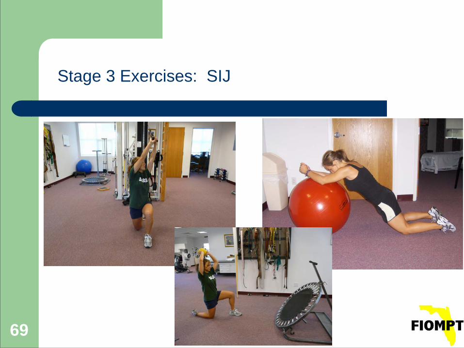
Stage 3 Exercises: SIJ
70
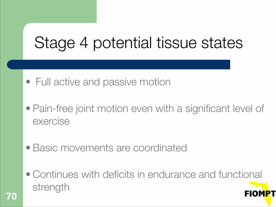
Stage 4 potential tissue states
• Full active and passive motion
• Pain-free joint motion even with a significant level of exercise
• Basic movements are coordinated
• Continues with deficits in endurance and functional strength
71
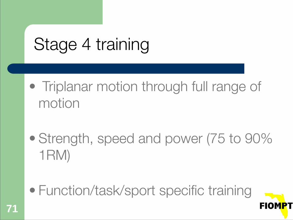
Stage 4 training
• Triplanar motion through full range of motion
• Strength, speed and power (75 to 90% 1RM)
• Function/task/sport specific training
72
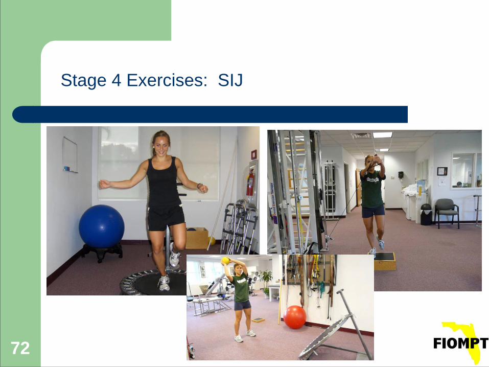
Stage 4 Exercises: SIJ
References
● Cleland J. Orthopaedic Clinical Examination: An Evidence-Based Approach for Physical Therapists. Philidephia, PA: Saunders Elsevier; 2007.
● Levangie, PK (Phys Ther 1999; 79: 1043-1057) ● Slipman et al (Archives of Physical Medicine and Rehabilitation,2000; 81:
334-338) ● Rivard J, Grimsby O, eds. Scientific Theory and Clinical Application in
Orthopaedic Manual Physical Therapy: Scientific Therapeutic Exercise Progressions (STEP): The Back and Lower Extremity. Vol 3. Taylorsville, UT: The Academy of Graduate Physical Therapy, Inc; 2008.