Embed Size (px)
Citation preview
Orthopadic cors
Topic :
-Cervical spondylitis.
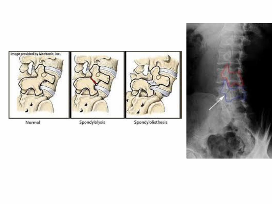
-Development disorders(Spondylolysis and Spodylolsithesis)
Cervical spondylitis.
Definition :
- a painful condition of the cervical spine resulting from the degeneration of intervertebral disks.
- is common from middle age onwards, even in people who have not been aware of any acute episode in former years.
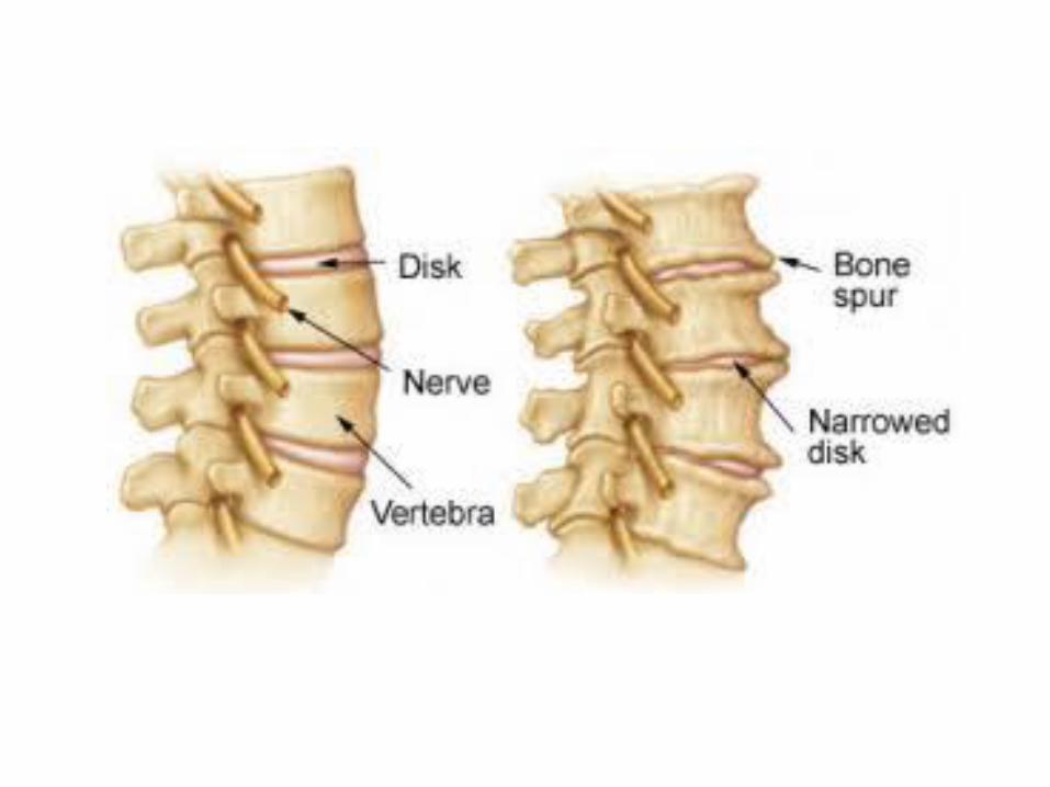
:pathophysiology the intervertebral disk lose hydration and elasticity with age , this starts in the nucleus pulposus and it buckling inward , the annulus fibrosis will become thinner and bulge outword.
This increase the mechanical stress at cartilaginous end plates , so the cartilage wears away and facets override and bony spurs appear at the anterior and posterior margins of the vertebral bodies on either side of the affected discs; those that develop posteriorly may encroach upon the intervertebral foramina
causing pressure on the nerve roots
Quick review of cervical nerves
In addition to the seven cervical vertebrae, cervical anatomy features eight cervical nerve roots (C1-C8) that branch from the spinal cord and control motor and sensory abilities for different parts of
the body.
•The top seven cervical nerves are named based on the lower of the two cervical vertebrae that it runs between. As an example, the C6 nerve root runs beneath the C5 vertebra and above the C6 vertebra. The C8 nerve root, however,
runs between the C7 vertebra and T1 vertebra.
Each level of the cervical spine actually has two nerve roots—one on each side—that branch off from the spinal cord.

Sensory function Motor function
The C2 dermatome handles sensation for the upper part of the head, and the C3 dermatome covers the side of the face and behind the head. (C1 does not have a dermatome.)
control the head and neck, including movements forward, backward, and to the sides. These nerves also play key roles in breathing
C1 C2 C3
The C4 dermatome covers the
neck and top of the shoulders. helps control the shoulders as well as the diaphragm
C4
The C5 dermatome covers the shoulders and outer part of the arm down to about the elbow
or close to the wrist.
controls upper body muscles like the deltoids and the biceps
C5
C6 dermatome covers the top of the shoulders and runs down the side of the arm and into the
thumb side of the hand.
controls the wrist extensors and also provides some innervation to the biceps
C6
The C7 dermatome goes from the shoulder down the back of the arm and into the middle
finger.
C7 controls the triceps C7
he C8 dermatome covers the lower part of the shoulder and goes down the arm into the
pinky side of the hand.
C8 controls the hands C8
Cervical Nerve Functions

symptoms
•The condition is not always symptomatic, and many people go throughout life
without experiencing anything more
than slight stiffness.
Troublesome symptoms come on gradually
Clinical features
…..The patient, usually aged over 40 years profile
chief complaint ….. neck pain and stiffness. .
The pain may radiate : to the occiput, the scapular muscles and down one or both arms.
Paraesthesia, weakness and are occasional symptoms.
Symptoms :
Signs :
•There may be tenderness in the soft tissues at the back of the neck and above the
scapulae
•neck movements are limited and painful at the extremes.
• Careful neurological examination may show abnormal signs in one or both upper limbs

Imaging
•Typical x-ray features •narrowing of several disc
spaces, bony spur formation at the anterior and posterior edges of the vertebral bodies
•Oblique views may show bony
encroachment on the intervertebral foramina.
• MRI:
• will show whether there is nerve root compression.

Differential diagnosis Other disorders associated with neck or arm pain and sensory symptoms must be excluded. Cervical vertebral spur formation is very common in older people and this
can be misleading in patients with other disorders. Rotator cuff lesions: pain around the shoulder may resemble the referred pain of cervical spondylosis. However, features such as rotator cuff tenderness and restricted shoulder movements should suggest a local problem. Nerve entrapment syndromes: median or ulnar nerve entrapment may give rise to intermittent symptoms of pain and paraesthesia in the hand.
•Cervical tumours: with tumours of the vertebrae, spinal cord, nerve roots or lymph nodes the symptoms are unremitting. Imaging studies should reveal the diagnosis.
Treatment
During painful episodes, heat and massage are soothing; some patients benefit from a period in
a restraining collar. Physiotherapy is the mainstay of treatment, patients usually being maintained
in relative comfort by various measures including exercises.
Surgical treatment is indicated if severe symptoms are relieved only by a rigid and irksome support,
particularly if there are neurological changes due to nerve root compression.
•Spondylolysis and spondylolisthesis are common causes of low back pain in young athletes
Spondylolisthesis
is the displacement of compared to another. vertebra one
Medical dictionaries define spondylolisthesis specifically as the forward or anterior displacement of a vertebra over the vertebra inferior to it
).sacrum (or the
Spondylolisthesis
Listhesis mean (slippage)
Spondylo mean ( spine )
prevention mechanism
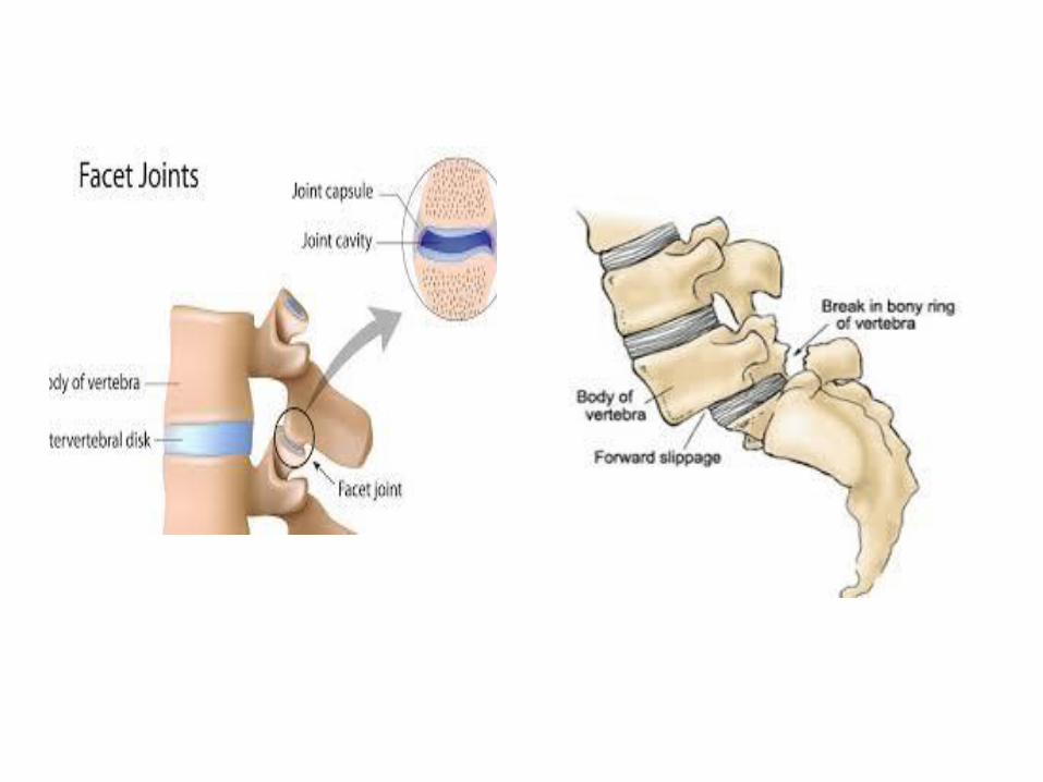
plane ,synovial are a set of facet joints : of processes articular between the joints
two adjacent vertebrae.
•Normal laminae and facets constitute a locking mechanism which prevents each vertebra from moving forwards on the one below. …… Forward shift (or slippage) occurs only when this mechanism fails
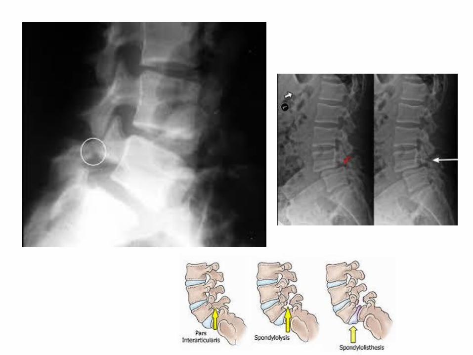
Listhesis (slippage) is nearly always between L4 and L5, or between L5 and the sacrum…… This usually happens for one of the following reasons:
■ Dysplasia of the lumbosacral facet joints (20% of cases).
■ Separation or stress fracture (lysis) through the neural arch (the pars interarticularis), allowing the anterior part of the vertebra to slip forward upon the one below (50% of cases).
■ Osteoarthritic degeneration of the facet joints, causing them to lose their normal stability. This usually occurs at L4/5 (25% of cases).
■ Destructive conditions such as fracture, TB and neoplasia (5% of cases).
Spondylolisthesis Grades •Spondylolisthesis can be described
according to its degree of severity. One commonly used description grades spondylolisthesis, with grade 1 being least advanced, and grade 5 being most advanced. The spondylolisthesis is graded by measuring how much of a vertebral body has slipped forward over the body beneath it.
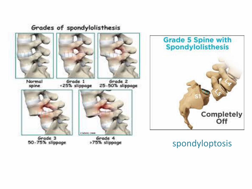
spondyloptosis
How do People Get Spondylolisthesis ?
•Approximately 5%-6% of males, and 2%-3% of females have a spondylolisthesis.
•It becomes apparent more often in people who are involved with
very physical activities such as weightlifting, gymnastics,or
football.
•Males are more likely than females to develop symptoms from the disorder, primarily due to their engaging in more physical
activities.
•Although some children under the age of five may be pre-disposed towards having a spondylolisthesis, or may indeed already have an undetected spondylolisthesis, it is rare that such young children are diagnosed with spondylolisthesis. Spondylolisthesis becomes more common among 7-10 year olds. The increased physical activities of adolescence and adulthood, along with the wear-and-tear of daily life, result in spondylolisthesis being most common among adolescents and adults.
Types of Spondylolisthesis •Developmental Spondylolisthesis: This type of
spondylolisthesis may exist at birth, or may develop during childhood, but generally is not noticed until later in childhood or even in adult life.
•Acquired Spondylolisthesis: •
•With all of the daily stresses that are put on a spine, such as carrying heavy items and physical sports, the spine may wear out (ie, degenerate). As the connections between the vertebrae weaken, this may lead to spondylolisthesis.
•A single or repeated force being applied to the spine can
cause spondylolisthesis; for example, the impact of falling off a ladder and landing on your feet

Clinical features Dysplastic spondylolisthesis is seen in children. It is usually painless but the mother may notice the unduly protruding abdomen. There may be an associated scoliosis.
Lytic spondylolisthesis is the commonest variety. It occurs in adults and intermittent backache is the usual presenting symptom. Pain may be initiated or exacerbated by exercise or strain. Movements are usually normal in younger patients but may be restricted in older people
Degenerative spondylolisthes is usually occurs in patients over 40 years with long-standing backache due to facet joint arthritis. Sometimes the presenting symptom is spinal ‘claudication’ due to narrowing of the spinal canal (see under spinal stenosis).
Also ……
•Pain in the low back, especially after exercise scoliosis
protruding abdomen ) ) increased lordosis.
•Pain and/or weakness in one or both thighs or legs
•Reduced ability to control bowel and bladder functions
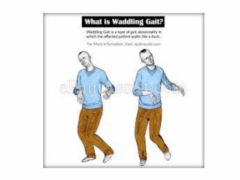
•In cases of advanced spondylolisthesis changes may occur in the way people stand and walk; for example, development of a waddling style of walking.
•may seem shorter;
• and muscle spasms in the lowback may occur
Imaging
•X-rays show the forward shift of the upper part of the spinal column on the stable vertebra below; elongation of the arch or defective facets may be seen. The gap in the pars interarticularis is more easily seen in oblique x-ray views, and best of all in CT scans.

Treatment
•conservative treatment, similar to that for other types of back pain.
• is suitable for most patients. Operative treatment is indicated: (1) if the symptoms are disabling and interfere significantly with work and recreational activities; (2) if the upper vertebra has slipped forwards over more than 50% of the vertebral body below; and (3) if neurological compression is significant.
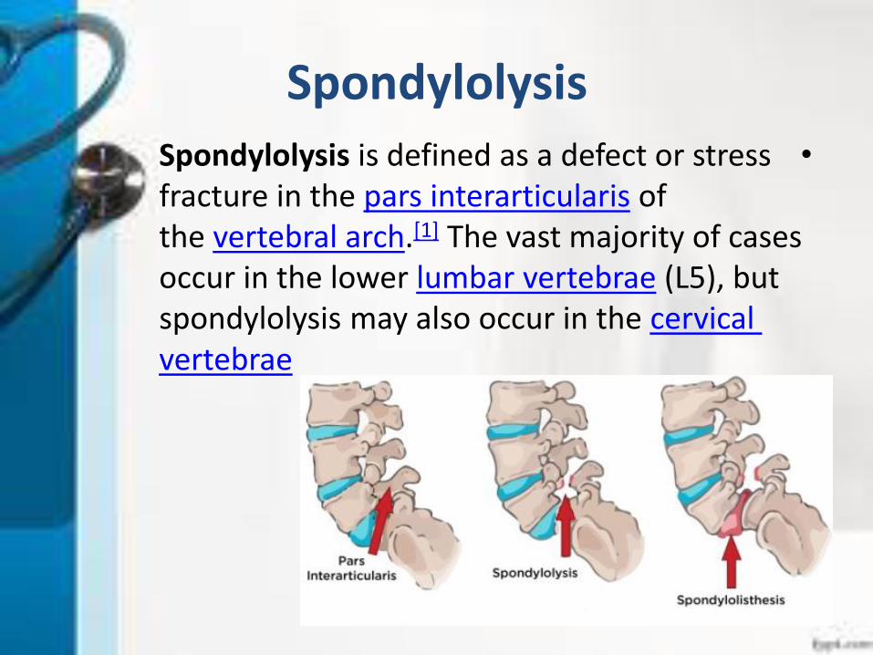
Spondylolysis
•Spondylolysis is defined as a defect or stress of interarticularispars fracture in the
The vast majority of cases ]1[.vertebral arch the), but 5(L lumbar vertebrae occur in the lower
cervical may also occur in the spondylolysisvertebrae
Cause
The cause of spondylolysis remains unknown, however many factors are thought to contribute to its development.
The condition is present in up to 6% of the population, majority of which usually present asymptomatically.
Research supports that there are hereditary and acquired risk factors that can make one more susceptible to the defect.
The disorder is generally more prevalent in males compared to females, and tends to occur earlier in males due to their involvement in more strenuous activities at a younger age.
In a young athlete, the spine is still growing which means there are
, leaving points of weakness in the ossification centers manyspine.
It is believed ]6[% of all low back pain.50as it accounts for about
that both repetitive trauma and an inherent genetic weakness can make an individual more susceptible to spondylolysis

In majority of cases, spondylolysis presents asymptomatically which
[.can make diagnosis both difficult and incidental •Symptoms:
•Unilateral low back pain •Pain that radiates into the buttocks or legs
•Onset of pain can be acute or gradual •Pain that can restrict daily activities
•Pain that worsens after stress activity •Pain aggravated with lumbar hyperextension
•Clinical Signs:[
•Excessive lordotic posture •Unilateral tenderness on palpation
•Scottie dog fractureVisible on diagnostic imaging (

Treatment
•Bracing to immobilize the spine for a short period (e.g. four months) to allow the pars defect to heal
•, as inflammatory medication-anti Pain medications and/orneeded
•Exercise that is controlled and builds gradually over time.
•On rare occasions, spondylolysis that is not healing or may
have neurological components can require surgery to provide internal fixation and stability to the area. Usually, two procedures are performed as part of the same surgery:
•, which reduces irritation and laminectomy decompressive Ainflammation in the area (but increases spinal instability)
•A spinal fusion to provide stabilization of the affected area.


![CORS Program FY0945]09... · 2009-10-01 · CORS Program FY09 Giovanni Sella CORS Program Manager NOAA- National Geodetic Survey giovanni.sella @ noaa.gov CGSIC Savannah, GA CORS](https://img.pdfslide.us/doc/110x75/5f09f82f7e708231d4296126/cors-program-4509-2009-10-01-cors-program-fy09-giovanni-sella-cors-program.jpg)