Embed Size (px)
Citation preview
Int J Anat Res 2016, 4(3):2518-21. ISSN 2321-4287 2518
Original Research Article
STUDY OF HUMAN ATLAS VERTEBRAE FOR THE PRESENCE OFRETROARTICULAR GROOVE OR CANAL IN NORTH INDIANSSeema *1, Poonam Verma 2, Anupama Mahajan 3.
ABSTRACT
Address for Correspondence: Dr. Seema, Professor, Department of Anatomy, Sri Guru Ram DasInstitute of Medical Sciences and Research, Vallah, Amritsar, Punjab, India.E-Mail: [email protected]
Background Atlas is the first cervical vertebra articulating above with the occipital condyles of the skull andbelow with the axis vertebra. Anatomy of this first cervical vertebra show great morphological variations. Atlasbony out growths known as Ponticles which can be lateral or posterior are commonly found. These ponticles cancompress the third part of vertebral artery leading to vertebrobasilar insufficiency. Atlas vertebra is approachedin a number of surgical procedures like transpedicular screw fixation, transarticular screw fixation interlaminarclamp and for interspinous wiring.Aim: To find out the incidence of retroarticular groove or canals in North Indian population in order to avoidpostoperative complications arising due to these anatomical variations. Material and Methods: The present study was done on fifty dry adult human atlas vertebrae of unknown sextaken from Department of Anatomy, Sri Guru Ram Das Institute of Medical Sciences and Research, Amritsar. Theywere carefully studied for the presence of retroarticular groove or canal.Results: The retroarticular groove or canal were found in eight vertebrae (16%). Presence of these retroarticularcanals predisposes to Barre –Lieou and Cervicogenic syndromes especially in neck movements.Conclusion: These bony bridges embracing the vertebral artery are responsible for vertigo and vertebrobasilarinsufficiency. The present study can be helpful to radiologists, anthropologists and neurosurgeons to avoidmany postoperative complications.KEY WORDS: Cervical, Axis, Occipital, Vertebrobasilar, Transpedicular.
INTRODUCTION
International Journal of Anatomy and Research,Int J Anat Res 2016, Vol 4(3):2518-21. ISSN 2321-4287
DOI: http://dx.doi.org/10.16965/ijar.2016.248
Access this Article online
Quick Response code Web site:
Received: 25 Jun 2016 Accepted: 14 Jul 2016Peer Review: 25 Jun 2016 Published (O): 31 Jul 2016Revised: None Published (P): 31 Jul 2016
International Journal of Anatomy and ResearchISSN 2321-4287
www.ijmhr.org/ijar.htm
DOI: 10.16965/ijar.2016.248
*1 Professor, Department of Anatomy, Sri Guru Ram Das Institute of Medical Sciences and Research,Amritsar, Punjab, India.2 Professor, Department of Anatomy, Sri Guru Ram Das Institute of Medical Sciences and Research,Amritsar, Punjab, India.3 Professor and Head, Department of Anatomy Sri Guru Ram Das Institute of Medical Sciences andResearch, Amritsar, Punjab, India.
artery, venous plexus and first cervical nerve [1].Frequently the bony spurs arise from theanterior and posterior margins of the grooveknown as ponticles converting the groove intoaforamen [2,3]. The third part of vertebralartery appears from foramen transversarium of
Atlas the first cervical vertebra supports thehead. It consists of short anterior and longerposterior arch. The superior surface of theposterior arch bears a wide groove for vertebral
Int J Anat Res 2016, 4(3):2518-21. ISSN 2321-4287 2519
Seema, Poonam Verma, Anupama Mahajan. STUDY OF HUMAN ATLAS VERTEBRAE FOR THE PRESENCE OF RETROARTICULAR GROOVE ORCANAL IN NORTH INDIANS.
the atlas, turns backwards and medially behindthe lateral mass of the atlas, and lies in theneurovascular groove on posterior arch of theatlas. It then passes through the opening inanterior part of posterior atlanto-occipital mem-brane and enters the foramen magnum. Thevertebral artery in its way from foramentransversarium to the cranial cavity is vulner-able to damage or distortion from externalfactors like bony or ligamentous structure likeretro-articular canal which may cause externalpressure on vertebral artery [4].The compressionof vertebral artery will cause vertebro basilarinsufficiency [5]. These indicate the ossificationof posterior atlantooccipital membrane [6]. Theaim of the study is to determine the incidenceof these ponticles in north Indians. This mighthelp to explain the correlation between theoccurrence of bony ponticles and vertebralartery entrapment. Recently, transarticular andtranspedicular screws fixation has been widelyused in stabilizing the cervical column. Inappro-priate insertion of pedicle screws can causedamages to the upper cervical spine, care hasto be taken to avoid injury to the vertebralartery lying in the retroarticular groove.Iatrogenic injury to the vertebral arteryadjoining vital structures such as the spinal cord,nerve roots, cranial nerves, and vertebralarteries [7]. When exposing is the mostcommonest intra-operative complication duringa posterior approach [8]. A short segmentposterior fixation technique is often adopted topreserve the motion of the atlanto-occipital joint[9,10]. The present study was done to find theincidence of retroarticular anatomical variationin the atlas vertebra either in the form ofretroarticular groove or canals in North Indians.
MATERIALS AND METHODS
Fifty dry adult human atlas vertebrae ofunknown age and sex were taken fromDepartment of Anatomy, Sri Guru Ram DasInstitute of Medical Sciences and Research,Amritsar. All atlas vertebrae were cleanedthoroughly. Before the present study it wasensured that all the vertebrae were intact andwere free of any trauma or metastatic growth.They were numbered from one to fifty. Eachvertebra was examined carefully for the
presence of retroarticular groove or bony spursforming canal or foramen on the right as well asleft side.
OBSERVATIONS
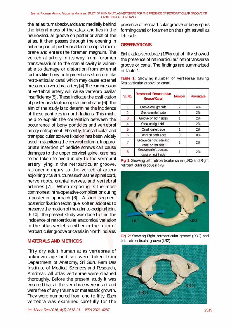
Eight atlas vertebrae (16%) out of fifty showedthe presence of retroarticular/ retrotransversegroove or canal. The findings are summarizedin Table 1.
Table 1: Showing number of vertebrae havingRetroarticular groove or canal.
Sr. No.Presence of Retroarticular
Groove/CanalNumber Percentage
1 Groove on right side 2 4%2 Groove on left side 1 2%3 Groove on both sides 1 2%4 Canal on right side 1 2%5 Canal on left side 1 2%6 Canal on both sides 0 0%
7 Groove on right side and canal on left side
1 2%
8 Groove on left side and canal on right side
1 2%
Fig. 1: Showing Left retroarticular canal (LRC) and Rightretroarticular groove (RRG).
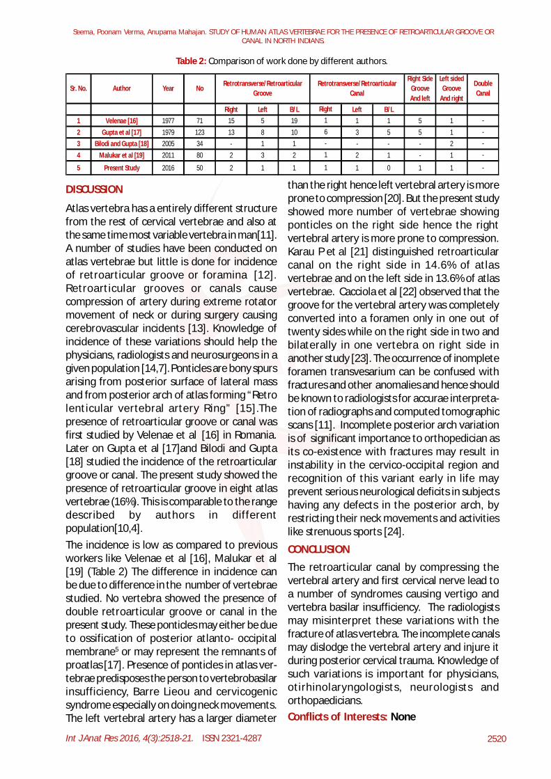
Fig. 2: Showing Right retroarticular groove (RRG) andLeft retroarticular groove (LRG).
Int J Anat Res 2016, 4(3):2518-21. ISSN 2321-4287 2520
Seema, Poonam Verma, Anupama Mahajan. STUDY OF HUMAN ATLAS VERTEBRAE FOR THE PRESENCE OF RETROARTICULAR GROOVE ORCANAL IN NORTH INDIANS.
DISCUSSION
Atlas vertebra has a entirely different structurefrom the rest of cervical vertebrae and also atthe same time most variable vertebra in man[11].A number of studies have been conducted onatlas vertebrae but little is done for incidenceof retroarticular groove or foramina [12].Retroarticular grooves or canals causecompression of artery during extreme rotatormovement of neck or during surgery causingcerebrovascular incidents [13]. Knowledge ofincidence of these variations should help thephysicians, radiologists and neurosurgeons in agiven population [14,7]. Ponticles are bony spursarising from posterior surface of lateral massand from posterior arch of atlas forming “Retrolenticular vertebral artery Ring” [15].Thepresence of retroarticular groove or canal wasfirst studied by Velenae et al [16] in Romania.Later on Gupta et al [17]and Bilodi and Gupta[18] studied the incidence of the retroarticulargroove or canal. The present study showed thepresence of retroarticular groove in eight atlasvertebrae (16%). This is comparable to the rangedescribed by authors in differentpopulation[10,4].The incidence is low as compared to previousworkers like Velenae et al [16], Malukar et al[19] (Table 2) The difference in incidence canbe due to difference in the number of vertebraestudied. No vertebra showed the presence ofdouble retroarticular groove or canal in thepresent study. These ponticles may either be dueto ossification of posterior atlanto- occipitalmembrane5 or may represent the remnants ofproatlas [17]. Presence of ponticles in atlas ver-tebrae predisposes the person to vertebrobasilarinsufficiency, Barre Lieou and cervicogenicsyndrome especially on doing neck movements.The left vertebral artery has a larger diameter
than the right hence left vertebral artery is moreprone to compression [20]. But the present studyshowed more number of vertebrae showingponticles on the right side hence the rightvertebral artery is more prone to compression.Karau P et al [21] distinguished retroarticularcanal on the right side in 14.6% of atlasvertebrae and on the left side in 13.6% of atlasvertebrae. Cacciola et al [22] observed that thegroove for the vertebral artery was completelyconverted into a foramen only in one out oftwenty sides while on the right side in two andbilaterally in one vertebra on right side inanother study [23]. The occurrence of inompleteforamen transvesarium can be confused withfractures and other anomalies and hence shouldbe known to radiologists for accurae interpreta-tion of radiographs and computed tomographicscans [11]. Incomplete posterior arch variationis of significant importance to orthopedician asits co-existence with fractures may result ininstability in the cervico-occipital region andrecognition of this variant early in life mayprevent serious neurological deficits in subjectshaving any defects in the posterior arch, byrestricting their neck movements and activitieslike strenuous sports [24].
Table 2: Comparison of work done by different authors.
Right Left B/L Left B/L1 Velenae [16] 1977 71 15 5 19 1 1 5 12 Gupta et al [17] 1979 123 13 8 10 3 5 5 13 Bilodi and Gupta [18] 2005 34 - 1 1 - - - 24 Malukar et al [19] 2011 80 2 3 2 2 1 - 1
5 Present Study 2016 50 2 1 1 1 0 1 1
1 -
1 -
Right1 -6 -
Retrotransverse/Retroarticular Canal
Retrotransverse/Retroarticular Groove
Sr. No.
- -
Double Canal
Left sided Groove
And right
Right Side Groove And left
Author Year No
CONCLUSION
The retroarticular canal by compressing thevertebral artery and first cervical nerve lead toa number of syndromes causing vertigo andvertebra basilar insufficiency. The radiologistsmay misinterpret these variations with thefracture of atlas vertebra. The incomplete canalsmay dislodge the vertebral artery and injure itduring posterior cervical trauma. Knowledge ofsuch variations is important for physicians,otirhinolaryngologists, neurologists andorthopaedicians.Conflicts of Interests: None
Int J Anat Res 2016, 4(3):2518-21. ISSN 2321-4287 2521
Seema, Poonam Verma, Anupama Mahajan. STUDY OF HUMAN ATLAS VERTEBRAE FOR THE PRESENCE OF RETROARTICULAR GROOVE ORCANAL IN NORTH INDIANS.
REFERENCES [13]. Krishnamurthy, Nayak SR, Khan S, Prabhu La Tha V,Lakshmi A, et al. References :Arcuate foramenofatlas: incidence, phylogenetic and clinicalsignificance. Romanian Journal of Morphology andEmbryology. 2007;48(3):263-66.
[14].Agrawal D, Mohanty BB, Shetty S, Parija B, HazarySK and China a PK. Variations in foramentransversarium: An osteological study in easternIndia. Int J of current research.2012;4(9):120-122.
[15]. Manjunath KY. Posterior bridging of the atlasvertebra in South Indians. Incl. J Med. Sci.2001;55(9):488-90.
[16]. Velenae C, Barzu S, Panescu S and Udroiu C,Retrotransverse groove or canal of atlas and itssignificance. Acta Anatemica Basel. 1997;97:400-2.
[17]. Gupta SC, Gupta CD, Arora HK, Maheshwari BB. Theretro transerse groove/canal in Indian Atlasvertebral. Anat Anz. 1979;145:514-16.
[18]. Bilodi AK and Gupta SC. Presence of retrotransversegroove or canal in atlas vertebrae. J. Anat Soc. Ind.2005;54(1):16-18.
[19]. Malukar O, Prajapati VP, Nagar SK. Ponticulus. J ofMed. Res. 2011;1(2):51-5.
[20]. Thiel H. Gross morphology and pathoanatomy ofthe vertebral arterier. J Manipulat Physiol, Ther.1991;14:133-41.
[21]. Karau, BP, Ogengo JA., Hassanali J, Odula PO.Morphometry and Variations of Bony Ponticles ofthe Atlas Vertebrae (CI) in Kenyans. Int J Morphol.2010; 28(4):1019-24.
[22].Cacciola F, Phalke U, Goel A. Vertebral artery inrelationship to C1 –C2 vertebra: An anatomic study.Neurology India. 2004;52(2):178–84.
[23]. Gosavi SN. Morphometric Study of the AtlasVertebra using Manual. Malays Orthop J.2012;6(3):18-20.
[23]. Kaushal P. Median deficiency in the posterior archof the atlas vertebra: a case report. Int J AnatVari, 2011;4:67-8.
[1]. Williams PL, Bannister LH, Berry MM, Collins P,Dyson M, Dussek JE, Ferguson MWJ. In: SkeletalSystem. 38th Edn. Churchill Living stone. New York.1995:519.
[2]. Lamberty BGH and Zivanovic S. The retro articularvertebral artery ring of the atlas and itssignificance. Acta Anat. 1973;85:113-122.
[3]. Long J. Cranio cervical region Osteology andarticulations. Neuro orthop. 1986;1:67-92.
[4]. Sylvia S, Kulkarni S, Hatti A. Bilateral Retro Articularring in Atlas vertebra – A Case Report. AnatomicaKarnataka. 2011;5(1):81-86.
[5]. Sun JY. Foramen arcuals and vertigo. Zhonghua WaiKe Za Zhi 1990;28(10):592-4,636-7.
[6]. Romanus T and Tovi A. A variation of the Atlas.Roetgenologic incidence of a bridge over the grooveon the atlas for the vertebral artery. Acta RadiolDiagn. (Stocks) 1964;2:289-97.
[7]. Senguel G, Kadioglu HH. Morphometric Anatomy ofthe atlas and axis vertebrae. TurkishNeuroanatomy. 2006;16(2):69-76.
[8]. Neo M, Sakamoto T, Fujibayashi S, Nakamura T. Theclinical risk of vertebral artery injury from cervicalpedicle screws which were inserted in degenerativevertebra. Spine. 2005;30(24):2800-5.
[9]. Leventhal MR. Campbell’s Operative OrthopaedicsIn: Spinal Anatomy and Surgical Approach. 9th edn;Vol. 3. Mosby, St. Louis; 1998;2:681-270.
[10]. Hanson PB, Montesano PX, Sharkey NA. Anatomicand biochemical assessment of transarticularscrew fixation for the atlanto-axial instability.Spine. 1991;16:1141–5.
[11]. Wysocki J Buhrowski M, Reymond J Kwiastkowski J.Anatomical variants of the cervical vertebrae andthe first thoracic vertebra in man. Folia Morpho.2003;62: 357-63.
[12]. Chauhan R, Khanna J. Absence of costal element ofthe foramen transversarium of atlas vertebrae. IntJ Res Med Sci. 2013;1:66-8.
How to cite this article:Seema, Poonam Verma, Anupama Mahajan. STUDY OF HUMANATLAS VERTEBRAE FOR THE PRESENCE OF RETROARTICULARGROOVE OR CANAL IN NORTH INDIANS. Int J Anat Res2016;4(3):2518-2521. DOI: 10.16965/ijar.2016.248


![Materials and Design - WebInstances/N/Publicn.ethz.ch/~jocmuell/download/Mueller_MechanicalPropertiesOfParts.pdf · to over-curing. In another article, Udroiu and Mihail [17] report](https://img.pdfslide.us/doc/110x75/5a7bd8dd7f8b9a49588c363f/materials-and-design-webinstancesn-jocmuelldownloadmuellermechanicalpropertiesofpartspdfto.jpg)