Embed Size (px)
Citation preview

Dwyer et al. Thrombosis Journal 2014, 12:10http://www.thrombosisjournal.com/content/12/1/10
ORIGINAL BASIC RESEARCH Open Access
Thrombin based gelatin matrix and fibrin sealantmediated clot formation in the presence ofclopidogrelJoseph F Dwyer1*, Jill A McCoy1, Ziping Yang1, Michael Husser1, Heinz Redl2, Mary Ann Murphy1,Martin Wolfsegger3, James P DiOrio1, Andreas Goppelt3 and Shane Donovan1ˆ
Abstract
Background: Platelet inhibitors are commonly used to reduce the risk of atherothrombotic events. The aim of thisstudy was to determine the impact of platelet inhibitors, specifically clopidogrel and aspirin, on clot kinetics,strength, and/or structure during the use of thrombin based gelatin matrices and fibrin sealants.
Methods: Blood was collected and heparinized from donors on clopidogrel (and aspirin) and age matched controldonors. Blood component analysis, whole blood platelet aggregometry, and activated clotting time (ACT) wereused to monitor compliance to therapy and identify any differences between donor groups. Clot kinetics andstrength were analyzed using thrombelastography (TEG). Field Emission Scanning Electron Microscopy (FESEM) wasused to analyze clot structure.
Results: Blood component profiles were similar for both donor groups. Aggregometry indicated that aggregationresponse to adenosine diphosphate (ADP) for clopidogrel donors was 12% of that for the controls (p = 0.0021), anexpected result of clopidogrel induced platelet inhibition. However, blood from both donor groups had anelevated thrombin induced aggregation response. Heparinization of donor blood resulted in similarly elevated ACTsfor both donor groups. TEG results indicated similar clot kinetics and strength between clopidogrel and controldonor groups for blood alone and when clotting was induced using thrombin based gelatin matrices and fibrinsealants. FESEM images supported TEG findings in that similar morphologies were observed in ex vivo formed clotsfrom both donor groups when thrombin based gelatin matrices and fibrin sealants were used.
Conclusion: These results suggest that platelet inhibitors do not negatively impact clot kinetics, strength, andstructure when clotting is initiated with thrombin based gelatin matrices and fibrin sealants.
Keywords: Floseal, Tisseel, Clopidogrel, Thrombelastography, Thrombin, Hemostasis
BackgroundPlatelets contribute to physiological hemostasis by aggre-gating at the site of a lesion, providing surfaces forhemostatic reactions, and supplying regulatory hemostaticfactors. They also contribute to atherosclerotic disease bystimulating and stabilizing thrombi. Following myocardialinfarction or stroke, the American Heart Association andAmerican College of Cardiology guidelines recommendthat patients take platelet inhibitors to reduce the risk of
* Correspondence: [email protected]ˆDeceased1Baxter Healthcare Corporation, Deerfield, IL, USAFull list of author information is available at the end of the article
© 2014 Dwyer et al.; licensee BioMed CentralCommons Attribution License (http://creativecreproduction in any medium, provided the orDedication waiver (http://creativecommons.orunless otherwise stated.
further atherothrombotic events [1,2]. Clopidogrel and as-pirin (acetylsalicylic acid) are commonly used platelet in-hibitors. Both drugs reduce mortality in patients withchronic atherosclerotic disease and are also effective drugsfor the acute treatment of infarcts [3].Platelet aggregation is disrupted by these inhibitors via
two distinct pathways. Clopidogrel acts as an inhibitor ofadenosine diphosphate (ADP) induced platelet aggrega-tion by covalently binding to its receptor, P2Y12. ADPmediated activation of the GPIIb/IIIa complex is there-fore abated [4-6]. Aspirin irreversibly inhibits cyclooxy-genase enzymes, COX1 and COX2 [7]. This preventsthe conversion of arachidonic acid to thromboxane A2
Ltd. This is an Open Access article distributed under the terms of the Creativeommons.org/licenses/by/2.0), which permits unrestricted use, distribution, andiginal work is properly credited. The Creative Commons Public Domaing/publicdomain/zero/1.0/) applies to the data made available in this article,

Dwyer et al. Thrombosis Journal 2014, 12:10 Page 2 of 13http://www.thrombosisjournal.com/content/12/1/10
and subsequent thromboxane stimulated platelet aggre-gation [8,9]. Clopidogrel and aspirin can be used indi-vidually or in combination for dual anti-platelet therapy.Several clinical studies have linked the use of platelet ac-
tivation inhibitors to increased bleeding during surgeryresulting in excess blood loss, increased transfusion rates,and reoperation [10-13]. These findings led the authors ofthese studies to recommend that surgery be delayed ifpossible to allow patients to withdraw from anti-platelettherapy [10,12]. Clopidogrel and aspirin both irreversiblyalter platelet function. As a result, platelet aggregation andbleeding time generally do not return to normal levelsuntil 5 days after discontinuing usage when sufficientquantities of new unaffected platelets have been produced[14]. Recommendations for and optimal time interval be-tween cessation of anti-platelet medications and surgeryvary from 3–10 days [15-18].Other studies have suggested that the risk of further
atherothrombotic events outweighs the risk of increasedbleeding during surgery and recommend that in certainsituations patients remain on either aspirin or dual anti-platelet therapy throughout the perioperative period[19,20]. For example, patients who are at an intermediateor high risk for cerebro- or cardiovascular events are rec-ommended to remain on both clopidogrel and aspirinduring many types of surgeries such as orthopedic surgery,reconstructive surgery, and endoscopy [19]. Consequently,many patients are undergoing surgical procedures withsignificant platelet inhibition as a result of a reluctance orinability (i.e. non-elective/emergency procedures) to with-hold anti-platelet therapy prior to surgery. In such cases,surgeons face concerns that adjunctive thrombin basedgelatin matrix and fibrin sealant hemostatic agents maynot work effectively for patients on anti-platelet therapy.Gelatin matrices and fibrin sealants that contain a
thrombin component have been used for many years aseffective adjunctive hemostats in surgery [21,22]. Both ex-ogenous hemostats stop bleeding by inducing physio-logical hemostasis in a number of distinct ways. Theycurtail bleeding on tissue surfaces by generating fibrin me-diated blood clots. As thrombin based gelatin matricesrely on incoming blood to supply the substrates forhemostatic clots, one potential clinical concern is thatthese hemostats may not work effectively for patients tak-ing platelet aggregation inhibitors such as clopidogrel andaspirin. They also provide a high dose of thrombin, noveltopologies, and substrates for blood and tissue derived fac-tors to regulate hemostasis, and, in the case of thrombinbased gelatin matrix hemostats, the gelatin particles pro-vide a tamponade effect at the wound site, as well as con-tact activation, which leads to platelet activation.The purpose of this study was to evaluate clots formed
when thrombin based gelatin matrices and fibrin seal-ants were mixed with heparinized blood from patients
who were taking clopidogrel to determine whether therewas a negative impact of this platelet activation inhibitoron clot kinetics, strength, and/or structure. Assaysemploying whole blood were selected where appropriatefor sample testing to more closely reflect the surgicalconditions during application of the hemostats. Clotkinetics and strength were analyzed using thrombelas-tography (TEG) and clot structure was analyzed usingField Emission Scanning Electron Microscopy (FESEM).TEG was deemed an appropriate assay since it useswhole human blood, and it has been used to monitorpatient hemostasis profiles during cardiac and generalsurgery [23,24].
MethodsDonor criteria and collection of whole bloodDonor samples were collected from individuals in ac-cordance with Baxter Healthcare’s Internal Review Board.Blood was collected from three donors on clopidogreland aspirin and one donor on only clopidogrel. Bloodwas also taken from four age matched control donorswho were not taking clopidogrel or aspirin (i.e. con-trol). The small number of donors is a consequence ofthe limited available donor pool and selection criteria(Table 1). These criteria were used isolate the impact ofclopidogrel on hemostat performance from other drugsor medical conditions that interfere with hemostasis.The first 2–3 ml of blood collected was discarded fromeach donor. Thereafter, 25 ml of blood was collected insodium citrate (3.2%). An additional 5 ml aliquot wascollected in EDTA (1.5 mg/ml). The blood collected inEDTA was used for analysis of the blood cells, platelets,hematocrit, and hemoglobin. Other analyses were per-formed using citrated blood. All researchers performingthe assays were blinded to donor status throughout thecourse of the study.
Blood component analysesTo investigate possible non-pharmaceutical related differ-ences between the donor groups, a series of blood ana-lyses were performed. Red and white blood cells, platelets,hemoglobin, and hematocrit were quantified for eachdonor sample with an ADVIA 2120 Hematology Sys-tem (Siemens Corporation; Malvern, PA) using analiquot of whole blood freshly collected in EDTA. Theremaining blood analyses were performed using plasmaprepared by centrifugation of citrated whole blood.The plasma was stored at −80°C. Fibrinogen concentra-tion was measured using a STA Compact HemostasisSystem (Diagnostica Stago; Parsippany, NJ), Factor VIIIactivity was measured using a Behring Coagulation Sys-tem (Siemens Corporation; Malvern, PA), and FactorXIII levels were measured by ELISA (AssayPro; St.Charles, MO).
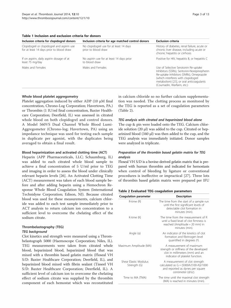
Table 1 Inclusion and exclusion criteria for donors
Inclusion criteria for clopidogrel donors Inclusion criteria for age matched control donors Exclusion criteria
Clopidogrel or clopidogrel and aspirin usefor at least 14 days prior to blood draw
No clopidogrel use for at least 14 daysprior to blood draw
History of diabetes, renal failure, acute orchronic liver disease, including acute orchronic hepatitis or cirrhosis
If on aspirin, daily aspirin dosage of atleast 75 mg/day
No aspirin use for at least 14 days priorto blood draw
Positive for HIV, hepatitis B, or hepatitis C
Males and Females Males and Females Use of Selective Serotonin Re-uptakeInhibitors (SSRIs), Seritonin-NorepinepherinRe-uptake Inhibitors (SNRIs), Omeprazole(which interferes with clopidogrelmetabolism) [25], or oral anticoagulants(Coumadin, Warfarin, etc.)
Table 2 Evaluated TEG coagulation parameters
Coagulation parameter Description
R-time (R) The time from the start of a sample rununtil the first significant levels ofdetectable clot formation in
minutes (min).
K-time (K) The time from the measurement of Runtil a fixed level of clot firmness isreached (Amplitude = 20 mm) in
minutes (min).
Angle (α) An indicator of the kinetics of clotformation and fibrinogen level
quantified in degrees (º).
Maximum Amplitude (MA) A measurement of maximumstrength or stiffness of the developed
clot in millimeters (mm) and anindicator of platelet function.
Shear Elastic ModulusStrength (G)
A measurement of clot strengthcalculated as G = (5000A/(100-A))/1000and reported as dynes per square
centimeter (d/sc).
Time to MA (TMA) The time until the maximal clot strength(MA) is reached in minutes (min).
Dwyer et al. Thrombosis Journal 2014, 12:10 Page 3 of 13http://www.thrombosisjournal.com/content/12/1/10
Whole blood platelet aggregometryPlatelet aggregation induced by either ADP (10 μM finalconcentration, Chrono-Log Corporation; Havertown, PA.)or Thrombin (1 IU/ml final concentration, Baxter Health-care Corporation; Deerfield, IL) was assessed in citratedwhole blood on both clopidogrel and control donors.A Model 560VS Dual Channel Whole Blood Lumi-Aggregometer (Chrono-log; Havertown, PA) using animpedance technique was used for testing each samplein duplicate per agonist, with the duplicates beingaveraged to obtain a final result.
Blood heparinization and activated clotting time (ACT)Heparin (APP Pharmaceuticals, LLC; Schaumburg, IL)was added to each citrated whole blood sample toachieve a final concentration of 5 U/ml prior to TEGand imaging in order to assess the blood under clinicallyrelevant heparin levels [26]. An Activated Clotting Time(ACT) measurement was taken of each blood sample be-fore and after adding heparin using a Hemochron Re-sponse Whole Blood Coagulation System (InternationalTechnidyne Corporation; Edison, NJ). Because citratedblood was used for these measurements, calcium chlor-ide was added to each test sample immediately prior toACT analysis to return calcium ion concentration to asufficient level to overcome the chelating effect of thesodium citrate.
Thrombelastography (TEG)TEG backgroundClot kinetics and strength were measured using a Throm-belastograph 5000 (Haemoscope Corporation; Niles, IL).TEG measurements were taken from citrated wholeblood, heparinized blood, heparinized blood that wasmixed with a thrombin based gelatin matrix (Floseal VHS/D: Baxter Healthcare Corporation; Deerfield, IL), andheparinized blood mixed with fibrin sealant (Tisseel VHS/D: Baxter Healthcare Corporation; Deerfield, IL). Asufficient level of calcium ion to overcome the chelatingeffect of sodium citrate was supplied by the thrombincomponent of each hemostat which was reconstituted
in calcium chloride so no further calcium supplementa-tion was needed. The clotting process as monitored bythe TEG is reported as a set of coagulation parameters(Table 2).
TEG analysis with citrated and heparinized blood aloneThe cup & pin were loaded onto the TEG. Calcium chlor-ide solution (20 μl) was added to the cup. Citrated or hep-arinized blood (340 μl) was then added to the cup, and theTEG analysis was immediately initiated. Donor sampleswere analyzed in triplicate.
Preparation of the thrombin based gelatin matrix for TEGanalysisFloseal VH S/D is a bovine-derived gelatin matrix that is pre-pared with human thrombin and indicated for hemostasiswhen control of bleeding by ligature or conventionalprocedures is ineffective or impractical [27]. Three lotsof thrombin based gelatin matrix were prepared per IFU

Dwyer et al. Thrombosis Journal 2014, 12:10 Page 4 of 13http://www.thrombosisjournal.com/content/12/1/10
(instructions for use) but on a reduced scale. The gelatin(0.100 g or ~1/8 of a 5 ml kit syringe) was weighed out onweigh paper and then back-loaded into a 1 ml tuberculinsyringe. To remove air, the granules were compressed to-wards the luer end of the syringe. In a second tuberculinsyringe, 0.5 ml of 500 IU/ml thrombin solution was with-drawn (prepared by adding 5 ml CaCl2 to the thrombinvial) and a syringe connector was attached (luer end facingup). The two syringes were connected (one with gelatinand one with thrombin solution) and ‘swooshed’ at least 20times to mix the gelatin and thrombin solution adequately.
TEG analysis with thrombin based gelatin matrixThe cup & pin were loaded onto the TEG. The cup wassubsequently removed from the TEG. The TEG cup wasplaced on the scale and the weight was zeroed. The first100 μl of mixed thrombin based gelatin matrix was dis-carded from the syringe. The tip of the syringe wascleaned with a Kimwipe to ensure that no excess throm-bin based gelatin matrix remained. Using the tuberculinsyringe, 150 μl of the mixed thrombin based gelatin
Figure 1 Blood component analyses results. The left panel shows dot-pwhere blood parameters are indicated on the y-axis and the ratios of averaCIs on the x-axis.
matrix was placed at the bottom of the TEG cup. Theweight of the added thrombin based gelatin matrix wasrecorded. If the weight was 0.1160 g to 0.1260 g, thesample was used. If not, the weight was adjusted so thatthe weight of sample in the cup was in this range. Thecup was impacted against the lab bench several times toconcentrate the thrombin based gelatin matrix evenly onthe bottom of the cup. Heparinized blood (210 μl) wasadded to the cup and mixed quickly with the thrombinbased gelatin matrix using a pipette tip. The cup wasthen loaded onto the TEG with analysis initiated imme-diately. Donor samples were analyzed in triplicate witheach of the three lots of thrombin based gelatin matrix.
Preparation of the fibrin sealant for TEG analysisTisseel VH S/D is a human-derived fibrin sealant indicatedfor hemostasis in surgeries involving cardiopulmonary by-pass and treatment of splenic injuries due to blunt or pene-trating trauma when control of bleeding by conventionaltechniques is ineffective or impractical [28]. Three lots of fi-brin sealant were prepared per IFU. The fibrinogen and
lots of all blood parameters and group. The right panel shows a CI plotges (clopidogrel relative to control) and corresponding two-sided 95%

Dwyer et al. Thrombosis Journal 2014, 12:10 Page 5 of 13http://www.thrombosisjournal.com/content/12/1/10
aprotinin components were warmed to 37°C, and the40 mM CaCl2 was brought to room temperature. The fi-brinogen was reconstituted in aprotinin (F.C. ~100 mg/ml).The re-suspended fibrinogen was placed in a Fibrinotherm(Baxter Healthcare Corporation; Deerfield, IL) until dis-solved and stored at 37°C until used. The thrombin wasreconstituted in 40 mM CaCl2 (F.C. ~500 IU/ml) andstored at room temperature until used.
TEG analysis with fibrin sealantThe cup & pin were loaded onto the TEG. The cup wassubsequently removed from the TEG. Heparinized blood(210 μl) was added to the cup. Fibrinogen solution(75 μl, ~7.5 mg) and thrombin solution (75 μl, ~37.5 IU)were added to the TEG cup simultaneously in conjunc-tion with the blood volume above to total 360 μl. Thesample was mixed quickly using a pipette tip and thecup loaded into the TEG. TEG analysis was immediatelyinitiated. Donor samples were analyzed in triplicate witheach of the three lots of fibrin sealant.
Figure 2 Whole blood aggregation results after activation with ADP oaggregation impedance per group for aggregation induced with ADP andis indicated on the y-axis and the ratios of averages (clopidogrel relative tothe x-axis. No bounded two-sided 95% CI for the ratio regarding ADP is avstatistically different from zero.
Field emission scanning electron microscopy (FESEM)Preparation of thrombin based gelatin matrix clotsThe thrombin based gelatin matrix was prepared perIFU as indicated above and 150 μl was dispensed into awell of a 24 well plate. Heparinized blood (210 μl) wasadded to the well and the blood and hemostat werequickly mixed. The clot was allowed to set for 30 mi-nutes at 37°C before the addition of 2 ml/well of phos-phate buffered saline (PBS). Clots were subsequentlydetached from the bottom of each well. After another30 minutes at 37°C, the clot was rinsed with 1 ml PBSand fixed overnight at 4°C in 2 ml of 2% glutaraldehyde(Electron Microscopy Sciences; Hatfield, PA) in 0.1 MHEPES buffer.
Preparation of fibrin sealant clotsThe fibrinogen and thrombin components of the fibrinsealant were prepared per IFU as indicated above. Hepa-rinized blood (210 μl) was added to the well of a 24 wellplate followed by 75 μl of fibrinogen solution and 75 μl
r thrombin. The left panel shows dot-plots of whole blood plateletthrombin. The right panel shows a CI plot where the type of inductioncontrol) and corresponding two-sided 95% CIs are presented onailable as the numerator of the ratio (i.e. mean of clopidrogel) is not

Dwyer et al. Thrombosis Journal 2014, 12:10 Page 6 of 13http://www.thrombosisjournal.com/content/12/1/10
of thrombin solution. The blood and hemostat werequickly mixed and the clot was allowed to set for 30 mi-nutes at 37°C. Two milliliters of PBS were added to thewell and the clot was detached from the bottom of thewell. After another 30 minutes at 37°C, the clot wasrinsed with 1 ml PBS and fixed overnight at 4°C in 2 mlof 2% glutaraldehyde in 0.1 M HEPES buffer.
Preparation of clots for FESEMAfter clot fixation, clots were rinsed with 0.1 M HEPESbuffer and dehydrated in a graded ethanol (EtOH) seriesto 100% EtOH. The specimens were placed into 2:1, 1:1,and 1:2 solutions of 100% EtOH to hexamethyldisilazane(HMDS, Electron Microscopy Sciences; Hatfield, PA),rinsed three times in 100% HMDS followed by a quickchange of 100% HMDS, and allowed to air dry. The speci-mens were then mounted onto FESEM stubs affixed withdouble-stick conductive carbon tape and sputter coatedwith palladium in a Desk IV Sputter/Etch Unit (DentonVacuum; Moorestown, NJ). The samples were examinedin a JSM-6300 F FESEM (JEOL; Tokyo, Japan) and repre-sentative areas were recorded as digital images.
Figure 3 ACT results pre and post-heparinization. The left panel showspost-heparinization. The right panel shows a CI plot for pre- and post-hepacontrol) and corresponding two-sided 95% CIs are presented on the x-axis
Statistical analysesComparison of clopidogrel versus control results wasconsidered to be exploratory; therefore, analysis focusedon estimation of group effects using confidence intervals(CIs) rather than on hypothesis testing. All analyses wereperformed with R version 2.15.3 (R Core Team; Vienna,Austria) [29].
Blood analysisDifferences in blood parameters between clopidrogeland control donors were assessed using ratios of aver-ages, corresponding two-sided 95% CIs, and two-sidedp-values obtained by R function “t.test.ratio (option var.equal = FALSE)” of R package “mratios” [30].
ThrombelastographyTEG data were analyzed using a linear mixed effectsmodel taking the replicate measurements per donor ad-equately into account. The model consisted of the fixedeffect group (clopidogrel and control) and donor asrandom effects, and was fitted using R function “lme”of R package “nlme” [31]. Differences in TEG data
dot-plots of activated clotting time per group and pre andrinization (y-axis) where the ratios of averages (clopidogrel relative to.
Dwyer et al. Thrombosis Journal 2014, 12:10 Page 7 of 13http://www.thrombosisjournal.com/content/12/1/10
between clopidogrel and control donors were assessedusing ratios of averages, corresponding two-sided 95%CIs and two-sided p-values where the two-sided 95% CIfor the ratio of averages were obtained using Fieller’stheorem [32].
ResultsDonor criteriaFour clopidogrel donors and four control donors thatmet the set criteria were used for this study. The fourclopidogrel donors were taking clopidogrel daily andthree of the four donors were also taking daily aspirin.The control donors had not taken clopidogrel or aspirinfor at least 14 days prior to blood collection. The medianage of clopidogrel and control donors was 55 (range: 47to 62) and 55 (range: 50 to 58) years, respectively.
Blood component analysesThe results of the blood component analyses weresimilar between donor groups (Figure 1). The largest dif-ference between donor groups was observed in WBC
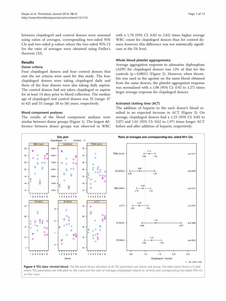
Figure 4 TEG data: citrated blood. The left panel shows dot-plots of all Twhere TEG parameters are indicated on the y-axis and the ratio of averageon the x-axis.
with a 1.78 (95% CI: 0.83 to 2.82) times higher averageWBC count for clopidogrel donors than for control do-nors; however, this difference was not statistically signifi-cant at the 5% level.
Whole blood platelet aggregometryAverage aggregation response to adenosine diphosphate(ADP) for clopidogrel donors was 12% of that for thecontrols (p = 0.0021) (Figure 2). However, when throm-bin was used as the agonist on the same blood obtainedfrom the same donors, the platelet aggregation responsewas normalized with a 1.08 (95% CI: 0.92 to 1.27) timeslarger average response for clopidogrel donors.
Activated clotting time (ACT)The addition of heparin to the each donor’s blood re-sulted in an expected increase in ACT (Figure 3). Onaverage, clopidogrel donors had a 1.23 (95% CI: 0.83 to2.07) and 1.01 (95% CI: 0.62 to 1.97) times longer ACTbefore and after addition of heparin, respectively.
EG parameters per donor and group. The right panel shows a CI plots (clopidogrel relative to control) and corresponding two-sided 95% CIs

Dwyer et al. Thrombosis Journal 2014, 12:10 Page 8 of 13http://www.thrombosisjournal.com/content/12/1/10
Thrombelastography (TEG)Citrated and heparinized blood aloneTEG data for citrated blood (Figure 4) indicates that acrossall TEG parameters there is a trend which suggests thatclopidogrel may impede hemostasis; however differencesbetween donor groups did not reveal statistical significanceat the 5% level for any of the parameters. Heparinizedblood alone samples did not clot for any donor and werestopped after 45 minutes in the TEG (data not shown).
Thrombin based gelatin matrix and fibrin sealantTEG parameter results for the thrombin based gelatinmatrix (Figure 5) and fibrin sealant (Figure 6) mixedwith donor blood were similar between donor groupsbased on the ratios of averages.
FESEM of thrombin based gelatin matrix and fibrinsealant clotsImages of thrombin based gelatin matrix clots (Figure 7)from both donor groups appeared morphologically similar.
Figure 5 TEG data: thrombin based gelatin matrix. The left panel showshows a CI plot where TEG parameters are indicated on the y-axis and thetwo-sided 95% CIs on the x-axis. Due to the rapid rate of clot formation whenough to generate values for R and K other than the lowest possible dataeters between donor groups.
The fibrin structure had similar fiber thickness, branch-ing, and porosity. Clots generated with the fibrin seal-ant (Figure 8) also had similar morphology for bothdonor groups.
DiscussionIn this study, clopidogrel had no significant impact onthe kinetics and visco-elastic clot strength of wholeblood clots formed ex vivo. This result is consistent withthe relative low risk of bleeding observed in patientstaking clopidogrel [14]. Perhaps clopidogrel has a biggermechanistic impact on a plaque stimulated thrombusthan it has on disrupting physiological hemostasisdue to differences in temporal activation, sequence ofthrombin activation, or the relative greater importanceof platelets.Blood component measurements indicated similar re-
sults between clopidogrel and control donors with theexception of average WBC count which was 1.78 (95%CI: 0.83 to 2.82) times higher for clopidogrel donors
s dot-plots of all TEG parameters per donor and group. The right panelratio of averages (clopidogrel relative to control) and correspondingen using the thrombin based gelatin matrix, the TEG is not sensitivevalue for most sample replicates preventing comparison these param-

Figure 6 TEG data: fibrin sealant. The left panel shows dot-plots of all TEG parameters per donor and group. The right panel shows a CI plotwhere TEG parameters are indicated on the y-axis and the ratio of averages (clopidogrel relative to control) and corresponding two-sided 95% CIson the x-axis. Due to the rapid rate of clot formation when using the fibrin sealant, the TEG is not sensitive enough to generate values for R otherthan the lowest possible data value for most sample replicates preventing comparison this parameters between donor groups.
Dwyer et al. Thrombosis Journal 2014, 12:10 Page 9 of 13http://www.thrombosisjournal.com/content/12/1/10
than for control donors. Further assessment by analysisof covariance of the difference in blood aggrega-tion after activation with ADP and thrombin betweendonor groups adjusted for potential differences in WBCcount resulted in two-sided p-values of p = 0.0046 andp = 0.3908 respectively leading to the same statisticalconclusion as without using WBC count as a covariate(Figure 2). The similarity in remaining blood parametermeasurements reduces the likelihood that a difference inanother coagulation factor or blood component influ-enced aggregation response to adenosine diphosphate orthrombin. The averages for ACT before and after additionof heparin were similar between both donor groups. How-ever, the addition of heparin to the blood samples didelevate the clotting time to clinically relevant values.The average clotting times for both donor groups werebetween 550 and 600 seconds following addition ofheparin compared to averages below 200 seconds at base-line. ACTs between 300 and 600 seconds are describedas the ‘safe zone’ for patients undergoing extracorporeal
circulation [33] while the ACT for cardiac surgery can betargeted at 350 seconds or greater [34].Although platelets are important for hemostasis, our
results using this ex vivo model suggest that impairedplatelet activation does not impact the effectiveness ofgelatin matrices and fibrin sealants incorporating theuse of thrombin. An explanation for this finding comesfrom one of our assays used to monitor patient compli-ance to therapy. A statistically significant difference atthe 5% level was detected using ADP activated wholeblood aggregometry which demonstrated that the clopi-dogrel donors had defective platelet activation com-pared to the control donors. This result also indicatesthat the donors do not harbor the polymorphism in theCYP2C19 gene that renders them poor clopidogrel re-sponders because they do not aggregate in response toADP [35]. However, when thrombin was used as theagonist, the average whole blood aggregometry resultsfor clopidrogel donors were similar to that for controldonors. Whole blood aggregometry is a well-established

Figure 7 Representative images of thrombin based gelatin matrix clots acquired with FESEM. Control (A,B), clopidogrel (C,D); 10 μm scalebars (A,C), 5 μm scale bars (B,D); Arrows: Gelatin (a), Fibrin (b), Red Blood Cells (c). No qualitative differences were observed in the fibrin structure(porosity, fiber thickness, and branching) or the cellular accumulation around the gelatin granules including large numbers of trapped RBCsbetween clopidogrel and control donor groups.
Dwyer et al. Thrombosis Journal 2014, 12:10 Page 10 of 13http://www.thrombosisjournal.com/content/12/1/10
method to measure platelet function and has beenfound to correlate well with clinical outcome [36,37].Therefore, these data demonstrate that thrombin caninitiate platelet aggregation in platelets that are unableto respond to ADP due to clopidogrel blockade of theP2Y12 receptor.The high concentration of thrombin in the thrombin
based gelatin matrix and the fibrin sealant hemostatsprovides a possible explanation for the lack of impact ofclopidogrel on their efficacy. The scientific literaturesupports this conclusion in that thrombin is the mostpotent activator of platelets and does so through a differ-ent signal transduction pathway than ADP. Thrombinactivates platelets through the Par1/Par4 and GpI 7 re-ceptors [38]. Once the signaling pathway is initiated, ac-tivation propagates though PI3 kinase and GPIIb/IIIa
Figure 8 Representative images of fibrin sealant clots acquired with FRed Blood Cells (b). As seen in the images of the thrombin based gelatin matand control donor groups. The density of the fibrin structure seen comparedfibrinogen supplied by the fibrin sealant.
leading to platelet aggregation and an increase in intra-platelet calcium concentration [39]. Thus, thrombin maynot only play a central role in controlling bleeding,but may also restore normal clot kinetics and strengthwhen used in combination with gelatin matrix and fibrinsealant hemostats in patients who use prophylactic clo-pidogrel as a platelet aggregation inhibitor. TEG analysisof blood from donors using prophylactic clopidogrelfound no detectable impact on hemostasis stimulated byeither gelatin matrix or fibrin sealant hemostats whichcontain thrombin when compared to the control donors.This is consistent with the platelet aggregation resultsand clot structure analysis.Morphological characterization of clots formed when
thrombin based gelatin matrix and fibrin sealant hemostatswere mixed with donor blood supports the quantitative
ESEM. Control (A), clopidogrel (B); 5 μm scale bars; Arrows: Fibrin (a),rix clots, no qualitative differences were observed between clopidogrelto the gelatin based hemostat clots may be due to the additional

Dwyer et al. Thrombosis Journal 2014, 12:10 Page 11 of 13http://www.thrombosisjournal.com/content/12/1/10
data accumulated during this study. Although differencescan be seen with FESEM based on the type of hemostatused, the fibrin morphology and incorporation of redblood cells between clots formed with clopidogrel donorblood and those of the controls were similar. The clotsformed with fibrin sealant were similar to those seen previ-ously [40,41] as well as those formed with the thrombinbased gelatin matrix hemostat [42]. Therefore, not onlydoes the use of thrombin attenuate the impact of clopido-grel by initiating platelet activation while retaining normalclot kinetics and strength, it also facilitates structurallysimilar clots when used in combination with gelatin matrixand fibrin sealant hemostats. However, this indicates onlythe potential to form a structurally adequate clot in vivoconsidering the artificial nature in which the clots werecreated ex vivo.Whole blood was selected for testing in that it more
closely reflects the conditions during surgical applicationof the thrombin based gelatin matrix and fibrin sealanthemostats, but there are considerations that must be ad-dressed due to the presence of high concentrationthrombin in the hemostats. While thrombin is a potentstimulator of platelets, it is also responsible for convert-ing fibrinogen to fibrin, a reaction which also influenceswhole blood platelet aggregometry and TEG results. Inthe case of whole blood platelet aggregometry, thethrombin concentration had to be reduced from the kitconcentration of 500 IU/ml to 1 IU/ml. For the purposesof simplicity it was also referred to as to platelet aggre-gometry even though a coagulation reaction was knownto have occurred. The same can be said for the TEGassay in that the high concentration of thrombin in thehemostats elicited the same reaction making it unlikelyto discern between coagulation and platelet aggregationeffects by this method. However, these tests were not de-signed to imply a direct comparative basis for ADP stimu-lated platelet aggregation to thrombin stimulated plateletaggregation or to discern between coagulation and plateletaggregation effects but to evaluate the effectiveness of thehemostats when used with donor blood in which plateletswere inhibited by clopidogrel.Based on these data, thrombin based gelatin matrices
and fibrin sealants are likely to be effective when usedduring surgical procedures involving patients currentlyon clopidogrel anti-platelet therapy. This is consistentwith recent clinical trial data demonstrating the effect-iveness of a fibrin sealant based hemostat in patients onplatelet inhibitors [43]. Despite the clinically meaningfulresults of this study, it was limited in scope and designedto encourage additional clinical studies to verify thebasic findings of this work. The small patient numbersare the main limitation of this exploratory study, redu-cing the power of the statistical analysis to detect differ-ences. Therefore, analysis focused on estimation rather
than on hypothesis testing. Lack of clinical correlationswith this study’s TEG data is another obvious limitation.However, other research using a rabbit bleeding modelto show that clopidogrel had no impact on the bleedingrate in vivo [44] is consistent with results in humanblood presented in this paper.This study also raises some interesting questions. In
the modern era of poly-pharmacy, the impact of multiplemedications such as SSRIs, SNRIs, platelet inhibitors,thrombin inhibitors, and Vitamin K antagonists as wellas congenital deficiencies in hemostatic factors mayhave unpredictable consequences on the regulation ofhemostasis. Consequently, surgical hemostats may haveto be carefully evaluated in the context of this growingcombinatorial complexity.
ConclusionThe use of thrombin based gelatin matrices and fibrinsealants initiated clot formation when mixed with bloodfrom donors on anti-platelet therapy that was similar tothat of control donor blood within the constraints of theTEG assay. This study provided a basis for future inves-tigation into the effectiveness of thrombin based hemo-stats during surgical procedures involving patients onanti-platelet therapy.
Abbreviationsα: Angle; ACT: Activated clotting time; ADP: Adenosine diphosphate;CI: Confidence interval; FESEM: Field emission scanning electron microscopy;G: Shear elastic modulus strength; IFU: Instructions for use; K: Clotting time;MA: Maximum amplitude; R: Reaction time; RBC: Red blood cell;SNRI: Seritonin-norepinepherin re-uptake inhibitor; SSRI: Selective serotoninre-uptake inhibitor; TEG: Thrombelastography; TMA: Time to MA; WBC: Whiteblood cell.
Competing interestsThe authors of this manuscript are current or former employees andconsultants of Baxter Healthcare Corporation.
Authors’ contributionsJFD was responsible for blood sample labeling and distribution,development and execution of TEG assays, organization of data for statisticalanalysis, drafting and submission of the manuscript, and was involved instudy design. JAM was responsible for study design, maintaining the blindfor the study, donor recruitment, and was involved in the drafting of themanuscript. ZY was involved in the study design, preparations of clots forFESEM, and assay execution. MH was responsible for the development andexecution of the hematological assays. HR was involved in the study designand development of particle based TEG assay. MM was involved in theprocessing of clots for FESEM. MW was responsible for the statistical analysis.JPD was responsible for FESEM. AG was involved in study design. SD wasinvolved in study design and drafting of the manuscript. All authorscontributed to, read, and approved the final manuscript.
AcknowledgementsThe authors dedicate this publication to the memory of Shane Donovan, Ph.D. so that his work lives on. The authors would also like to thank HuubKreuwel, Ph.D., Megan Francis-Sedlak, Ph.D., and Kevin Lewis, DVM for theirassistance in completing this publication and Anna Khadem for her assist-ance on the particle based TEG assay.

Dwyer et al. Thrombosis Journal 2014, 12:10 Page 12 of 13http://www.thrombosisjournal.com/content/12/1/10
Author details1Baxter Healthcare Corporation, Deerfield, IL, USA. 2Ludwig BoltzmannInstitute for Experimental and Clinical Traumatology, AUVA Research Center,Austrian Cluster for Tissue Regeneration, Vienna, Austria. 3Baxter InnovationsGmbH, Wagramerstrasse 17-19, 1220 Wien, Austria.
Received: 12 September 2013 Accepted: 21 April 2014Published: 7 May 2014
References1. Krumholz HM, Anderson JL, Bachelder BL, Fesmire FM, Fihn SD, Foody JM,
Ho PM, Kosiborod MN, Masoudi FA, Nallamothu BK, Masoudi FA, Bonow RO,DeLong E, Estes NAM, Goff DC, Grady K, Green LA, Loth A, Peterson ED,Radford MJ, Rumsfeld JS, Shahian DM: ACC/AHA 2008 performancemeasures for adults with ST-elevation and non-ST-elevation myocardialinfarction: a report of the American College of Cardiology/AmericanHeart Association Task Force on Performance Measures (writingcommittee to develop performance measures for ST-elevation andnon-ST-elevation myocardial infarction): developed in collaboration withthe American Academy of Family Physicians and the American Collegeof Emergency Physicians: endorsed by the American Association ofCardiovascular and Pulmonary Rehabilitation, Society for CardiovascularAngiography and Interventions, and Society of Hospital Medicine.Circulation 2008, 118:2596–2648.
2. Adams RJ, Albers G, Alberts MJ, Benavente O, Furie K, Goldstein LB, Gorelick P,Halperin J, Harbaugh R, Johnston SC, Katzan I, Kelly-Hayes M, Kenton EJ,Marks M, Sacco RL, Schwamm LH: Update to the AHA/ASA recommendationsfor the prevention of stroke in patients with stroke and transientischemic attack. Stroke 2008, 39:1647–1652.
3. Faxon DP: Use of antiplatelet agents and anticoagulants for cardiovasculardisease: current standards and best practices. Rev Cardiovasc Med 2005,6(Suppl 4):S3–S14.
4. Gachet C, Stierle A, Cazenave JP, Ohlmann P, Lanza F, Bouloux C, MaffrandJP: The thienopyridine PCR 4099 selectively inhibits ADP-induced plateletaggregation and fibrinogen binding without modifying the membraneglycoprotein IIb-IIIa complex in rat and in man. Biochem Pharmacol 1990,40:229–238.
5. Savi P, Labouret C, Delesque N, Guette F, Lupker J, Herbert JM: P2y(12),a new platelet ADP receptor, target of clopidogrel. Biochem Biophys ResCommun 2001, 283:379–383.
6. Savi P, Laplace MC, Herbert JM: Evidence for the existence of twodifferent ADP-binding sites on rat platelets. Thromb Res 1994, 76:157–169.
7. Vane JR, Botting RM: The mechanism of action of aspirin. Thromb Res2003, 110:255–258.
8. Tohgi H, Konno S, Tamura K, Kimura B, Kawano K: Effects of low-to-highdoses of aspirin on platelet aggregability and metabolites ofthromboxane A2 and prostacyclin. Stroke 1992, 23:1400–1403.
9. Patrignani P, Filabozzi P, Patrono C: Selective cumulative inhibition ofplatelet thromboxane production by low-dose aspirin in healthysubjects. J Clin Invest 1982, 69:1366–1372.
10. Badreldin A, Kroener A, Kamiya H, Lichtenberg A, Hekmat K: Effect ofclopidogrel on perioperative blood loss and transfusion in coronaryartery bypass graft surgery. Interact Cardiovasc Thorac Surg 2010, 10:48–52.
11. Herman CR, Buth KJ, Kent BA, Hirsch GM: Clopidogrel increases bloodtransfusion and hemorrhagic complications in patients undergoingcardiac surgery. Ann Thorac Surg 2010, 89:397–402.
12. Nurozler F, Kutlu T, Kucuk G, Okten C: Impact of clopidogrel onpostoperative blood loss after non-elective coronary bypass surgery.Interact Cardiovasc Thorac Surg 2005, 4:546–549.
13. Sun JC, Whitlock R, Cheng J, Eikelboom JW, Thabane L, Crowther MA, Teoh KH:The effect of pre-operative aspirin on bleeding, transfusion, myocardialinfarction, and mortality in coronary artery bypass surgery: a systematicreview of randomized and observational studies. Eur Heart J 2008,29:1057–1071.
14. Bristol-Meyers Squib/Sanofi Pharmaceuticals: Plavix (Clopidogrel Bisulfate)Tablet, Film Coated. Bridgewater, NJ; 2009.
15. Billett HH: Antiplatelet agents and arterial thrombosis. Cardiol Clin 2008,26:189–201. vi.
16. Ferrari E, Benhamou M, Cerboni P, Marcel B: Coronary syndromesfollowing aspirin withdrawal: a special risk for late stent thrombosis.J Am Coll Cardiol 2005, 45:456–459.
17. Vasudeva P, Goel A, Sengottayan VK, Sankhwar S, Dalela D: Antiplateletdrugs and the perioperative period: what every urologist needs to know.Indian J Urol 2009, 25:296–301.
18. Weber AA, Braun M, Hohlfeld T, Schwippert B, Tschope D, Schror K:Recovery of platelet function after discontinuation of clopidogreltreatment in healthy volunteers. Br J Clin Pharmacol 2001, 52:333–336.
19. Chassot PG, Delabays A, Spahn DR: Perioperative antiplatelet therapy:the case for continuing therapy in patients at risk of myocardialinfarction. Br J Anaesth 2007, 99:316–328.
20. O’Riordan JM, Margey RJ, Blake G, O’Connell PR: Antiplatelet agents in theperioperative period. Arch Surg 2009, 144:69–76. discussion 76.
21. Emilia M, Luca S, Francesca B, Luca B, Paolo S, Giuseppe F, Gianbattista B,Carmela M, Luigi M, Mauro L: Topical hemostatic agents in surgicalpractice. Transfus Apher Sci 2011, 45:305–311.
22. Oz MC, Rondinone JF, Shargill NS: FloSeal matrix: new generation topicalhemostatic sealant. J Card Surg 2003, 18:486–493.
23. Kaufmann CR, Dwyer KM, Crews JD, Dols SJ, Trask AL: Usefulness ofthrombelastography in assessment of trauma patient coagulation.J Trauma 1997, 42:716–720. discussion 720-712.
24. Tuman KJ, McCarthy RJ, Djuric M, Rizzo V, Ivankovich AD: Evaluation ofcoagulation during cardiopulmonary bypass with a heparinase-modifiedthromboelastographic assay. J Cardiothorac Vasc Anesth 1994, 8:144–149.
25. Ferreiro JL, Ueno M, Capodanno D, Desai B, Dharmashankar K, Darlington A,Charlton RK, Bass TA, Angiolillo DJ: Pharmacodynamic effects ofconcomitant versus staggered clopidogrel and omeprazole intake:results of a prospective randomized crossover study. Circ CardiovascInterv 2010, 3:436–441.
26. Hardy JF, Belisle S, Robitaille D, Perrault J, Roy M, Gagnon L: Measurementof heparin concentration in whole blood with the Hepcon/HMS devicedoes not agree with laboratory determination of plasma heparinconcentration using a chromogenic substrate for activated factor X.J Thorac Cardiovasc Surg 1996, 112:154–161.
27. Baxter Healthcare Corporation: FloSeal Hemostatic Matrix. Hayward, CA; 2009.28. Baxter Healthcare Corporation: TISSEEL [Fibrin Sealant]. Westlake Village,
CA; 2009.29. R-Core Team: R: A Language and Environment for Statistical Computing.
[http://www.R-project.org/].30. Dilba G, Hasler M, Gerhard D, Schaarschmidt F: mratios: Inferences for
Ratios of Coefficients in the General Linear Model. R Package Version1.3.17. [http://CRAN.R-project.org/package=mratios].
31. Pinheiro J, Bates D, DebRoy S, Sarkar D, R Development Core Team: nlme:Linear and Nonlinear Mixed Effects Models. R Package Version 3.1-108.[http://CRAN.R-project.org/package=nlme].
32. Fieller EC: Some problems in interval estimation. J R Stat Soc Ser B 1954,16:175–185.
33. Bull MH, Huse WM, Bull BS: Evaluation of tests used to monitor heparintherapy during extracorporeal circulation. Anesthesiology 1975, 43:346–353.
34. Fitzgerald DJ, Patel A, Body SC, Garvin S: The relationship between heparinlevel and activated clotting time in the adult cardiac surgery population.Perfusion 2009, 24:93–96.
35. Hulot JS, Bura A, Villard E, Azizi M, Remones V, Goyenvalle C, Aiach M, Lechat P,Gaussem P: Cytochrome P450 2C19 loss-of-function polymorphism is amajor determinant of clopidogrel responsiveness in healthy subjects.Blood 2006, 108:2244–2247.
36. Agarwal S, Coakley M, Reddy K, Riddell A, Mallett S: Quantifying theeffect of antiplatelet therapy: a comparison of the platelet functionanalyzer (PFA-100) and modified thromboelastography (mTEG) withlight transmission platelet aggregometry. Anesthesiology 2006,105:676–683.
37. Ivandic BT, Schlick P, Staritz P, Kurz K, Katus HA, Giannitsis E: Determinationof clopidogrel resistance by whole blood platelet aggregometry andinhibitors of the P2Y12 receptor. Clin Chem 2006, 52:383–388.
38. Kahn ML, Nakanishi-Matsui M, Shapiro MJ, Ishihara H, Coughlin SR:Protease-activated receptors 1 and 4 mediate activation of humanplatelets by thrombin. J Clin Invest 1999, 103:879–887.
39. Voss B, McLaughlin JN, Holinstat M, Zent R, Hamm HE: PAR1, but not PAR4,activates human platelets through a Gi/o/phosphoinositide-3 kinasesignaling axis. Mol Pharmacol 2007, 71:1399–1406.
40. Macasev D, Diorio JP, Gugerell A, Goppelt A, Gulle H, Bittner M: Cell compatibilityof fibrin sealants: in vitro study with cells involved in soft tissue repair.J Biomater Appl 2011, 26:129–149.

Dwyer et al. Thrombosis Journal 2014, 12:10 Page 13 of 13http://www.thrombosisjournal.com/content/12/1/10
41. Furst W, Banerjee A, Redl H: Comparison of structure, strength andcytocompatibility of a fibrin matrix supplemented either withtranexamic acid or aprotinin. J Biomed Mater Res B Appl Biomater 2007,82:109–114.
42. DiOrio JP, Stojanovic L, Yardimci A, Amrani DL, Helgerson S, Vega F:Electron microscopic characterization of a gelatin matrix/thrombinhemostat. Microsc Microanal 2005, 11:178–179.
43. Saha SP, Muluk S, Schenk W 3rd, Dennis JW, Ploder B, Grigorian A, Presch I,Goppelt A: A prospective randomized study comparing fibrin sealant tomanual compression for the treatment of anastomotic suture-holebleeding in expanded polytetrafluoroethylene grafts. J Vasc Surg 2012,56:134–141.
44. Hughes SD, Bishop PD, Garcia R, Zhang T, Alexander WA: Topical recombinantthrombin at a concentration of 1000 IU/mL reliably shortens in vivo TTHand delivers durable hemostasis in the presence of heparin anticoagulationand clopidogrel platelet inhibition in a rabbit model of vascular bleeding.Ann Surg Innov Res 2009, 3:14.
doi:10.1186/1477-9560-12-10Cite this article as: Dwyer et al.: Thrombin based gelatin matrix andfibrin sealant mediated clot formation in the presence of clopidogrel.Thrombosis Journal 2014 12:10.
Submit your next manuscript to BioMed Centraland take full advantage of:
• Convenient online submission
• Thorough peer review
• No space constraints or color figure charges
• Immediate publication on acceptance
• Inclusion in PubMed, CAS, Scopus and Google Scholar
• Research which is freely available for redistribution
Submit your manuscript at www.biomedcentral.com/submit