Embed Size (px)
Citation preview
Thrombin peptide Chrysalins stimulates healing of diabeticfoot ulcers in a placebo-controlled phase I/II study
Caroline Fife, MD1; Jon T. Mader, MD2; Jeffery Stone, MD3; Leon Brill, DPM3; Kathleen Satterfield, DPM4;Andrea Norfleet, PhD2,5n; Amber Zwernemann, BS2; James T. Ryaby, PhD5; Darrell H. Carney, PhD2
1. Department of Anesthesiology, University of Texas Health Science Center, Houston, Texas,
2. Department of Biochemistry and Molecular Biology, University of Texas Medical Branch, Galveston, Texas,
3. The Limb Salvage Center at the Brillstone Building, Dallas, Texas,
4. University of Texas Health Science Center, Texas Diabetes Institute, San Antonio, Texas, and
5. OrthoLogic Corp., Tempe, Arizona
Reprint requests:Darrell H. Carney, PhD, Department of
Biochemistry and Molecular Biology, The
University of Texas Medical Branch, 301
University Blvd., Galveston, TX 77555-0645.
Tel: 1409 772 3210;
Fax: 409-772-2880;
Email: [email protected]
nDr. Norfleet’s current address is: Genzyme
Corporation, Cambridge, MA 02142.
Manuscript received: January 31, 2006
Accepted in final form: September 13, 2006
DOI:10.1111/j.1524-475X.2006.00181.x
ABSTRACT
Thrombin and thrombin peptides play a role in initiating tissue repair. The po-tential safety and efficacy of TP508 (Chrysalins) treatment of diabetic foot ulcerswas evaluated in a 60-subject, prospective, randomized, double-blind, placebo-controlled phase I/II clinical trial. Chrysalins in saline or saline alone was ap-plied topically, twice weekly, to diabetic ulcers with standardized care and off-loading. A dose-dependent effect was seen in the per-protocol population where 1and 10 mg Chrysalins treatment resulted in 45 and 72% more subjects with com-plete healing than placebo treatment. Chrysalins treatment of foot ulcers morethan doubled the incidence of complete healing (p < 0.05), increased mean clo-sure rate � 80% (p < 0.05), and decreased the median time to 100% closure by� 40% (p < 0.05). Chrysalins treatment of heel ulcers within this population re-sulted in mean closure rates 165% higher than placebos (p < 0.02) and completehealing in 86% (6/7) of ulcers compared with 0% (0/5) of placebo ulcers(p < 0.03). Local wound reactions and adverse events (AEs) were equal betweengroups with no reported drug-related changes in laboratory tests or serious AEs.These results indicate the potential safety and efficacy of Chrysalins for treat-ment of diabetic foot ulcers.
Chronic diabetic ulcers of the lower extremities represent amajor healthcare problem today, with over 850,000 diag-noses made in the United States each year.1,2 Because ofthe increasing incidence of diabetes, the magnitude of thechallenge presented to the healthcare system by chronicfoot and leg ulcers is also expected to increase.1,2 Chronicdiabetic ulcers not only negatively impact the quality oflife but can also lead to amputation and an increased like-lihood of death.3
Healing a chronic diabetic ulcer is an expensive, timeconsuming, and complicated task. A significant fraction ofthese ulcers do not heal or do not remain healed, and be-come chronic wounds that endure for months or years.The cost to the healthcare system is estimated at greaterthan 10 billion dollars per year, in addition to untold lossesin workplace productivity.3,4 Diabetic ulcers are distin-guished from acute wounds in healthy individuals by sev-eral factors that stem from the underlying pathology ofdiabetes including the aging of tissues, hypoxia, and infec-tion.5–7 In addition, diabetics exhibit various degrees ofperipheral neuropathy. Diabetics also have a dysfunction-al endothelium, which fails to respond to various growthfactors and angiogenic stimuli,8,9 and increased levels ofmetalloproteinases that degrade matrix molecules and de-crease the half-life of growth factors in wound fluid.10–12
The impaired wound environment characteristic of chron-ic, nonhealing ulcers has led to development of a standardregimen of chronic wound care that includes appropriate
wound bed preparation, moist wound coverings, and pres-sure off-loading. For many larger ulcers, treatment inwound-care centers now often includes use of bioactiveskin substitutes, delivery of autologous platelet concen-trates, or application of a therapeutic agent such as be-caplermin (Regranexs OMJ Pharmaceutical Inc., SanGerman, Puerto Rico). Although these therapeutic op-tions have shown potential efficacy in clinical trials andreceived FDA approval, they do not appear to have beenwidely accepted as standard of care.
The reasons why therapeutic alternatives approved bythe FDA for diabetic foot ulcers have not been widely ac-cepted include limited benefit over standard of care, treat-ment regimens that require extensive debridement, dailytreatment, and/or repeated visits to specialized wound carefacilities. Clinical trials with becaplermin (Regranexs), us-ing a regimen that included daily treatment for 12 hours,rinsing with saline, and rebandaging, e.g., showed com-plete healing in � 50% of subjects relative to 35% whowere treated with good clinical practice and placebogel.13,14 A subsequent post hoc analysis suggested thatthe efficacy of becaplermin was seen in subjects whosewounds were debrided almost every week.15 In both thehome health setting and in many wound clinics, treatmentschedules and debridement fall short of the regimenthat may be required for becaplermin efficacy. Therefore,a more ideal therapeutic for diabetic foot ulcers may beone that can be easily applied in the home care
Wound Rep Reg (2007) 15 23–34 c� 2007 by the Wound Healing Society 23
Wound Repair and Regeneration
environment, can be applied less often without reducingefficacy, and is less dependent upon surgical debridementfor its efficacy.
Chrysalins (TP508) is a 23-amino acid peptide repre-senting the natural sequence of amino acids of humanthrombin identified as the thrombin-binding domain for aspecific class of thrombin receptors on fibroblasts andother cells.16 Early studies showed that thrombin, the ser-ine protease responsible for fibrin clot formation, initiatedcell proliferation and other cellular postclotting eventsthrough a growth factor-like mechanism that involved itsbinding and activation of specific thrombin receptors onthe surface of fibroblasts and other cells.17–19 Althoughmany of the cellular effects of thrombin appear to requireproteolytic activity and activation of proteolytically acti-vated receptors,20 studies show that binding of thrombinor thrombin derivatives without proteolytic activity pro-motes a number of cellular events involved in tissue repairand wound healing.21–24. These observations have led tothe hypothesis that nonproteolytic peptide fragments ofthrombin released from a fibrin clot during early stages ofwound repair may modulate inflammation and promotehealing.
Unlike thrombin, which is activated at the site of injury,the Chrysalins peptide has no enzymatic activity and doesnot promote or interfere with blood coagulation.16 Pre-clinical safety studies have shown that the peptide can beinjected intravenously or intraperitonealy at doses of up to25mg/kg with no adverse effects, that it is classified as anonsensitizer based on hamster skin sensitivity testing, andthat topical treatment of open porcine wounds (followedupon wound closure with dermal injection at the woundsite) of 100mg/day for 20 weeks had no apparent negativeeffects (OrthoLogic Corp., unpublished results).
In full-thickness incisional wounds in rats, a single top-ical application of Chrysalins increased the breakingstrength of wounds by approximately 80% over salinecontrols when measured at day-7 postincision.25 Relativeto control breaking strength, this single application ofChrysalins shifted the healing curve forward by approxi-mately 4 days. Significant effects of Chrysalins were alsoseen on incisional wounds in rats with radiation-inducedhealing impairment.26 In larger full-thickness excisionalanimal wounds in normal27 and ischemic skin,28 a singletopical application of Chrysalins also accelerated woundclosure. In all of these model studies, Chrysalins acceler-ated recruitment of inflammatory cells to the wound site,shortened the inflammatory phase, and promoted early re-vascularization of the tissues.27–29 Chrysalins also accel-erated repair of rat fresh fractures30 and promoted boneformation in rabbit critical size segmental bone defects31
and in a rabbit model of distraction osteogenesis.32 Thus,this molecule may serve as a natural initiator of tissue re-pair in a number of tissues.
Based on preclinical studies, we hypothesized thatChrysalins may improve repair quality and accelerate therate of tissue repair following surgical or traumatic acutetissue damage and reinitiate healing of chronic ulcerswhere normal repair processes are disrupted. We now re-port the results of the first human phase I/II pilot clinicaltrial designed to evaluate the safety and efficacy of Chrys-alins in the treatment of chronic diabetic lower extremityand foot ulcers. Modeled after other diabetic foot ulcer
clinical trials, but with expanded entry criterion includinginclusion of larger, more severe (Grade III) ulcers, ulcerson the leg and ankle, and those with O2 tension (TcPO2)levels of between 20 and 30mmHg, this trial was designedto determine whether twice-weekly application ofChrysalins could cause healing of diabetic ulcers and helpus better define the role of thrombin peptides in tissuerepair.
MATERIALS AND METHODS
Overall study design
This study was a multicenter (four sites), prospective, ran-domized, double-blind, placebo-controlled pilot clinicaltrial to evaluate the safety and potential efficacy of Chrys-alins topically applied to diabetic ulcers. The study wasdesigned as a three-arm, 60-subject trial including lowerextremity (below the knee) ulcers ranging from 0.9 to38.5 cm2 (� 1 to 7 cm in diameter) that had been presentfor more than 8 weeks and that were classified as WagnerGrades I, II, or early III33 (Grade III ulcers included deepischemic ulcers that exposed bone or tendon, but excludedthose considered to have eroded into the bone or tendon).The exclusion criteria included the following: clinical in-fection of the ulcer, the presence of uncontrolled systemicinfection or osteomyelitis; poor diabetes control, renalfailure, abnormal liver function; treatment with steroids,chemotherapeutics, or radiation within 6 months beforestudy enrollment; cancer; a history of drug or alcoholabuse; and wound oxygen tension (TcPO2) of < 20mm-Hg. Women who were pregnant, nursing, or of child-bear-ing potential and not using approved birth control wereexcluded.
The study included a preenrollment/screening visit, fol-lowed by twice-weekly office visits for up to 20 weeks oruntil the ulcer reached complete closure. Eligible patients,upon signing an informed consent, were randomized toone of three subject treatment groups: 1 mg Chrysalins,10 mg Chrysalins, or placebo. The wound bed was pre-pared by sharp debridement as deemed necessary by theinvestigator physician, irrigated with saline, and blottedwith gauze. Study treatment was administered topically ina volume of 0.1 cm3 of saline solution. After approximate-ly 1 minute, the wound was covered with CutinovaFoams (Beiersdorf, AG, Germany) and bandaged. If theulcer was on a pressure-bearing surface, the clinician pre-scribed offloading. In most cases, offloading was accom-plished by using sponsor-provided D.H. Walkeroffloading boots (made by Royce Medical), althoughsome subjects were prescribed crutches or wheelchairs without offloading boots. Bandages were removedduring the twice-weekly visits for ulcer evaluation, de-bridement as needed to remove necrotic tissue, and re-treatment for up to 20 weeks or complete wound closure.Subject compliance was monitored and treatmentcontrol was insured as study drug application and banda-ging were conducted in the clinic by the attending clinicianor nurse. Subjects were removed from the study if a clinicalinfection developed or if the wound condition significantlyworsened. Any such removals were counted as closurefailures.
Wound Rep Reg (2007) 15 23–34 c� 2007 by the Wound Healing Society24
Chrysalins diabetic foot ulcer phase I/II trial Fife et al.
Safety and efficacy endpoints
At each clinic visit, adverse events (AEs) were recorded,and local wound reactions were scored for erythema,edema, pain, and overall condition. At enrollment and atweeks 5, 10, 15, and 20, blood was drawn for chemical andhematological analyses, radiographs were obtained, andwound cultures were performed.
Before and after wound debridement, the ulcer perim-eter was traced onto acetate and photographed with adigital camera. The acetates were preprinted with a stand-ard ruler and 1 cm diameter circle. Each tracing was ana-lyzed using digital morphometric analysis software(Image-Pro, Media Cybernetics, Silver Spring, MD) todetermine both open ulcer area and the perimeter of theulcer. The primary efficacy endpoint was the incidenceof ulcers that progressed to complete closure during the20-week study. Secondary endpoints included the timeto 100% and to 80% closure of the study wounds.
A post hoc analysis was also performed to access thelinear rate of wound closure (wound healing rate [WHR]expressed in mm of edge closure per day) using the reduc-tion in area per day divided by the average wound perim-eter using the following formula.34,35
WHR ¼ ½ðAreaT0 �AreaTXÞ=ð½PerimeterT0
þ PerimeterTX �=2Þ�=daysðTXÞThis calculation ofWHR (mm/day) provides an average
vectoral rate of closure from the wound edge, which con-ceptually is the same as a decrease in wound radius. As de-bridement was performed as needed (in some cases, atalmost every visit), the WHR reflects the average closureper day excluding any tissue growth that was removed bydebridement.
Study drug
Chrysalins, also known as TP508 (CAS #497221-38-2), isa synthetic peptide representing the native 23 amino acidsequence of human thrombin that appears to bind to high-affinity thrombin receptors on cells to activate a sequenceof cellular events.16 The peptide was chemically synthe-sized and purified to > 95% by HPLC under cGMP(Peninsula Laboratories, Belmont, CA), and then, filter-sterilized, sterile-filled, and lyophilized in 2 cc glass vials(Ben Venue, Bedford, OH) and stored at 4 1C. Upon sub-ject enrollment, the peptide was dissolved and diluted insterile, pyrogen-free, saline (Abbott Laboratories, Chica-go, IL) by an unblinded pharmacist. To ensure blinding ofthe subject and clinician, treatment solution was deliveredto the clinic in a vial identified with only the subject’s IDnumber.
Study groups and statistical analyses
Two primary populations were defined for analysis beforeunblinding of subject treatments and were used in report-ing this study to the FDA. All subjects with study ulcerswho received at least one treatment were included in anintent-to-treat (ITT) population. Because a number ofsubjects were enrolled in this study with ulcers that shouldhave been excluded based on ulcer size, chronicity of theulcer (based on the length of time the ulcer had been pres-
ent before treatment), or Wagner entry criteria, a per-pro-tocol (PP) population was defined before unblinding andused for efficacy analysis. The PP population differed fromthe ITT population in that seven subjects were removedfor protocol deviations, eight had ulcers < 0.9 cm2 atbaseline, four had been present for < 8 weeks, and onedid not meet the Wagner entry criteria. This left a total of40 subjects in the PP population: 15 placebos, 11 treatedwith 1mg Chrysalins, and 14 treated with 10mg Chrys-alins. Subjects prematurely removed from the study forany reason, were counted as closure failures, but ulcer areameasurements were used up to the time of discontinuationfor healing rates and percent closure analysis.
Clinical monitoring of sites and statistical analysis wasperformed by Synergos Inc. (Woodlands, TX). Standardstatistical methods were used to analyze all data. These in-cluded Fisher’s exact test and Student’s T test using two-tailed tests with an a of 0.05 and Kaplan–Meier analysis oftime to 80 and 100% closure. No adjustment was made formultiple comparisons for either the efficacy analysis or thesafety analysis.
As the ITT and PP populations included individualswith ulcers located below the kneecap on the leg, ankle,and foot, a subset analysis was performed post hoc to de-termine the potential effects of Chrysalins on ulcers locat-ed on the foot. Ulcers located on the ankle and leg oftenhave very different etiologies and may arise from vascularinsufficiency and may not involve neuropathy or pressure.These lower limb ulcers are often distinguished from footulcers as they may require a different treatment regimenincluding pressure bandaging for optimal healing. Relativeto the ITT population, the foot ulcer population for thisanalysis excluded: nine ulcers not on the foot; 10 that were
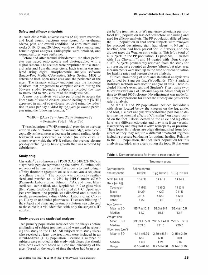
Table 1. Demographic data for intent-to-treat population
Demographic
characteristic
Treatment group
Saline control
(n521) 1mg (n520) 10 mg (n518)
Male [n (%)] 15 (71) 14 (70) 14 (78)
Race [n (%)]
Caucasian 11 (52) 12 (60) 11 (61)
Black 6 (29) 4 (20) 2 (11)
Hispanic 3 (14) 4 (20) 5 (28)
Other 1 (5) 0 (0) 0 (0)
Age (years)
Mean�SD 55.7� 12.8 59.3� 6.4 53.4� 10.5
Median 54.7 59.6 53.7
Weight (lbs)
Mean�SD 196.3� 77.3 206.5� 41.8 229.5� 58.8
Median 203.5 211.0 220.0
Ulcer area (cm2)
Mean�SD 4.11� 5.99
(20)
3.59� 5.31
(21)
3.15� 3.20
(18)
Median 1.63 1.21 2.02
Range 0.16–26.46 0.27–24.36 0.14–13.10
Wound Rep Reg (2007) 15 23–34 c� 2007 by the Wound Healing Society 25
Chrysalins diabetic foot ulcer phase I/II trialFife et al.
< 0.9 cm2; four that had been present for < 8 weeks; andfour that were removed from consideration due to unre-lated SAEs (one—Chrysalins 1mg, two—Chrysalins
10 mg, and one—saline placebo). This left a population of35 subjects: 13 placebo subjects; 12 Chrysalins 1mg sub-jects; and 10 Chrysalins 10mg subjects. All subjects re-moved from the study for infection, osteomyelitis, orworsening of ulcer condition were included and countedas closure failures. Thus, this population is similar to, butnot the same as the PP population. As described below, thedemographics of this population showed no significantdifferences between the groups and group means for sub-ject age and ulcer starting size were similar to values for theITT populations (see Tables 1 and 4).
A further subset analysis was performed to ensure thatthe location of ulcers on the foot was not biasing data tofavor groups treated with Chrysalins. Foot ulcers locatedon the heel of the foot, e.g., are among the most difficultulcers to heal.36,37 As shown in Table 5, heel ulcers madeup five of 13 (38%) of the placebo foot ulcers, three of 12(25%) of those treated with 1mg Chrysalins, and four of10 (40%) of those treated with 10 mg Chrysalins. Analysisof Chrysalins effects on this heel ulcer subpopulation wasperformed looking at the incidence of complete closureachieved by 20 weeks and the rate of healing as describedabove.
Human ethical considerations
This study was conducted as a part of US FDA IND #56,811. Patient consent to become subjects in this trial wasobtained before study treatment for each subject and theprotocol conformed to ethical guidelines of the 1975 Dec-laration of Helsinki as reflected in approval by the humanresearch review committee or appropriate InstitutionalReview Board at each clinical site.
RESULTS
Subject demographics
As shown in Table 1, randomization between ITT treat-ment groups resulted in three subject groups with similardemographic characteristics. In all groups, the number ofmales exceeded females (70–78%males, 22–30% females),with 52–62% being Caucasian. The median age for the sa-line placebo, 1, and 10 mg groups were 54.7, 59.6, and 53.7years, respectively. The median weights for these groupswere 204, 211, and 220 pounds, respectively. The averageulcer starting sizes for these groups were 4.11, 3.59, and3.15 cm2 with median sizes of 1.63, 1.21, and 2.02 cm2, re-spectively. There were no significant differences betweenthe randomized groups in this study.
Safety evaluation
A primary goal of this initial pilot clinical trial was to de-termine whether topical application of Chrysalins causedlocal effects on the ulcer, the adjacent dermal tissue, orsystemic effects that might be seen in hematology or bloodchemistry analysis. For this analysis, all subjects receivingat least one treatment were included and are described as
the ITT population. Laboratory values showed no statis-tically significant changes from baseline or significanttransitions from value groupings (low, normal, or high)for any of the treatment groups at any of the time points
Table 2. Incidence of local ulcer reactions throughout study
Local ulcer reaction
Saline
(n521)
1mg
(n520)
10 mg
(n518)
Well-defined to severe
erythema
2 (10) 3 (15) 2 (11)
Well-defined to severe
edema
3 (14) 3 (15) 4 (22)
Worsened pain 2 (10) 2 (10) 2 (11)
Values presented as: n, number of subjects experiencing a re-
action during the study; (%), percent of subjects in the study
group.
Table 3. Incidence of serious adverse events
Body system event
Saline
(n521)
1mg
(n520)
10 mg
(n518)
Body as a whole
Progressive disease 2 (10) 0 (0) 0 (0)
Infection 1 (5) 1 (5) 1 (6)
Fever 1 (5) 0 (0) 0 (0)
Chills 1 (5) 0 (0) 0 (0)
Pain 1 (5) 1 (5) 0 (0)
Sepsis 0 (0) 0 (0) 1 (6)
Metabolic/nutritional disorder
Hypervolemia 0 (0) 1 (5) 0 (0)
Edema 0 (0) 0 (0) 1 (6)
Respiratory
Dyspnea 0 (0) 1 (5) 1 (6)
Cardiovascular
Myocardial Infarction 1 (5) 0 (0) 1 (6)
Peripheral gangrene 0 (0) 0 (0) 1 (6)
Coronary artery disorder 0 (0) 1 (5) 0 (0)
Urogenital
Urinary tract infection 0 (0) 0 (0) 1 (6)
Oliguria 0 (0) 0 (0) 1 (6)
Acute kidney failure 0 (0) 1 (5) 0 (0)
Kidney failure 0 (0) 1 (5) 0 (0)
Hemic and lymphatic
Ecchymosis 0 (0) 0 (0) 1 (6)
WBC abnormal 0 (0) 0 (0) 1 (6)
Digestive
Gastrointestinal
hemorrhage
1 (5) 0 (0) 0 (0)
Musculoskeletal
Osteomyelitis 0 (0) 1 (5) 0 (0)
Wound Rep Reg (2007) 15 23–34 c� 2007 by the Wound Healing Society26
Chrysalins diabetic foot ulcer phase I/II trial Fife et al.
(data not shown). Chrysalins treatment did not result inany AEs that were probably or definitely drug related, al-though some subjects in all groups reported erythema,edema, and pain (Table 2). At least one non-drug–relatedAE was reported in 76% (16/21) of subjects in the salinetreatment group, 78% (14/18) in the 10mg treatmentgroup, and 75% (15/20) in the 1 mg treatment group (notshown). Serious adverse events (SAEs) were reported infive subjects (24%) in the saline control group, four sub-jects (22%) in the 10 mg group, and four subjects (20%) inthe 1 mg treatment group (Table 3).
Fourteen subjects discontinued treatment during thestudy due to SAEs, infections, or for nonmedical reasons.None of these discontinuations or AEs appeared to bedrug related (Table 4). In the saline control group, threesubjects discontinued because of infection, one subject dis-continued due to osteomyelitis, one subject discontinueddue to a fatal myocardial infarction, and one withdrew fornonmedical reasons. In the 1mg treatment group, two sub-jects discontinued due to osteomyelitis, one subject dis-continued due to an amputation of the contralateral(untreated) foot, one subject discontinued due to a coro-nary artery disorder, and one subject withdrew for non-medical reasons. In the 10mg treatment group, twosubjects discontinued due to infection and a third discon-tinued due to a nonfatal myocardial infarction. No signif-icant differences were found in the incidence of infection orother adverse effects among the groups.
Efficacy analysis
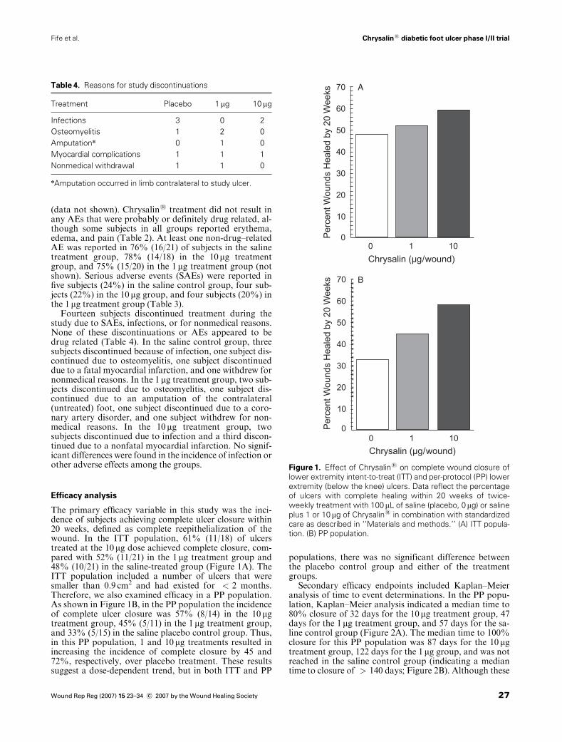
The primary efficacy variable in this study was the inci-dence of subjects achieving complete ulcer closure within20 weeks, defined as complete reepithelialization of thewound. In the ITT population, 61% (11/18) of ulcerstreated at the 10mg dose achieved complete closure, com-pared with 52% (11/21) in the 1mg treatment group and48% (10/21) in the saline-treated group (Figure 1A). TheITT population included a number of ulcers that weresmaller than 0.9 cm2 and had existed for < 2 months.Therefore, we also examined efficacy in a PP population.As shown in Figure 1B, in the PP population the incidenceof complete ulcer closure was 57% (8/14) in the 10mgtreatment group, 45% (5/11) in the 1 mg treatment group,and 33% (5/15) in the saline placebo control group. Thus,in this PP population, 1 and 10mg treatments resulted inincreasing the incidence of complete closure by 45 and72%, respectively, over placebo treatment. These resultssuggest a dose-dependent trend, but in both ITT and PP
populations, there was no significant difference betweenthe placebo control group and either of the treatmentgroups.
Secondary efficacy endpoints included Kaplan–Meieranalysis of time to event determinations. In the PP popu-lation, Kaplan–Meier analysis indicated a median time to80% closure of 32 days for the 10mg treatment group, 47days for the 1mg treatment group, and 57 days for the sa-line control group (Figure 2A). The median time to 100%closure for this PP population was 87 days for the 10 mgtreatment group, 122 days for the 1mg group, and was notreached in the saline control group (indicating a mediantime to closure of > 140 days; Figure 2B). Although these
Table 4. Reasons for study discontinuations
Treatment Placebo 1mg 10 mg
Infections 3 0 2
Osteomyelitis 1 2 0
Amputationn 0 1 0
Myocardial complications 1 1 1
Nonmedical withdrawal 1 1 0
nAmputation occurred in limb contralateral to study ulcer.
A
B
Figure 1. Effect of Chrysalins on complete wound closure of
lower extremity intent-to-treat (ITT) and per-protocol (PP) lower
extremity (below the knee) ulcers. Data reflect the percentage
of ulcers with complete healing within 20 weeks of twice-
weekly treatment with 100mL of saline (placebo, 0mg) or saline
plus 1 or 10mg of Chrysalins in combination with standardized
care as described in ‘‘Materials and methods.’’ (A) ITT popula-
tion. (B) PP population.
Wound Rep Reg (2007) 15 23–34 c� 2007 by the Wound Healing Society 27
Chrysalins diabetic foot ulcer phase I/II trialFife et al.
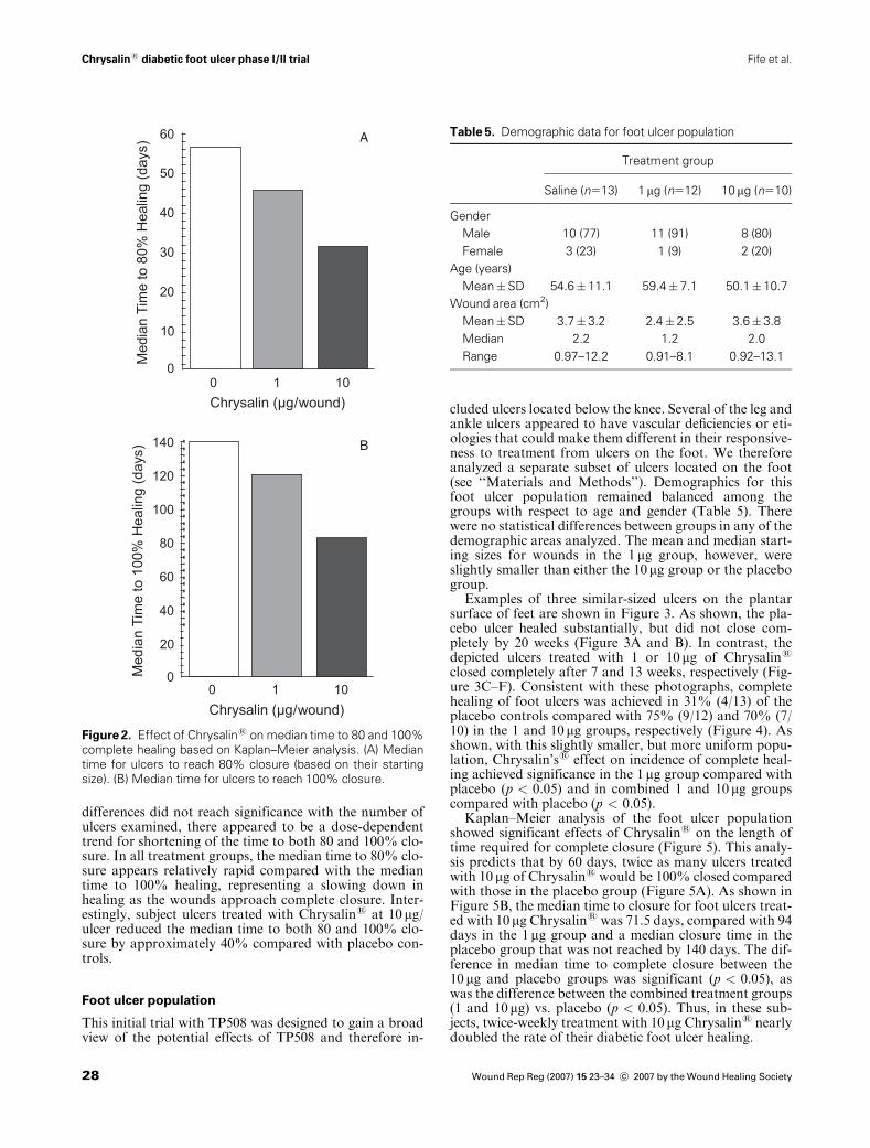
differences did not reach significance with the number ofulcers examined, there appeared to be a dose-dependenttrend for shortening of the time to both 80 and 100% clo-sure. In all treatment groups, the median time to 80% clo-sure appears relatively rapid compared with the mediantime to 100% healing, representing a slowing down inhealing as the wounds approach complete closure. Inter-estingly, subject ulcers treated with Chrysalins at 10 mg/ulcer reduced the median time to both 80 and 100% clo-sure by approximately 40% compared with placebo con-trols.
Foot ulcer population
This initial trial with TP508 was designed to gain a broadview of the potential effects of TP508 and therefore in-
cluded ulcers located below the knee. Several of the leg andankle ulcers appeared to have vascular deficiencies or eti-ologies that could make them different in their responsive-ness to treatment from ulcers on the foot. We thereforeanalyzed a separate subset of ulcers located on the foot(see ‘‘Materials and Methods’’). Demographics for thisfoot ulcer population remained balanced among thegroups with respect to age and gender (Table 5). Therewere no statistical differences between groups in any of thedemographic areas analyzed. The mean and median start-ing sizes for wounds in the 1 mg group, however, wereslightly smaller than either the 10mg group or the placebogroup.
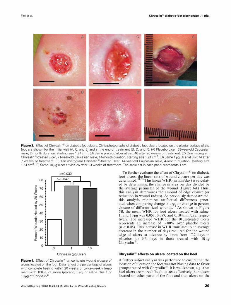
Examples of three similar-sized ulcers on the plantarsurface of feet are shown in Figure 3. As shown, the pla-cebo ulcer healed substantially, but did not close com-pletely by 20 weeks (Figure 3A and B). In contrast, thedepicted ulcers treated with 1 or 10 mg of Chrysalins
closed completely after 7 and 13 weeks, respectively (Fig-ure 3C–F). Consistent with these photographs, completehealing of foot ulcers was achieved in 31% (4/13) of theplacebo controls compared with 75% (9/12) and 70% (7/10) in the 1 and 10mg groups, respectively (Figure 4). Asshown, with this slightly smaller, but more uniform popu-lation, Chrysalin’ss effect on incidence of complete heal-ing achieved significance in the 1mg group compared withplacebo (p < 0.05) and in combined 1 and 10 mg groupscompared with placebo (p < 0.05).
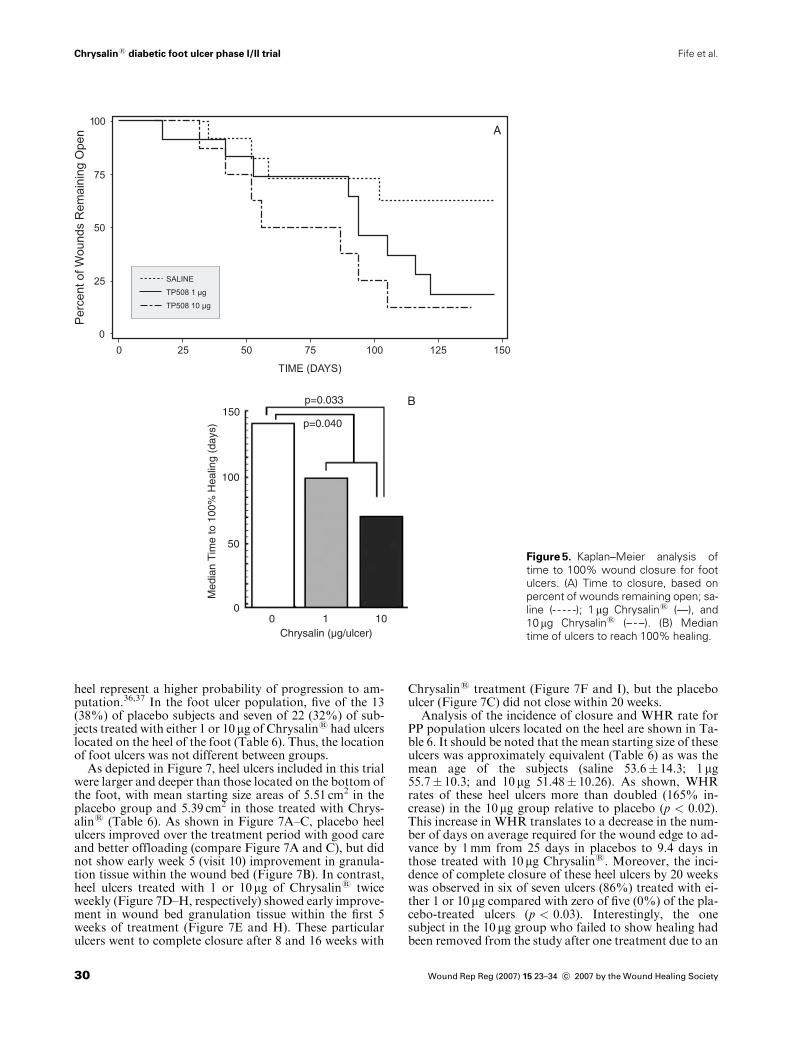
Kaplan–Meier analysis of the foot ulcer populationshowed significant effects of Chrysalins on the length oftime required for complete closure (Figure 5). This analy-sis predicts that by 60 days, twice as many ulcers treatedwith 10 mg of Chrysalins would be 100% closed comparedwith those in the placebo group (Figure 5A). As shown inFigure 5B, the median time to closure for foot ulcers treat-ed with 10mg Chrysalins was 71.5 days, compared with 94days in the 1mg group and a median closure time in theplacebo group that was not reached by 140 days. The dif-ference in median time to complete closure between the10 mg and placebo groups was significant (p < 0.05), aswas the difference between the combined treatment groups(1 and 10mg) vs. placebo (p < 0.05). Thus, in these sub-jects, twice-weekly treatment with 10 mg Chrysalins nearlydoubled the rate of their diabetic foot ulcer healing.
A
B
Figure 2. Effect of Chrysalins on median time to 80 and 100%
complete healing based on Kaplan–Meier analysis. (A) Median
time for ulcers to reach 80% closure (based on their starting
size). (B) Median time for ulcers to reach 100% closure.
Table 5. Demographic data for foot ulcer population
Treatment group
Saline (n513) 1mg (n512) 10 mg (n510)
Gender
Male 10 (77) 11 (91) 8 (80)
Female 3 (23) 1 (9) 2 (20)
Age (years)
Mean�SD 54.6� 11.1 59.4� 7.1 50.1� 10.7
Wound area (cm2)
Mean�SD 3.7� 3.2 2.4� 2.5 3.6� 3.8
Median 2.2 1.2 2.0
Range 0.97–12.2 0.91–8.1 0.92–13.1
Wound Rep Reg (2007) 15 23–34 c� 2007 by the Wound Healing Society28
Chrysalins diabetic foot ulcer phase I/II trial Fife et al.
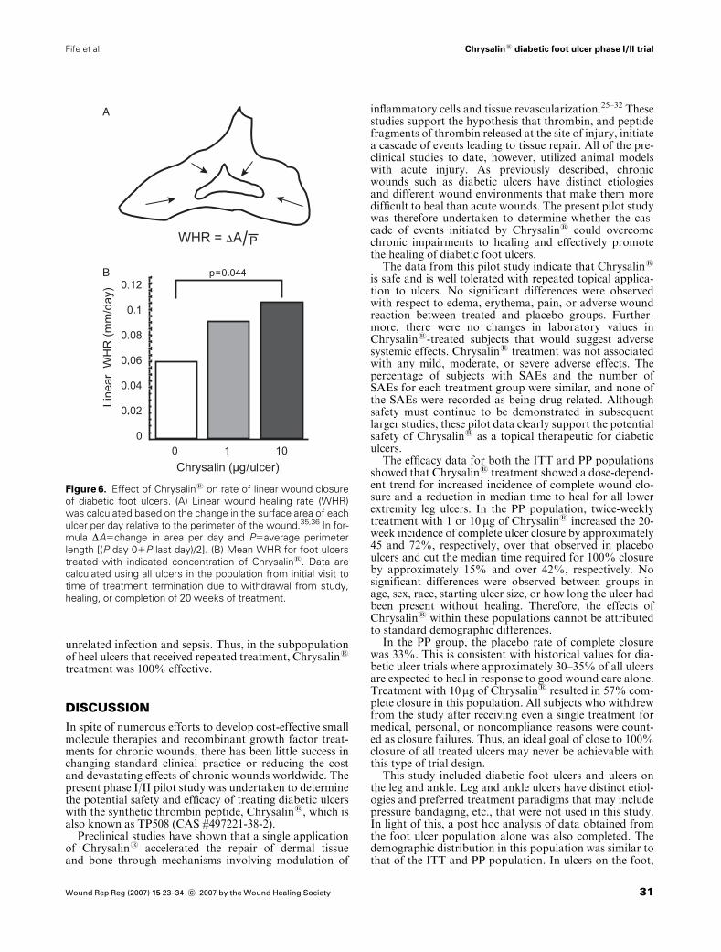
To further evaluate the effect of Chrysalins on diabeticfoot ulcers, the linear rate of wound closure per day wasdetermined.34,35 This linear WHR (in mm/day) is calculat-ed by determining the change in area per day divided bythe average perimeter of the wound (Figure 6A) Thus,this analysis determines the amount of edge closure (orreduction in wound radius). As previously demonstrated,this analysis minimizes artifactual differences gener-ated when comparing change in area or change in percentclosure of different-sized wounds.35 As shown in Figure6B, the mean WHR for foot ulcers treated with saline,1, and 10 mg was 0.058, 0.089, and 0.104mm/day, respec-tively. The increased WHR for the 10mg-treated ulcersrepresents an increase of � 80% over placebo ulcers(p < 0.05). This increase in WHR translates to an averagedecrease in the number of days required for the woundedge of ulcers to advance by 1mm from 17.2 days inplacebos to 9.6 days in those treated with 10 mgChrysalins.
Chrysalins effects on ulcers located on the heel
A further subset analysis was performed to ensure that thelocation of ulcers on the foot was not biasing data to favorgroups treated with Chrysalins. It is well known, e.g., thatheel ulcers are more difficult to treat effectively than ulcerslocated on other parts of the foot and that ulcers on the
Figure 3. Effect of Chrysalins on diabetic foot ulcers. Clinic photographs of diabetic foot ulcers located on the plantar surface of the
foot are shown for the initial visit (A, C, and E) and at the end of treatment (B, D, and F). (A) Placebo ulcer, 63-year-old Caucasian
male, 2-month duration, starting size 1.24 cm2. (B) Same placebo ulcer at visit 40 after 20 weeks of treatment. (C) One microgram
Chrysalins-treated ulcer, 71-year-old Caucasian male, 14-month duration, starting size 1.21 cm2. (D) Same 1mg ulcer at visit 14 after
7 weeks of treatment. (E) Ten microgram Chrysalins-treated ulcer, 44-year-old Caucasian male, 4-month duration, starting size
1.51 cm2. (F) Same 10 mg ulcer at visit 26 after 13 weeks of treatment. The scale bar in each panel represents 1 cm.
80
p=0.032
p=0.047
70
60
50
40
30
20
Per
cent
Wou
nds
Hea
led
by 2
0 W
eeks
10
00 1 10
Chrysalin (µg/ulcer)
Figure 4. Effect of Chrysalins on complete wound closure of
ulcers located on the foot. Data reflect the percentage of ulcers
with complete healing within 20 weeks of twice-weekly treat-
ment with 100mL of saline (placebo, 0 mg) or saline plus 1 or
10 mg of Chrysalins.
Wound Rep Reg (2007) 15 23–34 c� 2007 by the Wound Healing Society 29
Chrysalins diabetic foot ulcer phase I/II trialFife et al.
heel represent a higher probability of progression to am-putation.36,37 In the foot ulcer population, five of the 13(38%) of placebo subjects and seven of 22 (32%) of sub-jects treated with either 1 or 10 mg of Chrysalins had ulcerslocated on the heel of the foot (Table 6). Thus, the locationof foot ulcers was not different between groups.
As depicted in Figure 7, heel ulcers included in this trialwere larger and deeper than those located on the bottom ofthe foot, with mean starting size areas of 5.51 cm2 in theplacebo group and 5.39 cm2 in those treated with Chrys-alins (Table 6). As shown in Figure 7A–C, placebo heelulcers improved over the treatment period with good careand better offloading (compare Figure 7A and C), but didnot show early week 5 (visit 10) improvement in granula-tion tissue within the wound bed (Figure 7B). In contrast,heel ulcers treated with 1 or 10 mg of Chrysalins twiceweekly (Figure 7D–H, respectively) showed early improve-ment in wound bed granulation tissue within the first 5weeks of treatment (Figure 7E and H). These particularulcers went to complete closure after 8 and 16 weeks with
Chrysalins treatment (Figure 7F and I), but the placeboulcer (Figure 7C) did not close within 20 weeks.
Analysis of the incidence of closure and WHR rate forPP population ulcers located on the heel are shown in Ta-ble 6. It should be noted that the mean starting size of theseulcers was approximately equivalent (Table 6) as was themean age of the subjects (saline 53.6� 14.3; 1mg55.7� 10.3; and 10 mg 51.48� 10.26). As shown, WHRrates of these heel ulcers more than doubled (165% in-crease) in the 10mg group relative to placebo (p < 0.02).This increase in WHR translates to a decrease in the num-ber of days on average required for the wound edge to ad-vance by 1mm from 25 days in placebos to 9.4 days inthose treated with 10 mg Chrysalins. Moreover, the inci-dence of complete closure of these heel ulcers by 20 weekswas observed in six of seven ulcers (86%) treated with ei-ther 1 or 10 mg compared with zero of five (0%) of the pla-cebo-treated ulcers (p < 0.03). Interestingly, the onesubject in the 10 mg group who failed to show healing hadbeen removed from the study after one treatment due to an
150p=0.033
p=0.040
Med
ian
Tim
e to
100
% H
ealin
g (d
ays)
100
50
00 1 10
Chrysalin (µg/ulcer)
A
B
I
Figure 5. Kaplan–Meier analysis of
time to 100% wound closure for foot
ulcers. (A) Time to closure, based on
percent of wounds remaining open; sa-
line (- - - - -); 1mg Chrysalins (—), and
10 mg Chrysalins (– - –). (B) Median
time of ulcers to reach 100% healing.
Wound Rep Reg (2007) 15 23–34 c� 2007 by the Wound Healing Society30
Chrysalins diabetic foot ulcer phase I/II trial Fife et al.
unrelated infection and sepsis. Thus, in the subpopulationof heel ulcers that received repeated treatment, Chrysalins
treatment was 100% effective.
DISCUSSION
In spite of numerous efforts to develop cost-effective smallmolecule therapies and recombinant growth factor treat-ments for chronic wounds, there has been little success inchanging standard clinical practice or reducing the costand devastating effects of chronic wounds worldwide. Thepresent phase I/II pilot study was undertaken to determinethe potential safety and efficacy of treating diabetic ulcerswith the synthetic thrombin peptide, Chrysalins, which isalso known as TP508 (CAS #497221-38-2).
Preclinical studies have shown that a single applicationof Chrysalins accelerated the repair of dermal tissueand bone through mechanisms involving modulation of
inflammatory cells and tissue revascularization.25–32 Thesestudies support the hypothesis that thrombin, and peptidefragments of thrombin released at the site of injury, initiatea cascade of events leading to tissue repair. All of the pre-clinical studies to date, however, utilized animal modelswith acute injury. As previously described, chronicwounds such as diabetic ulcers have distinct etiologiesand different wound environments that make them moredifficult to heal than acute wounds. The present pilot studywas therefore undertaken to determine whether the cas-cade of events initiated by Chrysalins could overcomechronic impairments to healing and effectively promotethe healing of diabetic foot ulcers.
The data from this pilot study indicate that Chrysalins
is safe and is well tolerated with repeated topical applica-tion to ulcers. No significant differences were observedwith respect to edema, erythema, pain, or adverse woundreaction between treated and placebo groups. Further-more, there were no changes in laboratory values inChrysalins-treated subjects that would suggest adversesystemic effects. Chrysalins treatment was not associatedwith any mild, moderate, or severe adverse effects. Thepercentage of subjects with SAEs and the number ofSAEs for each treatment group were similar, and none ofthe SAEs were recorded as being drug related. Althoughsafety must continue to be demonstrated in subsequentlarger studies, these pilot data clearly support the potentialsafety of Chrysalins as a topical therapeutic for diabeticulcers.
The efficacy data for both the ITT and PP populationsshowed that Chrysalins treatment showed a dose-depend-ent trend for increased incidence of complete wound clo-sure and a reduction in median time to heal for all lowerextremity leg ulcers. In the PP population, twice-weeklytreatment with 1 or 10 mg of Chrysalins increased the 20-week incidence of complete ulcer closure by approximately45 and 72%, respectively, over that observed in placeboulcers and cut the median time required for 100% closureby approximately 15% and over 42%, respectively. Nosignificant differences were observed between groups inage, sex, race, starting ulcer size, or how long the ulcer hadbeen present without healing. Therefore, the effects ofChrysalins within these populations cannot be attributedto standard demographic differences.
In the PP group, the placebo rate of complete closurewas 33%. This is consistent with historical values for dia-betic ulcer trials where approximately 30–35% of all ulcersare expected to heal in response to good wound care alone.Treatment with 10mg of Chrysalins resulted in 57% com-plete closure in this population. All subjects who withdrewfrom the study after receiving even a single treatment formedical, personal, or noncompliance reasons were count-ed as closure failures. Thus, an ideal goal of close to 100%closure of all treated ulcers may never be achievable withthis type of trial design.
This study included diabetic foot ulcers and ulcers onthe leg and ankle. Leg and ankle ulcers have distinct etiol-ogies and preferred treatment paradigms that may includepressure bandaging, etc., that were not used in this study.In light of this, a post hoc analysis of data obtained fromthe foot ulcer population alone was also completed. Thedemographic distribution in this population was similar tothat of the ITT and PP population. In ulcers on the foot,
B
A
Figure 6. Effect of Chrysalins on rate of linear wound closure
of diabetic foot ulcers. (A) Linear wound healing rate (WHR)
was calculated based on the change in the surface area of each
ulcer per day relative to the perimeter of the wound.35,36 In for-
mula DA5change in area per day and P5average perimeter
length [(P day 01P last day)/2]. (B) Mean WHR for foot ulcers
treated with indicated concentration of Chrysalins. Data are
calculated using all ulcers in the population from initial visit to
time of treatment termination due to withdrawal from study,
healing, or completion of 20 weeks of treatment.
Wound Rep Reg (2007) 15 23–34 c� 2007 by the Wound Healing Society 31
Chrysalins diabetic foot ulcer phase I/II trialFife et al.
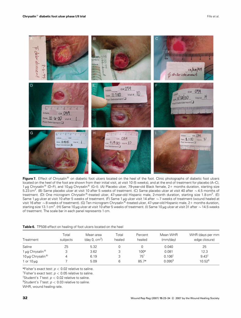
Figure 7. Effect of Chrysalins on diabetic foot ulcers located on the heel of the foot. Clinic photographs of diabetic foot ulcers
located on the heel of the foot are shown from their initial visit, at visit 10 (5 weeks), and at the end of treatment for placebo (A–C),
1mg Chrysalins (D–F), and 10mg Chrysalins (G–I). (A) Placebo ulcer, 79-year-old Black female, 21 months duration, starting size
5.23 cm2. (B) Same placebo ulcer at visit 10 after 5 weeks of treatment. (C) Same placebo ulcer at visit 40 after � 4.5 months of
treatment. (D) One microgram Chrysalins-treated ulcer, 47-year-old Hispanic male, 2-month duration, starting size 1.8 cm2. (E)
Same 1mg ulcer at visit 10 after 5 weeks of treatment. (F) Same 1mg ulcer visit 14 after � 7 weeks of treatment (wound healed at
visit 16 after � 8 weeks of treatment). (G) Ten microgram Chrysalins-treated ulcer, 47-year-old Hispanic male, 21 months duration,
starting size 13.1 cm2. (H) Same 10 mg ulcer at visit 10 after 5 weeks of treatment. (I) Same 10 mg ulcer at visit 31 after � 14.5 weeks
of treatment. The scale bar in each panel represents 1 cm.
Table 6. TP508 effect on healing of foot ulcers located on the heel
Treatment
Total
subjects
Mean area
(day 0, cm2)
Total
healed
Percent
healed
Mean WHR
(mm/day)
WHR (days per mm
edge closure)
Saline Z5 5.32 0 0 0.040 25
1mg Chrysalins 3 3.62 3 100n 0.081 12.3
10 mg Chrysalins 4 6.19 3 75w 0.106z 9.43z
1 or 10mg 7 5.09 6 85.7n 0.095§ 10.52§
nFisher’s exact test: p < 0.02 relative to saline.wFisher’s exact test: p < 0.05 relative to saline.zStudent’s T test: p < 0.02 relative to saline.§Student’s T test: p < 0.03 relative to saline.
WHR, wound healing rate.
Wound Rep Reg (2007) 15 23–34 c� 2007 by the Wound Healing Society32
Chrysalins diabetic foot ulcer phase I/II trial Fife et al.
complete closure was achieved in 75 and 70% in ulcerstreated with 1 and 10 mg of Chrysalins, respectively, com-pared with 31% of the placebo controls. These differencesbetween Chrysalins-treatment and placebo were signifi-cant (placebo vs. 1 mg p < 0.05; and placebo vs. 1 or 10mgp < 0.05). Of note, this effect of Chrysalins represents anincrease in the incidence of complete closure of greaterthan 125% over placebo controls with good standardwound care. Based on Kaplan–Meier analysis, treatmentof foot ulcers with 10 mg of Chrysalins produced a mediantime to closure of 71.5 days while in the control group themedian was not reached by 140 days (p < 0.05). Thus,Chrysalins-treated ulcers appeared to heal approximatelytwice as fast as the placebo ulcers. Consistent with the Ka-plan–Meier analysis, the linear WHR in the 10mg groupwas � 80% greater than ulcers treated with saline(p < 0.05).
A number of other clinical trials examining potentialtreatment modalities have only examined relatively well-vascularized Wagner Grades I and II diabetic foot ulcersand have not included ulcers on the leg or ankle. There-fore, having determined the effects of Chrysalins on thefoot ulcer subpopulation allows a better comparison withdata obtained in previous trials. For example, in the fourmajor becaplermin (Regranexs) trials that included a totalof 922 subjects, placebo healing was � 35% comparedwith � 50% healing in diabetic foot ulcers treated dailywith becaplermin.13,14 This represents an increase of ap-proximately 40% in complete healing over placebo con-trols. In the current study, the incidence of placebo healingwas comparable with that seen in the becaplermin studies,but Chrysalins treatment twice weekly with 1mg/ulcer re-sulted in a 75% incidence of complete closure by 20 weeks.This represents a 140% increase over the incidence of heal-ing in placebos. The linear WHR also nearly doubled withChrysalins treatment. These data suggest that the twice-weekly treatment with Chrysalins may be at least as ef-fective in closing diabetic foot ulcers as daily application ofbecaplermin.
The data from this study are especially encouraging,considering that the foot ulcer population in this study in-cluded a number of Wagner category III (deeper) ulcers,many of which were located on the heel of the foot. It isrecognized that ulcers located on the heel of the foot re-quire special treatment, are among the most difficult offoot ulcers to heal, and are the ones most likely to result inamputation of the foot.36,37 Subset analysis of heel ulcersin a recently published clinical trial with over 250 diabeticfoot ulcers, e.g., showed that only 8% (1/13) control heelulcers achieved complete closure compared with 33%(6/18) of heel ulcers treated with Dermagraft.38 In our footulcer population, wound closure rates of heel ulcers morethan doubled (165% increase) in the 10 mg Chrysalins
group relative to placebo (p < 0.02). This increase inWHR translates into a decrease in the average time re-quired for 1mm of edge closure from 25 days in placebosto 9.4 days in ulcers treated with 10 mg of Chrysalins.Moreover, complete closure of these heel ulcers was ob-served in six of seven ulcers (86%) treated with either 1 or10 mg compared with zero of five (0%) of the placebo-treated ulcers (p < 0.03). These data suggest that Chrys-alins treatment may be especially effective in larger andmore difficult chronic ulcers.
In this study, twice-weekly treatments with Chrysalins
or placebo were combined with a standardized regimen ofgood wound care, including a moisture-retentive primarydressing, initial and ongoing sharp debridement as deemednecessary by the attending physician, and pressure off-loading. Subjects were treated during office visits to ensuretreatment compliance and limit treatment variability; yet,with only twice-weekly application and inclusion of bothorthopedic wound centers and podiatry centers, the datamay provide a realistic prediction of real-world use. There-fore, the significant data obtained in this pilot study sug-gest that topical application of Chrysalins may haveconsiderable therapeutic value for diabetic foot ulcers.
ACKNOWLEDGMENTS
The authors wish to thank Dennis McWilliams, Dr. RogerCrowther, and all of the former employees of ChrysalisBioTechnology Inc. for their support and early manage-ment of this study, Dr. Jon Mader, who died in 2003, andhis group at the University of Texas Medical Branch(UTMB) who treated the majority of the subjects in thisstudy, the research coordinators and pharmacists withoutwhom clinical trials would not be possible, Jaye Thomp-son, Paul Lewis, and the staff at Synergos Inc. in Wood-lands, TX, for monitoring, data analysis, and statisticalsupport, Dr. Thomas A. Mustoe for his advice and criticalreview of this manuscript, and Drs. Gerald M. Fuller andGordon H. Otto, who provided outside reviewers of themanuscript. This phase I/II clinical trial was sponsored byChrysalis BioTechnology Inc., Galveston, TX, a spinoutof UTMB, Galveston. Chrysalis was acquired in 2004, byOrthoLogic Corp., Tempe, AZ (NASDAQ: OLGC), whonow owns the rights to the Chrysalins Technology for allapplications worldwide. Dr. J.T. Ryaby is currently em-ployed by and is an OrthoLogic Corp shareholder. Dr.D.H. Carney (the founder of Chrysalis BioTechnologyInc.) is an OrthoLogic shareholder and receives monetarycompensation as a consultant to OrthoLogic. Potentialconflicts of interest and oversight of Dr. Carney’s researchrelated to this technology are monitored and managed bythe UTMB Conflicts of Interest Committee.
REFERENCES
1. Gordois A, Scuffham P, Shearer A, Oglesby A, Tobian JA.The health care costs of diabetic peripheral neuropathy in the
US. Diabetes Care 2003; 26: 1790–5.2. Harrington C, Zagari MJ, Corea J, Klitenic J. A cost analysis
of diabetic lower-extremity ulcers. Diabetes Care 2000; 23:
1333–8.3. Girod I, Valensi P, Laforet C, Moreau-Defarges T, Guillon
P, Baron F. An economic evaluation of the cost of diabeticfoot ulcers: results of a retrospective study on 239 patients.
Diabetes Metab 2003; 293: 269–77.4. Redekop WK, Stolk EA, Kok E, Lovas K, Kalo Z, Bussch-
bach JJ. Diabetic foot ulcers and amputations: estimates of
health utility for use in cost-effectiveness analyses of newtreatments. Diabetes Metab 2004; 30: 549–56.
Wound Rep Reg (2007) 15 23–34 c� 2007 by the Wound Healing Society 33
Chrysalins diabetic foot ulcer phase I/II trialFife et al.
5. Mustoe T. Understanding chronic wounds: a unifying hy-pothesis on their pathogenesis and implications for therapy.Am J Surg 2004; 187: 65S–70S.
6. Jeffcoate WJ, Harding KG. Diabetic foot ulcers. Lancet2003; 361: 1545–51.
7. Greenhalgh DG. Wound healing and diabetes mellitus. ClinPlast Surg 2003; 30: 37–45.
8. Cosentino F, Luscher T. Endothelial dysfunction in diabetesmellitus. J Cardiovasc Pharmacol 1998; 32: 54–61.
9. Jude EB, Tentolouris N, Appleton I, Anderson S, BoultonAJ. Role of neuropathy and plasma nitric oxide in recurrentneuropathic and neuroischemic diabetic foot ulcers. WoundRepair Regen 2001; 9: 353–9.
10. Trengove NJ, Stacey MC,MacAuley S, Bennett N, Gibson J,Burslem F, Murphy G, Schultz G. Analysis of the acute andchronic wound environments: the role of proteases and theirinhibitors. Wound Repair Regen 1999; 7: 442–52.
11. Lobmann R, Ambrosch A, Schultz G, Waldmann K, Schi-weck S, Lehnert H. Expression of matrix-metalloproteinasesand their inhibitors in the wounds of diabetic and non-dia-betic patients. Diabetologia 2002; 45: 1011–6.
12. Wall SJ, SampsonMJ, Levell N, Murphy G. Elevated matrixmetalloproteinase-2 and -3 production from human diabeticdermal fibroblasts. Br J Dermatol 2003; 149: 13–6.
13. Wieman TJ, Smiell JM, Su Y. Efficacy and safety of a topicalgel formulation of recombinant human platelet-derivedgrowth factor-BB (becaplermin) in patients with chronic neu-ropathic diabetic ulcers. A phase III randomized placebo-con-trolled double-blind study.Diabetes Care 1998; 21: 822–7.
14. Smiell JM, Wieman TJ, Steed DL, Perry BH, Sampson AR,Schwab BH. Efficacy and safety of becaplermin (recombi-nant human platelet-derived growth factor-BB) in patientswith nonhealing, lower extremity diabetic ulcers: a combinedanalysis of four randomized studies. Wound Repair Regen1999; 7: 335–46.
15. Steed DL, Donohoe D, Webster MW, Lindsley L. Effect ofextensive debridement and treatment on the healing of dia-betic foot ulcers. Diabetic Ulcer Study Group. J Am CollSurg 1996; 183: 61–4.
16. Glenn KC, Frost GH, Bergmann JS, Carney DH. Syntheticpeptides bind to high-affinity thrombin receptors and modu-late thrombin mitogenesis. Peptide Res 1988; 1: 65–73.
17. Carney DH, Cunningham DD. Cell surface action of throm-bin is sufficient to initiate division of chick cells.Cell 1978; 14:811–23.
18. Carney DH, Cunningham DD. Role of specific cell surfacereceptors in thrombin-stimulated cell division. Cell 1978; 15:1341–9.
19. Carney DH, Bergmann JS. 125I-thrombin binds to clusteredreceptors on noncoated regions of mouse embryo cell sur-faces. J Cell Biol 1982; 95: 697–703.
20. Coughlin SR. Thrombin signalling and protease-activatedreceptors. Nature 2000; 407: 258–64.
21. Carney DH, Stiernberg J, Fenton JW II. Initiation of prolif-erative events by human alpha-thrombin requires both re-ceptor binding and enzymic activity. J Cell Biochem 1984; 26:181–95.
22. Carney DH, Herbosa GJ, Stiernberg J, Bergmann JS, Gor-don EA, Scott D, Fenton JW II. Double-signal hypothesis
for thrombin initiation of cell proliferation. Semin ThrombHemost 1986; 12: 231–40.
23. Carney DH. Postclotting cellular effects of thrombin medi-ated by interaction with high-affinity thrombin receptors. In:Berliner L, editor. Thrombin Structure and Function. NewYork: Plenum, 1992: 351–96.
24. Carney DH, Redin W, McCroskey L. Role of high-affinitythrombin receptors in postclotting cellular effects of throm-bin. Semin Thromb Hemost 1992; 18: 91–103.
25. Carney DH, Mann R, Redin WR, Pernia SD, Berry D,Heggers JP, Hayward PG, Robson MC, Christie H, AnnableC, Fenton JW II, Glenn KC. Enhancement of incisionalwound healing and neovascularization in normal rats bythrombin and synthetic thrombin receptor-activating pep-tides. J Clin Invest 1992; 89: 1469–77.
26. Cromack DT, Porras RBH, Wee SS, Glenn KC, Purdy JA,Carney DH, Mustoe TA. Acceleration of soft tissue repairby a thrombin-derived oligopeptide. J Surg Res 1992; 53:117–22.
27. Stiernberg J, Norfleet AM, Redin WR, Warner WS, FritzRR, Carney DH. Acceleration of full-thickness wound heal-ing in normal rats by the synthetic thrombin peptide, TP508.Wound Repair Regen 2000; 8: 204–15.
28. Norfleet AM, Huang Y, Sower LE, Redin WR, Fritz RR,Carney DH. Thrombin peptide TP508 accelerates closure ofdermal excisions in animal tissue with surgically inducedischemia. Wound Repair Regen 2000; 8: 517–29.
29. Norfleet AM, Bergmann JS, Carney DH. Thrombin peptide,TP508, stimulates angiogenic responses in animal models ofdermal wound healing, in chick chorioallantoic membranes,and in cultured human aortic and microvascular endothelialcells. Gen Pharmacol 2000; 35: 249–54.
30. Wang H, Li X, Tomin E, Doty SB, Lane JM, Carney DH,Ryaby JT. Thrombin peptide (TP508) promotes fracture re-pair by up-regulating inflammatory mediators, early growthfactors, and increasing angiogenesis. J Orthop Res 2005; 23:671–79.
31. Sheller MR, Crowther RS, Kinney JH, Yang J, Di Jorio S,Breunig T, Carney DH, Ryaby JT. Repair of rabbit segmen-tal defects with the thrombin peptide, TP508. J Orthop Res2004; 22: 1094–9.
32. Li G, Ryaby JT, Carney DH, Wang H. Bone formation isenhanced by thrombin-related peptide TP508 during distrac-tion osteogenesis. J Orthop Res 2005; 23: 196–202.
33. Wagner FW. The dysvascular foot: a system of diagnosis andtreatment. Foot Ankle 1981; 2: 64–122.
34. Gilman TH. Parameter for measurement of wound closure.Wounds 1990; 3: 95–101.
35. Gorin DR, Cordts PR, LaMorte WW, Menzoian JO. Theinfluence of wound geometry on the measurement of woundhealing rates in clinical trials. J Vasc Surg 1996; 23: 524–8.
36. Younes NA, Albsoul AM, Awad H. Diabetic heel ulcers: amajor risk factor for lower extremity amputation. OstomyWound Manage 2004; 50: 50–60.
37. Hampton S. The complexities of heel ulcers. Nurs Stand2003; 17: 68–70, 72, 74.
38. Marston WA, Hanft J, Norwood P, Pollak R. The efficacyand safety of Dermagraft in improving the healing of chronicdiabetic foot ulcers: results of a prospective randomized trial.Diabetes Care 2003; 26: 1701–5.
Wound Rep Reg (2007) 15 23–34 c� 2007 by the Wound Healing Society34
Chrysalins diabetic foot ulcer phase I/II trial Fife et al.