Embed Size (px)
Citation preview
Introduction Hepatocelluar carcinoma (HCC) is frequently observed in patients with liver cirrhosis due to HCV in Japan [1]. Although the underlying mechanism has not yet been clarified many administering anticancer drugs have been developed and used for the treatment in patients with HCC [2-7]. However, these anticancer drugs usually have severe side effects (such as nausea, vomiting, diarrhea, leucocytopenia thrombocytopenia and hepatorenal injury) Therefore, the development of new drug that inhibits the proliferation of cancer, which has less or no side effects are important and anticipated. In Japan, gabexate mesilate often is clinically used as a drug of acute pancreatitis and disseminated intravascular coagulation (DIC) [8-10]. We happened to administer gabexate mesilate (GM) to patients with pancreatitis complicated by HCC. In liver cirrhosis (C) and observed the inhibition of HCC and long survival period in
several cases. Therefore, an experimental study was performed to confirm this phenomenon and evaluate the mechanism of this inhibition. Materials and methods Cell culture
Frozen Hep G2 cells (RIKEN Cell Bank,Japan) were thawed at 37°C, placed in a centrifuge tube containing growth medium (E-MEN, 2mML-Gln, sodium hydrogen carbonate, 1% NEAA, 1.0mM Na-Pyr, and 10% FBS) , and centrifuged to remove the supernatant. After addition of growth medium, the cells were spread on two 100-mm Petri dishes , and cultured at 37°C for 6 days under 5% CO2 /95% air for stabilization. When the confluence reached 80 to 90% after subculture, the medium was removed, and the cells were washed in PBS (-). Subsequently, PBS (-) was removed, and dispersing agents (0.25% trypsin, 0.02% EDTA, and PBS (-) were added. When the cells became round/globular, growth
Int J Clin Exp Pathol 2010;3(7):710-717 www.ijcep.com /IJCEP1004003
Original Article The specific inhibition of HepG2 cells proliferation by apoptosis induced by gabexate mesilate
Tsuneo Ozeki1, Tsuneo Natori2 1Ozeki Internal Medicine Clinic, 10-30-10, Iseigaoka, Nishiku Yahata, Kitakyushu City, Japan; 2SRL, Inc, Hamura Laboratory, 3-5-5 Midorigaoka Hamura City, Tokyo, Japan. Received April 19, 2010; accepted August 12, 2010; available online August 15, 2010 Abstract: Many anticancer drugs are developing until now.However,conventional anticancer drugs causes damage to not only cancer cells but also non-cancerous tissues and cells. Therefore, the development of new drugs are antici-pated.HepG2 cell proliferation in cell culture was significantly inhibited by gabexate mesilate. In TUNEL method, a significant amount of HepG2 cells cultured with gabexate mesilate showed a decrease in the number of total cells and an increased in the number of positive cells. Further immunohistochemical staining for P-53,ss-DNA and caspase 3 showed samely a decrease in the number of total cells and an increase in the number of positive cells. The staining for bcl2 showed a decrease in the number of total cells and no remarkable change in the number of positive cells. The cell growth inhibition by gabexate mesilate was almost blocked by caspase 3 inhibitor. Therefore, the inhibition itself of HepG2 cell proliferation by gabexate mesilate was mainly due to the apoptosis. This agent causes mainly damage to HepG2 cell by apoptosis but does not cause side effects, differing from the above anticancer drugs, Gabexate mesilate is a useful drug. Keywords: Apoptosis,hepatocellular carcinoma, HepG2cell, gabexate mesilate
Gabexate mesilate-induced inhibition of HepG2 cells proliferation
711 Int J Clin Exp Pathol 2010;3(7):710-717
medium was added to arrest the actions of the dispersing agents. Subsequently, the cells were exfoliated by pipetting, and placed in a centrifuge tube. They were washed in PBS (-), and then centrifuged. After the supernatant was removed, growth medium was added. In a portion of the cell suspension, the cell count was measured employing the trypan blue exclusion method with a blood cell counter. The cells were spread on a 100-mm petri dish, and cultured with growth medium at 37°C for 1 week under 5% CO2 / 95% air for stabilization. Subsequently, the cell count was adjusted to 5.76 × 10⁴ cells / 0.1 ml / well using a 96-well plate. The cells were cultured as follows to examine the concentration of GM and cell counts at designated points:Control Group 1:24-hour culture with PBS (-) instead of GM (3 wells), Test Group 1:24-hour culture in the presence of 1,000 µM GM (3 wells), Control Group 2:48-hour culture with PBS (-) instead of GM (3 wells) , Test Group 2:48-hour culture in the presence of 1,000 µM GM (3 wells), Control Group 3:72-hour culture with PBS (-) instead of GM (3 wells), Test Group 3:72-hour culture in the presence of 100 µM GM (3 wells), Test Group 4:72-hour culture in the presence of 300 µM GM (3 wells), and Test Group 5:72-hour culture in the presence of 1.000 µM GM (3 wells). The cell count was measured employing the WST-8 method [11, 12]. Agarose electrophoresis GM (0 and 1.000 µM) was added to 5.76 × 10⁵ cells/ml, which had been stabilized, and cultured for 72 hours on 100-mm Petri dishes, respectively. Furthermore, in the presence of 1,000 µM GM, the cells were cultured for 12 hours. When the confluence reached 80 to 90%, trypsination was discontinued by dispersing agents / medium, as described in the “Cell culture” section, and the cell count was measured to adjust it to 5.76 x 10⁵ cells/ml. The cells were thoroughly washed in PBS (-). The precipitate was mixed with 0.5 ml of lysis buffer, which was included in an ApopLadder EX kit (Takara Bio Japan). According to the procedure for this kit, DNA fragments [13, 14] were extracted, and 10 microliters of sample was electrophoresed with 2.5% agarose at 100 volts for 30 minutes. Ladder formation of DNA fragments was confirmed using a UV illuminator.
TUNEL method After HepG2 cells were similarly cultured for a week and stabilized, cell suspensions (1×10⁵
cells/ml) were cultured at 37C under 5% CO2 and 95% air, for 24, 48 and 72 hours with final 1,000 µM GM. Cells were adequately washed with PBS (-) and 10% formalin was added. For TUNEL staining [15-17], cells were adequately washed with PBS (-) again, and suspended in 0.5ml PBS (-). On slide glasses, a circle (1.5cm in diameter) was drawn using a stylus and 50 µl suspension was placed within the circle, dried with cool air, immersed in 10% formal in for 5 min, adequately washed with PBS (-), air dried, and stained using an Apoptosis in situ Detection Kit( Wako, Japan). Immunohistochemical staining for P53, ss-DNA, bcl2 and caspase 3 HepG2 cells were similarly cultured and stabilized in Eagle’s MEM medium. After the same sampling method that was performed in TUNEL staining was carried out , the staining of P53, ss-DNA, bcl2 and caspase 3 was performed using a Daco Envision +Kit/HRP (DAKO, Denmark); the primary antibodies for P53 and bcl2 were mouse monoclonal antibody anti-human P53 protein (Novo UK) in the dilution of 1:100 and mouse monoclonal antibody anti-human bcl2 oncoprotein (DAKO Denmark) in the dilution of 1:100, the primary antibodies for ss-DNA and caspase 3 were rabbit polyclonal antibodies anti-human ss-DNA (DAKO Denmark) in the dilution of 1:100 and rabbit polyclonal antibodies anti-human caspase 3 (Cell signaling UK) in the dilution 1:100. Polymer method using DAKO Envision + Kit/HRP13 ( DAKO Denmark ) was performed. Secondary antibody was DAKO ENVISION + Kit/HPR (DAB) anti-rabbit or anti-mouse (DAKO Denmark) and finally incubation with DAB and counter stain with hematoxylin were performed. In addition to evaluate non-specific staining immunohistochemical staining was performed using PBS (-) instead of the primary antibody. Statistical analysis The time course of the number of Hep G2 Cells in culture with or without GM was evaluated.
Gabexate mesilate-induced inhibition of HepG2 cells proliferation
712 Int J Clin Exp Pathol 2010;3(7):710-717
Significances was analyzed using 12 samples each after 0, 24, 48, and 72 hours and 12 samples each for cells cultured with 0, 300 and 1,000 µM GM and those cultured without GM. Each sample was consist of 3 samples.
Effects of GM+Caspase inhibitor Frozen Hep G2 cells were thawed, similarly cultured for a week after seading (Cells in (0)), and the cells that became stable were further cultured for 24 hours (Cells in ( 1 ) ). Cells in (0) were cultured with GM at a final concentration of 1,000 µM for 24 hours ( Cells in (2) ). Cells in (0) were cultured with GM at a final concentration of 1,000 µM and caspase 3 inhibitor ( Ac –Asp – Asn – Leu – Asp – H (aldehyde) (Peptide Institute Inc, Japan) at a final concentration of 80 µM for 24 hours (Cells in (3)). Similarly, cells in (0) were cultured with GM at a final concentration of 1,000 µM and
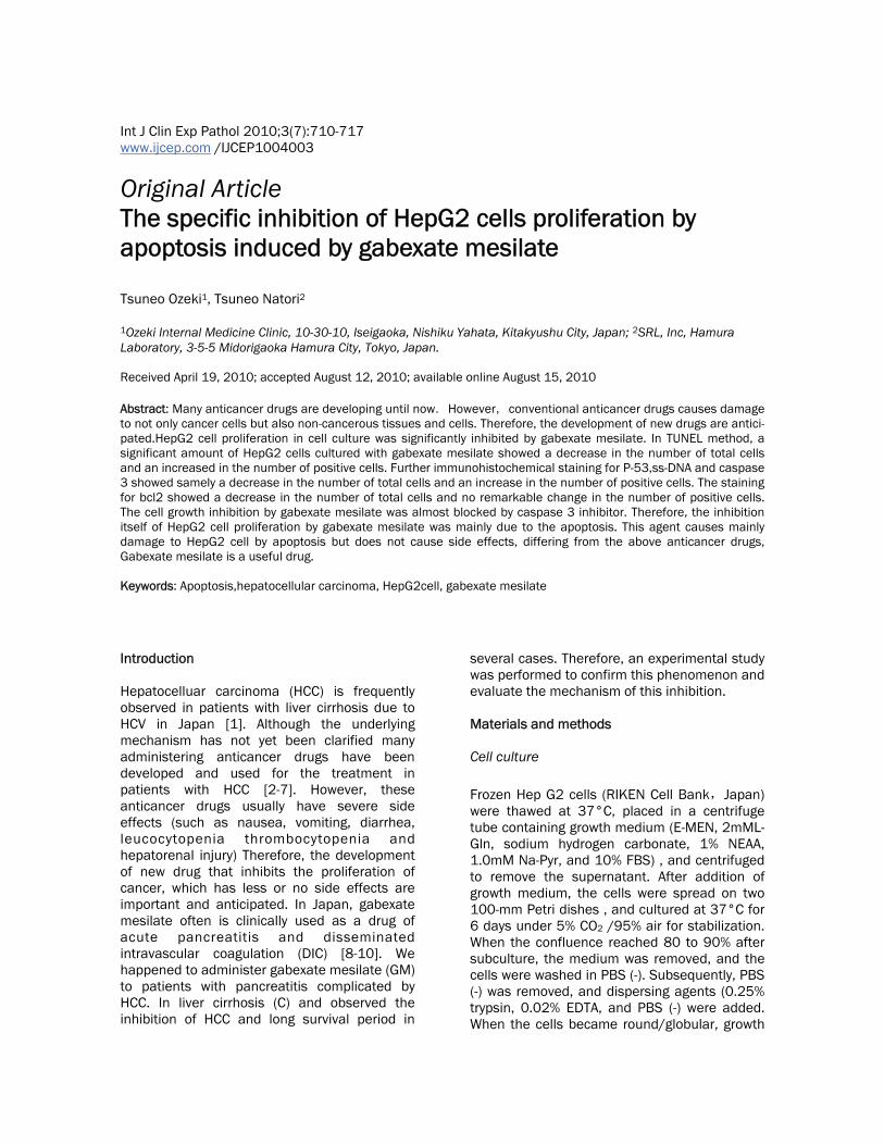
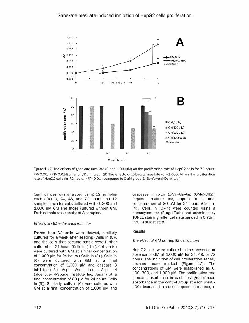
caspases inhibitor (Z-Val-Ala-Asp (OMe)-CH2F, Peptide Institute Inc, Japan) at a final concentration of 80 µM for 24 hours (Cells in (4)). Cells in (0)-(4) were counted using a hemocytometer (Burgel-Turk) and examined by TUNEL staining, after cells suspended in 0.75ml PBS (-) at last step. Results The effect of GM on HepG2 cell culture Hep G2 cells were cultured in the presence or absence of GM at 1,000 µM for 24, 48, or 72 hours. The inhibition of cell proliferation serially became more marked (Figure 1A). The concentrations of GM were established as 0, 100, 300, and 1,000 µM. The proliferation rate ( mean absorbance in each test group/mean absorbance in the control group at each point x 100) decreased in a dose-dependent manner, in
Figure 1. (A) The effects of gabexate mesilate (0 and 1,000μM) on the proliferation rate of HepG2 cells for 72 hours.
*P<0.05, **P<0.01(Bonferroni/Dunn test). (B) The effects of gabexate mesilate (0~1,000μM) on the proliferation rate of HepG2 cells for 72 hours. **P<0.01 : compared to 0 μM group 1 (Bonferroni/Dunn test).
Gabexate mesilate-induced inhibition of HepG2 cells proliferation
713 Int J Clin Exp Pathol 2010;3(7):710-717
the presence of GM at 1,000 µM, cell proliferation was most markedly inhibited (Figure 1B). Agarose electrophoresis When extracting DNA after 72-hour culture with GM 1,000 µM, there was no ladder formation pattern as a biochemical parameter of apoptosis. Most DNA extracts failed to migrate, showing a negligible electrophoresis pattern. No
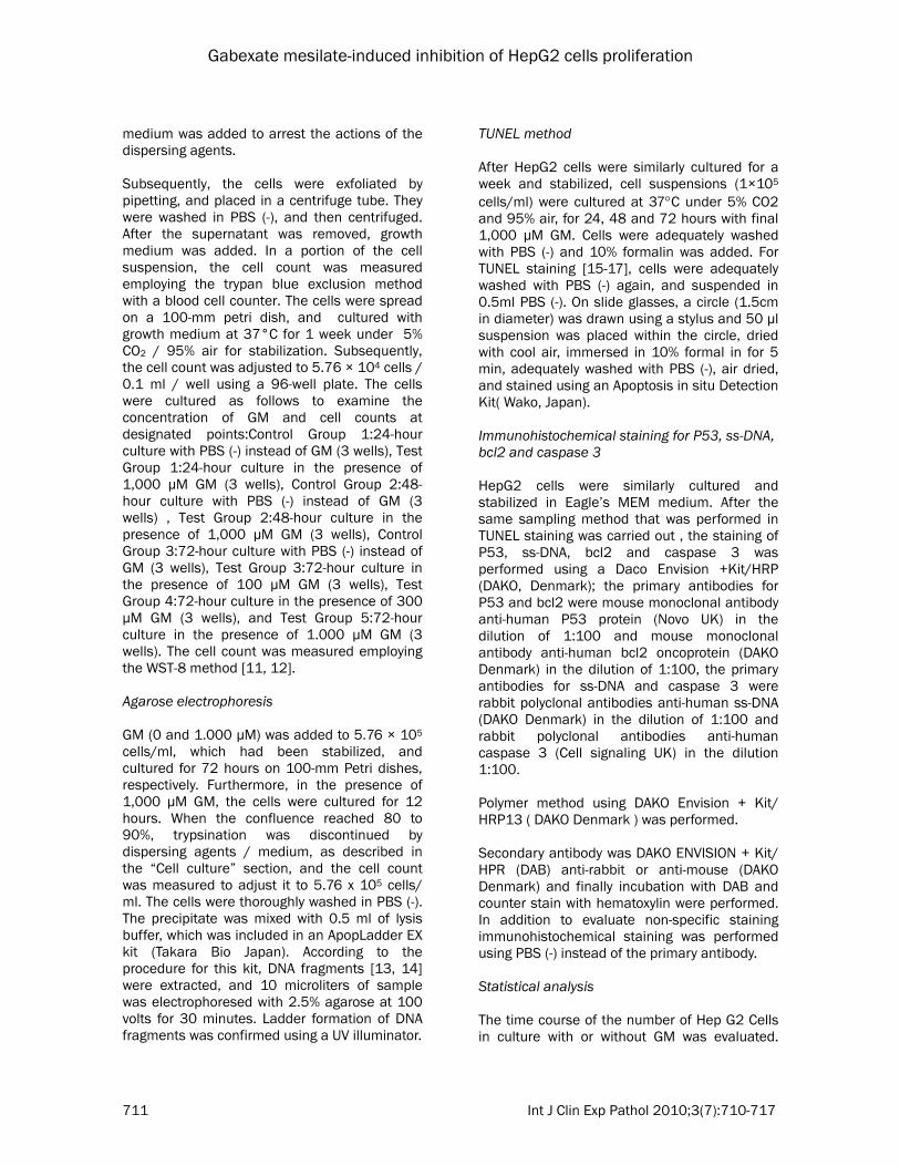
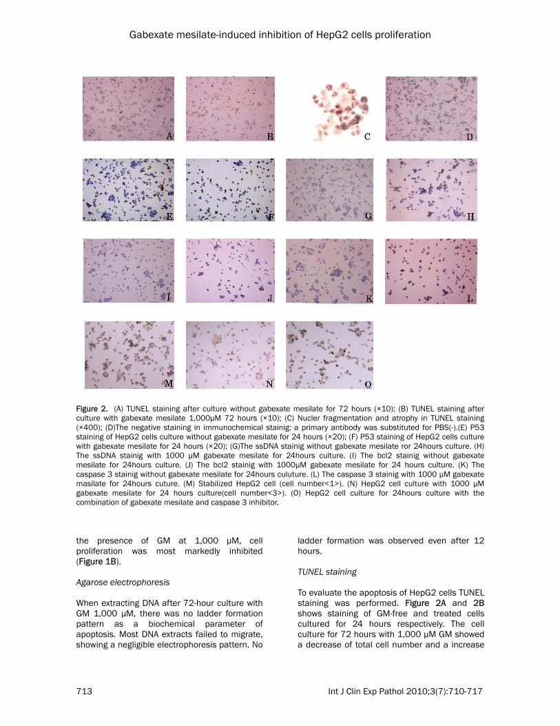
ladder formation was observed even after 12 hours. TUNEL staining To evaluate the apoptosis of HepG2 cells TUNEL staining was performed. Figure 2A and 2B shows staining of GM-free and treated cells cultured for 24 hours respectively. The cell culture for 72 hours with 1,000 µM GM showed a decrease of total cell number and a increase
Figure 2. (A) TUNEL staining after culture without gabexate mesilate for 72 hours (×10); (B) TUNEL staining after culture with gabexate mesilate 1,000μM 72 hours (×10); (C) Nucler fragmentation and atrophy in TUNEL staining (×400); (D)The negative staining in immunochemical stainig: a primary antibody was substituted for PBS(-).(E) P53 staining of HepG2 cells culture without gabexate mesilate for 24 hours (×20); (F) P53 staining of HepG2 cells culture with gabexate mesilate for 24 hours (×20); (G)The ssDNA stainig without gabexate mesilate ror 24hours culture. (H) The ssDNA stainig with 1000 µM gabexate mesilate for 24hours culture. (I) The bcl2 stainig without gabexate mesilate for 24hours culture. (J) The bcl2 stainig with 1000µM gabexate mesilate for 24 hours culture. (K) The caspase 3 stainig without gabexate mesilate for 24hours culuture. (L) The caspase 3 stainig with 1000 µM gabexate masilate for 24hours cuture. (M) Stabilized HepG2 cell (cell number<1>). (N) HepG2 cell culture with 1000 µM gabexate mesilate for 24 hours culture(cell number<3>). (O) HepG2 cell culture for 24hours culture with the combination of gabexate mesilate and caspase 3 inhibitor.
Gabexate mesilate-induced inhibition of HepG2 cells proliferation
714 Int J Clin Exp Pathol 2010;3(7):710-717
of positive cell number. Effects of GM on Hep G2 cell culture As show in Figure 1A, GM at 1,000 µM serially decreased the cell count in comparison with GM-free culture, showing significant differences. Figure 1B presents the influence of the GM concentrations on the 72-hour culture of Hep G2 cells. Cell proliferation was significantly inhibited in a dose-dependent manner (GM: 100, 300, and 1,000 µM). Immunohistochemical staining Figure 2D shows negative staining in which a primary antibody was substituted for PBS(-). There was no nonspecific reaction. Figures 2E and 2F show the P53 staining of GM- free and -treated cells cultured for 24 hours, respectively. The total number of cells decreased and positive cells increased. In the absence of GM, P53-positive cells were scattered. However, GM-treated cells were also strongly positive. The number of positive cells after 48-hour culture was larger than after 24-hour culture. Figures 2G and 2H show ssDNA staining of GM-free and -treated cells cultured for 24 hours, respectively. In the presence of GM, the total number of cells decreased. ssDNA-positive cells increased in total number of cells. After 48-hour culture, the proportion of positive cells in the total number of cells was higher than after 24-hour culture. Figures 2I and 2J show the bcl2 staining of GM-
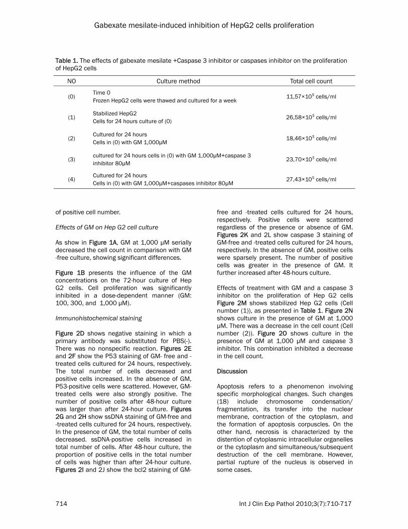
free and -treated cells cultured for 24 hours, respectively. Positive cells were scattered regardless of the presence or absence of GM. Figures 2K and 2L show caspase 3 staining of GM-free and -treated cells cultured for 24 hours, respectively. In the absence of GM, positive cells were sparsely present. The number of positive cells was greater in the presence of GM. It further increased after 48-hours culture. Effects of treatment with GM and a caspase 3 inhibitor on the proliferation of Hep G2 cells Figure 2M shows stabilized Hep G2 cells (Cell number (1)), as presented in Table 1. Figure 2N shows culture in the presence of GM at 1,000 µM. There was a decrease in the cell count (Cell number (2)). Figure 2O shows culture in the presence of GM at 1,000 µM and caspase 3 inhibitor. This combination inhibited a decrease in the cell count. Discussion Apoptosis refers to a phenomenon involving specific morphological changes. Such changes (18) include chromosome condensation/fragmentation, its transfer into the nuclear membrane, contraction of the cytoplasm, and the formation of apoptosis corpuscles. On the other hand, necrosis is characterized by the distention of cytoplasmic intracellular organelles or the cytoplasm and simultaneous/subsequent destruction of the cell membrane. However, partial rupture of the nucleus is observed in some cases.
Table 1. The effects of gabexate mesilate +Caspase 3 inhibitor or caspases inhibitor on the proliferation of HepG2 cells
NO Culture method Total cell count
(0) Time 0 Frozen HepG2 cells were thawed and cultured for a week
11,57×10⁵ cells/ml
(1) Stabilized HepG2 Cells for 24 hours culture of (0)
26,58×10⁵ cells/ml
(2) Cultured for 24 hours Cells in (0) with GM 1,000μM
18,46×10⁵ cells/ml
(3) cultured for 24 hours cells in (0) with GM 1,000μM+caspase 3 inhibitor 80μM
23,70×10⁵ cells/ml
(4) Cultured for 24 hours Cells in (0) with GM 1,000μM+caspases inhibitor 80μM
27,43×10⁵ cells/ml
Gabexate mesilate-induced inhibition of HepG2 cells proliferation
715 Int J Clin Exp Pathol 2010;3(7):710-717
In this case, even cells with necrosis show a positive reaction on TUNEL staining. Therefore, a diagnosis of apoptosis cannot be made based on a positive TUNEL staining reaction alone. However, in this study, Hoechst 33324 and orange acrylic staining of viable floating cells was conducted. There were a large number of nuclei with condensation, morphologically suggesting apoptosis [15-16]. On the other hand, DNA was extracted from apoptotic cells for biochemical analysis, in which the pattern of ladder formation is detected on agarose electrophoresis [17]. In this study, agarose electrophoresis revealed negligible “DNA extract “migration, and there was no ladder formation. However, usually, ladder formation is frequently achieved with floating cells such as leukemia cells, whereas there is often no ladder formation with adhesive cells. There is also no ladder formation on extraction in the early phase of apoptosis, this is not the most appropriate method to verify apoptosis. In this experiment, similar results were also obtained 12 hours after apoptosis induction. Currently, the usefulness of various staining procedures, agarose electrophoresis, and cytoflowmetry for the evaluation of cell apoptosis is controversial. Apoptosis is characterized by Caspase 3 activation-associated chromosome condensation, which is considered to be a specific change. To verify that a substance causes the apoptosis of cultured cells, it must be confirmed that there is a decrease in the cell count after the addition of the substance on cell culture. On the other hand, when cell culture in the presence of a substance causing apoptosis and a caspase 3 inhibitor inhibits a decrease in the cell count, the cells are regarded as showing apoptosis. This evaluation method may be the most accurate. It is hypothesized that there are two types of cell apoptosis (Types I and II). In type I cells, the death-inducing signaling complex (DISC) is formed, and caspase 8, which is activated in DISC, is released from DISC, effector caspase 3 as a performance factor and causing apoptosis. In type II cells, caspase 8 is involved in the fragmentation of a BH3 protein, Bid, acting on mitochondria, releasing cytochrome C, and finally causing apoptosis via the activation of caspase 3. Type I cells consist
of lymphocytes, and type II cells consist of hepatocytes. Hep G2 cells are hepatocytes, and may be classified as type II. The purpose of this study was to verify that cell death is apoptosis, not to investigate the activation route of apoptosis. However, we speculate the following route: P53 activation → BH3 activation → release of cytochrome c from mitochondria → Apaf-1 activation → caspase 3 activation [19,20]. P53 has apoptosis induction, stopping cell cycle and transform inhibition and acts as cancer-suppressing function. In this study, the death receptors on the cell surface membrane are not evaluated. However, P53activation is enhanced suggesting a rout associated with caspase 3. With respect to ssDNA, a change from double to single strands resulted in a decrease in the cell count and increase in the number of positive cells. The change in apoptosis was thought Concerning bcl2 families: apoptosis-promoting and-suppressing families. For bcl2 ICS, monoclonal staining is performed to detect bcl2.
Caspase 3 staining showed a marked decrease in the cell count and increase in the number of positive cells, suggesting apoptosis. bcl2 plays an important role in the inhibition of apoptosis. However, when apoptosis-promoting protein is more predominant than apoptosis-inhibiting protein in the apoptosis route, apoptosis may be induced. Many anticancer drugs, such as alkylating agents, metabolic antagonists, alkaloid antibiotics, topoisomerase inhibitors, hormone preparations (including analogues), and platinum preparations, have been developed. Among these, Adriacin and Farmorubicin induce apoptosis. However, these, conventional anticancer drugs cause apoptosis/cell necrosis, affecting not only cancer but also non-cancerous cells. This is the most important fault of anticancer drugs. On the other hand, GM may primarily affect cancer cells (hepatoma) vea apoptosis, but not non-cancerous cells. Usually, anticancer drugs exhibit antagonistic actions on cellular metabolism and cytotoxic effects; therefore, they cause both the necrosis of cancer cells and apoptosis, As anticancer drugs that induce apoptosis increase bcl2 activity, apoptosis is weakened. In this study, it was impossible to culture normal hepatocytes; therefore, no experiment involving normal hepatocyte culture was carried out.
Gabexate mesilate-induced inhibition of HepG2 cells proliferation
716 Int J Clin Exp Pathol 2010;3(7):710-717
It is unlikely that GM enhances and promotes apoptosis in normal cells. The reason is that previous clinical studies and treatment on GM administration at a high dose to patients with pancreatitis have shown no abnormalities in the liver. Therefore, GM may be involved only in the apoptosis of HCC. Martin-Remedo [22] el al reported that melatonin inhibits Hep G2 cell prostration by inducing apoptosis. However, the administration of a large quantity of the hormone may give some distortions physiologically, we happened to be able to administer intravenously GM to patients with pancreatitis and advanced multiple livers carcinoma without liver injury due to GM and the patients had long survival periods. After administration, AFP showed the lowering, and tumor size in picture was shortening or fixed, GM may be used in tran-scatheter arterial embolization. The administration method of GM Should be improved. Acknowledgments This author would like to thank Dr. Takao Ma-tsumata, a famous hepatologist and the presi-dent of Saiseikai Hospital in Kitakyushu City, Japan for his support to this study and critical reading of this manuscript. Please address correspondence to: Tsuneo Ozeki, Ozeki Internal Medicine Clinic, 10-30-10, Iseigaoka, Nishiku Yahata, Kitakyushu City, Japan. Tel: 093 693 0712, Fax: 093 693 0712, E-mail: [email protected] References [1] Ikeda.K, Saito.S, Koida.I, Arase.Y, Tsubota.A,
Chagama.K, Kumada.H, Kawanishi.M . A multi-variate analysis of risk factors for hepatocellular carcinogeneres : a prospective observation of 795 patients with viral and aleoholic cirrhosis. Hepatology. 1993 : 18:47-53.
[2] Bruix.J, Liovet JM, Castells, Montana.X, Bru.C, A y u s o M D C , V l l a n a . R , R o d s . J Transarterial cmbolization versus gymptomatic treatment in patients with advanced hepatocel-lular carcinoma: result of a yandomiged con-trolled trial in a single institusion. Hepatology 1995;27:1578-1583.
[3] Matsui.O, Kadoya.M, Yoshikawa.J, Gobata.T, Arai.K, Demachi.H, Miyayama.S, Takashima.T, Unoura.M, Kogayashi.K. Small hepatocellular carcinoma: Treatment with subsegmental tran-scatheter Asterial embolization. Radiology 1998;
188:79-83. [4] Takahashi.Y, Nishioka.K. Survival without tumor
shrimkage : Re-evaluation of Survival gain by cytostatic effect of chemotherapy.I Natl Cancer Institute. 1995: 262-1263.
[5] Ando.E, Tanaka.M, Yamashita.F, Kuromatsu.R, Yutani.S, Fukumori.K, Sumie.S, Yano.Y Okuda.K, Sata.M. Hepatic arterial infusion chemotherapy for advanced hepatocellular carcinoma with portal vein tumor thrombosis : analysis of 48 cases. Cancer.2002 :95 :588-595.
[6] Ishikawa.T, Ichida.T, Sugitani.S, Tsuboi.Y, Genda.T, Sugahara.S Uehara.K, Inayoshi.J, Yoko-yama.J Ishimoto.Y, Askakura.H, Improved sur-vival with oral administration of enteric –coated tagefur / uracil for advanced stage IV-A hepato-cellular carcinoma, J Gastroenterol Hepatol 2001:16 :452-459.
[7] Ikeda.M, Maeda.S, Shibata.J, Muta.R, Ashi-hara.H, Tanaka.M, Fujiyama.S. Tomita.K. Tran-scatheter arterial chemotherapy with and with-out embolization in patients with hepatocellular carcinoma. Omecogy .2004:65:24-31.
[8] Murakawa M, Okamura T, Shibuya T, Harada M, Otsuka T and Niho Y. Use of a synthetic protease inhibitor for the treatment of L-asparaginase-induced acute pancreatitis complicated by dis-seminated intravascular coagulation. Ann Hema-tol 1992; 64: 249-252.
[9] Nkano S, Mugikura M, Endoh M, Ogami Y and Otsuki M. Acute pancreatitis with diabetic keto-acidosis associated with hypermyoglobinemia, acute renal failure, and DIC. J Gastroenterol 1996; 31: 623-626.
[10] Sakurai M, Abe H, Okamura N, Inoue Y, Akiyoshi T, Matsuyama K, Uchida T and Otsuka M. Stabil-ity of gabexate mesilate products: Influence of the addition of mannitol. Biomed Mater Eng 2010; 20: 13-20.
[11] Kusunoki N, Ito T, Sakurai N, Suguro T, Handa H and Kawai S. A novel celecoxib derivative potently induces apoptosis of human synovial fibroblasts. J Pharmacol Exp Ther 2005; 314: 796-803.
[12] Oie S, Ono M, Fukushima H, Hosoi F, Yano H, Maruyama Y, Kojiro M, Terada T, Hirano K, Ku-wano M and Yamada Y. Alteration of dihydro-pyrimidine dehydrogenase expression by IFN-alpha affects the antiproliferative effects of 5-fluororuracil in human hepatocellular carcinoma cells. Mol Cancer Ther 2007; 6: 2310-2318.
[13] Oberhammer F, Wilson JW, Dive C, Morris ID, Hickman JA, Wakeling AE, Walker PR and Sikor-ska M. Apoptotic death in epithelial cells: cleav-age of DNA to 300 and/ or 50 kb fragments prior to or in the absence of internucleosomal fragmentation. Embo J 1993; 12: 3679-3684.
[14] Wei D, Gong W, Kanai M, Schlunk C, Wang L, Yao JC, Wu TT, Huang S and Xie K. Drastic down-regulation of Kruppel-like factor 4 expression is critical in human gastric cancer development and progression. Cancer Res 2005; 65: 2746-
Gabexate mesilate-induced inhibition of HepG2 cells proliferation
717 Int J Clin Exp Pathol 2010;3(7):710-717
2754. [15] Funaki N, Sasano H, Shizawa S, Nio M, Iwami D,
Ohi R and Nagura H. Apoptosis and cell prolifera-tion in biliary atresia. J Pathol 1998; 186: 429-433.
[16] Wijsman JH, Jonker RR, Keijzer R, van de Velde CJ, Cornelisse CJ and van Dierendonck JH. A new method to detect apoptosis in paraffin sections: in situ end-labeling of fragmented DNA. J Histo-chem Cytochem 1993; 41: 7-12.
[17] Gavrieli Y, Sherman Y and Ben-Sasson SA. Iden-tification of programmed cell death in situ via specific labeling of nuclear DNA fragmentation. J Cell Biol 1992; 119: 493-501.
[18] Kerr JF, Wyllie AH and Currie AR. Apoptosis: a basic biological phenomenon with wide-rangin implications in tissue kinetics. Br J Cancer 1972; 26: 239-257.
[19] Lakhani SA, Masud A, Kuida K, Porter GA, Jr., Booth CJ, Mehal WZ, Inayat I and Flavell RA. Caspases 3 and 7: key mediators of mitochon-drial events of apoptosis. Science 2006; 311:
847-851. [20] Mihara M, Erster S, Zaika A, Petrenko O,
Chittenden T, Pancoska P and Moll um. P53 has a direct apoptogenic role at the mitochondria. Mol Cell 2003; 11: 577-590.
[21] Tsujimoto Y. Cell death regulation by the Bcl-2 protein family in the mitochondria. J Cell Physiol 2003; 195: 158-167.
[22] Martin-Renedo J, Mauriz JL, Jorquera F, Ruiz-Andres O, Gonzalez P and Gonzalez-Gallego J. Melatonin induces cell cycle arrest and apop-tosis in hepatocarcinoma HepG2 cell line. J Pin-eal Res 2008; 45: 532-540.