Embed Size (px)
Citation preview

378
SUMMARYIntroduction/Objective Maxillary or mandibular retrognathism are common dentofacial deformities treated by combined orthodontic-surgical treatment. Surgical maxillary or mandibular advancement changes the position and strain of surrounding structures, which may also affect pharyngeal airway dimensions.The aim of this study was to evaluate and compare three-dimensional pharyngeal airway space changes in patients treated with maxillary advancement and those treated with mandibular advancement. Methods The sample consisted of 25 patients – 12 treated with maxillary advancement and 13 with mandibular advancement surgery. Nasopharyngeal (NP) volume, oropharyngeal (OP) volume, and the area of maximum constriction (AMC) in the OP were measured on cone beam computed tomography scans (2 mA / 120 kV / 12’’ FOV) taken before and at least three months after surgery. Paired samples t-test was used for analyzing statistical significance of changes (p ≤ 0.05). Results Postoperative OP and NP volumes, as well as the AMC, increased insignificantly in both groups. Conclusion Results suggest that mono-maxillary surgical advancement of the maxilla or the mandible increases pharyngeal airway dimensions. Keywords: Cone Beam CT; mono-maxillary advancement surgery; pharyngeal airways
ORIGINAL ARTICLE / ОРИГИНАЛНИ РАД
Pharyngeal airway changes after mono-maxillary advancement surgeryNeda Lj. Stefanović1, Marija Živković-Sandić1, Juan Martin Palomo2
1University of Belgrade, School of Dental Medicine, Department of Orthodontics, Belgrade, Serbia;2Case Western Reserve University, School of Dental Medicine, Department of Orthodontics and Craniofacial Imaging Center, Cleveland, Ohio, USA
DOI: https://doi.org/10.2298/SARH180109049S
UDC: 616.314.1/.2-089.168:616.32
Received • Примљено: January 9, 2018
Revised • Ревизија: June 14, 2018
Accepted • Прихваћено: June 18, 2018
Online first: August 7, 2018
Correspondence to:Neda STEFANOVIĆResavska 8811000 [email protected]
INTRODUCTION
Dentofacial deformities are handicapping devi-ations that compromise patients’ facial features, masticatory function, and pharyngeal airway space (PAS). Since orthodontic treatment alone rarely yields satisfactory results in these pa-tients, orthognathic surgery for repositioning the jaws is usually recommended. Surgical cor-rection changes the position and strain of sur-rounding structures, therefore improving facial esthetics and occlusion. Moreover, surgery may also affect the dimensions of the oral and nasal cavities, as well as the PAS dimensions, hence improving or impairing breathing [1–5].
Class II and Class III are common den-tofacial deformities treated by combined orthodontic-surgical treatment. Class II de-formity caused by mandibular retrognathism is treated with a combination of orthodontics and mandibular advancement, most commonly achieved by bilateral sagittal split osteotomy [6]. Class III deformity caused by maxillary retrognathism is treated with a combination of orthodontics and maxillary advancement, which makes up about one half of Class III skeletal deformity treatments [7].
The aim of this study was to analyze and compare three-dimensional (3D) pharyngeal airway changes in orthodontic-surgical pa-tients treated with maxillary advancement and in those treated with mandibular advancement.
METHODS
The sample of this retrospective study consisted of 25 non-growing subjects who underwent combined orthodontic-surgical treatment at the Case Western Reserve University in Cleveland, OH, USA. According to the type of surgery, the sample was divided into two groups. Group A consisted of 12 patients treated with maxillary advancement, and group B consisted of 13 pa-tients treated with mandibular advancement. The groups were matched for age and sex.
Patients from both groups were treated with standard edgewise appliances and had cone beam computed tomography (CBCT) scans taken before (T1) and at least three months after surgery (T2) using a custom Hitachi CB MercuRay scanner (Hitachi Medical Systems America Inc., Twinsburg, OH, USA). The CB MercuRay scanner used had custom settings in order to provide the lowest radiation exposure possible while maintaining acceptable diagnos-tic image quality [8, 9]. This modification was made in order to fully comply with the ALARA (as low as reasonably achievable) standards. All images were taken at 2 mA, 120 kV, and a 12-inch field of view (F Mode) setting. Each pa-tient’s image data consisted of 512 slices, with an isometric voxel size of 0.377 mm, a resolu-tion of 1024 × 1024 pixels and 12 bits per pixel (4,096 greyscale). The images were taken in the sitting position with the patient’s head in the

379
Srp Arh Celok Lek. 2018 Jul-Aug;146(7-8):378-383 www.srpskiarhiv.rs
natural head posture, teeth at maximum intercuspation and at the end of the exhalation period when the patient was not swallowing. Scanning time was 9.6 seconds.
InVivo Dental Software (Anatomage Inc., San Jose, CA, USA) was used to analyze DICOM (Digital Imaging and Communication in Medicine) images. The images were first oriented using the Patient Orientation tool in the Section View according to the axial, sagittal, and coronal slices. The midsagittal plane was determined according to the foramen incisivum on the axial slice (Figure 1a); the palatal plane was adjusted to coincide with the True Hori-zontal Plane on the sagittal slice (Figure 1b); infraorbitale points were aligned on the coronal slice (Figure 1c). The images were further processed in the Volume Render View section, by putting them in greyscale view, setting Recon-struction to Maximum Intensity and moving them upward or downward with the Patient Orientation tool in order to overlap the palatal plane with the central horizontal line of the grid. Airway volumes were calculated in the Vol-ume Render View. The images were kept in greyscale view and reoriented to Top View. Reconstruction was set back to Volume Rendering and images were inversed, opacity reduced until the internal structures became visible and unnecessary parts were removed with the Sculpting Tool. Opacity was increased and brightness and contrast were reset after isolating the desired airway in order to obtain a solid airway before calculating the volume.
Nasal passages
For the calculation of the nasal passage (NP) airway vol-ume, inferior border of the NP was determined by the horizontal line through the palatal plane (Figure 2a), and the superior border was determined by moving the axial reference plane on the sagittal slice until noting on the axial slice that it has reached the point where the nasal septum first touches the posterior wall of the pharynx (Figure 2b). The superior-to-inferior border distance was measured with the Distance Measuring Tool on the sagittal slice in the Section View. The 3D Volume Clipping Tool was used for cutting the airway along the axial plane in the Volume Render View. Scrolling the mouse wheel moved the clip-ping plane where needed until it coincided with the inferior NP border. The distance between the superior and inferior border transferred from the Section View was marked us-ing the Distance Measuring Tool and the part above the
superior NP border was removed with the Clipping Tool. After reorientation to Top View, maxillary sinuses were clipped from the final NP volume. The remaining borders were determined by the software since we used automatic segmentation for measuring all volumes, i.e. the Volumet-ric Measuring Tool, which calculates and displays volume measurements in cubic millimeters and cubic centimeters.
Oropharyngeal airways
For the calculation of the oropharyngeal passage (OP) airway volume, inferior border was determined by the horizontal line through the most of the antero-inferior point of the second cervical vertebrae (Figure 2c), and the horizontal line through the palatal plane was used as the superior border (Figure 2a). The NP airway volume view was flipped to the opposite side, making the palatal plane the superior border. The distance between the superior and inferior border transferred from the Section View was marked using the Distance Measuring Tool and the part below the inferior border was cut using the Sculpting Tool. The Volumetric Measuring Tool was used for obtaining the OP volume value.
Area of maximum constriction in the oropharyngeal airways
The point of maximum constriction in the pharynx was de-termined on the sagittal slice by moving the axial reference
Figure 1. Image orientation on the axial, sagittal, and coronal slice
Figure 2. Pharyngeal airway borders
Pharyngeal airway changes after mono-maxillary advancement surgery

380
Srp Arh Celok Lek. 2018 Jul-Aug;146(7-8):378-383
DOI: https://doi.org/10.2298/SARH180109049S
plane on the corresponding axial slice. The area of maxi-mum pharyngeal constriction was measured on the axial slices using the Area Measuring Tool.
Cephalometric analysis
Cephalograms were generated from the DICOM files and analyzed using the Dolphin Imaging software version 11 (Dolphin Imaging, Chatsworth, CA, USA). SNA, SNB, and ANB angles and A-Nperp and B-Nperp linear measure-ments were used for determining sagittal jaw positions and relationships.
This methodology has previously been proven success-ful [10, 11, 12]. All measuring was done and re-tested by an experienced operator (NLjS) trained by an expert in the field (JMP).
Ethics
The images used were pre-existing, taken as a part of the standard diagnostic procedure. All the patients signed the informed consent form allowing the use of their records for research and publication purposes. The Human Re-search Ethics Committee of the School of Dental Medicine of the University of Belgrade approved this research (reso-lution number 36/20 from December 14, 2009).
Statistical analysis
The obtained data was organized and descriptive statis-tics [means, standard deviations and ranges for pretreat-ment (T1) and post-treatment (T2) records] was done using Microsoft Office Excel 2010 (Microsoft Corpora-
tion, Redmond, WA, USA). Detailed statistical analysis was performed in the SPSS software Version 12 (SPSS Inc., Chicago, IL, USA). The intraclass correlation coef-ficient was used for determining intra-operator reliability for each measurement. Since the Kolmogorov–Smirnov test revealed the normality of distribution for all data, parametric tests were employed. Statistical significance of changes between T1 and T2 was analyzed with the paired-samples t-test. The level of significance was set at p < 0.05.
RESULTS
The intraclass correlation coefficient values revealed high reproducibility and reliability of all parameter measure-ments (r > 0.95).
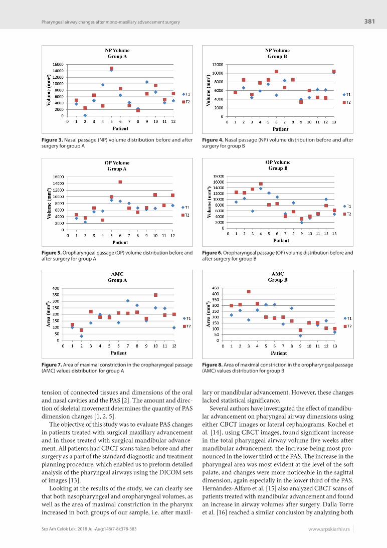
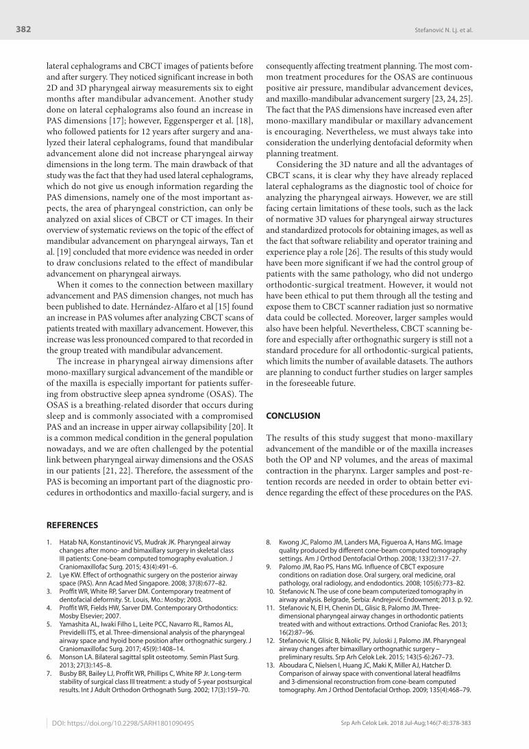
Cephalometric measurements and mean ages at T1 and T2 are presented in Table 1 for both groups. Pharyngeal airway measurements are shown in Table 2. Postoperative volumes of the OP and NP, as well as the AMC, increased in both groups without statistical significance (Table 2).
NP volume distribution before and after surgery is pre-sented in Figure 3 for group A and in Figure 4 for group B. OP volume distribution before and after surgery in pre-sented in Figure 5 for group A and in Figure 6 for group B. AMC values distribution is shown in Figure 7 for group A and in Figure 8 for group B.
DISCUSSION
Orthognathic surgery changes the position and strain of the surrounding structures, which affects the location and
Table 1. Average age and sagittal parameters for groups A and B
Age (years) SNA SNB ANB
T1 T1 T2 T1 T2 T1 T2
Mean ± SD Mean ± SD Mean ± SD Mean ± SD
Group An = 12 20.93 ± 9.87 77.98 ± 5.93 82.17 ± 6.34 80.32 ± 5.19 80.13 ± 5.40 -2.14 ± 2.14 2.02 ± 1.93
Group Bn = 13 22.48 ± 10.37 82.81 ± 3.57 82.75 ± 3.54 75.91 ± 3.09 79.02 ± 3.37 6.9 ± 2.84 3.81 ± 2.89
Table 2. Descriptive statistics and comparison of pharyngeal airway measurements at T1 and T2 for groups A and B
T1 T2
P valueΔ
Minimum Maximum Mean SD Minimum Maximum Mean SD T2 – T1
Group A n = 12
NP volume (mm³) 186 14,302 6,033.58 3,908.87 1,715 1,4834 6,145.00 3,644.96 .889 111.42 ± 2,709.43
OP volume (mm³) 2,392 8,993 6,399.08 1,957.25 3,005 1,4491 7,473.67 3,351.08 .149 1,074.58 ± 2,400.11
AMC (mm²) 32.56 304.5 175.81 81.44 79.09 348.15 193.83 64.01 .364 18.02 ± 65.91
Group Bn = 13
NP volume (mm³) 3,993 10,154 6,371.77 1,838.37 3,355 10,458 6,890.46 2,314.34 0.364 581.69 ± 1,983.99
OP volume (mm³) 1,965 13,742 7,762.92 3,655.67 3,240 15,358 8,214.46 4,012.35 0.608 451.54 ± 3,088.16
AMC (mm²) 41.77 310.02 194.31 86.29 90.09 419.41 208.62 99.40 0.609 14.31 ± 98.23
NP – nasal passage; OP – oropharyngeal passage; AMC – area of maximal constriction in the OP; *p < 0.05; **p < 0.01; ***p < 0.001
Stefanović N. Lj. et al.
381
Srp Arh Celok Lek. 2018 Jul-Aug;146(7-8):378-383 www.srpskiarhiv.rs
tension of connected tissues and dimensions of the oral and nasal cavities and the PAS [2]. The amount and direc-tion of skeletal movement determines the quantity of PAS dimension changes [1, 2, 5].
The objective of this study was to evaluate PAS changes in patients treated with surgical maxillary advancement and in those treated with surgical mandibular advance-ment. All patients had CBCT scans taken before and after surgery as a part of the standard diagnostic and treatment planning procedure, which enabled us to preform detailed analysis of the pharyngeal airways using the DICOM sets of images [13].
Looking at the results of the study, we can clearly see that both nasopharyngeal and oropharyngeal volumes, as well as the area of maximal constriction in the pharynx increased in both groups of our sample, i.e. after maxil-
lary or mandibular advancement. However, these changes lacked statistical significance.
Several authors have investigated the effect of mandibu-lar advancement on pharyngeal airway dimensions using either CBCT images or lateral cephalograms. Kochel et al. [14], using CBCT images, found significant increase in the total pharyngeal airway volume five weeks after mandibular advancement, the increase being most pro-nounced in the lower third of the PAS. The increase in the pharyngeal area was most evident at the level of the soft palate, and changes were more noticeable in the sagittal dimension, again especially in the lower third of the PAS. Hernández-Alfaro et al. [15] also analyzed CBCT scans of patients treated with mandibular advancement and found an increase in airway volumes after surgery. Dalla Torre et al. [16] reached a similar conclusion by analyzing both
Figure 3. Nasal passage (NP) volume distribution before and after surgery for group A
Figure 4. Nasal passage (NP) volume distribution before and after surgery for group B
Figure 5. Oropharyngeal passage (OP) volume distribution before and after surgery for group A
Figure 6. Oropharyngeal passage (OP) volume distribution before and after surgery for group B
Figure 7. Area of maximal constriction in the oropharyngeal passage (AMC) values distribution for group A
Figure 8. Area of maximal constriction in the oropharyngeal passage (AMC) values distribution for group B
Pharyngeal airway changes after mono-maxillary advancement surgery
382
Srp Arh Celok Lek. 2018 Jul-Aug;146(7-8):378-383
lateral cephalograms and CBCT images of patients before and after surgery. They noticed significant increase in both 2D and 3D pharyngeal airway measurements six to eight months after mandibular advancement. Another study done on lateral cephalograms also found an increase in PAS dimensions [17]; however, Eggensperger et al. [18], who followed patients for 12 years after surgery and ana-lyzed their lateral cephalograms, found that mandibular advancement alone did not increase pharyngeal airway dimensions in the long term. The main drawback of that study was the fact that they had used lateral cephalograms, which do not give us enough information regarding the PAS dimensions, namely one of the most important as-pects, the area of pharyngeal constriction, can only be analyzed on axial slices of CBCT or CT images. In their overview of systematic reviews on the topic of the effect of mandibular advancement on pharyngeal airways, Tan et al. [19] concluded that more evidence was needed in order to draw conclusions related to the effect of mandibular advancement on pharyngeal airways.
When it comes to the connection between maxillary advancement and PAS dimension changes, not much has been published to date. Hernández-Alfaro et al [15] found an increase in PAS volumes after analyzing CBCT scans of patients treated with maxillary advancement. However, this increase was less pronounced compared to that recorded in the group treated with mandibular advancement.
The increase in pharyngeal airway dimensions after mono-maxillary surgical advancement of the mandible or of the maxilla is especially important for patients suffer-ing from obstructive sleep apnea syndrome (OSAS). The OSAS is a breathing-related disorder that occurs during sleep and is commonly associated with a compromised PAS and an increase in upper airway collapsibility [20]. It is a common medical condition in the general population nowadays, and we are often challenged by the potential link between pharyngeal airway dimensions and the OSAS in our patients [21, 22]. Therefore, the assessment of the PAS is becoming an important part of the diagnostic pro-cedures in orthodontics and maxillo-facial surgery, and is
consequently affecting treatment planning. The most com-mon treatment procedures for the OSAS are continuous positive air pressure, mandibular advancement devices, and maxillo-mandibular advancement surgery [23, 24, 25]. The fact that the PAS dimensions have increased even after mono-maxillary mandibular or maxillary advancement is encouraging. Nevertheless, we must always take into consideration the underlying dentofacial deformity when planning treatment.
Considering the 3D nature and all the advantages of CBCT scans, it is clear why they have already replaced lateral cephalograms as the diagnostic tool of choice for analyzing the pharyngeal airways. However, we are still facing certain limitations of these tools, such as the lack of normative 3D values for pharyngeal airway structures and standardized protocols for obtaining images, as well as the fact that software reliability and operator training and experience play a role [26]. The results of this study would have been more significant if we had the control group of patients with the same pathology, who did not undergo orthodontic-surgical treatment. However, it would not have been ethical to put them through all the testing and expose them to CBCT scanner radiation just so normative data could be collected. Moreover, larger samples would also have been helpful. Nevertheless, CBCT scanning be-fore and especially after orthognathic surgery is still not a standard procedure for all orthodontic-surgical patients, which limits the number of available datasets. The authors are planning to conduct further studies on larger samples in the foreseeable future.
CONCLUSION
The results of this study suggest that mono-maxillary advancement of the mandible or of the maxilla increases both the OP and NP volumes, and the areas of maximal contraction in the pharynx. Larger samples and post-re-tention records are needed in order to obtain better evi-dence regarding the effect of these procedures on the PAS.
REFERENCES
1. Hatab NA, Konstantinović VS, Mudrak JK. Pharyngeal airway changes after mono- and bimaxillary surgery in skeletal class III patients: Cone-beam computed tomography evaluation. J Craniomaxillofac Surg. 2015; 43(4):491–6.
2. Lye KW. Effect of orthognathic surgery on the posterior airway space (PAS). Ann Acad Med Singapore. 2008; 37(8):677–82.
3. Proffit WR, White RP, Sarver DM. Contemporary treatment of dentofacial deformity. St. Louis, Mo.: Mosby; 2003.
4. Proffit WR, Fields HW, Sarver DM. Contemporary Orthodontics: Mosby Elsevier; 2007.
5. Yamashita AL, Iwaki Filho L, Leite PCC, Navarro RL, Ramos AL, Previdelli ITS, et al. Three-dimensional analysis of the pharyngeal airway space and hyoid bone position after orthognathic surgery. J Craniomaxillofac Surg. 2017; 45(9):1408–14.
6. Monson LA. Bilateral sagittal split osteotomy. Semin Plast Surg. 2013; 27(3):145–8.
7. Busby BR, Bailey LJ, Proffit WR, Phillips C, White RP Jr. Long-term stability of surgical class III treatment: a study of 5-year postsurgical results. Int J Adult Orthodon Orthognath Surg. 2002; 17(3):159–70.
8. Kwong JC, Palomo JM, Landers MA, Figueroa A, Hans MG. Image quality produced by different cone-beam computed tomography settings. Am J Orthod Dentofacial Orthop. 2008; 133(2):317–27.
9. Palomo JM, Rao PS, Hans MG. Influence of CBCT exposure conditions on radiation dose. Oral surgery, oral medicine, oral pathology, oral radiology, and endodontics. 2008; 105(6):773–82.
10. Stefanovic N. The use of cone beam computerized tomography in airway analysis. Belgrade, Serbia: Andrejević Endowment; 2013. p. 92.
11. Stefanovic N, El H, Chenin DL, Glisic B, Palomo JM. Three-dimensional pharyngeal airway changes in orthodontic patients treated with and without extractions. Orthod Craniofac Res. 2013; 16(2):87–96.
12. Stefanovic N, Glisic B, Nikolic PV, Juloski J, Palomo JM. Pharyngeal airway changes after bimaxillary orthognathic surgery – preliminary results. Srp Arh Celok Lek. 2015; 143(5-6):267–73.
13. Aboudara C, Nielsen I, Huang JC, Maki K, Miller AJ, Hatcher D. Comparison of airway space with conventional lateral headfilms and 3-dimensional reconstruction from cone-beam computed tomography. Am J Orthod Dentofacial Orthop. 2009; 135(4):468–79.
Stefanović N. Lj. et al.
DOI: https://doi.org/10.2298/SARH180109049S

383
Srp Arh Celok Lek. 2018 Jul-Aug;146(7-8):378-383 www.srpskiarhiv.rs
14. Kochel J, Meyer-Marcotty P, Sickel F, Lindorf H, Stellzig-Eisenhauer A. Short-term pharyngeal airway changes after mandibular advancement surgery in adult Class II-Patients – a three-dimensional retrospective study. J Orofac Orthop. 2013; 74(2):137–52.
15. Hernández-Alfaro F, Guijarro-Martínez R, Mareque-Bueno J. Effect of mono- and bimaxillary advancement on pharyngeal airway volume: cone-beam computed tomography evaluation. J Oral Maxillofac Surg. 2011; 69(11):e395–400.
16. Dalla Torre D, Burtscher D, Widmann G, Rasse M, Puelacher T, Puelacher W. Long-term influence of mandibular advancement on the volume of the posterior airway in skeletal Class II-patients: a retrospective analysis. Br J Oral Maxillofac Surg. 2017; 55(8):780–6.
17. Achilleos S, Krogstad O, Lyberg T. Surgical mandibular advancement and changes in uvuloglossopharyngeal morphology and head posture: a short- and long-term cephalometric study in males. Eur J Orthod. 2000; 22(4):367–81.
18. Eggensperger N, Smolka K, Johner A, Rahal A, Thüer U, Iizuka T. Long-term changes of hyoid bone and pharyngeal airway size following advancement of the mandible. Oral Surg Oral Med Oral Pathol Oral Radiol Endod. 2005; 99(4):404–10.
19. Tan SK, Leung WK, Tang ATH, Zwahlen RA. How does mandibular advancement with or without maxillary procedures affect
pharyngeal airways? An overview of systematic reviews. PLoS One. 2017; 12(7):e0181146.
20. Ryan CM, Bradley TD. Pathogenesis of obstructive sleep apnea. J Appl Physiol (1985). 2005; 99(6):2440–50.
21. Madani M, Madani F. The pandemic of obesity and its relationship to sleep apnea. Atlas Oral Maxillofac Surg Clin North Am. 2007; 15(2):81–8.
22. Abu Allhaija ES, Al-Khateeb SN. Uvulo-glosso-pharyngeal dimensions in different anteroposterior skeletal patterns. Angle Orthod. 2005; 75(6):1012–8.
23. Sullivan CE, Issa FG, Berthon-Jones M, Eves L. Reversal of obstructive sleep apnoea by continuous positive airway pressure applied through the nares. Lancet. 1981; 1(8225):862–5.
24. Ferguson KA, Cartwright R, Rogers R, Schmidt-Nowara W. Oral appliances for snoring and obstructive sleep apnea: a review. Sleep. 2006; 29(2):244–62.
25. Riley RW, Powell NB, Guilleminault C. Maxillofacial surgery and nasal CPAP. A comparison of treatment for obstructive sleep apnea syndrome. Chest. 1990; 98(6):1421–5.
26. El H, Palomo JM. Measuring the airway in 3 dimensions: a reliability and accuracy study. Am J Orthod Dentofacial Orthop. 2010; 137(4 Suppl):S50.e1–9; discussion S50–2.
САЖЕТАКУвод/Циљ Ретрогнатизам горње вилице и ретрогнатизам доње вилице су чести дентофацијални деформитети, који се лече комбинованом ортодонтско-хируршком терапијом. Хируршко померање горње или доње вилице унапред мења положај и напетост околних структура, што такође утиче на димензије фарингеалних ваздушних путева.Циљ истраживања био је да се процене и упореде троди-мензионалне промене фарингеалних ваздушних путева код болесника лечених хируршким померањем горње или доње вилице унапред.Методе Узорак истраживања се састојао од 25 болесника – 12 лечених хируршким померањем горње вилице и 13 лече-них хируршким померањем доње вилице унапред. Запреми-
не назофаринкса и орофаринкса и површине најужег дела фаринкса су мерене на CBCT снимцима (2 mA / 120 kV / 12'' FOV) направљеним пре и бар три месеца после хируршке корекције. Студентов т-тест за упарене узорке је коришћен за анализу статистичке значајности промена (p ≤ 0,05).Резултати Запремине назофаринкса и орофаринкса и повр-шине најужег дела фаринка повећале су се после хируршког померања горње или доње вилице унапред. Статистичка значајност није забележена. Закључак Резултати указују на то да хируршко померање горње или доње вилице унапред доводи до повећања ди-мензија фарингеалних ваздушних путева. Кључне речи: CBCT; мономаксиларна ортогнатска хирур-гија; фарингеални ваздушни путеви
Промене фарингеалних ваздушних путева након мономаксиларне ортогнатске хирургијеНеда Љ. Стефановић1, Марија Живковић-Сандић1, Хуан Мартин Паломо2
1Универзитет у Београду, Стоматолошки факултет, Клиника за ортопедију вилица, Београд, Србија; 2Универзитет Case Western Reserve, Стоматолошки факултет, Клиника за ортодонцију и краниофацијални имиџинг центар, Кливленд, Охајо, САД
Pharyngeal airway changes after mono-maxillary advancement surgery