Embed Size (px)
Citation preview
Int J Clin Exp Pathol 2016;9(7):6835-6845www.ijcep.com /ISSN:1936-2625/IJCEP0031162
Original Article Development and characterization of pig xenograft model for human hepatocellular carcinoma
Yan-Shuang Wu1, Xiao-Nan Zhou1, Jun Li1, Xiao Du1, Yi-Yi Zhang1, Hua Zhang2, Rui-Zhen Sun1, Chun-Jia Liu1, Na Li1, Zhi-Yan Shan1, Xing-Hui Shen1, Jing-Ling Shen1, Tao Liu2, Hong-Bin Wang2, Zhong-Hua Liu3, Lei Lei1
1Department of Histology and Embryology, Harbin Medical University, Harbin 150081, China; 2Department of Veterinary Surgery, College of Veterinary Medicine, Northeast Agricultural University, Harbin 150030, China; 3College of Life Science, Northeast Agricultural University, Harbin 150030, China
Received April 25, 2016; Accepted May 18, 2016; Epub July 1, 2016; Published July 15, 2016
Abstract: The lack of large animal transplantable tumor models has limited the study of novel therapeutic strategies for the treatment of liver cancer. Pig is an ideal animal for applying clinical research as it has similar anatomical and physiological characteristics to humans. However, there is no human hepatocellular carcinoma (HCC) model available in pigs. The aim of our study is to develop a large-animal human HCC xenograft model in pigs. Under laparoscopic guidance, HepG2-GFP cells were delivered into the liver of fifteen three-month-old Bama mini pigs and saline was injected into two pigs as control. Thirteen pigs received human hepatoma cells at the time of partial hep-atectomy, in which five of them were given immunosuppressants after treatment (PH+HepG2+IA group). Peripheral blood was harvested from the recipients for human-specific protein tests, liver function test and analysis of immune cells at different time points. At the sacrifice, liver tissues were completely excised for histological and immunohis-tochemistry analysis. Human-specific AFP was increased in porcine serum in the first week after implantation, and was still detectable at the end of experiment. Immunohistochemical staining showed that a few GFP-positive HepG2 cells were scattered as individual cells or nodules containing 2-5 cells in porcine liver, and the repopulation rate was significant higher in PH+HepG2+IA group than that in other groups (P<0.05). There was a rise in the levels of hepatic enzymes and bilirubin at first day after treatment, and the levels recovered in 2 weeks. A pig model of HCC can be rapidly achieved by orthotopic implantation of human hepatoma cells after partial hepatectomy.
Keywords: Hepatocellular carcinoma, partial hepatectomy, laparoscopy, pigs
Introduction
Hepatocellular carcinoma (HCC) is one of the most common primary tumor, the third leading cause of cancer-related death, and represents a challenge for clinicians [1]. Because of low survival rate, better preventive, diagnostic and therapeutic approaches are urgently needed. Cancer animal models play an important role in better understanding the pathophysiology, tumor characteristic and drug treatment [2]. Over the last few years a broad number of in vivo models of HCC have been developed [3]. Several chemicals were used to damage the liver and induce the development of tumors [4]. However, chemicals induced hepatocarcino-genesis shows a few limits such as long-time of
the administration, animal strain-related effica-cy and high mortality.
The rodent model of HCC is commonly used in cancer studies. Tumor xenograft models induced by injection human cancer cells into immune deficient mice are applied for the study of drug treatment and cancer cell proliferation [5]. Numerous investigations have established subcutaneous [6] and orthotopic [7] xenograft model of human hepatoma cells in severe com-bined immunodeficiency mice. However, it is limited when considering preclinical applica-tions, including minimally invasive laparoscopic surgery, ablative treatment and transarterial chemoembolization [8]. Thus, alternative ani-mal models are required to overcome this size limitation. Mini pigs as ideal animals are used
Liver tumor model in pigs
6836 Int J Clin Exp Pathol 2016;9(7):6835-6845
to establish a xenograft model for investigation of human liver cancer [9], on account of their anatomical and physiological characteristics similar to humans. Unfortunately, few research-ers have focused on the engraftment of human hepatoma cells in pigs [10, 11].
Several relatively recent reports created differ-ent tumor models in large animal by orthotopic implantation of human tumor cells [12]. High grade glioma have been presented in fifteen pigs by implanted in the parietal lobe with human glioblastoma cell lineage under a chem-ical immunosuppression [13]. In terms of liver anatomy and physiology, pigs represent a fairly close to humans. A current study was carried out to create hepatocellular carcinoma in the pig. Human hepatoma cell line was delivered in two immunosuppressed pigs by portal vein injection. Although serum alpha-fetoprotein (AFP) level was increased in the first week, there was no evidence of liver tumor at the end of experiment [11].
The present study describes a pig xenograft model of HCC. We performed orthotopic implan-tation of human hepatoma cells by laparoscop-ic technique at the time of partial hepatectomy. Engrafted human hepatoma cells were discov-ered in porcine liver, and human-specific albu-min (ALB) and AFP were detected in the serum. In addition, our data indicated that using of combined immunosuppressive agents could reduce immune rejection and prolongate sur-vival of engrafted human hepatoma cells in the pig.
Material and methods
Animals
Seventeen healthy male Bama mini pigs three-month old and weighing 16.8~25.5 kg were used for this study. They were obtained from the laboratory animal center of Harbin veteri-nary research institute (number of animal license SYXK 2011-0039). All experimental procedures and animal care were performed in accordance with the national animal research guidelines (Approved by the State Council on October 31, 1988 and promulgated by Decree No. 2 of the State Science and Technology Commission on November 14, 1988) and were approved by the Experimental Animal Ethics Committee of Northeast Agricultural University
and Harbin Medical University Ethics Com- mittees, China. The pigs were housed individu-ally, and were fed a standard piglet diet (Shenzhen Jinxinnong Feed, China) along with tap water ad libitum.
The animals were randomly divided into three groups. In SHAM group (control group), left par-tial hepatectomy (40%) was performed without any treatment in two pigs. In PH+HepG2 group, pigs received HepG2 cells after partial hepa-tectomy (PH). In PH+HepG2+IA Group, pigs received both HepG2 cells and immunosup-pressive agents after surgery.
Cell culture and preparation
Human hepatoma cell line HepG2 expressing GFP (Shanghai SBO medical biotechnology co., LTD), was maintained at 37°C with 95%/5% air/CO2 in Dulbecco’s modified eagle medium (DMEM, Life Technologies, USA) containing 10% (v/v) heat-inactivated fetal bovine serum (FBS, Biological Industries, Israel), 100 U/ml penicillin, 100 U/ml streptomycin (P/S, Life Technologies, USA), 2 mM L-Glutamine (Life Technologies, USA) and 2 μg/ml puromycin (Life Technologies, USA).
Prior to implantation, cells were harvested with a 0.25% solution of trypsin/EDTA (Life Tech- nologies, USA) and resuspended at a final con-centration of 300 million cells in 100 μl PBS (Life Technologies, USA).
Establishment of partially hepatectomized pig model
Pigs were pre-medicated with an intramuscular injection of atropine sulfate (0.04 mg/kg) along with intramuscular injection of 1 mg/kg xyla-zine and 10 mg/kg ketamine hydrochloride. Once unconscious, orotracheal intubation was performed and sedation was maintained by a continuous inhalation of isoflurane 2%. All sur-gical procedures were performed under aseptic conditions. The PH model which was left hepa-tectomy by laparoscopy in pigs was applied as described by Zhang [14]. Liver attachments were freed, and left hepatic lobes were dissect-ed and removed after ligating hepatic hilum. The weights of removed lobes were recorded, and tissue specimens were taken for further investigations. After surgery, the animal was monitored in a recovery room. A 5 mg fentanyl
Liver tumor model in pigs
6837 Int J Clin Exp Pathol 2016;9(7):6835-6845
patch was given every 3 days for 6 days, and ampicillin (20 mg/kg; Shandong Lukang Record Pharmaceutical, China) was administered intra-muscularly every 8 hours for 3 days.
Cell transplantation
Under laparoscopic guidance, freshly prepared cells were implanted into liver parenchyma at the time of PH surgery. The cells were slowly injected into right anterior lobe at ten spots via trocar-cannula unit at the dose of 1×10⁸ or 3×10⁸. Animals were kept in recovery room after surgery, and the pigs were transferred to a 25°C hog house until they could stand on their own. At the different time points, animals were sacrificed under general anesthesia and the liver was completely excised. Removed livers were flushed with saline solution and weighed after gently drying. The liver tissues were taken for further investigations
Immunosuppressant treatment
To prevent rejection of the grafted cells, com-bined immunosuppressive agents were given at 12 hours after transplantation. Mycophe- nolate mofetil (0.25 g; Shanghai Roche Ltd), methylprednisolone (1 mg/kg; Pfizer Italia Srl) and tacrolimus (0.3 mg/kg; Astellas Ireland Co., Ltd) were administered orally twice a day.
ture. DAB substrate solution (BOSTER) was applied to the sections, and the color develop-ment was allowed for 1 minute. All the sections were counterstained with hematoxylin solution (Sigma). The percent of positive areas in sam-ples were determined by counting the positive stained areas in ten randomly selected sec-tions on slide and normalized for the total area. Quantification of GFP-positive areas was per-formed using Photoshop 5.0 and Image-Pro Plus 6.0 software.
Human-specific AFP and ALB secretion analy-sis
Human AFP ELISA kits (RayBiotech, USA) and human ALB ELISA kits (RayBiotech, USA) were used to measure human-specific AFP and ALB protein serum levels in accordance with the manufacturer’s instructions. Briefly, the stan-dards and samples were added into the anti-body coated 96 well plate, incubated at 4°C for 16 hours; after repeated washes, the substrate was incubated in each well at 37°C for 20 min-utes. Absorbance was measured immediately at 450 nm using a SpectraMax M5e (Molecular Devices, USA).
Biochemical analysis of liver metabolic func-tion
The serum was separated from blood by cen-trifugation of 3000 rpm at 4°C for 10 minutes,
Table 1. The summary of Bama miniature pigs in this studyPig number
Age (month)
Weight (kg)
Cell type
Injected cell number PH IA Duration of
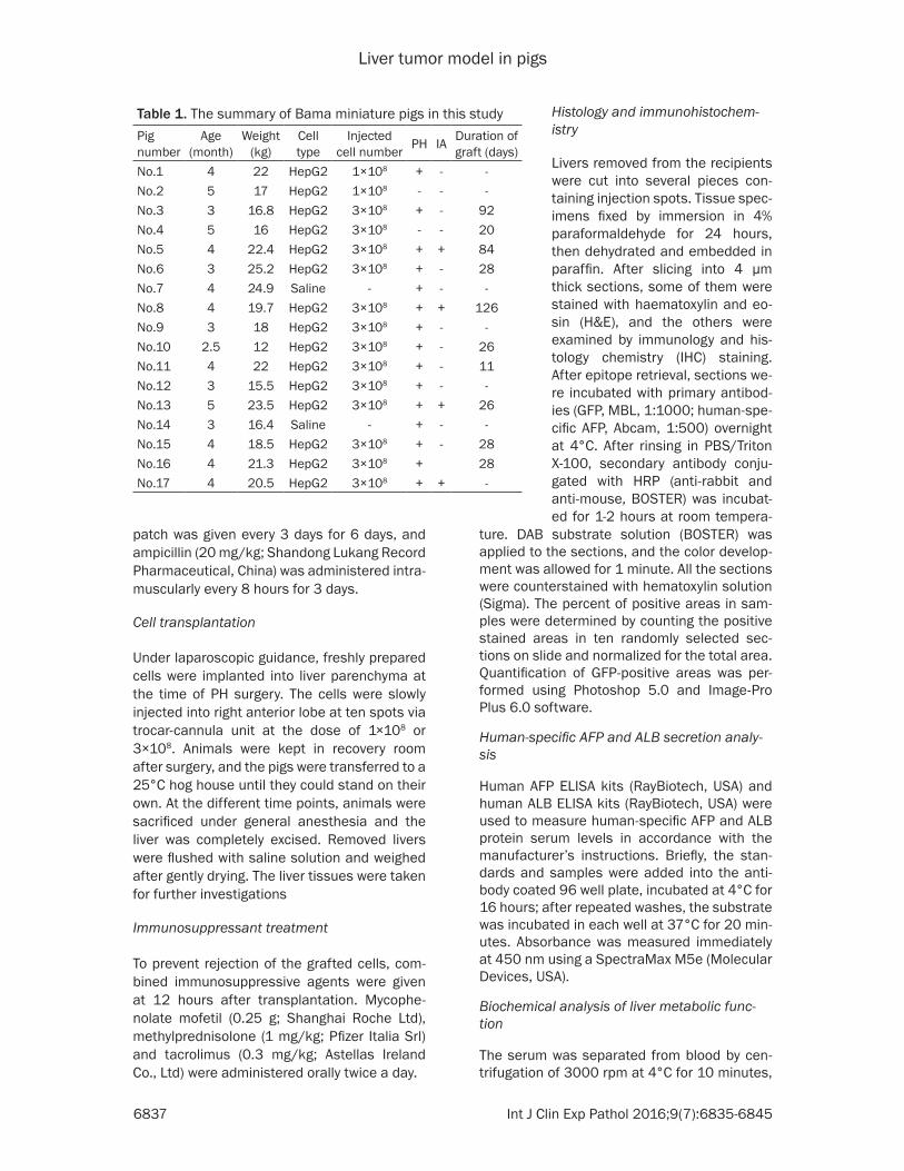
graft (days)No.1 4 22 HepG2 1×10⁸ + - -No.2 5 17 HepG2 1×10⁸ - - -No.3 3 16.8 HepG2 3×10⁸ + - 92No.4 5 16 HepG2 3×10⁸ - - 20No.5 4 22.4 HepG2 3×10⁸ + + 84No.6 3 25.2 HepG2 3×10⁸ + - 28No.7 4 24.9 Saline - + - -No.8 4 19.7 HepG2 3×10⁸ + + 126No.9 3 18 HepG2 3×10⁸ + - -No.10 2.5 12 HepG2 3×10⁸ + - 26No.11 4 22 HepG2 3×10⁸ + - 11No.12 3 15.5 HepG2 3×10⁸ + - -No.13 5 23.5 HepG2 3×10⁸ + + 26No.14 3 16.4 Saline - + - -No.15 4 18.5 HepG2 3×10⁸ + - 28No.16 4 21.3 HepG2 3×10⁸ + 28No.17 4 20.5 HepG2 3×10⁸ + + -
Histology and immunohistochem-istry
Livers removed from the recipients were cut into several pieces con-taining injection spots. Tissue spec-imens fixed by immersion in 4% paraformaldehyde for 24 hours, then dehydrated and embedded in paraffin. After slicing into 4 μm thick sections, some of them were stained with haematoxylin and eo- sin (H&E), and the others were examined by immunology and his-tology chemistry (IHC) staining. After epitope retrieval, sections we- re incubated with primary antibod-ies (GFP, MBL, 1:1000; human-spe-cific AFP, Abcam, 1:500) overnight at 4°C. After rinsing in PBS/Triton X-100, secondary antibody conju-gated with HRP (anti-rabbit and anti-mouse, BOSTER) was incubat-ed for 1-2 hours at room tempera-
Liver tumor model in pigs
6838 Int J Clin Exp Pathol 2016;9(7):6835-6845
and then stored at -80°C for further analysis. Hepatic enzymes (aspartate aminotransfera- se, alanine aminotransferase, gamma-glutam-yltransferase and alkaline phosphatase), pro-teins (total protein, ALB, and globulin) and bili-rubin (total bilirubin and direct bilirubin) were determined as previously described [15].
Statistical analyses
Data between different groups were analyzed by Student’s t-test, and were expressed as the mean ± standard deviation (SD). A value of P<0.05 was considered significant.
Results
Engraftment of human hepatoma cells into pig liver
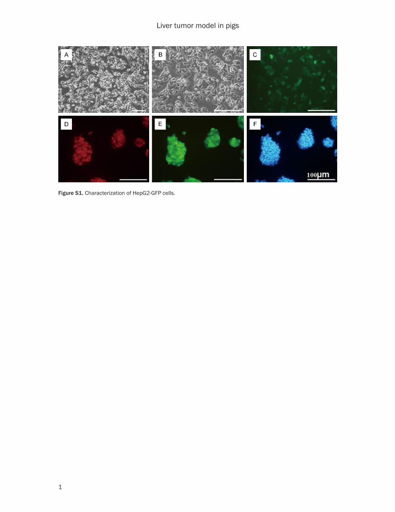
We at first investigated if human hepatoma cells could survive and function in pigs. It has been shown that the growth of engrafted cells could be enhanced in partially hepatectomized rats, in which regenerating liver constitutes a highly active anabolic focus [16-18]. As shown in Table 1, HepG2-GFP cells under growth sta-tus (Figure S1) were injected into 2 pigs (No.1/No.3) after PH, and 2 pigs (No.2/No.4) received
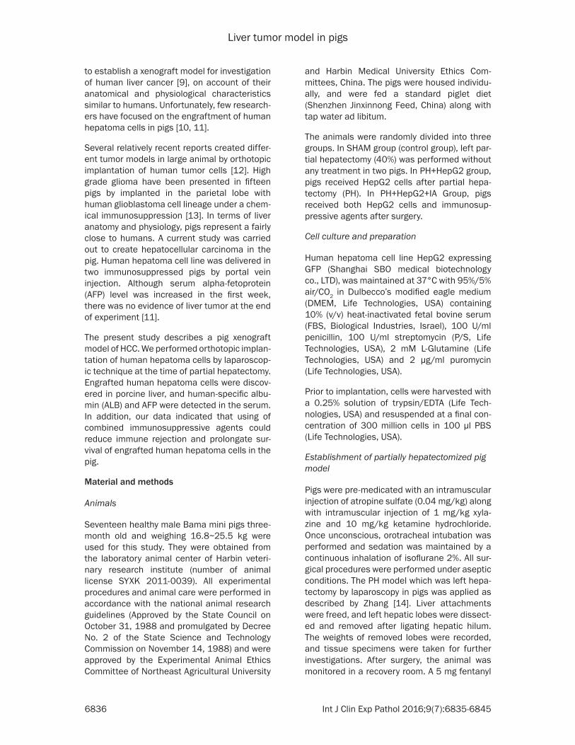
HepG2-GFP cells only. Two pigs (No.1/No.2) sacrificed at 4 months after transplantation, and the other pigs (No.3/No.4) were sacrificed 3 months and 3 weeks after surgery respec-tively. Engrafted HepG2-GFP cells were detect-ed by IHC staining using antibodies specifically against GFP. GFP-positive HepG2 cells were hardly observed in pigs which obtained only the cells, while a few engrafted surviving HepG2 cells were discovered in two recipients which treated by both cell implantation and PH. A few GFP-positive HepG2 cells were found as scat-tered individual cells or nodules containing 2-5 cells in liver, and human-specific AFP were detectable in the serum of the two recipients (Figure 1A).
Improving long-term graft survival by combined immunosuppressive agents
Although AFP serum levels had been increased at the first week, there was subsequent de- crease in AFP level until the end of experiment (Figure 1B). These results indicated engrafted tumor cells may have been rejected by porcine immune system. Immunosuppressive agents are considered capable of inhibiting prolifera-tion of T- and B-cells as well as prevention of allograft rejection [19]. We wondered whether
Figure 1. Engraftment of human hep-atoma cells in porcine liver. A. GFP-positive HepG2 cells were seen as individual cells or nodules containing 2-5 cells in the liver of No. 3 and No. 4 at the sacrifice (arrows). B. Human AFP showed higher serum level at the first week after transplantation, how-ever, decreased subsequently. Scale bars = 50 μm.
Liver tumor model in pigs
6839 Int J Clin Exp Pathol 2016;9(7):6835-6845
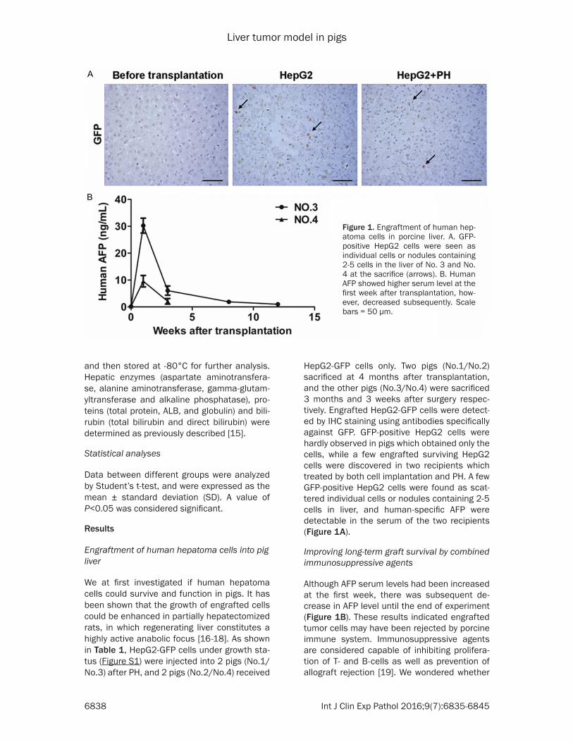
immunosuppressive agents could prolong xenograft survival. Therefore, 13 pigs were divided into three groups and the PH were per-formed on the recipients. Two pigs in the first group did not receive any treatment during the experiment (SHAM group). Six pigs in the sec-ond group received HepG2 cells (PH+HepG2 group). In the last group, 5 pigs received HepG2 cells with combined immunosuppressive ag- ents (PH+HepG+IA group). To analyze the sur-vival of grafted cells, a cohort of recipients (SHAM group, n = 1; PH+HepG2 group, n = 5; PH+HepG2+IA group, n = 3) was assessed by histologic analysis 28 days after transplanta-tion. At sacrifice, the recipients in PH+HepG2 group and PH+HepG2+IA group showed GFP-positive of engrafted cells in liver specimens, and no GFP-positive HepG2 cells were found in SHAM group. IHC staining showed that HepG2 cells repopulated up to 2.79%±1.17% of the liver parenchyma in PH+HepG2+IA group, and 0.98%±0.62% in PH+HepG2 group (Figure 2). The rest of the recipients were sacrificed from 84 days to 137 days after transplantation res- pectively. GFP-positive cells were still detect-able in the grafted livers 126 days after trans-plantation in PH+HepG2+IA group. In contrast, HepG2 cells were not observed in PH+HepG2 group. Table 1 summarizes the state of trans-plantation in 17 recipients. IHC staining indi-cated that 5/8 in PH+HepG2 group and 4/5 in
PH+HepG2+IA group recipients showed GFP-positive of engrafted cells. These data indicat-ed that using of combined immunosuppressive agents could prolongate survival of engrafted human hepatoma cells in pigs.
Human-specific AFP and ALB secretion analy-sis
To determine whether HepG2-GFP cells in chi-meric livers were functional, serial section with IHC staining of GFP and human-specific AFP was carried out. AFP-positive cells were ob- served in GFP-positive liver tissues (Figure 3A). However, no positive signal was found in SHAM group. This was further confirmed by detection of human-specific AFP and ALB secretion of engrafted cells in the serum. Human-specific ALB and AFP were detectable in the serum of the recipients until the end of experiments. As shown in Figure 3B, the serum of the recipients in PH+HepG2+IA group contained 39.53±20.28 ng/ml human-specific AFP in 2 weeks after transplantation, and level decreased over the next few weeks. Notably, PH+HepG2+IA group showed higher level of AFP than PH+HepG2 group at the different time points over 12 weeks after transplantation. There was no sig-nificant difference in serum level of human-specific ALB between the two groups (Figure 3C). These results showed that grafted HepG2 cells were proliferating and expressing func-tional proteins in porcine liver.
Histologic characterization of liver tissue
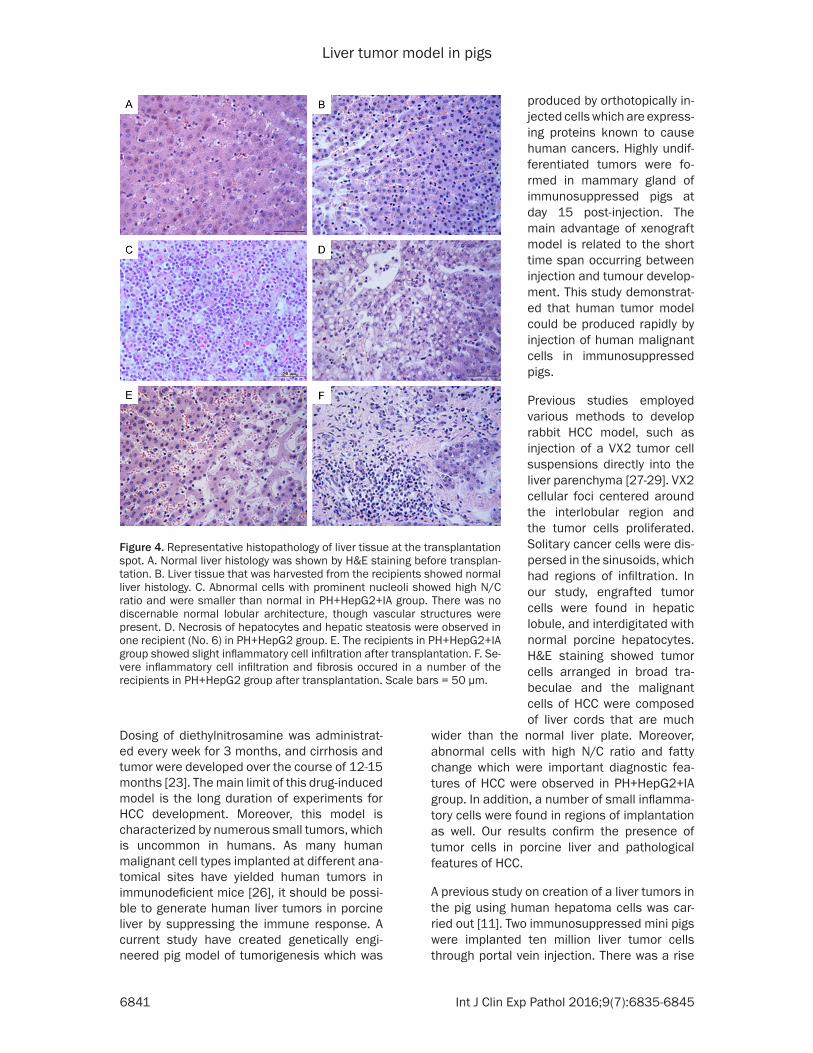
Normal liver histology was confirmed by H&E staining before treatment (Figure 4A). Hepatic tissue of SHAM group was basically normal after operation and no rejection feature was seen under microscope (Figure 4B). Shown in Figure 4C, trabecular pattern composed of tumor cells were much wider than the normal liver plate that is two cells thick, and abnormal cells with prominent nucleoli have high N/C ratio in PH+HepG2+IA group. There was no dis-cernable normal lobular architecture, though vascular structures were present. Necrosis of hepatocytes and hepatic steatosis were ob- served in one recipient (No. 6) in PH+HepG2 group (Figure 4D). All the animals in this study did not have any severe complications including abdominal abscess. Compared to PH+HepG2+IA group, severe inflammatory cell infiltration and fibrosis which are rejection fea-
Figure 2. Comparison of xeno-repopulation between three groups in four weeks after transplantation. The engraftment percentages in porcine liver for survival HepG2 cells were calculated on the ba-sis of GFP-positive staining and the area scan im-ages of multiple different liver lobules obtained around injection spots from the recipients in three groups. IHC staining showed that HepG2 cells re-populated up to 2.79%±1.17% of the liver paren-chyma in PH+HepG2+IA group, and 0.98%±0.62% in PH+HepG2 group. The difference between three groups had significant statistical significance. *P<0.05.

Liver tumor model in pigs
6840 Int J Clin Exp Pathol 2016;9(7):6835-6845
tures were found in a number of the recipients in PH+HepG2 group after transplantation (Figure 4E and 4F).
Evaluation of liver function after transplanta-tion
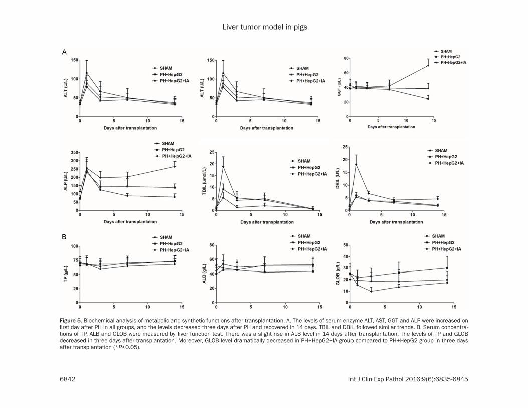
Blood biochemical measurements indicated that there was an increase in alanine amino-transferase, aspartate aminotransferase, alka-line phosphatase and total bilirubin levels on
teristics of pigs are similar to those of humans. In particular, larger animal models that might be developed by minimally invasive image-guid-ed interventions seem a step closer to the human scale [22]. In this paper, we described an HCC model in large animals, induced by implantation of human hepatoma cells.
Several relatively recent reports have devel-oped porcine liver tumors, and these models mimic the human condition of HCC [23-25].
Figure 3. Human-specific AFP were measured by ELISA. A. IHC staining of GFP and human-specific AFP in the recipients of PH+HepG2+IA group (ar-rows). However, no positive signal was found in SHAM group. Assay of hu-man-specific AFP and ALB in the serum of the recipients. Scale bars = 50 μm. B. AFP levels were significant higher in PH+HepG2+IA group than those in PH+HepG2 group at the different time points over 12 weeks after transplan-tation (*P<0.05). C. Human-specific ALB were detectable in the serum of the recipients in PH+HepG2 and PH+HepG2+IA group. There was an increase in four weeks after transplantation.
the first day after PH perfor-mance in all three groups. The decline tendency of these lev-els were already evident on day 3 and all the recipients recovered in 14 days after the PH (Figure 5A). We also found that serum level of alkaline phosphatase, gamma-gluta- myltransferase, direct biliru-bin and indirect bilirubin were in the same trend. Serum con-centrations of total protein, ALB and globulin were mea-sured by liver function test. There was a slight rise in ALB level in 14 days after trans-plantation. The levels of total protein and globulin decrea- sed in three days after trans-plantation. Moreover, globulin level dramatically decreased in PH+HepG2+IA group com-pared to PH+HepG2 group in three days after transplanta-tion (Figure 5B).
Discussion
Xenograft animal models are widely used to improve our knowledge of HCC, in particu-lar the rodent model [20]. Although such models help to understand the development of HCC and the therapeutic approaches, no rodent mod-els was ideal for developing surgical procedures and de- vices, due to their small body size relative to human [21]. The pig model is one of the best candidates, as anatomi-cal and physiological charac-
Liver tumor model in pigs
6841 Int J Clin Exp Pathol 2016;9(7):6835-6845
Dosing of diethylnitrosamine was administrat-ed every week for 3 months, and cirrhosis and tumor were developed over the course of 12-15 months [23]. The main limit of this drug-induced model is the long duration of experiments for HCC development. Moreover, this model is characterized by numerous small tumors, which is uncommon in humans. As many human malignant cell types implanted at different ana-tomical sites have yielded human tumors in immunodeficient mice [26], it should be possi-ble to generate human liver tumors in porcine liver by suppressing the immune response. A current study have created genetically engi-neered pig model of tumorigenesis which was
wider than the normal liver plate. Moreover, abnormal cells with high N/C ratio and fatty change which were important diagnostic fea-tures of HCC were observed in PH+HepG2+IA group. In addition, a number of small inflamma-tory cells were found in regions of implantation as well. Our results confirm the presence of tumor cells in porcine liver and pathological features of HCC.
A previous study on creation of a liver tumors in the pig using human hepatoma cells was car-ried out [11]. Two immunosuppressed mini pigs were implanted ten million liver tumor cells through portal vein injection. There was a rise
Figure 4. Representative histopathology of liver tissue at the transplantation spot. A. Normal liver histology was shown by H&E staining before transplan-tation. B. Liver tissue that was harvested from the recipients showed normal liver histology. C. Abnormal cells with prominent nucleoli showed high N/C ratio and were smaller than normal in PH+HepG2+IA group. There was no discernable normal lobular architecture, though vascular structures were present. D. Necrosis of hepatocytes and hepatic steatosis were observed in one recipient (No. 6) in PH+HepG2 group. E. The recipients in PH+HepG2+IA group showed slight inflammatory cell infiltration after transplantation. F. Se-vere inflammatory cell infiltration and fibrosis occured in a number of the recipients in PH+HepG2 group after transplantation. Scale bars = 50 μm.
produced by orthotopically in- jected cells which are express-ing proteins known to cause human cancers. Highly undif-ferentiated tumors were fo- rmed in mammary gland of immunosuppressed pigs at day 15 post-injection. The main advantage of xenograft model is related to the short time span occurring between injection and tumour develop-ment. This study demonstrat-ed that human tumor model could be produced rapidly by injection of human malignant cells in immunosuppressed pigs.
Previous studies employed various methods to develop rabbit HCC model, such as injection of a VX2 tumor cell suspensions directly into the liver parenchyma [27-29]. VX2 cellular foci centered around the interlobular region and the tumor cells proliferated. Solitary cancer cells were dis-persed in the sinusoids, which had regions of infiltration. In our study, engrafted tumor cells were found in hepatic lobule, and interdigitated with normal porcine hepatocytes. H&E staining showed tumor cells arranged in broad tra-beculae and the malignant cells of HCC were composed of liver cords that are much
Liver tumor model in pigs
6842 Int J Clin Exp Pathol 2016;9(6):6835-6845
Figure 5. Biochemical analysis of metabolic and synthetic functions after transplantation. A. The levels of serum enzyme ALT, AST, GGT and ALP were increased on first day after PH in all groups, and the levels decreased three days after PH and recovered in 14 days. TBIL and DBIL followed similar trends. B. Serum concentra-tions of TP, ALB and GLOB were measured by liver function test. There was a slight rise in ALB level in 14 days after transplantation. The levels of TP and GLOB decreased in three days after transplantation. Moreover, GLOB level dramatically decreased in PH+HepG2+IA group compared to PH+HepG2 group in three days after transplantation (*P<0.05).
Liver tumor model in pigs
6843 Int J Clin Exp Pathol 2016;9(7):6835-6845
in serum AFP level in the first week. Unfor- tunately, tumor cells did not implant in porcine livers at the end of six weeks. Our results were in general agreement with this study. In this study, human-specific AFP which are known to be secreted by human hepatoma cells was up to 39.53±20.28 ng/ml, and then the levels decreased slightly. AFP were detectable at the end of experiments even at very low level in serum. In view of raised AFP level at the first week, the fact of repopulation of engrafted cells in porcine livers is evident. Moreover, GFP-positive cells were found as individual cells or nodules in porcine liver until the end of experi-ments. In our study, 10/15 pigs showed evi-dence of tumor growth. The reason of our high-er success rate might be a larger volume of hepG2 suspension cells.
Laparoscopic surgery has shown advantages to open surgery, including decreased hospital stays, less postoperative discomfort and com-plications. A previous study has shown that laparoscopic surgery could be safely used for islet cell transplantation [30]. It can be inferred that laparoscopic surgery may be applicable for other cell transplantation procedures. Our study indicates that laparoscopic surgery sh- ould be appropriate to implant human hepato-ma cells in pigs.
Subsequent decrease in AFP value was proba-bly related to rejection of tumor cells by porcine immune system, and this was confirmed by his-tology characteristics of liver tissues. Severe inflammatory cell infiltration which is a rejection feature was found in a number of the recipients in PH+HepG2 group after transplantation. Previously study showed that cell-mediated immunity played an important role in the rejec-tion of xenografts [31]. Immunosuppressive agents are not only used in allograft rejection prevention, but also in pig-to-nonhuman pri-mate xenotransplantation models [32]. There- fore, combined immunosuppressive agents were used to prolong survival of xenografts [33]. In this study, inflammatory cell infiltration was hardly observed in PH+HepG2+IA group after transplantation. IHC staining of GFP exhib-ited that HepG2 cells repopulated up to 2.79%± 1.17% of the liver parenchyma in PH+HepG2+ IA group, which was significant higher than PH+HepG2 group. Moreover, PH+HepG2+IA group showed higher level of AFP than PH+ HepG2 group in 12 weeks after transplanta-
tion. These results demonstrate that using of combined immunosuppressive agents could attenuate inflammation and prolongate surviv-al of engrafted human hepatoma cells in por-cine liver.
In summary, we have developed a large-animal human liver tumor xenograft model in the pig. Orthotopic implantation of human hepatoma cells using immunosuppressive agents leads to engraftment of tumor cells in porcine liver. Although we found that engrafted cells were still detectable in the recipient 126 days after transplantation, there was no macroscopic solid tumor at the end of experiment. Our study could be applied for the study of tumor cell pro-liferation and drug treatment. For successfully introducing pig liver models into the clinic, fur-ther works on multiple genetic modifications pigs (to overcome innate and adaptive immune responses) and more effective immunosup-pressants are required.
Acknowledgements
This study was supported by the National Natural Science Foundation of China (3127- 1590 and 81301023) and the State Key Development Program of Basic Research of China (2012CBA01303).
Disclosure of conflict of interest
None.
Address correspondence to: Dr. Lei Lei, Department of Histology and Embryology, Harbin Medical University, Xuefu Road 194, Harbin 150081, China. Tel: 86-451-86674518; Fax: 86-451-87503326; E-mail: [email protected]
References
[1] Bakiri LandWagner EF. Mouse models for liver cancer. Mol Oncol 2013; 7: 206-223.
[2] Huynh H, Soo KC, Chow PK, Panasci L and Tran E. Xenografts of human hepatocellular carci-noma: a useful model for testing drugs. Clin Cancer Res 2006; 12: 4306-4314.
[3] De Minicis S, Kisseleva T, Francis H, Baroni GS, Benedetti A, Brenner D, Alvaro D, Alpini G, Marzioni M. Liver carcinogenesis: rodent mod-els of hepatocarcinoma and cholangiocarcino-ma. Dig Liver Dis 2013; 45: 450-459.
[4] Chen X, Yamamoto M, Fujii K, Nagahama Y, Ooshio T, Xin B, Okada Y, Furukawa H, Nishikawa Y. Differential reactivation of fetal/
Liver tumor model in pigs
6844 Int J Clin Exp Pathol 2016;9(7):6835-6845
neonatal genes in mouse liver tumors indu- ced in cirrhotic and non-cirrhotic conditions. Cancer Sci 2015; 106: 972-981.
[5] Zhao GJ, Xu LX, Chu ES, Zhang N, Shen JY, Damirin A and Li XX. Establishment of an or-thotopic transplantation tumor model of hepa-tocellular carcinoma in mice. World J Gas- troenterol 2012; 18: 7087-7092.
[6] Tang TC, Man S, Xu P, Francia G, Hashimoto K, Emmenegger U, Kerbel RS. Development of a Resistance-like Phenotype to Sorafenib by Human Hepatocellular Carcinoma Cells Is Reversible and Can Be Delayed by Metronomic UFT Chemotherapy. Neoplasia 2010; 12: 928-940.
[7] Lee TK, Na KS, Kim J, Jeong HJ. Establishment of animal models with orthotopic hepatocellu-lar carcinoma. Nucl Med Mol Imaging 2014; 48: 173-179.
[8] Misra SK, Ghoshal G, Gartia MR, Wu Z, De AK, Ye M, Bromfield CR, Williams EM, Singh K, Tangella KV, Rund L, Schulten K, Schook LB, Ray PS, Burdette EC and Pan D. Trimodal Therapy: Combining Hyperthermia with Re- purposed Bexarotene and Ultrasound for Treating Liver Cancer. ACS Nano 2015; 9: 10695-10718.
[9] Flisikowska T, Kind A and Schnieke A. The new pig on the block: modelling cancer in pigs. Transgenic Res 2013; 22: 673-680.
[10] Reymond MA, Tannapfel A, Schneider C, Scheidbach H, Kover S, Jung A, Reck T, Lippert H, Kockerling F. Description of an intraperito-neal tumour xenograft survival model in the pig. Eur J Surg Oncol 2000; 26: 393-397.
[11] Rai R, Flecknell P, Richardson C and Manas DM. Creation of porcine liver tumour using hu-man hepatoma cell lines: Experimental study. Cancer Biol Ther 2014; 4: 635-637.
[12] Krushelnycky BW, Farr-Jones MA, Mielke B, McKean JD, Weir BK and Petruk KC. Develop- ment of a large-animal human brain tumor xe-nograft model in immunosuppressed cats. Cancer Res 1991; 51: 2430-2437.
[13] Selek L, Seigneuret E, Nugue G, Wion D, Nissou MF, Salon C, Seurin MJ, Carozzo C, Ponce F, Roger T and Berger F. Imaging and histological characterization of a human brain xenograft in pig: the first induced glioma model in a large animal. J Neurosci Methods 2014; 221: 159-165.
[14] Zhang H, Liu T, Wang Y, Liu HF, Zhang JT, Wu YS, Lei L and Wang HB. Laparoscopic left hep-atectomy in swine: a safe and feasible tech-nique. J Vet Sci 2014; 15: 417.
[15] Ekser B, Echeverri GJ, Hassett AC, Yazer MH, Long C, Meyer M, Ezzelarab M, Lin CC, Hara H, van der Windt DJ, Dons EM, Phelps C, Ayares D, Cooper DK and Gridelli B. Hepatic function
after genetically engineered pig liver trans-plantation in baboons. Transplantation 2010; 90: 483-493.
[16] Paschkis KE, Cantarow A, Stasney J and Hobbs JH. Tumor growth in partially hepatectomized rats. Cancer Res 1955; 15: 579-582.
[17] Basma H, Soto-Gutierrez A, Yannam GR, Liu L, Ito R, Yamamoto T, Ellis E, Carson SD, Sato S, Chen Y, Muirhead D, Navarro-Alvarez N, Wong RJ, Roy-Chowdhury J, Platt JL, Mercer DF, Miller JD, Strom SC, Kobayashi N and Fox IJ. Diffe- rentiation and transplantation of human em-bryonic stem cell-derived hepatocytes. Gas- troenterology 2009; 136: 990-999.
[18] Okay E, Simsek T, Subasi C, Gunes A, Duruksu G, Gurbuz Y, Gacar G and Karaoz E. Cross ef-fects of resveratrol and mesenchymal stem cells on liver regeneration and homing in par-tially hepatectomized rats. Stem Cell Rev 2015; 11: 322-331.
[19] Mohiuddin MM, Corcoran PC, Singh AK, Azimzadeh A, Hoyt RF Jr, Thomas ML, Eckhaus MA, Seavey C, Ayares D, Pierson RN 3rd and Horvath KA. B-cell depletion extends the sur-vival of GTKO. hCD46Tg pig heart xenografts in baboons for up to 8 months. Am J Transplant 2012; 12: 763-771.
[20] Yao X, Hu JF, Daniels M, Yien H, Lu H, Sharan H, Zhou X, Zeng Z, Li T, Yang Y and Hoffman AR. A novel orthotopic tumor model to study growth factors and oncogenes in hepatocarcinogene-sis. Clin Cancer Res 2003; 9: 2719-2726.
[21] Kim T, Kim DY, Cho MJ, Kim SC, Seo JJ and Kim IK. Surgery for hepatoblastoma: from laparo-scopic resection to liver transplantation. He- patogastroenterology 2011; 58: 896-899.
[22] Pinto PA, Montgomery RA, Ryan B, Roberts W, Hsu T, Kavoussi P, Klein AS, Kavoussi LR and Molmenti EP. Laparoscopic procurement mod-el for living donor liver transplantation. Clin Transplant 2003; 17 Suppl 9: 39-43.
[23] Li X, Zhou X, Guan Y, Wang YX, Scutt D and Gong QY. N-nitrosodiethylamine-induced pig liver hepatocellular carcinoma model: radio-logical and histopathological studies. Cardio- vasc Intervent Radiol 2006; 29: 420-428.
[24] Adam SJ, Rund LA, Kuzmuk KN, Zachary JF, Schook LB and Counter CM. Genetic induction of tumorigenesis in swine. Oncogene 2007; 26: 1038-1045.
[25] Kim J, Ahn H, Woo HM, Lee E and Lee GS. Generation of liver-specific TGF-alpha and c-Myc-overexpressing fibroblasts for future cre-ation of a liver cancer porcine model. Mol Med Rep 2014; 10: 329-335.
[26] Moskaluk CA, Baras AS, Mancuso SA, Fan H, Davidson RJ, Dirks DC, Golden WL and Frierson HF Jr. Development and characterization of xe-
Liver tumor model in pigs
6845 Int J Clin Exp Pathol 2016;9(7):6835-6845
nograft model systems for adenoid cystic car-cinoma. Lab Invest 2011; 91: 1480-1490.
[27] Zhang L, Liu FY, Fu JX, Duan F, Fan QS and Wang MQ. Hepatic arterial administration of sorafenib and iodized oil effectively attenuates tumor growth and intrahepatic metastasis in rabbit VX2 hepatocellular carcinoma model. Int J Clin Exp Pathol 2014; 7: 7775-7781.
[28] Ma R, Li X, Kong F, Ma J, Ma Z, Zhao Z, Wen Z and Hu W. Effect of ischemia reperfusion on rabbit VX2 cells in a hepatocellular carcinoma model. Int J Clin Exp Pathol 2015; 8: 497-503.
[29] Izumi B, Tashiro S and Miyauchi Y. Anticancer effects of local administration of mitomycin C via the hepatic artery or portal vein on implan-tation and growth of VX2 cancer injected into rabbit liver. Cancer Res 1986; 46: 4167-4170.
[30] Casavilla A, Rilo HL, Julian TB, Fontes PA, Starzl TE and Ricordi C. Laparoscopic approach for islet cell transplantation. Transplant Proc 1992; 24: 2800.
[31] Kawahara T, Douglas DN, Lewis J, Lund G, Addison W, Tyrrell DL, Churchill TA and Kneteman NM. Critical role of natural killer cells in the rejection of human hepatocytes af-ter xenotransplantation into immunodeficient mice. Transpl Int 2010; 23: 934-943.
[32] Thompson P, Badell IR, Lowe M, Turner A, Cano J, Avila J, Azimzadeh A, Cheng X, Pierson RN 3rd, Johnson B, Robertson J, Song M, Leopardi F, Strobert E, Korbutt G, Rayat G, Rajotte R, Larsen CP and Kirk AD. Alternative immuno-modulatory strategies for xenotransplantation: CD40/154 pathway-sparing regimens pro-mote xenograft survival. Am J Transplant 2012; 12: 1765-1775.
[33] Candinas D, Belliveau S, Koyamada N, Miyatake T, Hechenleitner P, Mark W, Bach FH and Hancock WW. T cell independence of mac-rophage and natural killer cell infiltration, cyto-kine production, and endothelial activation during delayed xenograft rejection. Trans- plantation 1996; 62: 1920-1927.
Liver tumor model in pigs
1
Figure S1. Characterization of HepG2-GFP cells.


![Whole transcriptome profiling of patient-derived xenograft ...eprints.whiterose.ac.uk/96695/1/WRRO_96695.pdf · xenograft models or specific cancer type [8–9]. In this paper, we](https://img.pdfslide.us/doc/110x75/5f0337437e708231d4081c1a/whole-transcriptome-profiling-of-patient-derived-xenograft-xenograft-models.jpg)