Embed Size (px)
Citation preview

Organogenesis in deep time: A problem in genomics,development, and paleontologyJoyce Pierettia,1, Andrew R. Gehrkea,1, Igor Schneiderb,1, Noritaka Adachia, Tetsuya Nakamuraa, and Neil H. Shubina,2
aDepartment of Organismal Biology and Anatomy, The University of Chicago, Chicago, IL 60637; and bInstituto de Ciencias Biologicas, Universidade Federaldo Para, 66075, Belem, Brazil
Edited by David Jablonski, The University of Chicago, Chicago, IL, and approved February 6, 2015 (received for review November 25, 2014)
The fossil record is a unique repository of information on majormorphological transitions. Increasingly, developmental, embryol-ogical, and functional genomic approaches have also conspired toreveal evolutionary trajectory of phenotypic shifts. Here, we usethe vertebrate appendage to demonstrate how these disciplinescan mutually reinforce each other to facilitate the generation andtesting of hypotheses of morphological evolution. We discussclassical theories on the origins of paired fins, recent data onregulatory modulations of fish fins and tetrapod limbs, and casestudies exploring the mechanisms of digit loss in tetrapods. Weenvision an era of research in which the deep history of morphologicalevolution can be revealed by integrating fossils of transitional formswith direct experimentation in the laboratory via genome manipu-lation, thereby shedding light on the relationship between genes,developmental processes, and the evolving phenotype.
fossil record | development | genomics | evolution | limb
Paleontologists in recent decades have discovered a host ofnew taxa that reveal transitional stages in the evolution of
birds, whales, mammals, tetrapods, frogs, salamanders, and arthro-pods (1–9). This pulse of discovery is not an accident, but the resultof an elaboration of our ability to identify likely sites for fossil re-covery by using increasingly refined phylogenies, stratigraphic maps,and geological records. Likewise, imaging techniques, such as high-energy CT, have opened up old and understudied fossil collectionsas new vehicles for discovery. With advances in both fieldwork andimaging, the discovery of the phenotypic basis for morphologicalinnovation is at a critical moment in its long history: Novel per-spectives on classical questions of anatomical evolution are withinour reach.Fossils, when placed in a phylogenetic context, can reveal taxa
with novel combinations of characters that could not be pre-dicted by studying extant creatures alone. If we lacked fossil evi-dence of mammal-like reptiles, for example, then the physiologicaland morphological similarities of birds and mammals would likelybe interpreted as homologies rather than examples of parallel evo-lution (e.g., the discredited “Haemothermia” clade) (10, 11). Inaddition to identifying solid taxonomic groupings, these same fossilsreveal transitional series in the origin of the mammalian dentition,ear, and cranium (3). Our understanding of numerous other trans-formations, from the origin of birds to the origin of tetrapods, isseriously limited without the knowledge of extinct stem taxa.A rich fossil record permits us to document robustly supported
transformation series in the evolution of an anatomical feature,organ system, or body plan. However, to understand the patternand process of evolutionary transitions, paleontologists have in-creasingly turned their attention to development. In recent years,the combination of technologies from developmental biologyand abundant genomic resources for a multitude of model andnonmodel organisms has greatly enriched our understanding ofthe genetic and developmental processes underlying organo-genesis. This broad set of tools provides a new framework fortesting hypotheses derived from paleontological findings, therebyforming an interdisciplinary research program with comparative
genomics as well as genetic manipulation of embryonic de-velopment (12–15).Here, we use the evolution and diversification of the vertebrate
limb as an exemplar to reveal how discoveries in paleontology canleverage experimental and comparative work in molecular biology,genomics, and embryology. First, we review how fossil analyses ofearly gnathostomes, coupled with embryological studies, offer thefoundation for hypotheses on the origin of paired appendages.Then, we discuss current research on model and nonmodelspecies that shed light on the origin of digits by comparing geneexpression and regulatory mechanisms underlying fin and limbdevelopment. Next, we examine recent studies that identify thegenetic and developmental basis for digit reduction in tetrapods.Finally, we highlight novel technologies that are enabling biolo-gists to solve century-old evolutionary puzzles with state-of-the-artmolecular approaches. The synthesis of modern technology withpaleontological findings has been an ongoing topic of interest(16–18). Continued advances in technology now give morphol-ogists an ever-expanding toolkit to test genome function and,ultimately, manipulate genomes in a phylogenetic framework.When these new technologies are coupled with paleontologicaldiscovery, new insights into classical questions in evolutionarymorphology lie in the offing.
Origin of Paired AppendagesThe origin of paired fins is one of the critical events in the historyof vertebrates. Two hypotheses, dating back to the 19th century,have been generated to explain this transition: (i) the gill-archhypothesis, in which the posterior-most gill arch is considered to bea precursor to the pectoral girdle and paired fins (Fig. 1A) (19),and (ii) the fin-fold hypothesis, which holds that paired fins arederived from lateral longitudinal folds that appear early in de-velopment and evolution (Fig. 1B) (21–23). Both hypotheses wereoriginally proposed from observations of comparative embryologyand anatomy of extant sharks. Here, we review how evidence
Significance
Phylogenetic data inclusive of rich paleontological records can beused to inform hypotheses on evolutionary transformations.These data, when combined with developmental studies andfunctional genomic assays in model and nonmodel organisms,expand our understanding of the evolutionary processes thatbuild and pattern the vertebrate body plan. Here, we highlighta direction of the fossil record, one of “experimental paleontology,”where morphological transformations inferred from the fossil re-cord can be experimentally assayed in the lab. With the addition ofgenomic techniques to test hypotheses, researchers can now beginto explore genomic states that have influenced both past andpresent morphological diversity.
Author contributions: J.P., A.R.G., I.S., N.A., T.N., and N.H.S. wrote the paper.
The authors declare no conflict of interest.
This article is a PNAS Direct Submission.1J.P., A.R.G., and I.S. contributed equally to this work.2To whom correspondence should be addressed. Email: [email protected].
www.pnas.org/cgi/doi/10.1073/pnas.1403665112 PNAS | April 21, 2015 | vol. 112 | no. 16 | 4871–4876
EVOLU
TION
SPEC
IALFEATU
RE
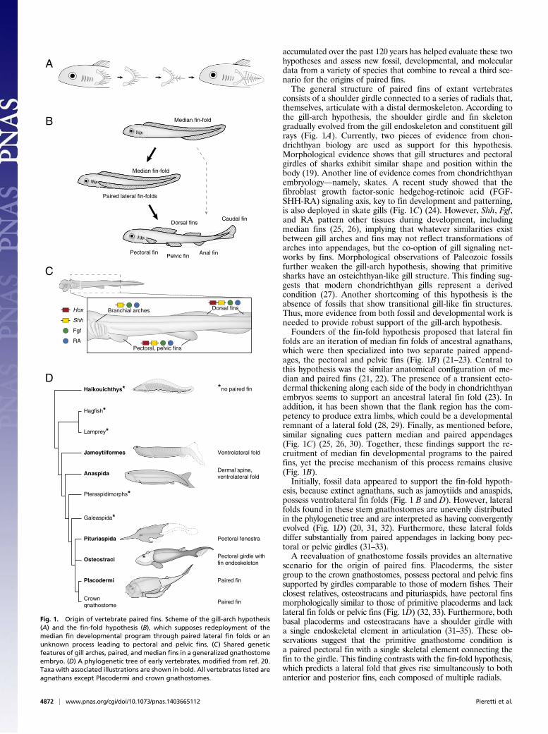
accumulated over the past 120 years has helped evaluate these twohypotheses and assess new fossil, developmental, and moleculardata from a variety of species that combine to reveal a third sce-nario for the origins of paired fins.The general structure of paired fins of extant vertebrates
consists of a shoulder girdle connected to a series of radials that,themselves, articulate with a distal dermoskeleton. According tothe gill-arch hypothesis, the shoulder girdle and fin skeletongradually evolved from the gill endoskeleton and constituent gillrays (Fig. 1A). Currently, two pieces of evidence from chon-drichthyan biology are used as support for this hypothesis.Morphological evidence shows that gill structures and pectoralgirdles of sharks exhibit similar shape and position within thebody (19). Another line of evidence comes from chondrichthyanembryology—namely, skates. A recent study showed that thefibroblast growth factor-sonic hedgehog-retinoic acid (FGF-SHH-RA) signaling axis, key to fin development and patterning,is also deployed in skate gills (Fig. 1C) (24). However, Shh, Fgf,and RA pattern other tissues during development, includingmedian fins (25, 26), implying that whatever similarities existbetween gill arches and fins may not reflect transformations ofarches into appendages, but the co-option of gill signaling net-works by fins. Morphological observations of Paleozoic fossilsfurther weaken the gill-arch hypothesis, showing that primitivesharks have an osteichthyan-like gill structure. This finding sug-gests that modern chondrichthyan gills represent a derivedcondition (27). Another shortcoming of this hypothesis is theabsence of fossils that show transitional gill-like fin structures.Thus, more evidence from both fossil and developmental work isneeded to provide robust support of the gill-arch hypothesis.Founders of the fin-fold hypothesis proposed that lateral fin
folds are an iteration of median fin folds of ancestral agnathans,which were then specialized into two separate paired append-ages, the pectoral and pelvic fins (Fig. 1B) (21–23). Central tothis hypothesis was the similar anatomical configuration of me-dian and paired fins (21, 22). The presence of a transient ecto-dermal thickening along each side of the body in chondrichthyanembryos seems to support an ancestral lateral fin fold (23). Inaddition, it has been shown that the flank region has the com-petency to produce extra limbs, which could be a developmentalremnant of a lateral fold (28, 29). Finally, as mentioned before,similar signaling cues pattern median and paired appendages(Fig. 1C) (25, 26, 30). Together, these findings support the re-cruitment of median fin developmental programs to the pairedfins, yet the precise mechanism of this process remains elusive(Fig. 1B).Initially, fossil data appeared to support the fin-fold hypoth-
esis, because extinct agnathans, such as jamoytiids and anaspids,possess ventrolateral fin folds (Fig. 1 B and D). However, lateralfolds found in these stem gnathostomes are unevenly distributedin the phylogenetic tree and are interpreted as having convergentlyevolved (Fig. 1D) (20, 31, 32). Furthermore, these lateral foldsdiffer substantially from paired appendages in lacking bony pec-toral or pelvic girdles (31–33).A reevaluation of gnathostome fossils provides an alternative
scenario for the origin of paired fins. Placoderms, the sistergroup to the crown gnathostomes, possess pectoral and pelvic finssupported by girdles comparable to those of modern fishes. Theirclosest relatives, osteostracans and pituriaspids, have pectoral finsmorphologically similar to those of primitive placoderms and lacklateral fin folds or pelvic fins (Fig. 1D) (32, 33). Furthermore, bothbasal placoderms and osteostracans have a shoulder girdle witha single endoskeletal element in articulation (31–35). These ob-servations suggest that the primitive gnathostome condition isa paired pectoral fin with a single skeletal element connecting thefin to the girdle. This finding contrasts with the fin-fold hypothesis,which predicts a lateral fold that gives rise simultaneously to bothanterior and posterior fins, each composed of multiple radials.
Median fin-fold
Paired lateral fin-folds
Pelvic finPectoral fin
Hagfish*
Lamprey*
Jamoytiiformes
Anaspida
Galeaspida*
Pituriaspida
Osteostraci
Placodermi
Crown gnathostome
Dorsal finsCaudal fin
Anal fin
Pectoral, pelvic fins
Dorsal fins
A
B
C
D
Median fin-fold
Haikouichthys*
Ventrolateral fold
Dermal spine,ventrolateral fold
Pectoral fenestra
Pectoral girdle withfin endoskeleton
Paired fin
Pteraspidimorphs*
Paired fin
*no paired fin
Branchial archesHox
Shh
Fgf
RA
Fig. 1. Origin of vertebrate paired fins. Scheme of the gill-arch hypothesis(A) and the fin-fold hypothesis (B), which supposes redeployment of themedian fin developmental program through paired lateral fin folds or anunknown process leading to pectoral and pelvic fins. (C) Shared geneticfeatures of gill arches, paired, and median fins in a generalized gnathostomeembryo. (D) A phylogenetic tree of early vertebrates, modified from ref. 20.Taxa with associated illustrations are shown in bold. All vertebrates listed areagnathans except Placodermi and crown gnathostomes.
4872 | www.pnas.org/cgi/doi/10.1073/pnas.1403665112 Pieretti et al.

Although the full details of the origin of paired fins, whetherdirectly from median fin folds or via lateral fin folds (Fig. 1B),remain to be determined, it seems likely that a redeployment ofthe median fin developmental program occurred in the origin ofpaired appendages. Although we know a great deal about pairedappendage development, little is known of median fin initiationand patterning. Comparative analyses of gene expression andregulation in median fins may provide us with new clues as to theorigins of paired fins.
Fins to LimbsAs we move crownward along the gnathostome tree of life, bothfossil and molecular data conspire to reveal transformations inthe structure of fins and limbs. In particular, considerable at-tention has been focused on the evolution of the defining featureof limbs: the presence of the wrist and digits (autopod). Theautopod has held a particular fascination with evolutionary biol-ogists for two main reasons. First, this structure provides a level offlexibility and precise tactile motion that was likely crucial to theradiation of tetrapods in diverse environments. Second, theautopod appears to be an anatomical novelty, in that there is noobvious counterpart to the wrist and digits in living fishes basedon morphology (36, 37). The fossil record has greatly improvedour understanding of the evolution of the autopod, with extinctintermediate forms that reveal an aquatic origin of tetrapodapomorphies in sarcoptyergian fish that was progressively builtthrough elaboration of the distal endochondral skeleton and re-duction of the dermal one (36). These insights from fossils lead toa number of questions: Notably, do the fins of living fishes havethe equivalent of an autopod? Is the autopod a true anatomicalnovelty that first appeared in extinct sarcopterygian fish such asTiktaalik (4), or is it a part of even more ancient fins? Do thegenetic mechanisms that build the wrists and digits of extanttetrapods have antecedents in fish? These questions are difficultto answer through morphology alone, but they can be addressedin concert with data from developmental biology and functionalgenomics.
The molecular mechanisms of tetrapod appendage develop-ment have been studied in detail in mouse limbs and provide aframework for comparison with fish fins. Limb development inmouse relies on expression of the Hox family of transcriptionfactors, where specific deletions of Hox activity manifest as lossesof discrete portions of the limb (38). The autopod is built via adistinct “late” phase of HoxD and HoxA gene expression that iscontrolled by a series of enhancers that lie 5′ to the clusters(Fig. 2) (39, 40). Studies in a variety of fish species (i.e., paddlefish,catshark, zebrafish) have found a late-phase-like pattern of Hoxgene expression in the distal portion of developing pectoral fins(30, 45, 46). Although these patterns are intriguing, comparisonsof gene expression patterns alone can be misleading (47). Thus,dissecting the regulatory architecture underlying the expression ofthese genes is necessary to define homologous domains of activity.Recent work has sought to elucidate this regulatory landscape
using functional genomics and developmental biology in a varietyof fish species (41). Woltering and colleagues reasoned that if fishdo contain a late-phase cis-regulatory apparatus, their chromatinstate at the 5′ end of theHox clusters should be “open” later in findevelopment, an epigenetic state that has been well documentedduring mouse digit formation (Fig. 2) (39, 41). This hypothesis isreasonable: Only open chromatin can be transcribed during de-velopment. The authors performed chromatin conformationcapture experiments on whole-body zebrafish embryos and foundthat the 5′ genomic region was indeed in an open and accessibleconformation (implying regulatory action) in comparison with theregion 3′ to the cluster (Fig. 2) (41). However, when tested intransgenic mice, these cis-regulatory domains were not able todrive reporter expression in developing digits (Fig. 2) (41). Theseresults led the authors to conclude that the late-phase regulatoryregion present in fishes is insufficient to build an elaborate distalendoskeleton comparable to an autopod, making it an innovationof tetrapods (41).Further insight has come from nonmodel species. The majority
of genomic work in this area has been performed by using modelspecies (Fugu, Tetraodon, and zebrafish), all of which are teleosts.This group may not be the ideal genomic model because the
mouse Hoxa13 4C contacts
zebrafish hoxa13 4C contacts
Island I Island II Island III Island IV CsB CsC
e19 e18 e16 e13 e10 e4 e3
Atp5g3 Lnp Evx2 HoxD
Jazf1 Hibadh Evx1HoxATaxbp1
5’ 3’
5’ 3’
mouse gar zebrafish mouse gar zebrafish
mouse gar zebrafish(via BAC)
HoxD
HoxA
mouse Hoxd13 4C contacts
zebrafish hoxd13 4C contacts
Enhancer present in: mouse mouse and fishor
Fig. 2. Epigenetic profiles and enhancer conservation of the vertebrate “autopod” Hox regulatory region. HoxD13 and HoxA13 show extensive contacts (asdefined by 4C-seq and shown generally as red and blue regions above genomic areas) with the region 5′ to the cluster, defining an “autopod building” regulatorytopology that is shared in both mouse and zebrafish (blue and red regions above the clusters, respectively) (39–42). A number of individual enhancers that driveexpression in the wrists and digits of mouse are also present in fish genomes (Island I, CsB, e16, e13, and e10) (42–44). Both zebrafish and pufferfish sequenceswere unable to drive reporter activity in the digits of transgenic mice, whereas those of gar (a nonteleost fish with an unduplicated genome) were able to driverobust expression throughout the autopod of transgenic mice (41, 42).
Pieretti et al. PNAS | April 21, 2015 | vol. 112 | no. 16 | 4873
EVOLU
TION
SPEC
IALFEATU
RE
teleost lineage-specific genome duplication could potentially allowreshuffling of genomic sequence around the duplicated Hoxclusters. Gehrke and colleagues used the genome of a nonmodelbony fish that diverged before the teleost genome duplication—the spotted gar—to identify specific enhancers that are commonbetween mouse and fish, which was not possible using the genomicsequence of teleosts (Fig. 2) (42). The authors found that these garenhancers were able to drive robust expression of the wrist anddigits of transgenic mice, in a pattern nearly identical to theirorthologs in mouse (Fig. 2) (42). These findings suggest that theinability of teleost fish enhancers to drive expression in the digitsof transgenic mice is due to the derived nature of teleost genomes,and the unduplicated genome of the gar better represents theancestral condition. These regulatory data define the late-phaseHox expression in fish fins and tetrapod limbs as homologous, inturn suggesting that at least a portion of the autopod is an ancestralfeature that is represented by the distal bones of fish pectoral fins,particularly Devonian sarcopterygians (42). This example revealsthe reciprocal illumination of paleontological, phylogenetic, andmolecular approaches: Fossils revealed that an autopod is de-finitively present in at least one lineage of ancient fish, therebysuggesting new molecular studies of phylogenetically relevantnonmodel organisms.With such conserved developmental networks, we can now ask
the question: How does morphological disparity in limbs arise?Recent work in digit evolution suggests that subtle modificationsto ancient networks may be the answer.
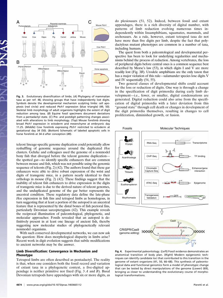
Limb Diversification: Convergence in Mechanism andPhenotypeTetrapod limbs are often described as pentadactyl. The realityis that, when one considers both the fossil record and variationof extant taxa in a phylogenetic context, a five-digited ap-pendage is neither primitive nor fixed (Fig. 3 A and B). BasalDevonian tetrapods have appendages with six or more digits, as
do plesiosaurs (51, 52). Indeed, between fossil and extantappendages, there is a rich diversity of digital number, withpatterns of limb reduction evolving numerous times in-dependently within lissamphibians, squamates, mammals, andarchosaurs. As a rule, however, extant tetrapod taxa do nothave more than five digits per limb, despite the fact that poly-dactylous mutant phenotypes are common in a number of taxa,including humans.The quest from both a paleontological and developmental per-
spective has been to look for underlying regularities and mecha-nisms behind the process of reduction. Among vertebrates, the lossof peripheral digits before central ones is a common sequence bestdescribed by Morse’s law (53), in which digits I and V are morereadily lost (Fig. 3B). Urodele amphibians are the only taxon thathas a major violation of this rule—salamander species lose digits Vand IV sequentially (54, 55).Two general classes of developmental shifts could account
for the loss or reduction of digits. One way is through a changein the specification of digit primordia during early limb de-velopment—i.e., fewer, or smaller, digital condensations aregenerated. Digital reduction could also arise from the specifi-cation of digital primordia with a later deviation from this“ground state” through cell death or changes in development ofthe digit primordia themselves, resulting in changes to cellproliferation, diminished growth, or fusion.
Horse Cow Hippo Pig Camel Jerboa Mouse
Placental mammalsArtiodactyls
A
B
CPerissodactyls
IIIII
IVV
IIIII
IV II
III
IV
III
†Hyracotherium†Mesohippus
†MerychippusEquus
Mouse Ptch1
Cow Ptch1
Horse apoptosis
Fig. 3. Evolutionary diversification of limbs. (A) Phylogeny of mammaliantaxa as per ref. 48, showing groups that have independently lost digits.Symbols denote the developmental mechanism sculpting limbs: cell apo-ptosis (red circle) and reduced Ptch1 expression (blue triangle) (49, 50).Skeletal limb morphology of adult organisms highlights the extent of digitreduction among taxa. (B) Equine fossil specimens document deviationsfrom a pentadactyl state. (C) Pre- and postdigit patterning changes associ-ated with alterations to limb morphology. (Top) Mouse forelimb showingbroad Ptch1 expression in ectoderm and mesenchyme at embryonic day11.25. (Middle) Cow forelimb expressing Ptch1 restricted to ectoderm atgestational day 34 (50). (Bottom) Schematic of labeled apoptotic cells inhorse forelimb at 34 d after conception (49).
Eryops
Acanthostega
Tiktaalik
Eusthenopteron
Moythomasia
RNA-Seq
ChIP-Seq
ChromatinCapture-Seq
ATAC-Seq
TransgenesisValidation
GFP
Transcriptome
TF binding
Enhancer/geneinteraction
Epigenome
GNNNNNNNNNNNNNNN NNNNGG
CNNNNNNNNNNNNNNN NNNNCC
5’
3’
GNNNNNNNNNNNNNNN NNNgRNA
3’
5’
Cas9
Fossils Molecular Techniques
CRISPR/Cas9(genome editing)
Fig. 4. Experimental paleontology. (Left) Fossil evidence demonstrates ananatomical transition of body plan. (Right) Modern epigenomic tech-niques can identify candidate loci that contributed to this transition in thegenome of extant organisms (41, 50, 66–68). This synthesis of paleonto-logical data and functional genomics form a model of phenotypic changethat can be tested by direct manipulations of the genome (Lower) (69),bringing us closer to understanding the evolutionary course of morpho-logical transformations.
4874 | www.pnas.org/cgi/doi/10.1073/pnas.1403665112 Pieretti et al.

Classic experiments by Alberch and Gale sought to exploretaxon-specific developmental mechanisms of digit loss by usinganurans and urodeles as exemplars (55, 56). Digit loss in these taxaappears unrelated to cell death, but dependent on cell number inthe developing limb bud; mitotic inhibitors brought about taxon-specific patterns of reduction. More recent analyses in urodelesextended these results to show that the duration of Shh expression,and likely extent of cell proliferation, is correlated to the number ofdigits that ultimately form (54). A similar relation between Shh ex-pression and digit loss was uncovered in squamates (e.g., Hemiergis)(57). Together, these data reveal one likely pattern of parallelevolution in lissamphibians and amniotes: a relation of digitalloss to changes in the duration of Shh activity and cell numberduring the specification of digit primordia (58, 59).However, how general are these mechanisms phylogenetically
and developmentally? Digital reduction has happened multipletimes in amniotes; mammals with reduced appendages, for ex-ample, reveal a range of cursorial and saltatory adaptations.Comparative studies of perissodactyls, artiodactlys, and rodentsoffer clues to likely genetic factors involved. In mouse, the SHHreceptor Patched1 (Ptch1) is expressed in both the posteriormesenchyme and ectoderm of developing limb buds and restrictsthe movement of SHH across the limb (Fig. 3C, Top) (60, 61). Inthe highly reduced digital pattern of a bovid (Bos taurus), Ptch1expression is restricted to the posterior ectoderm, resulting inattenuation of SHH signaling (Fig. 3C, Middle) (50). Experi-mentally disrupting Ptch1 during mouse limb development resultedin both a change in the central axis (a kind of paraxonic patternseen in artiodactyls) and oligodactyly, indicating that loss ofPtch1 is sufficient to phenocopy digital features of the cow limb(50). As such, the reduction of Ptch1 expression in cow providesa molecular clue for the loss of digit asymmetry in bovids. Pigs,another artiodactyl taxon, also reveal posteriorly restricted Ptch1expression in forelimbs, suggesting a role for alterations of theSHH pathway in mammalian limb reduction.As more taxa are added to the analysis, other mechanisms for
reduction emerge. In a basal artiodactyl, such as a camel, Cooperet al. discovered that Ptch1 expression is unaltered in camel limbmesenchyme. Rather, programmed cell death sculpts the auto-pod after digit patterning has taken place (49). Investigation ofperissodactyl (horse; Equus) forelimbs (Fig. 3C, Bottom) androdent (three-toed jerboa; Dipis saggita) hind limbs found thatautopodial remodeling results from cell apoptosis and expansionof Msx2 expression, a transcription factor associated with apo-ptotic pathways (62). This finding suggests the possibility that themechanisms for digit reduction were coopted from pathwayscontrolling interdigital cell death in the limb (49).These studies show that digit reduction through cell apoptosis
appears to be a convergently evolved trait among rodents (jerboa),perissodactyls (horse), and some artiodactyls (camel) (Fig. 3A).However, among other amniotes—squamates (e.g., Hemiergis),derived artiodactyls such as cow, and pig—alterations to SHHsignaling causes early patterning changes during limb development.The message from both paleontology and development is one ofextraordinary flexibility: The independent evolution of commonpatterns of digital reduction can result from the parallel evolutionof different kinds of genetic and developmental perturbations indiverse taxa.
A Future of the Fossil RecordFor decades, paleontologists have, in fits and starts, discussedways to synthesize molecular and geological data to understand therates and patterns of evolution (16, 17, 63). This interdisciplinary
integration can conspire to explore a range of issues including theanalysis of rates of evolution, topologies of phylogenetic trees, themechanics of evolutionary diversification, and the evolution of nov-elties, whether genetic, developmental, or morphological. Fossils,when placed in a phylogenetic context, can reveal extinct conditionsof stem taxa, unique combinations of characters, and the temporalsequence in the development of novelties (17, 64, 65). These featuresgive paleontology the power to shape experiments on the genome,epigenome, and development and explore the patterns and pro-cesses of morphological transformations (65).The arsenal of genomic tools available to understand
morphological diversity is ever growing, putting evolutionary-developmental biologists in a position to rapidly identify—andultimately characterize—the developmental and morphologicalroles of candidate genes and their regulatory elements in bothmodel and nonmodel organisms (Fig. 4). However, these epi-genomic and transgenic techniques offer a “passive” snapshotof a particular time point and locus in the organism of interest,begging the question of functional assays. Recent revolutionarytechniques in genome editing may finally allow biologists to modifygenomes and access an unprecedented new level of functional data.Until recently, rigorous genetic approaches to modify endog-
enous loci were limited for use only in model organisms, and ina time-consuming and expensive manner. Jinek et al. have usedthe breakthrough CRISPR/Cas9 (Clustered Regularly InterspacedShort Palindromic Repeats) system to cause double-strandedbreaks at targeted genomic sites by taking advantage of theadaptive immune response discovered in bacteria (69). CRISPR/Cas9 has been rapidly applied to produce targeted knockouts ina range of organisms (e.g., mouse, zebrafish, Xenopus, Drosophila,and human cell lines) (70), allowing researchers to directly ma-nipulate genomes and test hypotheses of morphological evolution.We are at an age in which expeditionary paleontological in-
vestigation for transitional forms, and high-resolution fossil im-aging, yielding new insights into previously hidden parts of thefossil record, can be part of a research program that encompassesgenomic and developmental biology (Fig. 4). Paleontologicaldiscovery of critical stem taxa with intermediate conditions orcharacter combinations and the elucidation of the sequence ofthe assembly of complex morphological novelties can shape mo-lecular research. Genomic and developmental biology, with anever-expanding array of experimental tools, can be used to testpaleontological hypotheses, amplify them, or reveal where criticalfossils may be lacking in the tree of life. A kind of “experimentalpaleontology” is on the horizon, in which morphological trans-formations revealed by the phylogenetic analysis of the fossilrecord may be physically assayed in the laboratory (71–73). Whatdo we need for this future to happen? It will take new or newlyinterpreted fossils from critical nodes of the tree of life, genomesfrom diverse species, and the further expansion of our moleculartoolkit for the identification, characterization, and modificationof genomes from nonmodel organisms. This synthesis is one ofmany new and promising futures of the fossil record.
ACKNOWLEDGMENTS. We thank John Westlund for aid in illustration andgraphics, and David Jablonski, Cliff Tabin, Michael Coates, and two anonymousreferees for comments. This work was supported by Graduate Assistance in Areasof National Need Grant P200A120178 (to J.P.); National Institutes of Health GrantT32 HD055164 and National Science Foundation Doctoral Dissertation Improve-ment Grant 1311436 (to A.R.G.); Uehara Memorial Foundation Research Fellow-ship 2013, Japan Society for the Promotion of Science Postdoctoral ResearchFellowship 2012-127, and Marine Biological Laboratory Research Award 2014(to T.N.); Brazilian National Council for Scientific and Technological Development(CNPq) Grants 402754/2012-3 and 477658/2012-1 (to I.S.); the Brinson Foundation(N.H.S.); and the University of Chicago Biological Sciences Division.
1. Foth C, Tischlinger H, Rauhut OWM (2014) New specimen of Archaeopteryx provides
insights into the evolution of pennaceous feathers. Nature 511(7507):79–82.2. Bajpai S, Gingerich PD (1998) A new Eocene archaeocete (Mammalia, Cetacea) from
India and the time of origin of whales. Proc Natl Acad Sci USA 95(26):15464–15468.
3. Luo Z-X, Chen P, Li G, Chen M (2007) A new eutriconodont mammal and evolutionary
development in early mammals. Nature 446(7133):288–293.4. Shubin NH, Daeschler EB, Jenkins FA, Jr (2006) The pectoral fin of Tiktaalik roseae and
the origin of the tetrapod limb. Nature 440(7085):764–771.
Pieretti et al. PNAS | April 21, 2015 | vol. 112 | no. 16 | 4875
EVOLU
TION
SPEC
IALFEATU
RE

5. Shubin NH, Jenkins FA (1995) An early Jurassic jumping frog. Nature 337(7):49–51.6. Gao K-Q, Shubin NH (2012) Late Jurassic salamandroid from western Liaoning, China.
Proc Natl Acad Sci USA 109(15):5767–5772.7. Gao T, et al. (2014) The first flea with fully distended abdomen from the Early Cre-
taceous of China. BMC Evol Biol 14:168.8. LaPolla JS, Dlussky GM, Perrichot V (2013) Ants and the fossil record. Annu Rev En-
tomol 58:609–630.9. Hughes NC (2007) The evolution of trilobite body patterning. Annu Rev Earth Planet
Sci 35:401–434.10. Kemp TS (1988) Haemothermia or Archosauria? The interrelationships of mammals,
birds and crocodiles. Zool J Linn Soc 92(1):67–104.11. Chiappe LM, Dyke GJ (2006) The early evolutionary history of birds. J Paleont Soc
Korea 22(1):133–151.12. Thewissen JGM, Cooper LN, Clementz MT, Bajpai S, Tiwari BN (2007)Whales originated
from aquatic artiodactyls in the Eocene epoch of India. Nature 450(7173):1190–1194.13. Müller J, et al. (2010) Homeotic effects, somitogenesis and the evolution of vertebral
numbers in recent and fossil amniotes. Proc Natl Acad Sci USA 107(5):2118–2123.14. Piekarski N, Gross JB, Hanken J (2014) Evolutionary innovation and conservation in
the embryonic derivation of the vertebrate skull. Nat Commun 5(5661):5661.15. Fusco G, Hong PS, Hughes NC (2014) Positional specification in the segmental growth
pattern of an early arthropod. Proc Biol Sci 281(1781):20133037.16. Runnegar B (1986) Molecular palaeontology. Palaeontology 29:1–24.17. Peterson KJ, Summons RE, Donoghue PCJ (2007) Molecular palaeobiology. Palae-
ontology 50(4):775–809.18. Urdy S, Wilson LAB, Haug JT, Sánchez-Villagra MR (2013) On the unique perspective
of paleontology in the study of developmental evolution and biases. Biol Theory8(3):293–311.
19. Gegenbaur C (1878) Elements of Comparative Anatomy (MacMillan, London).20. Sansom RS (2010) Taphonomy and affinity of an enigmatic Silurian vertebrate,
Jamoytius kerwoodi White. Palaeontology 53(6):1393–1409.21. Thacher JK (1877) Median and paired fins, a contribution to the history of the ver-
tebrate limbs. Trans Connect Acad Arts Sci 3:281–310.22. Mivart SG (1879) Notes on the fins of elasmobranchs, with considerations on the
nature and homologues of vertebrate limbs. Trans Zool Soc London 10(10):439–484.23. Balfour MF (1881) On the development of the skeleton of the paired fins of Elas-
mobranchii, considered in relation to its bearings on the nature of the limbs of thevertebrata. Proc Zool Soc Lond 1881:656–671.
24. Gillis JA, Dahn RD, Shubin NH (2009) Shared developmental mechanisms pattern thevertebrate gill arch and paired fin skeletons. Proc Natl Acad Sci USA 106(14):5720–5724.
25. Freitas R, Zhang G, Cohn MJ (2006) Evidence that mechanisms of fin developmentevolved in the midline of early vertebrates. Nature 442(7106):1033–1037.
26. Dahn RD, Davis MC, Pappano WN, Shubin NH (2007) Sonic hedgehog function inchondrichthyan fins and the evolution of appendage patterning. Nature 445(7125):311–314.
27. Pradel A, Maisey JG, Tafforeau P, Mapes RH, Mallatt J (2014) A Palaeozoic shark withosteichthyan-like branchial arches. Nature 509(7502):608–611.
28. Cohn MJ, Izpisúa-Belmonte JC, Abud H, Heath JK, Tickle C (1995) Fibroblast growthfactors induce additional limb development from the flank of chick embryos. Cell80(5):739–746.
29. Yonei-Tamura S, et al. (2008) Competent stripes for diverse positions of limbs/fins ingnathostome embryos. Evol Dev 10(6):737–745.
30. Davis MC, Dahn RD, Shubin NH (2007) An autopodial-like pattern of Hox expression inthe fins of a basal actinopterygian fish. Nature 447(7143):473–476.
31. Coates M (2003) The evolution of paired fins. Theory Biosci 122:266–287.32. Janvier P (2007) Major transitions in Vertebrate Evolution, eds Anderson JS, Sues H-D
(Indiana Univ Press, Bloomington, IN), pp 57–121.33. Janvier P (1996) Early Vertebrates (Oxford Univ Press, Oxford).34. Zhu M, et al. (2013) A Silurian placoderm with osteichthyan-like marginal jaw bones.
Nature 502(7470):188–193.35. Goujet D, Young G (2004) Recent Advances in the Origin and Early Radiation of
Vertebrates, eds Arratia G, Wilson MVH, Cloutier R (Dr Friedrich Pfeil, Munich), pp109–126.
36. Schneider I, Shubin NH (2013) The origin of the tetrapod limb: From expeditions toenhancers. Trends Genet 29(7):419–426.
37. Wagner GP (2014) Homology, Genes, and Evolutionary Innovation (Princeton UnivPress, Princeton).
38. Zakany J, Duboule D (2007) The role of Hox genes during vertebrate limb de-velopment. Curr Opin Genet Dev 17(4):359–366.
39. Montavon T, et al. (2011) A regulatory archipelago controls Hox genes transcriptionin digits. Cell 147(5):1132–1145.
40. Berlivet S, et al. (2013) Clustering of tissue-specific sub-TADs accompanies the regu-lation of HoxA genes in developing limbs. PLoS Genet 9(12):e1004018.
41. Woltering JM, Noordermeer D, Leleu M, Duboule D (2014) Conservation and di-vergence of regulatory strategies at Hox Loci and the origin of tetrapod digits. PLoSBiol 12(1):e1001773.
42. Gehrke AR, et al. (2015) Deep conservation of wrist and digit enhancers in fish. Proc
Natl Acad Sci USA 112(3):803–808.43. Schneider I, et al. (2011) Appendage expression driven by the Hoxd Global Control
Region is an ancient gnathostome feature. Proc Natl Acad Sci USA 108(31):
12782–12786.44. Amemiya CT, et al. (2013) The African coelacanth genome provides insights into
tetrapod evolution. Nature 496(7445):311–316.45. Ahn D, Ho RK (2008) Tri-phasic expression of posterior Hox genes during de-
velopment of pectoral fins in zebrafish: Implications for the evolution of vertebrate
paired appendages. Dev Biol 322(1):220–233.46. Freitas R, Zhang G, Cohn MJ (2007) Biphasic Hoxd gene expression in shark paired fins
reveals an ancient origin of the distal limb domain. PLoS ONE 2(8):e754.47. Woltering JM, Duboule D (2010) The origin of digits: Expression patterns versus
regulatory mechanisms. Dev Cell 18(4):526–532.48. Meredith RW, et al. (2011) Impacts of the Cretaceous Terrestrial Revolution and KPg
extinction on mammal diversification. Science 334(6055):521–524.49. Cooper KL, et al. (2014) Patterning and post-patterning modes of evolutionary digit
loss in mammals. Nature 511(7507):41–45.50. Lopez-Rios J, et al. (2014) Attenuated sensing of SHH by Ptch1 underlies evolution of
bovine limbs. Nature 511(7507):46–51.51. Coates MI, Clack JA (1990) Polydactyly in the earliest known tetrapod limbs. Nature
347:66–69.52. Richardson MK, Chipman AD (2003) Developmental constraints in a comparative
framework: A test case using variations in phalanx number during amniote evolution.
J Exp Zoolog B Mol Dev Evol 296(1):8–22.53. Morse ES (1872) On the tarsus and carpus of birds. Ann Lyc Nat Hist NY 10:141–158.54. Stopper GF, Wagner GP (2007) Inhibition of Sonic hedgehog signaling leads to pos-
terior digit loss in Ambystoma mexicanum: Parallels to natural digit reduction in ur-
odeles. Dev Dyn 236(1):321–331.55. Alberch P, Gale EA (1985) A developmental analysis of an evolutionary trend: Digital
reduction in amphibians. Evolution 39(1):8–23.56. Alberch P, Gale EA (1983) Size dependence during the development of the amphibian
foot. Colchicine-induced digital loss and reduction. J Embryol Exp Morphol 76:
177–197.57. Shapiro MD, Hanken J, Rosenthal N (2003) Developmental basis of evolutionary digit
loss in the Australian lizard Hemiergis. J Exp Zoolog B Mol Dev Evol 297(1):48–56.58. Zhu J, et al. (2008) Uncoupling Sonic hedgehog control of pattern and expansion of
the developing limb bud. Dev Cell 14(4):624–632.59. Harfe BD, et al. (2004) Evidence for an expansion-based temporal Shh gradient in
specifying vertebrate digit identities. Cell 118(4):517–528.60. Goodrich LV, Johnson RL, Milenkovic L, McMahon JA, Scott MP (1996) Conservation of
the hedgehog/patched signaling pathway from flies to mice: Induction of a mouse
patched gene by Hedgehog. Genes Dev 10(3):301–312.61. Butterfield NC, et al. (2009) Patched 1 is a crucial determinant of asymmetry and digit
number in the vertebrate limb. Development 136(20):3515–3524.62. Ferrari D, et al. (1998) Ectopic expression of Msx-2 in posterior limb bud mesoderm
impairs limb morphogenesis while inducing BMP-4 expression, inhibiting cell pro-
liferation, and promoting apoptosis. Dev Biol 197(1):12–24.63. Valentine JW, Campbell CA (1975) Genetic regulation and the fossil record. Am Sci
63(6):673–680.64. Patterson C (1981) Significance of fossils in determining evolutionary relationships.
Annu Rev Ecol Syst 12(1):195–223.65. Sánchez-Villagra MR (2012) Embryos in Deep Time: The Rock Record of Biological
Development (Univ of California Press, Oakland, CA).66. Wang Z, Gerstein M, Snyder M (2009) RNA-Seq: A revolutionary tool for tran-
scriptomics. Nat Rev Genet 10(1):57–63.67. Tena JJ, et al. (2011) An evolutionarily conserved three-dimensional structure in the
vertebrate Irx clusters facilitates enhancer sharing and coregulation. Nat Commun
2:310.68. Buenrostro JD, Giresi PG, Zaba LC, Chang HY, Greenleaf WJ (2013) Transposition of
native chromatin for fast and sensitive epigenomic profiling of open chromatin, DNA-
binding proteins and nucleosome position. Nat Methods 10(12):1213–1218.69. Jinek M, et al. (2012) A programmable dual-RNA-guided DNA endonuclease in
adaptive bacterial immunity. Science 337(6096):816–821.70. Sander JD, Joung JK (2014) CRISPR-Cas systems for editing, regulating and targeting
genomes. Nat Biotechnol 32(4):347–355.71. Harjunmaa E, et al. (2014) Replaying evolutionary transitions from the dental fossil
record. Nature 512(7512):44–48.72. Abitua PB, Wagner E, Navarrete IA, Levine M (2012) Identification of a rudimentary
neural crest in a non-vertebrate chordate. Nature 492(7427):104–107.73. Davidson EH, Erwin DH (2006) Gene regulatory networks and the evolution of animal
body plans. Science 311(5762):796–800.
4876 | www.pnas.org/cgi/doi/10.1073/pnas.1403665112 Pieretti et al.


![Decision Trees for Hierarchical Multilabel Classification ... · text classification [1] and functional genomics [2]. In functional genomics, an important problem is predicting](https://img.pdfslide.us/doc/110x75/5fb675a0ec941d1cdc0931b7/decision-trees-for-hierarchical-multilabel-classiication-text-classiication.jpg)