Embed Size (px)
Citation preview

Oral treatments for amoebic gill disease (AGD)
in Atlantic salmon, Salmo salar
A Thesis
Submitted to the Graduate Faculty
in Fulfilment of the Requirements
for the Degree of
Doctor of Philosophy
In the National Centre for
. Marine Conservation and Resource Sustainability
University of Tasmania
Renee L. Louwen-Skovdam (nee Florent)
Launceston, Tasmania
November, 2008
2008. R.L. Louwen-Skovdam (nee Florent)

Declaration and Authority of Access
Declaration
This thesis contains no material which has been accepted for a degree or diploma
by the university or any other institution, except by way of background information
and duly acknowledged in the thesis and to the best of my knowledge and belief no
material previously published or written by another person except where due
acknowledgements are made in the text of this thesis. At the time of publication,
Chapter 2 had been submitted, Chapter 5 was in press and Chapters 3 and 4 had
been published within peer reviewed journals.
klo-uk,u-cAkaucta.rv\
Renee Louwen-Skovdam (nee Florent)
Authority of Access
This thesis may be available for loan and limited copying in accordance with the
Copyrights act 1968.
kitat,LLAY-e.nifk.,v
Renee Louwen-Skovdam (nee Florent)

Statement of Co-Authorship
Statement of Co-Authorship
The following people and institutions Contributed to the publication of the work
undertaken as part of this thesis:
Chapter 2 Florent, R.L., Becker, J.A., Powell, M. D. Submitted. In vitro efficacy
of bithionol and bithionol sulphoxide to Neoparamoeba spp. the
causative agent of amoebic gill disease (AGD). Dis. Aquat. Org ..
Renee L. Florent (90%), Dr. Joy A. Becker (5%), Prof. Mark D.
Powell (5%)
Chapter 3 Florent, R.L., Becker, J.A., Powell, M. D., 2007. Evaluation of
bithionol as a bath treatment for amoebic gill disease caused by
Neoparamoeba spp. Vet. Par. 144, 197-207. Renee L. Florent (80%),
Dr. Joy A. Becker (10%), Prof. Mark D. Powell (10%)
Chapter 4 Florent, R.L., Becker, J.A., Powell, M. D., 2007. Efficacy of bithionol
as an oral treatment for amoebic gill disease in Atlantic salmon, Salm°
salar. Aquaculture 270, 15-22. Renee L. Florent (80%), Dr. Joy A. .
Becker (10%), Prof. Mark D. Powell (10%)
Chapter 5 Florent, R.L., Becker, J.A., Powell, M. D. In press. Further
development of bithionol therapy as a treatment for AGD. J. Fish Dis.
Renee L. Florent (80%), Dr. Joy A. Becker (10%), Prof. Mark D.
' Powell (10%)
• Dr. Joy A. Becker and Prof. Mark D. Powell contributed to the idea, its formalisation and development, and assisted with refinement and presentation.
We the undersigned agree with the above stated "proportion of work undertaken" for each of the above published (or submitted) peer-reviewed manuscripts contributing to this thesis:
Statement of Co-Authorship
Signed: 01. John Purser) Supervisor National Centre for Marine Conservation and Resource Sustainability University of Tasmania
(Prof Chad Hewitt) Director National Centre for Marine Conservation and Resource Sustainability University of Tasmania
Date . ze0/, los?
iv

Abstract
Abstract
Neoparamoeba spp. is a marine amphizoic protozoan parasite which infects the
gills of marine cultured Atlantic salmon, Salmo salar, worldwide causing amoebic
gill disease (AGD). Amoebic gill disease is a significant health issue affecting the
production of sea-caged Atlantic salmon in Australia with farms experiencing
outbreaks regularly throughout the year. It accounts for 10-20% of the gross value
of production due not only to the cost of treating and managing the disease, but also
to loss of fish condition, increased feed conversion ratio (FCR), lost growth and
sometimes mortalities. The current mitigation strategy for AGD is the
administration of a freshwater bath to affected sea-caged fish. However, this
method is becoming less effective with an apparent increase in bathing frequency
over the past few years. The increase in baths has fuelled a rise in already high
production costs to the Australian Atlantic salmon industry to approximately 20%
annually. This thesis aims to identify an improved method of treatment for AGD
either through development of a stand alone in-feed treatment or an in-feed
treatment used in conjunction with the current freshwater treatment strategy.
This thesis investigates in vitro and in vivo effects of bithionol and bithionol
sulphoxide on both Neoparamoeba spp. and Atlantic salmon. Initially, toxicity to
Neoparamoeba spp. was examined in vitro using isolated gill amoeba and exposing
them to seawater, freshwater, alumina (10 mg 1: 1 ), bithionol and bithionol
sulphoxide at 10, 5, 1, 0.5 and 0.1 mg L. The assays were observed for 72 h with
viable amoeba counts using trypan blue exclusion conducted at 0, 24, 48 and 72 h.
Both bithionol and bithionol sulphoxide were toxic to Neoparamoeba spp. in vitro

Abstract
at all concentrations examined. A similar toxicity to freshwater water was observed
with bithionol and bithionol sulphoxide at 10 and 5 mg L-1 following a 72 h
treatment. However, freshwater was the most effective with only 6% viable
amoebae seen after 24 h and no viable amoeba observed a further 24 h later.
Once identified as toxic to Neoparamoeba spp. in vitro, an assessment of the
toxicity of bithionol to Atlantic salmon and the efficacy as an AGD treatment was
evaluated. This was conducted via a bath treatment to Atlantic salmon and rainbow
trout, Oncorhynchus mykiss, held in either fresh or seawater using concentrations
between 1 and 35 mg L-1 to examine toxicity. To examine efficacy, a bath treatment
of AGD-affected Atlantic salmon and rainbow trout at 1 to 25 mg L -1 was also
evaluated. To examine toxicity, fish were bathed for 1, 3 and 6 h in bithionol, an
anti-protozoal at 0, 1, 5, 10, 25 and 35 mg L -1 , with toxicity determined by time to
morbidity and histological examination of internal organs. Efficacy was examined
by bathing AGD-affected Atlantic salmon and rainbow trout for I h at bithionol
concentrations of 1 to 25 mg L -1 . Efficacy was determined by examining gill
amoeba counts and identifying percent lesioned gill filaments at I and 24 h after
bath exposure to bithionol. Only bithionol at 1 mg L-1 was considered non-toxic
with no signs of morbidity. Bithionol appeared to be more toxic in seawater than
freshwater, exhibiting a higher rate of morbidity, and had no acute effects on gill
Na+/K+ ATPase and succinic dehydrogenase, or plasma osmolality and chloride
concentration. Bithionol reduced the percentage of lesioned gill filaments to the
same level as freshwater.
vi
Abstract
Bithionol was examined as an in-feed treatment for AGD with and without the
administration of a freshwater bath. Bithionol when fed as a two week prophylactic
or therapeutic treatment at 25 mg kg' feed delayed the onset of AGD pathology and
reduced the percent lesioned gill filaments. Administration of a 3 h freshwater bath
at 28 days post-exposure significantly reduced amoebae numbers to a similar level
across all treatments; in contrast gross gill score and percent lesioned filaments
were reduced proportionally. Hence, the control was significantly higher than both
bithionol treatments. Following the freshwater bath, clinical signs of AGD recurred
at a similar level across all treatments although controls clinical signs were
significantly higher than the bithionol treatments to begin with. Palatability was not
a problem with mean feed intake of bithionol over the trial duration higher
compared to both the oil and plain controls.
This thesis has identified that bithionol at 25 mg kg -I feed, when fed as a two
week prophylactic or a therapeutic treatment, delayed and reduced the intensity of
AGD pathology. Such findings as the identification of bithionol as a possible in-
feed treatment for AGD and its effectiveness against numerous other parasites
suggests that bithionol could be worth examining in other aquatic animal diseases.
Furthermore, bithionol warrants further investigation as a potential in-feed
treatment for AGD in Atlantic salmon especially in regards to a combination
therapy with the current freshwater mitigation.
vii
Acknowledgements
Acknowledgments
Undertaking a PhD is a mammoth task and it requires a team of dedicated people
to provide support both at work and at home. I am forever indebted to each of the
following people and organisations that have made it possible to survive my PhD.
First and foremost I would like to thank my supervisor, Prof Mark Powell, for an
immeasurable amount of assistance, guidance, encouragement and opportunity over
the last three years. Your enthusiasm, and motivation as a researcher are
inspirational and I am truly grateful for everything. Thank you so much.
Various components of this work would not have been possible without my co-
supervisors, Dr. Joy Becker and Dr. John Purser your knowledge, assistance and
guidance through the project has been greatly appreciated, without you some of the
work would not have been possible. Thank you.
Thanks to Dr. Mark Adams for histological and image analysis advice — what
would I do without Mark!
Also thanks to many people and organisations that have assisted me, whether it is
technically, financially or otherwise over the duration of my candidature: Paul
Cassidy, Helen Stratham, Jan Daniels, Debbie Wagner, Annabel Tyson, Detlef
Planko, Greg Kent and Matt Foale who did their up-most to help all graduate
students during their studies and whom always have the time to help. Furthermore,
Aquafin CRC for their generous stipend, the UTAS AGD research group, Julie
viii

Acknowledgements
Ransome, Megan Barney, Matthew Jones, FRDC, EAFP, Dr. Melanie Leef, Dr.
Kally Gross and anyone else that I have forgotten to mention.
To my family you have been wonderful, not only now but your support
throughout the years has been without limits. I owe the warmest of thanks to my
mum and dad for making the big trip to Tassie for without you I would not have
come. To my grandmother Judith, for proof reading of all manuscripts, correcting
all grammar and spelling. To my sister Michelle, thank you very much for all your
labelling of jars and folding of foil envelopes, you made setting up for experiments
so much easier and to Maite, for your love and support and many a night playing
canasta to relieve the stress I thank you.
Lastly to my beloved husband Torben, it is you that has made this all worthwhile,
without you I would not have made it through. I began this without you and to
finish it with you is a real joy. You were my strength and I am very grateful to have
had you in my life these past few years. Thank you for being my emotional
punching bag and my Dacron cleaner extraordinaire. It is to you that I dedicate this
thesis.
ix

List of Abbreviations
List of Abbreviations
AGD amoebic gill disease ANOVA analysis of variance AS Atlantic salmon ATP adenosine 5'-triphosphate BW body weight
control feed CRC Cooperative Research Centre EC50 median effective concentration EDTA ethylenediaminetetraacetic acid FCR feed conversion ratio FDA Food and Drug Administration FRDC Fisheries Research and Development Corporation fw freshwater H & E haematoxylin and eosin IFAT indirect fluorescent antibody test ILU inter lamellae units IQR inter-quartile range
Fulton's condition factor LC50 median lethal concentration LCEE L-cysteine ethyl ester LT lethal time LT50 median lethal time Na not applicable NADH reduced nicotinamide adenine dinucleotide Na±/K+ ATP sodium potassium adenosine 5'-triphosphate PB prophylactic bithionol feed
. PCO2 partial pressure of carbon dioxide PCR polymerase chain reaction PE post exposure PLOs Perkinsiella amoebae like organisms P02 partial pressure of oxygen RBT rainbow trout RPR relative percent reduction R & D research and development SALTAS Salmon Enterprises of Tasmania Pty Ltd SDH succinic dehydrogenase SEID sucrose, Na2EDTA, imidazole and Natdeoxycholic acid SEM standard error of the mean SGR specific growth rate sw seawater TB therapeutic bithionol feed

Table of Contents
Table of Contents Declaration . ii Authority of Access ii - Statement of Co-Authorship iii Abstract Acknowledgments viii List of Abbreviations • x Table of Contents xi List of Figures xiv List of Tables xvi
1 General Introduction - 2 -
1.1 The impact of amoebic gill disease (AGD) on Atlantic salmon, Salmo salar, in Australia - 2 - 1.2 The etiology of amoebic gill disease - 3 -
1.2.1 The agents associated with amoebic gill disease -3 - 1.2.2 Morphology and identification - 5 -
1.3 Pathology and pathophysiology of amoebic gill disease in Atlantic salmon - 8 - 1.4 Freshwater therapy for amoebic gill disease - 11 -
1.4.1 History -11- 1.4.2 Effects of freshwater bathing - 12 -
1.5 Alternative treatments for amoebic gill disease - 14 - 1.5.1 Chemotherapeutant drugs tested to date - 14 - 1.5.2 Bithionol and bithionol sulphoxide as novel treatments - 17 -
1.6
Fish health management - the host, pathogen and environment interaction - 20 -
1.7 Research objectives and specific aims - 23 - 1.7.1 Specific aims - 23 -
2 In vitro efficacy of bithionol and bithionol sulphoxide to Neoparamoeba spp. the causative agent of amoebic gill disease (AGD) - 27 -
2.1 Abstract - 27 - 2.2 Introduction - 28 - 2.3 Materials and Methods - 30 -
2.3.1 Amoeba isolation by adherence - 30 - 2.3.2 In vitro toxicity assay - 31 - 2.3.3 Treatments - 32 - 2.3.4 Statistical Analyses - 33 -
2.4 Results - 33 - 2.5 Discussion - 39 - 2.6 Acknowledgements - 43 -
xi

Table of Contents
3 Evaluation of bithionol as a bath treatment for amoebic gill disease caused by Neoparamoeba spp. - 45 -
3.1 Abstract -45- 3.2 Introduction - 46 - 3.3 Materials and Methods - 48 -
3.3.1 Toxicity Study - 48 - 3.3.1.1 Fish Husbandry -48- 3.3.1.2 Bath Administration - 49 - 3.3.1.3 Data Collection -50- 3.3.1.4 Histology -51 -
3.3.2 Efficacy Study - 52 - 3.3.2.1 Fish Husbandry - 52 - 3.3.2.2 Amoeba isolation and exposure - 53 - 3.3.2.3 'Bath administration - 54 - 3.3.2.4 Data Collection - 54 - 3.3.2.5 Histology -55 -
3.3.3 Statistical Analysis - 55 - 3.4 Results - 56 -
3.4.1 Toxicity Study - 56 - 3.4.2 Efficacy - 61 -
3.5 Discussion - 64 - 3.6 Acknowledgements - 70 -
4 Efficacy of bithionol as an oral treatment for amoebic gill disease in Atlantic salmon, Salmo salar - 72 -
4.1 Abstract - 72 - 4.2 Introduction - 73 - 4.3 Materials and Methods - 76 -
4.3.1 Fish husbandry and maintenance - 76 - 4.3.2 Experimental design and challenge method - 76 - 4.3.3 Feed preparation - 78 - 4.3.4 Data Collection - 79 - 4.3.5 Histology - 80 - 4.3.6 Statistical Analysis - 81 -
4.4 Results - 81 - 4.5 Discussion - 87 - 4.6 Conclusion - 90 - 4.7 Acknowledgements - 91 -
xii

Table of Contents
5 Further development of bithionol therapy as a treatment for AGD - 93 -
5.1 Abstract - 93 - 5.2 Introduction - 94 - 5.3 Materials and Methods - 98 -
5.3.1 Fish husbandry and maintenance -98- 5.3.2 Experimental design and challenge method - 98 - 5.3.3 Bath Administration - 100 - 5.3.4 Feed preparation - 101 - 5.3.5 Data Collection - 101 -
5.4 Histology - 103 - 5.4.1 Statistical Analyses - 104 -
5.5 Results - 105 - 5.6 Discussion - 114 - 5.7 Acknowledgements - 118 -
6 General Discussion - 120 -
6.1 Conclusion - 130 -
7 References - 133 -
8 Appendix I - 165 -
8.1 Trypsin-EDTA solution in Hanks Balanced Salts Contents and concentrations - 165 -

List of Figures
List of Figures Number Page
Fig 1.2-1. Attached trophozoites of Neoparamoeba pemaquidensis from a sediment isolate showing pseudopodia (fine arrow), nucleus (unfilled arrow), and "parasome" (filled arrow) (plate courtesy of Dr Bret Robinson) Bar = 201.1m - 6 -
Fig 1.5-1. Chemical structure of bithionol (2,2'-thiobis (4,6-dichlorophenol))..- 17 -
Fig 1.5-2. Chemical structure of bithionol sulphoxide (Bis (2-hydroxy-3,5- dichlorophenyl) sulphoxide) - 18 -
Fig 2.4-1. Effect of time and treatment on mean (± SEM) number of viable isolated amoeba (as a percentage of the seawater control) when exposed to either freshwater
), 10 mg L' 1 alumina ('' 4), seawater (EWE), or bithionol at 0.1, 0.5, 1,5 and 10 mg L-1 (1 1,1 1, 1T 1, 1 I, and ....DD. respectively) (n = 24). Seawater controls remained at 10 000 cells well' and are presented as 100% survival. Common letter across both time and treatment indicates no significant different using a Tukey's test (p > 0.05). - 36 -
Fig 2.4-2. Effect of time and treatment on mean (± SEM) number of viable isolated amoeba (as a percentage of the seawater control) when exposed to either freshwater
), 10 mg L' 1 alumina (21w), seawater ( ), or bithionol sulphoxide at 0.1, 0.5, 1, 5 and 10 mg L' (I 1,1 I , 1 1, 1 I, and respectively) (n = 24). Seawater controls remained at 10 000 cells well' and are presented as 100% survival. Common letter across both time and treatment indicates no significant different using a Tukey's test (p > 0.05). - 38 -
Fig 3.4-1. Mean (± SEM) (a) plasma osmolality and (b) plasma chloride for freshwater (open circle) and seawater (closed circle) (i) Atlantic salmon and (ii) rainbow trout when exposed to varying bithionol bath concentrations. Asterisks denotes a significant difference between fresh and seawater. - 59 -
Fig 3.4-2. Mean (± SEM) (a) gill Na+ / KtATPase activity and (b) gill succinic dehydrogenase (SDH) activity for freshwater (open circle) and seawater (closed circle) (i) Atlantic salmon and (ii) rainbow trout when exposed to varying bithionol bath concentrations. Asterisks denotes a significant difference between fresh and seawater. - 60 -
Fig 3.4-3. Mean (± SEM) (a) percent lesioned gill filaments, (b) crude amoeba numbers and (c) plasma osmolality for rainbow trout (open circle) and Atlantic salmon (closed circle) when administered varying bithionol bath concentrations. Lower and upper case letters denote significant differences within Atlantic salmon and rainbow trout, respectively. - 63 -
Fig 4.4-1. (a) percent lesioned gill filaments and (b) gross gill score for Atlantic salmon with amoebic gill disease (AGD) when fed either control feed (solid line) (n = 12) or bithionol at 25 mg kg' feed (broken line) (n = 6). No error bars indicate that all replicates exhibited the same value. Values are expressed as mean ± SEM..
xiv .

List of Figures
Letters denote significant differences among treatments at that sampling time (p < 0.05) - 84 -
Fig 4.4-2. (a) weekly tank feed intake (n = 3) and (b) weekly tank biomass (n = 3) for Atlantic salmon exposed to Neoparamoeba spp. when fed either commercial feed (closed circles), oil-coated commercial feed (open circles) or bithionol at 25 mg kg -I feed (broken line and open squares). Values are expressed as mean ± SEM. Letters denote significant differences among treatments over the trial duration (p < 0.05) - 85 -
Fig 5.5-1. Mean (± SEM) weekly tank feed intake (n = 3) for Atlantic salmon exposed to Neoparamoeba spp. when fed either control feed (dotted line open circle), prophylactic bithionol at 25 mg kg' feed (solid line closed circle) or therapeutic bithionol at 25 mg kg' feed (solid line open square). No error bars indicate that all replicates within a treatment exhibited the same value. The vertical broken line indicates administration of a 3 h freshwater bath. - 106 -
Fig 5.5-2. (a) gross gill score and (b) percent lesioned gill filaments for Atlantic salmon with amoebic gill disease (AGD) when fed either control feed (dotted line open circle), prophylactic bithionol at 25 mg kg -I feed (solid line closed circle) or therapeutic bithionol at 25 mg kg' feed (solid line open square) (n = 12 pre-bath or 9 post-bath). All values are expressed as mean ± SEM. No error bars indicate that all replicates within a treatment exhibited the same value. The vertical broken line indicates administration of a 3 h freshwater bath. Lower and uppercase letters denote significant differences among days and treatments, respectively (p < 0.05). ... - 110 -
Fig 5.5-3. Lesion size in inter lamellae units (ILU) for Atlantic salmon with amoebic gill disease (AGD) when fed either control feed (dotted line open circle), prophylactic bithionol at 25 mg kg -I feed (solid line closed circle) or therapeutic bithionol at 25 mg kg -I feed (solid line open square) (n = 12 pre-bath or 9 post-bath). Values are expressed as mean ± SEM. No error bars indicate that all replicates within a treatment exhibited the same value. The vertical broken line indicates administration of a 3 h freshwater bath. Lower and uppercase letters denote significant differences among days and treatments, respectively (p < 0.05). ... - 112 -
XV

List Of Tables
List of Tables Number Page
Table 1.4-1. Huon Aquaculture Company Dover, Australia method for AGD gross gill lesion scoring scheme - 12 -
Table 1.5-1. Compounds examined as possible treatmzents for amoebic gill disease. - 16 -
Table 2.4-1. The effective concentration (mg L -1 ) needed to cause 50% mortality (EC50) in Neoparamoeba spp. when exposed to bithionol (BT) or bithionol sulphoxide (BTS) for 0, 24, 48 or 72 h. - 34 -
Table 3.4-1. Median lethal time and inter-quartile range (IQR) for seawater (sw) and freshwater (fw) Atlantic salmon (AS) and rainbow trout (RBT) exposed to a 1 hour bithionol bath at a concentration of 25 or 35 mg L -1 - 57 -
Table 3.4-2. The number of moribund Atlantic salmon and rainbow trout in both freshwater (fw) and seawater (sw) observed at varying bithionol concentrations over a 6 hour (h) period. - 58 -
Table 3.4-3. The number of moribund Atlantic salmon (AS) and rainbow trout (RBT) with amoebic gill disease observed between 0 and 1 hour (0-1 h) and 1 and 24 hours (1-24 h) when exposed to either a freshwater control bath or a 1 hour bath at varying bithionol concentrations. - 61 -
Table 4.3-1. Scoring scheme for gross signs of amoebic gill disease on Atlantic salmon modified from Adams and Nowak (2003). - 80 -
Table 4.4-1. Mean tank specific growth rate (SGR) (n = 3) and individual fish condition factor (K) (n = 6) for plain control, oil control and bithionol treated Atlantic salmon feed from Day -14 to 0 and Day 0 to Day 10 Neoparamoeba spp. exposure. Values are mean ± SEM. Asterisks indicate significant difference between treatments within each time point (p<0.05) - 86 -
Table 5.3-1. Scoring scheme for gross signs of amoebic gill disease on Atlantic salmon modified from Adams and Nowak (2003). - 103 -
Table 5.5-1. Tank specific growth rate (SGR) (n = 3), individual fish condition factor (K) (n = 9) and tank feed conversion ratio (n = 3) for Atlantic salmon fed either oil coated commercial feed (C), prophylactic (PB) or therapeutic (TB) bithionol at 25 mg kg -1 feed from Day -14 to 0, Day 0 to 14, Day 14 to 28 and Day 28 to 42 from Neoparamoeba spp. exposure. All values are expressed as mean ± SEM • - 107 -
Table 5.5-2. Mean (± SEM) crude amoeba numbers and relative percent reduction pre- and post- 3 h freshwater bath for Atlantic salmon with amoebic gill disease (AGD) when fed either control feed, prophylactic bithionol at 25 mg kg' feed or therapeutic bithionol at 25 mg kg-1 feed. Letters superscripts denote significant differences among treatments (p < 0.05) (n = 9). - 113 -
xvi

CHAPTER 1
GENERAL INTRODUCTION

Chapter One General Introduction
1 General Introduction
1.1 The impact of amoebic gill disease (AGO) on Atlantic salmon, Salmo
salar, in Australia
Amoebic gill disease is a significant health issue affecting the production of sea-
caged salmon in Tasmania, Australia, with farms experiencing regular outbreaks
throughout the year (Clark and Nowak, 1999; Munday et al., 2001; Nowak et al.,
2002) accounting for 10-20% of the gross value of production (Munday et al.,
2001). However, AGD outbreaks are generally most severe during the summer
months; previous studies have shown that salinity and temperature are significant
environmental factors influencing the outbreaks of AGD (Clark and Nowak, 1999;
Douglas-Helders etal., 2001b; Adams and Nowak, 2003). Other factors that are
thought to influence AGD outbreaks include rainfall (Munday etal., 1993; Clark
and Nowak, 1999; Nowak, 2001), dissolved oxygen (Clark and Nowak, 1999),
biofouling on cages (Tan etal., 2002), husbandry techniques (Clark and Nowak,
1999), removal of mortalities (Douglas-Helders et al., 2000), the general health
status of the fish (Nowak, 2001) and possibly bacterial gill populations (Bowman
and Nowak, 2004; Embar-Gopinath etal., 2005).
The increase gross production costs mentioned above is due not only to the cost of
treating and managing the disease, but also to loss of fish condition, increased feed
conversion ratio (FCR) and lost growth attributed to an increase in standard
metabolic rate (Leef et al., 2007a). If left untreated fish mortalities can reach over
50% (Munday et al., 1990) and furthermore pose a greater financial loss to the
2

Chapter One General Introduction
industry. As well as mortalities, reduced growth can also be an issue taking a longer
time to reach market size (Rodger and McArdle, 1996; Dykova etal., 1998).
1.2 The etiology of amoebic gill disease
1.2.1 The agents associated with amoebic gill disease
The presumptive causative agent of AGD is a marine amphizoic protozoan
parasite, identified as Paramoeba pemaquidensis (Page, 1970) and then reclassified
to Neoparamoeba pemaquidensis (Page, 1987). More recently other amoeba
species, N. branchiphila and N aestuarina, were cultured from AGD affected fish,
but it was not known if one or all of these species were the aetiological agents of
AGD (Dykoyd etal., 2005). Subsequently, a new amoeba species designated
Neoparamoeba perurans n. sp. has been identified as the predominate aetiological
agent of AGD affecting Atlantic salmon, Salmo salar, culture in Tasmania
invalidating previous classifications (Young et al., 2007).
Neoparamoeba (Page, 1987) are small, naked and lobose amoebae that form
dactylopodiate subpseudopodia in their locomotive state and belong to the family
Vexilliferidae. Some defining characteristics enabling classification of this genus
include the fact that forms lack the well organised cell-surface structures of other
vexilliferids including surface scales and hexagonal glycostyles. They also possess
a nucleus plus one or more `parasomes'. These `parasomes' are described as
Perkinsiella amoebae like organisms (PLOs) and are eukaryotic endosymbionts,
closely related to the kinetoplastid khthyobodo (Dykova et al., 2003).
3

Chapter One General Introduction
Neoparamoeba spp. is not confined to Australian waters, with AGD outbreaks
worldwide including Ireland, Spain, New Zealand, USA (Washington State and
California), Chile, and more recently Norway (reviewed by Munday et al., 2001;
Nowak etal., 2002; Steinum etal., 2008). Amoebic gill disease has also been found
to affect a number of other species including Atlantic salmon (Kent etal., 1988;
Munday etal., 1990), rainbow trout, Oncorhynchus mykiss, (Munday etal., 1990),
brown trout, S. Trutta, (Munday etal., 2001), coho salmon, 0. Kisutch, (Kent etal.,
1988), chinook salmon, 0. Tshawytscha, (Kent et al., 1988), turbot, Scophthalmus
maximus, sharpsnout seabream, Diplodus puntazzo, and European sea bass,
Dicentrarchus labrax, (Dykova et al., 2000; Dykova and Novoa, 2001). Atlantic
salmon are reported to be the most susceptible of cultured species (Munday et al.,
2001), but interestingly AGD has not been reported in other wild fish species
including red cod, Pseudophycis bachus, sand flathead, Platycephalus bassensis,
and jack mackerel, Trachurus declivus, even when present around farms with
severely affected salmon (Nowak et al., 2004).
Throughout this thesis, the causative agent of AGD is referred to as
Neoparamoeba spp. Furthermore, this work was conducted prior to the discovery
of N. perurans and Neoparamoeba spp. is used in reference to the aetiological agent
of AGD. However, irrespective of the term used to describe the organism, it is
referring to the isolated gill amoebae and hence the species of Neoparamoeba
which are the aetiological agents of AGD.
4

Chapter One General Introduction
1.2.2 Morphology and identification
When freshly isolated from the gills of infected fish, Neoparamoeba spp. appear
in their free floating form as spherical and possess multiple pseudopodia (Kent et
al., 1988; Munday etal., 1990; Rodger and McArdle, 1996; Dykova etal., 1998).
However, a more lobose form is assumed when Neoparamoeba spp. are attached to
a substrate with the nucleus and "parasome(s)" generally visible (Fig 1.2-1). In
histology sections using either wax or resin, both the nucleus and "parasome(s)" are
visible and the trophozoites often appear highly vacuolated (Roubal et al., 1989;
Munday et al., 1990; Dykova etal., 1995). While morphological characteristics
distinguish Neoparamoeba from other vexilliferids, attempts to distinguish
members of the genus Neoparamoeba using morphological characteristics alone
have been unsuccessful (Dykova et al., 2000; Dykova et al., 2005).
-5-

Chapter One General Introduction
Fig 1.2-1. Attached trophozoites of Neoparamoeba pemaquidensis from a
sediment isolate showing pseudopodia (fine arrow), nucleus (unfilled arrow), and
"parasome" (filled arrow) (plate courtesy of Dr Bret Robinson) Bar = 20pm.
6

Chapter One General Introduction
Detection of Neoparamoeba spp. can be achieved using both pathogen specific
and non-specific tests with samples being obtained either lethally or non-lethally to
the fish. A wet mount preparation, whereby gill mucus is smeared onto a glass slide,
dried, stained and examined microscopically, is based primarily on the morphology
of the pathogen. Histology is reliable but the obtaining of histology samples is lethal
to the fish and once again reliant upon morphology. Specific stains, such as indirect
fluorescent antibody test (IFAT) (Howard and Carson, 1993), immuno-
cytochemistry (Zilberg and Munday, 2000; Howard, 2001) and immune-dot blot
(Douglas-Helders etal., 2001a), can also be used on histological sections and gill
smears, although not particularly reliable for assessing infections on some farms or
assessing treatment efficacy (Harris et al., 2004; Harris et al., 2005). Immunological
detection of Neoparamoeba using anti-N. pemaquidensis antiserum was reported
successful in identifying different Neoparamoeba spp. (Douglas-Helders et al.,
2001a). However, the anti-N pemaquidensis antiserum was later shown to bind
non-specifically to other marine amoebae (Morrison etal., 2005). Recently, N.
branchiphila was characterised using a combination of morphological and
molecular phylogenetic analyses inferred from 18S rRNA gene sequences (Fiala
and Dykova, 2003; Dykova et al., 2005) and was clearly differentiated from N.
pemaquidensis and N. aestuarina. Furthermore, this resolved inter-specific
relationships within the Neoparamoeba group (Fiala and Dykova, 2003; Dykova et
al., 2005). Hence, species-specific diagnostic tools were developed, based upon 18S
rRNA gene amplification by polymerase chain reaction (PCR) to study disease
aetiology where Neop" aramoeba spp. were the presumptive pathogens (Elliott et al.,
7

Chapter One General Introduction
2001; Fiala and Dykova, 2003; Wong etal., 2004; Dykova etal., 2005; Mullen et
• at, 2005; Young etal., 2007).
1.3 Pathology and pathophysiology of amoebic gill disease in Atlantic salmon
Amoebic gill disease is characterised clinically or macroscopically by the
presence of gross gill lesions which are characterised by focal or multifocal, raised
white mucoid patches, profuse mucus production and mucous cell proliferation
(Clark and Nowak, 1999; Adams and Nowak, 2001; Roberts and Powell, 2003b).
Gross examination of gills for the presence of white patches is routinely used by the
Ta.smanian Atlantic salmon industry for gross tentative diagnosis of AGD and
determination of treatment times. Other clinical signs reported for AGD include
lethargy, loss of appetite and respiratory distress manifested as rising to the water
surface and an increased ventilation frequency; however, due to routine farm
monitoring and early treatment, disease rarely progresses to the point at which such
behaviour is elicited (Munday etal., 1990; Munday etal., 2001). The prominent
microscopic feature of AGD is multi-focal hyperplasia of the lamellar epithelium
which, in turn, results in the , ftsion of secondary lamellae and formation of
interlamellar vesicles, which coincides with a reduction in chloride cell number and
an increase in mucous cells (Dykova etal., 1995; Powell etal., 2001; Adams and
Nowak, 2003; Roberts and Powell, 2003b). Typically AGD lesions are associated
with Neoparamoeba spp. present within the vicinity of the hyperplastic tissue
(Nowak and Munday, 1994). As the disease progresses, the hyperplastic tissue
along with associated amoeba are sloughed off, which has been thought to be a
8
Chapter One General Introduction
possible 'self-cleaning' action important for the recovery from the disease (Munday
etal., 2001).
Physiologically, Munday et al. (1990) reported elevated blood sodium levels in
Atlantic salmon severely affected with AGD; however, Powell et al. (2001)
reported that there were no clinical observations regarding this. Fish affected by
AGD are known to have lower blood oxygen partial pressure (P02) despite the fact
that oxygen uptake rates are not affected under normoxic conditions in clinically
affected fish (Powell etal., 2000). Elevated blood carbon dioxide (tensions) (PCO2)
are also seen in AGD-affected Atlantic salmon resulting in respiratory acidosis
(Powell etal., 2000; Leef et al., 2005b). It has been suggested that this respiratory
acidosis exhibited in AGD-affected fish maybe a result of the presence of AGD
lesions and/or an increase in branchial mucous secretion which in turn leads to a
reduced gill surface area and diffusive conductance (Powell et al., 2000). Even
though it has been shown that AGD-affected fish are acidotic, the presence of AGD
did not appear to contribute to respiratory failure when AGD-affected fish were
exposed to hypoxic condition at approximately 25% saturation. Furthermore,
suggesting that although physiological mechanisms including increased blood flow
and perfusion within the gill, AGD-affected fish were able to maintain oxygen
transport (Powell et al., 2000).
Amoebic gill disease is associated with a chronic vascular hypertension as well as
compensatory cardiac remodelling. Whilst examining the cardiovascular effects of
AGD, Powell et al. (2002b) hypothesised that in cases of chronic AGD, a
9

Chapter One General Introduction
compensatory response for high ventral (afterload) and dorsal (preload) aortic
pressure (as seen by Powell et al. (2002a)) may be to promote morphological
changes in both ventricle length and the thickening of ventricular compact muscle.
Atlantic salmon affected with AGD have been shown to have high systemic
resistance and lowered cardiac output when compared to naïve counterparts, a
characteristic of Atlantic salmon not seen in either rainbow or brown trout (Leef et
al., 2005a). There is evidence to suggest that AGD may possibly be associated with
cardiovascular dysfunction (Powell etal., 2002a; Powell et al., 2002b); this was
supported by Leef et al. (2005a; 2007b).
When affected with AGD, the critical swimming speed and subsequent recovery
of Atlantic salmon is significantly affected. Furthermore, following a freshwater
bath the swimming performance was found to increase with the suggestion that this
post-bath increase observed is due to the removal of amoeba and hyperplastic gill
tissue (M. Jones pers. comm.). The excess post-exercise oxygen consumption
doubled in AGD-affected fish compared to uninfected controls, hence infected fish
need longer to repay their oxygen debt and recover oxygen consumption rates to
routine levels (M. Jones pers. comm.). This supports earlier observations of Powell
et al. (2002a) where freshwater bathing also led to a decrease in dorsal aortic
pressure in clinically affected fish. Oxygen consumptions rates have been used
indirectly to measure metabolic rate in AGD-affected Atlantic salmon. It was
reported that both routine metabolic rate and metabolic scope are significantly
affected, with the magnitude of effect linked to the severity of AGD. Suggesting
-1 0-

Chapter One General Introduction
that AGD infection does have a significant metabolic cost associated however, only
at high infection levels (M. Jones pers. comm.).
1.4 Freshwater therapy for amoebic gill disease
1.4.1 History
The mitigation of AGD in Tasmania, Australia, is mostly due to freshwater
bathing for 2-4 h, which was first recommended by Foster and Percival (1988). On
the farm, fish are routinely sampled non-lethally to examine the presentation of
raised white mucoid patches, and given a gill score based upon the presence of
patches, mucus and colouration of the gills (Table 1.4-1) (Powell etal., 2001; Fisk
etal., 2002). Following gross diagnosis, fish are bathed for 2-4 h if required
(Parsons etal., 2001a). It is suspected that a combination of osmotic challenge to
amoeba, removal of seawater stable gill mucus and the dissolution of gill lesions
contributes to treatment success (Munday etal., 2001; Parsons et al., 2001a;
Roberts and Powell, 2003b; Adams and Nowak, 2004a). However, the efficacy is
variable and has notably become increasingly less effective (Parsons et al., 2001a;
Powell and Clark, 2003), possibly due to differing water chemistries (Parsons et al.,
2001b). This was supported both in vitro and in vivo with survival of gill amoeba
reported as lower in soft freshwater (low total hardness) compared to hard
freshwater (high total hardness) (Powell and Clark, 2003; Roberts and Powell,
2003a). When freshwater bathing was first introduced in the late 1980s, two to three
baths Were sufficient in providing alleviation from AGD during the marine
production cycle (Foster and Percival, 1988; Clark and Nowak, 1999). Presently,
.however, fish may require up to ten baths in the same period to achieve sufficient

Chapter One General Introduction
alleviation of AGD (Mitchell, 2001). It has been demonstrated that there was
survival of amoebae within mucous samples following bathing and amoebae have
been observed within inter-lamellar Vesicles suggesting a potential source of
recurrent infection (Parsons etal., 2001a). However, this was later refuted by
Adams and Nowak (2001) identifying the vesicles as closed structures containing
dead or dying amoeba. Furthermore, Clark et al. (2003) reported gill associated
amoeba numbers return to pre-bath levels ten days following treatment.
Table 1.4-1. Huon Aquaculture Company Dover, Australia method for AGD
gross gill lesion scoring scheme.
Infection level Score Gross signs
Clear 0 Gills appear clean, healthy and red in colour Very Light 1 1 mucoid patch, light mucus accumulations Light 2 2-3 mucoid patches, some paling colour Medium 3 Established thickened mucoid patches and mucus Heavy 4 > 3 mucoid patches or a single large patch resulting from
patch accumulation
1.4.2 Effects of freshwater bathing
The two major events that occur during commercial freshwater bath treatment
include: 1) removal of attached and/or associated trophozoites from the gills, and 2)
the osmotic killing of amoebae flushed from the gills to the treatment medium. It
was hypothesised that freshwater sloughs mucus and, to some degree, the
epithelium from the gills, thus removing amoebae (Munday etal., 2001). Nowak et
al. (2007) hypothesised that it is likely that amoebae "let go" of a given substrate as
- 12 -

Chapter One General Introduction
trophozoites succumb rapidly to the osmotic effects of freshwater through swelling
or "balling up" and disruption to cytoplasmic organelles.
A number of studies have shown that freshwater bathing has therapeutic effects of
promoting repair of the amoeba-induced damage to the gills via the sloughing off of
hyperplastic tissue, reducing the number of gill lesions (Munday et al., 2001;
Roberts and Powell, 2003a) and the hydration and removal of mucus from the gills
(Roberts and Powell, 2003b). The ability of a bath to flush amoebae from the gills
into the surrounding treatment medium is most likely to be dependent upon many
factors such as: water chemistry, bath duration, velocity of the water and the
severity of infection, as well as others not yet considered. The fact that re-infection
under experimental conditions can occur after a 3 h freshwater bath treatment
without further addition of infective material post-bath suggests amoebae are able to
survive within the gill environment during treatment and reproduce disease
following return to full salinity (Gross et al., 2004).
During a short-term exposure to freshwater, as is experienced in commercial
freshwater bathing, there appear to be no adverse physiological effects on AGD-
affected Atlantic salmon and no effects on plasma ions or branchial chloride cells
(Powell etal., 2001). However, the number of branchial mucous cells increased
coinciding with a change in their histochemical staining and the gill mitochondrial
marker succinic dehydrogenase (SDH) activity significantly decreased, suggesting
that freshwater bathing has the potential to reduce hyper-ionregulatory capacity of
AGD-affected marine Atlantic salmon (Powell et al., 2001). Thus, it was concluded
- 13 -

Chapter One General Introduction
that when used as a treatment for AGD, a minimum 2 h freshwater bath poses little
side effects, neither positive or negative, with regard to the physiological status of
the salmon (Powell et al., 2001)
1.5 Alternative treatments for amoebic gill disease
1.5.1 Chemotherapeutant drugs tested to date
A large variety of antimicrobials, disinfectants and detergents have been
examined both in vitro on isolated gill amoebae and in vivo as both bath and feed
additives for AGD mitigation. The chemicals that have been examined in vitro for
amoebicidal or amoebistatic properties include quinacrine, pyrimethamine,
levamisole, quinoline, hydroxyquinoline, narasin, napthoquinone, hydrogen
peroxide, chlorine dioxide, chloramine-T, L-cysteine ethyl ester, CitroxTM,
amprolium (Amprolium200Tm), toltrazuril (BaycoxTm ), bithionol, albendazole
(AlbenTm), caprylic acid, monensin, salinomycin (Bio-CoxTm), lasalocid acid
(BovatecTm), maduramycin (CygroTm), metronidazole, and bronopol (Pyceze TM)
(Table 1.5-1) (Alexander, 1991; Howard and Carson, 1993; Powell etal., 2003;
Powell and Clark, 2003; 2004; Powell etal., 2005; Powell.et al., 2008). Compounds
examined as feed and bath chemical additives in vivo have generally been identified
as ineffective in relieving clinical signs of AGD at the concentrations used (Table
1.5-1). Narasin when fed at 50-60 mg kg -I body weight (BW) for 7 days was found
to reduce AGD gill lesions; however, palatability problems were identified and
trials were discontinued (Cameron, 1992). Levamisole has been previously
examined with varying results; it was found to be ineffective when fed every third
day for 15 days at 15 mg kg -I BW (Cameron, 1992). In contrast, 1.25 to 5 ppm
- 14 -

Chapter One General Introduction
levamisole in freshwater for 2-3 h was reported effective as it significantly
augmented the efficacy of the freshwater bath (Findlay et al., 2000). Furthermore,
when levamisole was provided as a bath supplement under field conditions, Clark
and Nowak (1999) were unable to detect a beneficial effect.
Another chemotherapeutant that has been previously examined with mixed results
is chloramine-T. Although listed as ineffective by Munday et al. (2001), subsequent
studies using chlorarnine-T as a bath additive have produced some promising yet
varied results. Chloramine-T has been found to significantly reduce gill amoeba
numbers when added to freshwater during a commercial bathing trial on one farm
but not on another and its efficacy depends on factors such as water chemistry
(Powell and Clark, 2004). Recently, chloramine-T as an additive to seawater has
been shown to be an effective AGD treatment under experimental conditions
(Harris etal., 2004; Harris et al., 2005) although it was found to be niore acutely
toxic to Atlantic salmon in seawater as opposed to freshwater (Powell and Harris,
2004).
- 15-

Chapter One General Introduction
Table 1.5-1. Compounds examined ds possible treatmzents for amoebic gill disease.
Compound Type of drug Type of test Type of treatment Relative efficacy Relative toxicity*
Referencet
Amprolium Antiprotozoal In vitro N/A 1 1 Toltiazuril Antiprotozo al In vitro N/A 1 1 Albendazole Antiprotozoal In vitro N/A 1 1 Caprylic acid Anti-fungal In vitro N/A 1 1 Bronopol Biocide In vitro N/A 1 2 Formalin Biocide In vitro N/A 1 2 Monensin Ionophore In vitro N/A 1 2 Salinomycin Ionophore In vitro N/A 2 3
In vivo In-feed Low 1 3 Lasalocid acid Ionophore In vitro N/A 1 3
In vivo In-feed Low 1 3 Maduramycin Ionophore In vitro N/A 1 3 Metronidazole Metabolic inhibitor In vitro N/A 1 2 Bithionol Antiprotozoal In vivo Bath Moderate 2 4 Bithionol Antiprotozo al In vivo In-feed Moderate 1 5 Chloramine-T Disinfectant In vivo Bath Moderate 1 6 and 7 Chlorine Dioxide Disinfectant In vivo Bath Moderate Variable 7 Hydrogen Peroxide Disinfectant In vivo Bath Moderate 2 7 Levami sole Immuno stimul ant In vivo Bath Moderate 1 8 and 9 L-cysteine ethyl ester Mucolytic In vivo In-feed Moderate N/A 10 * In vitro toxicity to amoebae, in vivo toxicity to fish 1 = low to moderate toxicity; 2 = high toxicity I 1 = (Powell etal., 2003); 2 = (Powell et al., 2005); 3 = (Powell etal., 2008); 4 = (Florent etal., 2007a); 5 = (Florent etal., 2007b); 6 = (Harris etal., 2004); 7 = (Powell and Clark, 2004); 8 = (Findlay et al., 2000); 9 = (Munday and Zilberg, 2003); 10 = (Roberts and Powell, 2005)

Chapter One General Introduction
1.5.2 Bithionol and bithionol sulphoxide as novel treatments
Bithionol is a white crystalline powder with a faint phenolic odour. It is insoluble
in water and has the chemical structure as seen in Fig 1.5-1 (Enzie and Colglazier,
1960; Yang and Lin, 1967). It was examined in 1960 as a treatment for
paragonimiasis (lung fluke disease) in humans (Yang and Lin, 1967). In 1962, the
discovery of bithionol sulphoxide (Fig 1.5-2) created another opportunity for
disease treatment using a compound with allegedly less toxicity than bithionol
(Yang and Lin, 1967). There appears to be some conflict in the literature with
respect to bithionol and bithionol sulphoxide and how they relate to each other.
Durbize et al. (2003) suggested that bithionol sulphoxide is a photoproduct of
bithionol, whereas Mourot et al. (1987) hypothesised that in cows bithionol is a
metabolite of bithionol sulphoxide via the reduction pathway. Similarly Meshi et al.
(1970) found that in rats bithionol sulphoxide was oxidized to both bithionol
sulphone and bithionol. Furthermore, they identified that the metabolic fate of these
compounds was somewhat different. Bithionol sulphone was excreted mainly in
urine as 3,5-dichioro-2-hydroxyphenylsulfonic acid, whereas bithionol was excreted
mainly in bile as a glucuronide conjugate.
CI CI
CI CI
Fig 1.5-1. Chemical structure of bithionol (2,2'-thiobis (4,6-dichlorophenol)).
- 17-

Chapter One General Introduction
C I CI
CI CI
Fig 1.5-2. Chemical structure of bithionol sulphoxide (Bis (2-hydroxy-3,5-
dichlorophenyl) sulphoxide).
Bithionol and bithionol sulphoxide have been used as a successful treatment for
numerous human disease including paragonimiasis and fascioliasis (Yang and Lin,
1967; Bacq etal., 1991). However, bithionol sulphoxide was found to have stronger
anthelminthic activity than bithionol when examined in rats (Meshi etal., 1970).
Bithionol and bithionol sulphoxide have also been examined as possible treatments
for natural rumen fluke infections in cattle and tapeworm infections in cats, dogs,
sheep and chickens (Enzie and Colglazier, 1960; Prasittirat etal., 1997). Bithionol
was identified as toxic to medaka, Oryzias latipes, with a 96 h LC50 of 0.24 mg L -I ,
. whilst the cladoceran, Daphnia magna, and the rotifer, Brachionus calyciflorus,
were both found to be susceptible to bithionol with 48 h and 24 h EC50 values of 0.3
and 0.063 mg L -I respectively (Yoshimura and Endoh, 2005).
Bithionol and bithionol sulphoxide have been examined with mixed results as
both bath and oral treatments for numerous fish parasites including the flagellates,
Ichthyobodo necator and Hexamita salmonis (Tojo etal., 1994a; Tojo and
Santamarina, 1998a), the mongeneans, Microcotyle sebai tis (Kim and Choi, 1998),
- 18-
Chapter One General Introduction
Gyrodactylus spp. (Tojo et al., 1993) and Pseudodactylogyi-us spp. (Buchmann et
al., 1992), and the protozoan, Trichodina jadranica (Madsen et al., 2000).
Santamarina etal. (1991) observed limited toxicity and complete in vitro efficacy
against Gyrodactylus sp. in rainbow trout at 12.5 mg L-1 , with a minimum
20 mg L -1 reported as efficacious in vivo. Tojo etal. (1994b) stated that bithionol
was efficacious in vivo against I. necator in rainbow trout at 25 mg L for a 3 h bath
on two consecutive days; however, higher concentrations exhibited some mortality.
Finally, Madsen etal. (2000) determined that bithionol at 0.1 mg L-1 was an
effective treatment against trichodiniasis in European eels, Anguilla Anguilla; they
found that bithionol had a relatively narrow therapeutic index. Bithionol has also
been examined for its toxicity to Neoparamoeba spp. in vitro and was found to be
amoebicidal at 1 and 10 mg L (Powell etal., 2003); however, when examined in
vivo there were palatability issues (Powell unpublished).
Bithionol at 40 g kg' feed was 'offered for 10 days at 2% BW per day to rainbow
trout infected with Spironucleus salmonis formely known as (H. salmonis),
Gyrodactylus sp. or I. necator and resulted in a reduction in parasite load. Bithionol
eliminated approximately 80% of S. salmonis from rainbow trout while both
Gyrodactylus sp.. and I. necator infections were reduced from a high to low
intensity (Tojo and Santamarina, 1998a; b; c). Kim and Choi (1998) reported
bithionol administered in feed at 100-200 mg kg ' significantly reduced the
number of monogeneans, M. Sebastis, on the gills of cultured rock fish, Sebastes
schlegeli, with a 20 day feeding duration being most effective.
- 19-

Chapter One General Introduction
1.6
Fish health management - the host, pathogen and environment
interaction
In common with mammalian and poultry farming, the aquaculture industry is
subject to a . wide range of diseases; in fact Hedrick (1998) describes disease as an
integral part of the existence of all animals. Some of these diseases can be
controlled. However aquaculture involves intensive animal husbandry, maintaining
large numbers of animals in a relatively limited space; it is these large numbers and
limited space which has led to the exacerbation of diseases and an increased risk of
disease outbreaks (Stoffregen et al., 1996). In contrast to mammalian therapeutics,
the use of pharmaceutical substances, in particular antiparasitic drugs, in fishes is
limited (Athanassopoulou etal., 2004). It is restricted to the use of anaesthetic
agents and anti-infective agents for parasitic and microbial diseases (Burka et al.,
1997).
Veterinarians, fish biologists, and ecologists differ significantly in their approach
to infectious disease. Veterinary and human medicine believes that for a particular
disease, Koch's postulates must be satisfied (Hill, 1965; Evans, 1976). However, in
aquaculture the pathogenicity of infectious agents may be so severe that infected
fishes die before they can be detected; consequently satisfying Koch's postulates
may riot be possible. This does not, however, mean that their potential to cause
disease should be ignored (Bakke and Harris, 1998).
Disease covers a wide spectrum from acute mortality to benign syndromes;
however, they all display a deviation from the normal structure Or function of the
- 20 -
Chapter One General Introduction
host (Hedrick, 1998). Generally, diseases among cultured fish will result in poor
growth and food conversion, which in turn increases production costs and interrupts
production schedules (Hedrick, 1998). It is well known that fish diseases are not
necessarily isolated events but are the end result of the relationship between
pathogen, host and environment. A balanced relationship leads to good health and
growth and a poor one to disease (Sanmartin Duran et al., 1991). The severity of the
disease is dependent upon the interaction of numerous variables including, but not
limited to, the host, the pathogen and the environment, labelled the "triad". This
triad is described as three interlocking sections with disease occurring at the
intersection (Martin etal., 1987; Thrusfield, 1995; Hedrick, 1998).
Parasites often cause little damage to fish in their natural habitat; however, their
presence in the aquaculture environment may result in disease, pathological
changes, decreased condition and/or reduction in market value of the cultured
species (Dickerson and Clark, 1998; Scholz, 1999; Kent, 2000; Jones, 2001;
Buchmann and Lindenstrom, 2002). Mortality or morbidity of parasitised fish may
occur due to osmoregulatory disturbances (Grimnes and Jakobsen, 1996),
pathological changes (Dezfuli et al., 2002), immunosupression (Scharsack et al.,
2003), secondary infections (Mustafa etal., 2000) or stress (Bowers etal., 2000).
For AGD the pathogen is N. perurans with susceptible hosts being certain fish
species and the environment being the seawater that the fish are in. The interactions
within and among the pathogen, host and environment are complex with many
- 21 -

Chapter One General Introduction
variables involved (Thrusfield, 1995). The impact of this disease is dependent upon
the interactions of these variables for the host, pathogen and environment.
Host factors such as host species, fish size, population size and nutritional status
are considered to be present constantly (Hedrick, 1998). Although AGD has been
observed in numerous fish species, Atlantic salmon appear to be the most
susceptible of cultured species (Munday etal., 2001). Furthermore, the increased
stocking densities that are seen in aquaculture are also reported to contribute to an
increased virulence of microorganisms (Murray and Peeler, 2005)
The pathogen N. perurans has only recently been identified as the causative agent
of AGD (Young et al., 2007) and as yet not a lot is known regarding this pathogen.
Factors associated with the pathogen are known to include the delivery to the host,
• duration of exposure, infectivity and number of pathogens which all in turn
influence the severity of the disease (Hedrick, 1998; LaPatra, 1998). Douglas-
Helders etal. (2003) reported that N pemaquidensis remained infective for up to 14
days with no host contact. Furthermore, transmission of AGD has been successful
through co-habitation of affected salmon with naïve salmon (Zilberg and Munday,
2000) as well as the exposure of fish to freshly harvested Neoparamoeba spp. from
gills of fish known to have AGD (Zilberg etal., 2001). However, AGD has not
been achieved when exposing fish to cultured Neoparamoeba spp. (Kent et al.,
1988; Howard etal., 1993). Since the discovery of N. perurans there is a need to
investigate the culturing of the pathogen and the infectivity of this cultured
pathogen.
- 22 -

Chapter One General Introduction
Environment would probably be the least defined of the disease triad (Hedrick,
1998) and environmental factors can often significantly contribute to disease
outbreaks (Nowak, 1999). The two main environmental factors considered
important to AGD outbreaks are salinity and temperature (Clark and Nowak, 1999;
Munday et al., 2001). Other factors that are thought to influence AGD outbreaks
include rainfall (Munday et al., 1993; Clark and Nowak, 1999; Nowak, 2001),
dissolved oxygen (Clark and Nowak, 1999) and biofouling on cages (Tan et al.,
2002), although these are not considered as important as salinity and temperature.
1.7 Research objectives and specific aims
The objective of this thesis was to investigate possible compounds for an oral
treatment for AGD in Atlantic salmon.
1.7.1 Specific aims
Bithionol has previously been reported as amoebicidal at 1 and 10 mg U l
over a period of six days and this study examined the toxicity of bithionol and
bithionol sulphoxide to Neoparamoeba spp. at varying concentrations. It was
hypothesised that bithionol would be amoebicidal at a wider range of concentrations
than the 1 and 10 mg L -1 previously examined and that bithionol sulphoxide would
exhibit similar amoebicidal tendency as that seen with bithionol. This was achieved
through using the previously developed in vitro toxicity assay with 72 h duration
and comparing bithionol and bithionol sulphoxide with seawater (positive control),
alumina (particulate control) and the current freshwater mitigation (negative
- 23 -

Chapter One General Introduction
control). The results were expected to provide compounds that were toxic in vitro to
Neoparamoeba spp. that could then been examined in vivo for efficacy as an AGD
treatment and serve as a basis for the research of subsequent chapters.
It was hypothesised that bithionol would be efficacious as a bath treatment for
AGD whilst being non-toxic to Atlantic salmon. The efficacy and toxicity of
bithionol to both fresh and seawater AGD-affected and non-affected Atlantic
salmon and rainbow trout; when administered as a maximum 3h bath treatment at
varying concentrations in vivo was assessed in laboratory trials. This was to identify
the possibility of using bithionol to either improve or replace the current
commercial freshwater mitigation strategy. Pathology, blood plasma electrolytes,
tissue SDH and Na±/K+ ATPase as well as time to morbidity were quantified to
assess the toxicity of bithionol on Atlantic salmon and rainbow trout in fresh and
seawater.
Bithionol was assessed for its potential in alleviating AGD in Atlantic salmon.
Bithionol was examined in vivo as an in-feed treatment to evaluate and describe the
-1 efficacy and toxicity of bithionol when administered orally at 25 mg kg feed to
Atlantic salmon 14 days prior to and 28 days post Neoparamoeba spp. exposure.
Fish pathology, blood osmolality, and AGD parameters along with the feed intake
of medicated feed pellets were quantified to assess the effects of bithionol as an oral
treatment for AGD in Atlantic salmon. It was hypothesised that bithionol would be
biotransfomed enabling its excretion across the gills which would in turn reduce the
severity of AGD without any significant physiological consequences. The results
- 24 -
Chapter One General Introduction
were expected to demOnstrate the potential of bithionol as either an alternative or
combined mitigation strategy for AGD.
Bithionol was assessed for its potential in alleviating AGD in Atlantic salmon in
conjunction with the current freshwater bath mitigation. Prophylactic and
- therapeutic oral administration of bithionol at 25 mg kg' feed was assessed prior to
and post freshwater bath. Fish pathology, including gross gill score, percent
lesioned filaments, lesion size, specific growth rate, feed intake, condition factor
and feed conversion ratio, were quantified to assess the effects of treatment
administration. It was hypothesised that prophylactic administration of bithionol
would provide an enhanced treatment efficacy and that, when combined with a
freshwater bath, bithionol would provide a cumulative effect of treatment. The
results were expected to highlight the most effective strategy for the oral
administration of bithionol and elucidate the effectiveness of combining oral
bithionol therapy and freshwater bath administration.
This chapter provides a summation of the main results and conclusions from all
research chapters. The results and conclusions are discussed in terms of their
relevance in light of the current literature regarding bithionol and its current uses as
well as the development and use of treatments for AGD and other diseases such as
sea lice. Consideration is given to future directions in regard to bithionol
pharmacokinetics, environmental impacts, as well as combination therapy with
freshwater and field trials.
- 25 -
CHAPTER 2
BITHIONOL AND BITHIONOL
SULPHOXIDE EFFICACY IN VITRO
Submitted to Diseases of Aquatic Organisms

Chapter Two Bithionol and bithionol sulphoxide efficacy in vitro
2 In vitro efficacy of bithionol and bithionol sulphoxide to
Neopartunoeba spp. the causative agent of amoebic gill disease
(AGD)
Renee L. Florent, Joy A. Becker, Mark D. Powell.
2.1 Abstract
The objective of the study was to evaluate the in vitro toxicity of bithionol and
bithionol sulphoxide to Neoparamoeba spp. Neoparamoeba spp. are the causative
agent of amoebic gill disease (AGD) and the current treatment for AGD-affected
Atlantic salmon, Salmo salar, involving bathing sea-caged fish in freshwater for a
minimum of 3 h. This process is labour intensive and the number of baths needed
appears to be increasing; hence there is an effort to identify alternative treatments.
Toxicity to Neoparamoeba spp. was examined in vitro using amoeba isolated from
the gills of Atlantic salmon and exposing them to freshwater, alumina (10 mg L -1 ),
seawater, bithionol and bithionol sulphoxide at 0.1, 0.5, 1, 5 and 10 mg L i . The
numbers of viable amoeba were counted using the trypan blue exclusion method at
0, 24, 48 and 72 h. Both bithionol and bithionol sulphoxide demonstrated in vitro
toxicity to Neoparamoeba spp. at all concentrations examined. A similar toxicity to
freshwater was observed with both chemicals at concentrations >5 mg L -1 following
a 72 h treatment. Freshwater was the most effective with only 6% viable amoebae
seen after 24 h and no viable amoeba observed after 48 h. Bithionol and bithionol
sulphoxide were toxic to Neoparamoeba spp. at concentrations ranging from 0.1 to
10 mg L -1 over 72 h; however, freshwater still remained the most toxic with
complete mortality seen at 48 h.
- 27 -

Chapter Two Bithionol and bithionol sulphoxide efficacy in vitro
2.2 Introduction
Bithionol, 2,2'-thiobis (4,6-dichlorophenol), and bithionol sulphoxide, bis (2-
hydroxy-3,5-dichlorophenyl) sulphoxide, are halogenated anthelminthics that are
known to uncouple electron transport (Rew, 1978). They act on the mitochondrial
respiratory chain (Iglesias etal., 2002) and aid in the suppression of adenosine-5'-
triphosphate (ATP) synthesis by uncoupling oxidative phosphorylation (Harder,
2002). They are effective against trematode and cestocle infections in humans
(Harder, 2002). Bithionol has been reported as effective for the treatment of
metagonimiasis and paragonimiasis in humans and for killing the worms in vitro
(Yokogawa etal., 1961a; Yokogawa etal., 1961b; Sawatari and HaMajima, 1967).
Furthermore, bithionol is reported to kill the human parasite, Entamoeba histolytica,
in vitro by inhibiting the endogenous and 2-propanol-supported respiration, but not
the formation of ethanol in the parasite (Takeuchi et al., 1984). Bithionol and
bithionol sulphoxide have been examined as possible treatments for natural rumen
fluke infection in cattle and tapeworm infections in cats, dogs, sheep and chickens
(Enzie and Colglazier, 1960; Prasittirat et al., 1997).
Both bithionol and bithionol sulphoxide have been examined as treatments for
numerous fish parasites and showed mixed results. Santamarina etal. (1991)
observed limited toxicity and complete in vitro efficacy against Gyrodactylus sp. in
rainbow trout at 12.5 mg L -1 , with a minimum 20 mg L -1 reported as efficacious in
vivo. Tojo etal. (1994b) stated that bithionol was efficacious in vivo against
khthyobodo necator in rainbow trout at 25 mg L -1 for a 3 h freshwater bath on two
consecutive days; however, higher concentrations exhibited some mortality. Finally,
- 28 -

Chapter Two Bithionol and bithionol sulphoxide efficacy in vitro
Madsen et al. (2000) determined that bithionol at 0.1 mg L -1 was an effective
treatment against trichodiniasis in European eels, Anguilla anguilla, but found
bithionol to have a relatively narrow therapeutic index. More recently, bithionol has
displayed efficacy for the treatment of Neoparamoeba spp., the causative agent of
amoebic gill disease (AGD) (Florent etal., 2007a; b).
In Tasmania, amoebic gill disease is the primary disease affecting the production
of Atlantic salmon. The presumptive causative agent of AGD is a marine amphizoic
protozoan parasite. Page (1987) had previously classified the aetiological agent as
Neoparamoeba pemaquidensis. More recently another amoeba species
Neoparamoeba branchiphila was cultured from AGD affected fish, but it was not
known if one or both of these species were the aetiological agents of AGD (Dykova
et al., 2005). Furthermore, a new amoeba species designated Neoparamoeba
perurans is believed to be the predominate aetiological agent of AGD affecting
Atlantic salmon culture in Tasmania, invalidating previous classifications (Young et
al., 2007). Neoparamoeba perurans has recently been reported in Norway,
highlighting the cosmopolitan nature of this pathogen (Steinum etal., 2008)
Commercial mitigation of AGD uses a freshwater bath for 3 h, which is said to
remove the amoeba and promote improved gill health (Parsons et al., 2001a).
However, the frequency of freshwater bathing has increased as it appears that each
bath is proving to be less effective (Parsons etal., 2001a). The process of freshwater
bathing relies upon freshwater killing the amoebae and promoting their removal
from the gills of AGD-affected fish. It has been reported that different water sources
- 29 -

Chapter Two Bithionol and bithionol sulphoxide efficacy in vitro
have different treatment efficacies and these appear to correlate with water hardness
or total ionic concentration (Clark, 2002). Of all chemicals screened as alternative
treatments for AGD, only a small handful have achieve similar results seen with
freshwater, including chlormaine-T (Harris et al., 2004), hydrogen peroxide (Powell
and Clark, 2003), levamisole (Findlay et al., 2000) and bithionol (Powell et al.,
2003; Florent et al., 2007b; 2007a).
The ability to study amoeba in isolation is useful in evaluating and identifying
parameters affecting amoeba in vivo. Studies have been undertaken in vitro to
determine the capacity of freshwater to inactive species of amoeba under different
conditions (Howard and Carson, 1995; Powell and Clark, 2003). The aim of this
study was to determine the effect of bithionol and bithionol sulphoxide on the
survival of isolated gill amoebae in vitro. We hypothesised that both bithionol and
bithionol sulphoxide would decrease the survival of isolated gill amoeba when
compared to seawater.
2.3 Materials and Methods
2.3.1 Amoeba isolation by adherence
The isolation of Neoparamoeba spp. for toxicity assays followed the method
develop by Morrison et al. (2004). Briefly, donor Atlantic salmon (AS) were
obtained from an experimental AGD infection tank post-mortem (School of
Aquaculture, University of Tasmania). All Atlantic salmon used displayed gross
signs of AGD (raised white mucoid patches on the gills). Gill baskets were excised
- 30 -

Chapter Two Bithionol and bithionol sulphoxide efficacy in vitro
from the AS, centrifuged at 400 g for 2 min in distilled water and rinsed with clean
seawater three times, dislodging amoeba from the gills. The amoebae in seawater
were allowed to adhere to Petri dishes for approximately 2 h at 18°C. Following,
plates were washed with seawater and approximately 20 mL of seawater was added.
The amoebae were allowed to adhere to Petri dishes overnight at 18°C. The
adherent cells were removed by the addition of 1 mL Hanks balanced salt solution
with trypsin and ethylenediaminetetraacetic acid (EDTA) (Appendix 1), washed,
centrifuged at 400 g for 5 min and concentrated. An aliquot of amoeba isolate was
stained with 0.5% trypan-blue-seawater mix at a dilution of 1:1 and live amoeba
(those not taking up the stain) counts were determined using a haemocytometer
(Neubauer, BS 748). Three replicate counts were made with 18 large squares
counted per replicate.
2.3.2 In vitro toxicity assay
The in vitro toxicity assays used a modified version of the assay developed by
Powell etal. (2003). The in vitro toxicity assay involved the use of previously
isolated live amoeba which were adhered to flat bottom, 96 well plates and then
exposed to different treatments. The amoeba solution was prepared by either
concentrating or diluting to create approximately 10 000 cells in 150 p.1, per well
and allowed to adhere for 1.5 h at 18°C.
Eight treatments were examined and assays ran for a period of 72 h. The number
of live amoeba was determined at 0, 24, 48 and 72 h of exposure using the trypan-
blue exclusion assay (described above). Times were chosen based on previous
-31 -

Chapter Two Bithionol and bithionol sulphoxide efficacy in vitro
literature (Powell etal., 2003). All 96 wells were used allowing each treatment
three replicates per day and each experiment was repeated eight times to give n
equal to 24 per treatment. Survival of amoeba were calculated as a percentage of
seawater control (conducted at the same time) to ensure consistency among
treatments. For consistency with literature, the effective concentration (EC) at each
time point was calculated. At each time point, concentration was plotted against
percent survival for all, axes were logged (base 10 log) and a regression line was
fitted. From this regression line, the time for 50% of the population to reach
morbidity (EC50) was determined for all treatments (Sprague, 1969).
2.3.3 Treatments
All test solutions were aerated to 100% air saturation and brought to 18°C before
commencement of each experiment. Amoebae were exposed to either bithionol
(Experiment 1) or bithionol sulphoxide (Experiment 2) (Sigma-Aldrich, Sydney,
Australia) at 0, 0.1, 0.5 1, 5, or 10 mg L -1 . Concentrations were chosen to include
and expand upon previous literature (Powell et al., 2003). Bithionol and bithionol
sulphoxide treatments were prepared making a stock solution using a mortar and
pestle in order to create a suspension as they are both insoluble in water and then
diluting to make the necessary concentrations. There were several controls
examined including seawater (35%o, negative control), freshwater (de-chlorinated
municipal source, positive control) and alumina at 10 mg L 1 (Sigma-Aldrich,
particulate control).
-32-

Bithionol and bithionol sulphoxide efficacy in vitro Chapter Two
2.3.4 Statistical Analyses
Statistical analysis was conducted using SPSS for Windows ® (version 15.0). A
one-way analysis of variance (ANOVA) was used to determine differences between
assays. If no significant differences were found, assays were pooled and a two-way
ANOVA was used to analyse means with time and treatment as factors. The
interaction between time and treatment was examined first; if p> 0.05, there was an
interaction and the factors of time and treatment were combined and a Tukey's
post-hoc test conducted on the combined variable-to identify where the differences
occurred. Homogeneity was determined using a residual plot and Levene's test;
where the data were not normally distributed or variance homogeneous, a square
root transformation was used. A result was considered significant if p < 0.05 and
results are presented as mean ± standard error of the mean (SEM). Survival of
amoeba was calculated as a percentage of seawater control (conducted at the same
time) to ensure consistency among treatments using the following equation.
number of amoeba in treated group % of seawater control = [ lx 100
number of amoeba in seawater control group
2.4 Results
Survival in seawater controls was equal to or better than initial concentrations
observed at time 0 and are given as 100% survival. Amoebae survived when
exposed to 10 mg L-1 alumina for the 72 h duration and were determined to be not
significantly different from the seawater controls for both Experiment 1 and 2
(F31,736= 413.356, p <0.001, Fig 2.4-1, F31,736 = 280.358, p <0.001, Fig 2.4-2,
- 33 -

Chapter Two Bithionol and bithionol sulphoxide efficacy in vitro
respectively). Amoebae numbers declined rapidly when exposed to freshwater with
a 94% relative reduction from the seawater control seen within the first 24 h in
Experiment 1 and no viable amoebae were observed at any time points after time 0
(Fig 2.4-1). A similar result was seen in Experiment 2 with a 96% relative reduction
in freshwater amoeba numbers from 0 to 24 h when compared to the seawater
control (Fig 2.4-2). For both Experiment 1 and 2 EC50 values were calculated at
each time point (Table 2.4-1).
Table 2.4-1. The effective concentration (mg L -1 ) needed to cause 50% mortality
(EC50) in Neoparamoeba spp. when exposed to bithionol (BT) or bithionol
sulphoxide (BTS) for 0, 24, 48 or 72 h.
Oh 24h 48h 72h
BT n/a 7 mg L-1 0.65 mg L -1 0.32 mg L -1
BTS n/a n/a 0.35 mg L -1 0.11 mg L 1
Note: Not applicable (n/a) indicates that 50% mortality had not occurred at the
time point.
In Experiment 1 bithionol was effective at reducing amoebae numbers
significantly at all concentrations and time points (F31,736= 413.356, p <0.001, Fig
2.4-1). When amoebae were treated with 10 mg L -1 of bithionol, there was a 53%
relative reduction from 0 to 24 h when compared to seawater and by the
culmination of the assay there was a final reduction of amoeba similar to that seen
with freshwater. For bithionol at 5 mg I: 1 a 95% relative reduction from seawater in
amoeba numbers was observed which is similar to freshwater and bithionol at 10
-34-

Chapter Two Bithionol and bithionol sulphoxide efficacy in vitro
and 1 mg L-1 following a 72 h treatment (Fig 2.4-1). There was a continuing
decrease in amoeba numbers as time progressed across all bithionol treatments. At
the culmination of the assay, the greatest reduction in amoeba numbers from
seawater of 100,95 and 92% was seen in the 10,5 and 1 mg L -1 treatments,
respectively. Both the 0.5 and 0.1 mg L -1 significantly reduced amoeba number
compared to the seawater control; however the relative percent reduction at 72 h
was 87 and 82%, respectively, which was significantly higher than the other
bithionol treatments (F3 1 ,736— 413.356, p < 0.001, Fig 2.4-1).
- 35 -

120
a a a a a
a a
-
>4:
pl
a.
a a
gg
48
Time (h)
71
Chapter Two Bithionol and bithionol sulphoxide efficacy in vitro
Fig 2.4-1. Effect of time and treatment on mean (± SEM) number of viable
isolated amoeba (as a percentage of the seawater control) when exposed to either
-1 freshwater (■), 10 mg L alumina (EZZ:1), seawater (
), or bithionol at 0.1,
0.5, 1, 5 and 10 mg Li (1 1,1 I,
I I , and Rum respectively) (n = 24).
Seawater controls remained at 10 000 cells well' and are presented as 100%
survival. Common letter across both time and treatment indicates no significant
different using a Tukey's test (p > 0.05).
- 36 -
Chapter Two Bithionol and bithionol sulphoxide efficacy in vitro
Bithionol sulphoxide examined in Experiment 2 reduced amoebae numbers
significantly when compared to the seawater control at all concentrations and time
points (F31,736 = 280.358, p < 0.001, Fig 2.4-2). When treated with 10 mg U l there
was a 57% relative reduction compared to seawater in amoeba from 0 to 24 h and
by the culmination of the assay this relative reduction of amoebae was 99% when
compared to seawater, which was similar to that seen with freshwater. For the
5 mg U i treatment there was a 96% relative reduction in amoeba compared with
seawater, which is similar to freshwater and bithionol at 10 and 1 mg U l following
a 72 h treatment (Fig 2.4-2). Similar to bithionol, there was a continuing decrease in
amoeba numbers as time progressed across bithionol sulphoxide treatments with the
exception of the 0.5 and 0.1 mg 1_, -1 treatments. At the culmination of the assay, the
greatest reduction in amoeba when compared to the seawater control of 99, 96 and
91% was seen in the 10, 5 and 1 mg U l treatments, respectively. Both the 0.5 and
0.1 mg U l significantly reduced amoeba numbers compared to the control.
However, the relative percent reduction at 72 h was 59 and 60%, respectively,
which was significantly higher than the other bithionol treatments (F31,736 =
280.358, p < 0.001, Fig 2.4-2). Both the 0.5 and 0.1 mg U l treatments appeared to
plateau out after 48 h with no further decrease observed after 72 h.
- 37 -

Amoe
ba n
umbe
rs ° 0
coil
t )1)
0 24 48
-
00
20 -
a a
0 0 0.
cc d d
ii
Chapter Two Bithionol and bithionol sulphoxide efficacy in vitro
Tune (h)
Fig 2.4-2. Effect of time and treatment on mean (± SEM) number of viable
isolated amoeba (as a percentage of the seawater control) when exposed to either
freshwater (■), 10 mg L-1 alumina (EZZ:1), seawater (ordw), or bithionol
sulphoxide at 0.1, 0.5, 1, 5 and 10 mg L 1 (1 I, I I I I and
respectively) (n = 24). Seawater controls remained at 10 000 cells well -1 and are
presented as 100% survival. Common letter across both time and treatment
indicates no significant different using a Tukey's test (p > 0.05).
- 38 -

Chapter Two Bithionol and bithionol sulphoxide efficacy in vitro
2.5 Discussion
Across all in vitro toxicity assays seawater controls were equal to or better than •
initial controls indicating that conditions were appropriate to observe growth.
Present investigations identified that bithionol and bithionol sulphoxide used in
vitro were successful at reducing amoeba numbers relative to seawater controls.
When examining the alumina (particulate control) amoeba numbers did not differ
from the seawater controls, indicating that the particulate matter in the treatment did
not adversely affect amoebae survival over a 72 h period which is consistent with
other studies (Powell etal., 2003). The lowest dose able to kill a significant number
of amoeba (>50%) was 0.1 mg L' 1 mg 1_," for bithionol and bithionol
sulphoxide, respectively.
In Experiment 1 and 2 when comparing freshwater to the seawater controls a 94
and 96% relative reduction were observed, respectively. The amoeba numbers
declined rapidly in freshwater which is similar to other studies (Howard and
Carson, 1993; Powell and Clark, 2003) indicating that freshwater was the most
effective fast acting treatment. However, following the 72 hr toxicity assay
bithionol successfully reduced amoebae numbers to similar levels as freshwater at
concentrations of 5 and 10 mg Li . A similar result was seen with bithionol
sulphoxide at 1, 5 and 10 mg L -1 , where amoebae numbers were reduced to a
similar level as that seen with freshwater. Bithionol when used at 0.1, 0.5 or
1 mg 1_, -1 was successful in reducing amoebae numbers albeit not as effective as
freshwater or higher bithionol concentrations. Bithionol sulphoxide at the lower
concentrations of 0.1 and 0.5 mg 1_, -1 were the least effective reducing amoebae
-39-
Chapter Two Bithionol and bithionol sulphoxide efficacy in vitro
numbers by 50%. However, even reducing the amoebae numbers by half would
theoretically half the number of baths. When examining the cost of bithionol and
bithionol sulphoxide the price difference is considerable with bithionol eight times
more expensive than bithionol sulphoxide. Thus bithionol sulphoxide if as effective
as bithionol is a significantly cheaper alternative.
To date, the only commercially viable and effective treatment for AGD is
freshwater bathing. Several compounds have been screened for in vitro toxicity
including various antimicrobials, antiparasiti .c, disinfectants and detergents with
varying success. Of all chemicals screened only a small handful have achieved
similar results seen with freshwater, including chlormaine-T (Harris et al., 2004),
hydrogen peroxide (Powell and Clark, 2003), levamisole (Findlay etal., 2000) and
bithionol (Powell etal., 2003; Florent et al., 2007b; 2007a). However, for some of
these treatments they were most effective.when added to a freshwater bath, hence
still maintaining reliance upon freshwater bathing to treat AGD.
Studies examining the chemical control and treatment of Neoparamoeba spp.
(Powell etal., 2003), Hexamita salmonis (Tojo and Santamarina, 1998a),
Gyrodactylus spp. (Tojo and Santamarina, 1998c), Ichthyobodo necator (Tojo etal.,
1994a; Tojo and Santamarina, 1998b), Microcotyle sebastis (Kim and Choi, 1998),
Pseudodactylogyrus spp. (Buchmann et al., 1992) and Trichodina jadranica
(Madsen etal., 2000) have all examined bithionol and in some cases bithionol
sulphoxide in vitro and in vivo as bath and oral treatments. When examining the
literature, studies show that bithionol exhibited mixed results of toxicity; however,
- 40 -

Chapter Two Bithionol and bithionol sulphoxide efficacy in vitro
it was reported to be effective in vitro and in vivo as both bath and oral treatments,
successfully reducing the parasite load. However, with the scale and intensity of
salmonid farming occurring in Australia, a bithionol bath treatment would be
impractical, as it is insoluble and would require a large amount of the compound to
treat the bath water, thus making it labour and cost intensive. On the other hand,
with the determination of toxicity to the target animal and its efficacy .with respect
to AGD it would be possible to incorporate bithionol in-feed and assess as a
treatment for the control of AGD in Atlantic salmon. Furthermore, with the
increasing need for the use of chemotherapeutants in aquaculture, it is very
important to minimise the accumulation of chemicals in food for human
consumption and the effect on the environment. Producing an in-feed treatment
would assist in alleviating the release of large amounts of the compound into the
surrounding water (Findlay et al., 2000).
Bithionol and bithionol sulphoxide have been used as a successful treatment for
numerous human disease including paragonimiasis (oriental lung fluke) and
fascioliasis (liver fluke) (Yang and Lin, 1967; Bacq et al., 1991). However,
bithionol sulphoxide was found to have stronger anthelminthic activity than
bithionol when examined in rats (Meshi et al., 1970). Interestingly in this study,
bithionol and bithionol sulphoxide exhibited similar reductions at high
concentrations; however, at concentrations 0.5 mg L -1 or lower bithionol sulphoxide
was not as effective as bithionol. Bithionol and bithionol sulphoxide have also been
examined as possible treatments for natural rumen fluke infection in cattle and
tapeworm infections in cats, dogs, sheep and chickens (Enzie and Colglazier, 1960;
- 41 -

Chapter Two Bithionol and bithionol sulphoxide efficacy in vitro
Prasittirat etal., 1997). Neoparamoeba spp. were susceptible to both bithionol and
bithionol sulphoxide with median effective concentration (ECH) at 48 h of 0.35 and
0.65 mg L -1 respectively and 0.11 and 0.32 mg L -1 at 72 h. The cladoceran, Daphnia
magna, and the rotifer, Brachionus calyciflorus, were both found to be susceptible
to bithionol with 48 h and 24 h EC50 values of 0.063 and 0.3 mg L -1 respectively
(Yoshimura and Endoh, 2005). Bithionol was identified as toxic to medaka, Oryzias
latipes, with a 96 h median lethal concentration (LC50) of 0.24 mg L -1 , toxicity will
be analysed further in Chapter 3 with the indication that medaka's LC50 is well
within the therapeutic range in salmonids. In order for bithionol or bithionol
sulphoxide to be administered as a treatment it would need to be licensed. Bithionol
is used as an ingredient in deodorants, shampoos and surgical soaps, however has
been banned from these products by the U.S. Food and Drug Administration (FDA)
for safety reasons (Anonymous, 2002). However, it has been used as a parasiticide
for cattle, chicken and sheep (Enzie and Colglazier, 1960; Mourot etal., 1987).
Bithionol is currently an investigatory drug for the Centre for Disease Control in
America for therapeutic use in treating human paragonimiasis and fascioliasis
(Anonymous, 2008). It is not currently licensed in Australia for use in animals or
humans and in order to obtain a license a lot more research into the
pharmacokinetics, tissue and blood residues and human impacts is required.
The screening of compounds in vitro for toxicity toward Neoparamoeba spp.
enables the identification without considerable input of possible AGD treatment
candidates. Stage I of drug screening, the development of single- and multi-day in
vitro toxicity assays, has allowed for the bulk testing of numerous disinfectants,
- 42 -

Chapter Two Bithionol and bithionol sulphoxide efficacy in vitro
'antibiotics and antiprotozoal drugs. To date, few candidate drugs have moved to
Stage II of testing, whereby salmon are offered drug-coated feed and subsequently
experimentally challenged with Neoparamoeba spp. to determine fish toxicity and
efficacy for preventing clinical signs of AGD. Even fewer have moved to Stage III
testing involving field trials. This three-tiered approach has allowed for a strategic
system for screening and identifying candidate drugs from a large pharmacopeia
whilst maintaining effective resource management.
Bithionol has been identified as successful in stage I and II of the three-tiered
approach mentioned above. However, it would be advantageous to examine the in
vitro effects of bithionol and bithionol sulphoxide on Neoparamoeba spp. in more
detail especially looking at shorter times and higher concentrations. In order to
continue through to stage III involving field testing it would be advantageous to
examine the pharmacology and residue levels of bithionol and its metabolites.
Previous studies show that bithionol may work more effectively when high
infections pressure is observed such as that in a laboratory trial as opposed to a low
infection pressure seen typically in a farm situation (Chapter 5). Thus examining the
efficacy of both bithionol and bithionol sulphoxide at lower infections pressure
would be beneficial and aid in collating data to apply for an experimental license.
2.6 Acknowledgements
This research formed part of a project of the Aquafin CRC; and received funds
from the Australian Government's CRC Programme, Fisheries R & D Corporation
- and other CRC participants.
- 43 -

CHAPTER 3
BITHIONOL BATH TOXICITY AND
EFFICACY
Published in Veterinary Parasitology 144 (2007): 197-207

Chapter Three Bithionol bath toxicity and efficacy
3 Evaluation of bithionol as a bath treatment for amoebic gill
disease caused by Neoparamoeba spp.
Renee L. Florent, Joy A. Becker, Mark D. Powell.
3.1 Abstract
This study examined the toxicity of bithionol to Atlantic salmon, Salmo salar, and
rainbow trout, Oncorhynchus mykiss, in fresh and seawater and the efficacy of
bithionol as a 1 h seawater bath treatment for amoebic gill disease (AGD). To
examine toxicity, fish were bathed for 1, 3 and 6 h in bithionol, an anti-protozoal at
0, 1, 5, 10, 25 and 35 mg U 1 with toxicity determined by time to morbidity.
Efficacy was examined by bathing AGD-affected Atlantic salmon and rainbow
trout for 1 h at bithionol concentrations of 1 to 25 mg U 1 . Efficacy was determined
by examining gill amoeba counts and identifying percent lesioned gill filaments at 1
and 24 h after bath exposure to bithionol. For both species, bithionol was
determined to be toxic at 25 and 35 mg L exhibiting median lethal times (LT50)
ranging from 21 to 84 min. Morbidity occurred in the 5 and 10 mg U 1 treatments.
However, due to the sampling regime there were not enough fish available to
calculate LT50. Only bithionol at 1 mg L was considered non-toxic with no signs
of morbidity. Bithionol appeared to be more toxic in seawater than freshwater and
had no acute effects on gill Na +/K+ ATPase and succinic dehydrogenase (SDH), or
plasma osmolality and chloride concentration. Bithionol at 1 mg U 1 reduced
percent lesioned gill filaments in Atlantic salmon and rainbow trout by 33 and 27%
respectively, compared to the seawater control. Similarly, numbers of amoeba were
- 45 -
Chapter Three Bithionol bath toxicity and efficacy
reduced by 33 and 43% for Atlantic salmon and rainbow trout respectively, when
compared to the seawater control. Furthermore, bithionol reduced percent lesioned
gill filaments as much as did the current industry standard of freshwater. This study
demon§trated that a 1 h seawater bath containing 1 mg 1_, -1 bithionol could be an
improvement to the current method of treatment for AGD-affected Atlantic salmon
. and rainbow trout.
3.2 Introduction
Aquaculture involves intensive animal husbandry, maintaining large numbers of
animals in a relatively limited space; these large numbers and limited space have
lead to an increased risk of disease outbreaks (Stoffregen etal., 1996). Parasites are
provided with optimal conditions, as culture conditions contribute to rising water
temperature and poor water quality, thus leading to significant fish loss (Schmahl et
al., 1989). Hence, the ongoing need for control of such diseases is paramount.
Recent investigations have focused on antiprotozoals to keep farmed fish such as
rainbow trout and Atlantic salmon disease free (Santamarina etal., 1991).
Successful elimination of endoparasites, such as nematodes and cestodes, has been
achieved using oral administration of drugs including levamisole and praziquantel,
whereas bath administration is generally used in treating ectoparasites, such as a
formalin bath to treat trichodinads and monogeneans (Noga, 2000).
Significantly problematic to Australian salmon aquaculture, amoebic gill disease
is caused by the protozoan parasite Neoparamoeba spp. (Munday etal., 2001).
Neoparamoeba spp. is a free-living marine amphizoic amoeba that attaches itself to
- 46 -
Chapter Three Bithionol bath toxicity and efficacy
the gills particularly the secondary lamellae (Adams and Nowak, 2003). It is
characterised macroscopically by the presence of raised, white mucoid patches with
histological presentation of single or multi-focal epithelial hyperplasia leading to
lamellar fusion (Parsons etal., 2001a; Adams etal., 2004). Freshwater bathing of
affected fish is the current commercial management strategy for AGD; it lowers
gross gill lesions, mortalities and is environmentally friendly (Munday and Zilberg,
2003); however, it is labour and cost intensive as well as stressful to the fish
(Parsons etal., 2001a; Munday and Zilberg, 2003). Attempts to identify potential
chemotherapeutic agents have been limited due to either target fish toxicity or the
cost of treatment (Alexander, 1991; Howard and Carson, 1994). Toxicity of several
compounds to Neoparamoeba spp. has been examined in vitro including levamisole
(Howard and Carson, 1995), chlorine dioxide, chloramine-T, hydrogen peroxide
(Powell etal., 2003; Powell and Clark, 2003), amprolium, albendazole, toltrazuril,
and bithionol (Powell et al., 2003). Howard and Carson (1994) reported that
levamisole at concentrations? 10 ppm in vitro were lethal to N. pemaquidensis and
chloramine-T concentration of 25 and 50 ppm effectively reduced amoeba numbers
to deionised water equivalents after 2 h (Powell and Clark, 2003). Powell et al.
(2003) found amprolium at 1 mg 1_, -1 and bithionol at 1 and 10 mg L i to be
amoebicidal in vitro. From these studies, it was stated that chloramine-T and
bithionol showed promise and would be suitable AGD treatments for in vivo
examination (Powell and Clark, 2003).
Bithionol has been examined in vitro and in vivo as a bath treatment for other
salmonid parasites, such as Gyrodactylus sp. and Ichthyobodo necator. Santamarina
- 47 -

Chapter Three Bithionol bath toxicity and efficacy
et al. (1991) observed limited toxicity and complete in vitro efficacy against
Gyrodactylus sp. in rainbow trout at 12.5 mg L-1 , with a minimum 20 mg L -1
reported as efficacious in vivo. Tojo etal. (1994b) stated that bithionol was
efficacious in vivo against I. necator in rainbow trout at 25 mg L -1 for a 3 h bath on
two consecutive days, higher concentrations exhibited some mortality. Finally,
Madsen etal. (2000) determined that bithionol at 0.1 mg L -1 was an effective
treatment against trichodiniasis in European eels, Anguilla anguilla, but found
bithionol to have a relatively narrow therapeutic index.
This study aimed to determine the efficacy of bithionol as a bath treatment for
AGD. The first objective was to evaluate the toxicity of bithionol administered via a
bath treatment to Atlantic salmon and rainbow trout held in either fresh or seawater
using concentrations between 1 and 35 mg L -1 . The second objective was to
evaluate the efficacy of bithionol at 1 to 25 mg L -1 as a bath treatment for AGD-
affected Atlantic salmon and rainbow trout.
, 3.3 Materials and Methods
3.3.1 Toxicity Study
3.3.1.1 Fish Husbandry
Juvenile diploid mixed sex rainbow trout (RBT), with a mass of 74.5 ± 1.0 g and a
fork length of 18.5 ± 0.1 cm (N=234) were obtained from Sevrup Fisheries
(Tasmania, AuStralia). Atlantic salmon (AS) diploid mixed sex spring smolts, with
a mass of 74.1 ± 0.9 g and a fork length of 18.8 ± 0.1 cm (N = 234) were obtained
- 48 -

Chapter Three Bithionol bath toxicity and efficacy
from SALTAS salmon hatchery (Tasmania, Australia). Both groups of fish were
maintained at the University of Tasmania Aquaculture Centre for a minimum of
three weeks prior to experimental use. Fish were housed in two 3000 L Rathburn
tanks with recirculated water and individual biofilter systems. Half of each species
were housed in freshwater (de-chlorinated municipal source, 15.5 ± 1.0°C) and the
other half of each species were maintained in seawater (35%o, 1 p.m filtered, 15.5 ±
1.0°C). The tanks received constant aeration and oxygen levels were monitored
daily using a Handy Gamma Oxy Guard (Birkerod, Denmark). Fish were fed twice
daily to satiation and feed was withheld one day prior to bath administration.
Immediately prior to bath treatment, nine fish were sampled as pre-experimental
controls (see below).
3.3.1.2 Bath Administration
Bithionol concentrations of 0, 1, 5, 10, 25, and 35 mg L -I were examined for
Atlantic salmon and rainbow trout toxicity using a maximum 6 h bath duration.
Toxicity was examined in both freshwater (fw) (municipal source, 15.5°C, pH 7)
and seawater (sw) (35%0, 15.5°C, pH 8.2). Baths were conducted in triplicate with
six fish in 20 L plastic tubs at stocking densities ranging between 19.8 and
24.4 g Tubs received constant aeration, with oxygen levels monitored every 15
min and maintained at 95% saturation using oxygen, if required. Temperature,
- salinity, pH, ammonia, nitrate and nitrite were measured hourly throughout bath
duration. Bithionol (Sigma-Aldrich Pty. Ltd, Castle Hill, Australia) is insoluble in
water, so it was administered as a suspension prepared using 1 mL of bath water
and a mortar and pestle (Tojo et al., 1994b), with some precipitation occurring at
- 49 -

Chapter Three Bithionol bath toxicity and efficacy
concentrations > 10 mg In all cases, fish were euthanized using clove oil at a
concentration of 0.02% w/v. All procedures were conducted in accordance with the
Australian code of practices for the care and use of animals for scientific papers (7 th
edition) under administration of the University of Tasmania Animal Ethics
Committee.
3.3.1.3 Data Collection
Fish were sampled (two fish per tub) at 1, 3 and 6 h, unless fish were identified as
moribund. Morbidity was determined as fish that were swimming slowly, close to
the water surface or exhibiting loss of equilibrium. For each fish, mass, fork length
and if appropriate time to morbidity were recorded and blood was sampled for
plasma analysis. Gill tissue was processed for histology and biochemical analysis
(see below) and liver tissue was processed for histology. Immediately following
collection, blood was centrifuged for 2 min at 8 000 x g using a Spinwin MC-01
(Tarsons Products Pty. Ltd. Minto, Australia), the serum was decanted and frozen.
Gill tissue was snap frozen in liquid nitrogen for biochemical analysis and stored at
-80°C until analysis could be performed. Plasma samples were thawed and
osmolality determined using a Wescor Vapro 5520 vapour pressure osmometer
(Helena Laboratories Pty. Ltd., Mount Waverly, Australia). Chloride levels were
determined using plasma diluted 1000 X with deionised water following the
spectrophotometric method of Zall et al. (1956).
Thawed tissue samples for the 0, 10 and 35 mg L-I were examined for Na ±/K+
ATPase and succinic dehydrogenase (SDH) activity. If differences were found
- 50 -

Chapter Three Bithionol bath toxicity and efficacy
between the above treatments then other concentrations were examined. Na ÷/K+
ATPase activity was determined according to a method modified from McCormick
(1993). Briefly, tissue was homogenized using an Eppendorf micro pestle
homogeniser in ice-cold SEID (3X concentrate) (250 mM sucrose, 10 mM sodium
ethylene-diaminetetraacetic acid (EDTA), 50 mM imidazole, 0.1 g sodium
deoxycholic acid) and centrifuged at 5 000 x g for 30 seconds. ATPase activity was
determined in the presence or absence of 0.5 mM ouabain (Sigma-Aldrich Pty. Ltd,
Castle Hill, Australia) at 25°C and absorbance was measured at a wavelength of 340
nm for ten minutes at ten second intervals. Succinic dehydrogenase was determined
using the spectrophotometric method as used by Powell et al. (2001).
For consistency with literature, the time for individuals to reach morbidity was
determined lethal time (L-T) assuming there was no recovery from morbidity. Time
to morbidity was plotted against percent cumulative morbidity for all groups at 25
and 35 mg L' i , axes were logged (base 10 log) and a regression line was fitted.
From this regression line, the time for 50% of the population to reach morbidity
(LT50) was determined for all groups (Sprague, 1969). Median lethal times were
obtained for bithionol concentrations 25 and 35 mg L' I for fresh and seawater
Atlantic salmon and rainbow trout as morbidity occurred prior to first sampling
period.
3.3.1.4 Histology
The left gill basket was excised, rinsed gently in 0.211M filtered seawater and
fixed in seawater Davidson's fixative and the liver was fixed in 10% neutrally
-51 -

Chapter Three Bithionol bath toxicity and efficacy
buffered formalin for 24 h and then transferred to 70% ethanol. The second left
anterior hemibranch was removed, along with a small section of liver, dehydrated,
embedded in paraffin wax, sectioned at 5 um, and stained with haematoxylin and
eosin (H & E). The sections were viewed under a light microscope (Olympus,
Hamburg, Germany) at X100 to X400 magnifications. Sections were examined for
pathological changes including epithelial separation, aneurisms, change in
pavement cells, chloride cells, hyperplasia, and inflammation (MaHatt, 1985;
Takashima and Hibiya, 1994).
3.3.2 Efficacy Study
3.3.2.1 Fish Husbandry
Juvenile diploid mixed sex RBT, with a mass of 133.9 ± 6.0 g and a fork length of
24.0 ± 0.3 cm (N = 42) were obtained from Sevrup Fisheries. Atlantic salmon
diploid mixed sex spring smolts, with a mass of 92.0 ± 2.4 g and a fork length of
20.3 ± 0.2 cm (N = 108) were obtained from SALTAS salmon hatchery. Following
laboratory exposure to Neoparamoeba spp., (see below) all fish exhibited gross
signs of AGD according to Munday et al. (2001). Atlantic salmon and RBT were
housed separately in two seawater (35%o, 1 um filtered, 17.0 ± 0.8°C) re-circulation
systems, consisting of three 590 L tanks, a 500 L header and a 500 L sump
providing a total system volume of 2770 L. The tanks received constant aeration
and oxygen levels were monitored daily. Fish were fed twice daily to satiation and
feed was withheld one day prior to bath administration. Immediately prior to bath
treatment six fish were sampled as pre-experimental controls (see below).
- 52 -

Chapter Three Bithionol bath toxicity and efficacy
3.3.2.2 Amoeba isolation and exposure
Naïve experimental AS and RBT were exposed to Neoparamoeba spp. according
to Morrison et al. (2004). Briefly, eight donor Atlantic salmon were obtained from
an experimental AGD infection tank post-mortem (School of Aquaculture,
University of Tasmania, Launceston, Tasmania, Australia). Gill baskets were
excised from the AS, centrifuged in distilled water, and rinsed with seawater three
times, dislodging amoeba from the gills. The amoebae in seawater were allowed to
adhere to Petri dishes for approximately 2 h at 18°C. Plates were then washed with
seawater and approximately 20 mL of seawater was added. The amoebae were
allowed to adhere to Petri dishes overnight at 18°C. The adherent cells were
removed by the addition of 1000 uL Hanks balanced salt solution with trypsin and
EDTA (Appendix 1), washed, centrifuged and concentrated. Live amoeba counts
were determined using a haemocytometer (Neubauer, BS 748). Three replicate
counts were made with 18 large squares counted per replicate. The isolation
obtained 2 500 000 amoebae delivering a final concentration of approximately 300
cells U' persystem.
Following bath treatments, Neoparamoeba'spp. were re-isolated from the gills
using a technique modified from Howard and Carson (1995) and Powell and Clark
(2003). Briefly, the right gill basket was excised and rinsed gently in 0.2 gm
filtered seawater and individual arches were scraped with a bacterial spreader to
remove mucus. Mucus was collected in individual 50 mL centrifuge tubes and re-
suspended in sterile seawater up to 10 mL. A 100 1.11_, aliquot of mucus-amoeba
suspension was sampled and stained with 0.05% trypan blue viability stain at a
- 53 -

Chapter Three Bithionol bath toxicity and efficacy
dilution of 1:1. Live amoeba counts were determined using a haemocytometer.
Three replicate counts were made with 18 large squares counted per replicate. The
total number of live amoebae per fish was then calculated and divided by the natural
log of the fish mass to account for scaling differences in gill surface area with fish
of different mass (Palzenberger and Pohla, 1992).
3.3.2.3 Bath administration
To determine bath efficacy, AGD-affected Atlantic salmon and rainbow trout
were exposed to either a 1 h seawater bath containing bithionol concentrations of 1,
5, 10, or 25 mg Li , a 3 h freshwater bath or a 1 h seawater bath containing alumina
(25 mg L -1 , a particulate control). The alumina was prepared using the bithionol
procedure described above. Baths were conducted in triplicate with six fish in 20 L
plastic tubs at stocking densities ranging from 11.7 to 29.9 g Li . Following the
bath, fish were returned to the 2770 L recirculation system (as described above) and
monitored for morbidity until sampling occurred 24 h later. The baths were
conducted using the procedure mentioned previously for toxicity trial.
3.3.2.4 Data Collection
Fish were sampled (two fish per tub) immediately following the bath and after
24 h, unless fish were identified as moribund. As in the previous study, for each
fish mass, fork length and if appropriate time to morbidity were recorded and blood
was sampled for plasma analysis. Gill tissue was processed for histology and
amoeba counts. Immediately following collection, blood was centrifuged for 2 min
at 8 000 x g using a Spinwin MC-01, the serum was decanted and frozen. Plasma
- 54 -

Chapter Three Bithionol bath toxicity and efficacy
samples were thawed and osmolality determined using a Wescor Vapro 5520
vapour pressure osmometer.
3.3.2.5 Histology
The left gill basket was excised and processed for histology as described in
section 3.3.1.4. The sections were viewed under a light microscope at X100 to
X400 magnifications. The number of filaments exhibiting AGD lesions (Kent et al.,
1988) were counted and expressed as proportions of the total number of filaments in
each section (Parsons et al., 2001a) and presented as percent lesioned gill filaments.
A filament was counted only when the central venous sinus was visible in at least
two-thirds of the filament and lamellae were of equal length bilaterally present to
'near the tip of the filament (Speare et al., 1997).
3.3.3 Statistical Analysis
All values are expressed as mean ± standard error of the mean (SEM). Statistrcal
analysis was conducted using SPSS for Windows® (version 11.5). A two-way
analysis of variance (ANOVA) was used to determine differences between fixed
factors of sampling time and treatment. If p? 0.05, a two-way ANOVA was used to
determine differences between fixed factors of day and treatment. If no significant
differences were found, results were pooled and either a two-way ANOVA was
used to determine differences between water and bithionol concentration or a one-
way ANOVA used to determine differences between efficacy treatments.
Homogeneity was determined using a residual plot and Levene's test; if the data
were not normally distributed a log base 10 transformation was used. A result was
- 55 -

Chapter Three Bithionol bath toxicity and efficacy
considered significant if p < 0.05. Relative percent reduction (RPR) was calculated
for crude number of amoebae and percent lesioned gill filaments using the
following equation.
number of amoeba in treated group RPR = 1 I x100
number of amoeba in control group
3.4 Results
3.4.1 Toxicity Study
Median morbidity times were determined in all groups at bithionol concentrations
of 25 and 35 mg L -1 (Table 3.4-1). Atlantic salmon in freshwater at 25 and 35 mg L -1
had an LT50 of 86 and 44 min compared to seawater values of 35 and 21 min,
respectively.
A similar trend was seen with RBT at 25 and 35 mg L -1 in freshwater having
median lethal times of 51 and 46 min compared to seawater RBT of 29 and 22.5
min, respectively. For all groups, the number of moribund fish over the 6 h bath
duration was recorded in Table 3.4-2. At concentrations of 25 and 35 mg L -1 all
groups exhibited 100% morbidity within 3 h. One hundred percent morbidity was
observed at 10 mg L -1 within 3 h in both fw and sw RBT, which was the same for
sw AS but not fw AS, which had five (27%) moribund fish after 6 h. At 5 mg L -1
there were indications that bithionol may be more toxic in seawater with 12 (66%)
and 5 (27°/0) . RBT and AS moribund compared to freshwater with I (5%) and 0
- 56 -

Chapter Three Bithionol bath toxicity and efficacy
RBT and AS moribund, respectively. There was no morbidity observed in the 0 and
1 mg 1_, -1 treatments across all groups.
Table 3.4-1. Median lethal time and inter-quartile range (IQR) for seawater (sw)
and freshwater (fw) Atlantic salmon (AS) and rainbow trout (RBT) exposed to a 1
hour bithionol bath at a concentration of 25 or 35 mg L i .
Median lethal time (mm± IQR) Concentration (mg L -1 ) sw AS sw RBT fw AS fw RBT
35 ± 13 29±10 86±47 51 ± 15
35 21 ± 12 22.5±11 44±10 46±15 Note: (n = 18)
With increasing bithionol concentration, plasma osmolality, plasma chloride, gill
Na+/K+ ATPase activity and gill SDH were not significantly different for both
species within each water source (Fig 3.4-1 and Fig 3.4-2) (all p> 0.122). This was
comparable with histological results in which no pathology was seen in any of the
treatments (histology not shown). As expected, plasma osmolality increased from
freshwater to seawater observed in both species ranging from 30-100 mmol kg' (p
<0.001, Fig 3.4-1 ia, iia). Similarly, differences were found between freshwater and
seawater in both AS and RBT gill Na47K+ ATPase activity exhibiting an
approximate 2.5 times increase across all treatments from fw to sw (p < 0.001, Fig
3.4-2. ia, iia).
- 57 -

Chapter Three Bithionol bath toxicity and efficacy
Table 3.4-2. The number of moribund Atlantic salmon and rainbow trout in both
freshwater (fw) and seawater (sw) observed at varying bithionol concentrations
over a 6 hour (h) period.
Concentration (mg L -1 ) Number of moribund (sw, fw) Atlantic salmon 0-1 h 1-3 h 3-6 h
0 0,0 0,0 0,0 1 0,0 0,0 0, 0 5 4,0 1,0 0,0 10 18, 0 na, 3 na, 2 25 18, 6 na, 12 na, na 35 18, 18 na, na na, na
Rainbow trout 0 0,0 0,0 0,0 1 0,0 0,0 0, 0 5 2,0 6,1 4,0 10 18,4 na, 14 na, na 25 18,9 na, 9 na, na 35 18, 18 na, na na, na
Note: Not applicable (na) indicates there were no fish left at the time point (n =
'18)
- 58 -
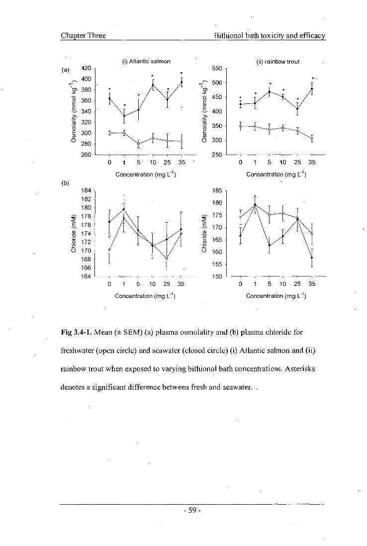
(i) Atlantic salmon (a)
-1
Osm
ola
lity (
mm
ol k
g 1)
420
400
380
360
340
320
300
280
260
(b) 185
180
175
170 -
165
160
155
Chl
orid
e (m
M)
150 0 1 5 10 25 35
Chl
orid
e (m
M)
184 182 180 178 176 174 172 170 168 166 164
0 1 5 10 25 35
Chapter Three
0 1 5 10 25 35 •
Concentration (mg L -1 )
Bithionol bath toxicity and efficacy
(ii) rainbow trout 550 -
500 - cr) (7) 450 - E
400
350
0) 300
250 0 1 5 10 25 35
Concentration (mg L -1 )
Concentration (mg L -1 )
Concentration (mg LI)
Fig 3.4-1. Mean (± SEM) (a) plasma osmolality and (b) plasma chloride for
freshwater (open circle) and seawater (closed circle) (i) Atlantic salmon and (ii)
rainbow trout when exposed to varying bithionol bath concentrations. Asterisks
denotes a significant difference between fresh and seawater.
- 59 -

0.10
0.16
0.15
Li 0.14
' 03 0.13
0.12
0.11 a
0.10
E 0.14
0.13
'70) 0.12 - Z
0.11 a cn
0.15
Chapter Three Bithionol bath toxicity and efficacy
(i) Atlantic salmon (ii) rainbow trout
7 30 (a) • c (Ti 2 25 o_ cn E 20
15
10 a)
5 - o_
0
7c 30
•
"(T)
0- 25 0.)
o_ 20
(7) § 15 — a)
10 0-
5
0 1 5 10 25 35
Concentration (mg L-1 )
0 1 5 10 25 35
Concentration (mg L-1 )
0 1 5 10 25 35
Concentration (mg 1_ - ')
0 1 5 10 25 35
Concentration (mg L )
Fig 3.4-2. Mean (± SEM) (a) gill Na + / KtATPase activity and (b) gill succinic
dehydrogenase (SDH) activity for freshwater (open circle) and seawater (closed
circle) (i) Atlantic salmon and (ii) rainbow trout when exposed to varying
bithionol bath concentrations. Asterisks denotes a significant difference between
fresh and seawater.
- 60 -
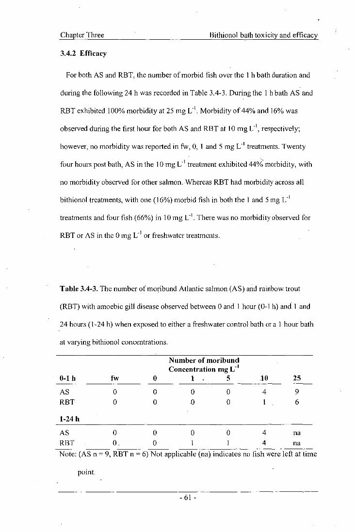
Chapter Three
3.4.2 Efficacy
Bithionol bath toxicity and efficacy
For both AS and RBT, the number of morbid fish over the 1 h bath duration and
during the following 24 h was recorded in Table 3.4-3. During the 1 h bath AS and
RBT exhibited 100% morbidity at 25 mg Morbidity of 44% and 16% was
observed during the first hour for both AS and RBT at 10 mg L -1 , respectively;
however, no morbidity was reported in fw, 0, 1 and 5 mg L treatments. Twenty
four hours post bath, AS in the 10 mg L -I treatment exhibited 44% morbidity, with
no morbidity observed for other salmon. Whereas RBT had morbidity across all
bithionol treatments, with one (16%) morbid fish in both the 1 and 5 mg L -I
treatments and four fish (66%) in 10 mg L -I . There was no morbidity observed for
RBT or AS in the 0 mg L -I or freshwater treatments.
Table 3.4-3. The number of moribund Atlantic salmon (AS) and rainbow trout
(RBT) with amoebic gill disease observed between 0 and 1 hour (0-1 h) and 1 and
24 hours (1-24 h) when exposed to either a freshwater control bath or a 1 hour bath
at varying bithionol concentrations.
Number of moribund Concentration mg L -1
0-1 h fw 0 1 . 5 10 25
AS 0 0 0 0 4 9 RBT 0 0 .0 0 1 . 6
1-24 h
AS 0 0 0 0 4 na RBT 0 0 1 1 4 na Note: (AS n = 9, RBT n = 6) Not applicable (na) indicates no fish were left at time
point.
- 61 -

Chapter Three Bithionol bath toxicity and efficacy
There was a concentration dependant decrease in percent lesioned gill filaments in
both Atlantic salmon and rainbow trout (Fig 3.4-3a). For AS, the lowest
concentration of bithiohol tested (1 mg 1: 1 ) significantly reduced percent lesioned
gill filaments to similar levels of the fw control, with all other bithionol
concentrations having significantly reduced percent lesioned gill filaments in
comparison to the sw control (p < 0.001, Fig 3.4-3a). A similar pattern was
observed in RBT, with all bithionol concentrations significantly reducing percent
lesioned gill filaments compared to the sw control, although they were not reduced
to a similar level seen in the fw control (Fig 3.4-3a).
Concurrently, there was a bithionol concentration dependant decrease in amoeba
numbers on the gills of AS and RBT following the 1 h bath (p <0.001, Fig 3.4-3b).
Atlantic salmon exposed to a 1 h bithionol bath at 1, 5, 10 or 25 mg 1_, -1 had a
relative percent reduction in amoeba numbers when compared to the sw control of
of 33, 46, 47 and 60 %. respectively. Rainbow trout similarly at corresponding
concentrations exhibited relative percent reductions of amoeba numbers compared
to sw control of 43, 49, 56 and 60%, respectively. There were no differences in
plasma osmolality at any of the treatment concentrations for both AS and RBT (p >
0.078, Fig 3.4-3c).
- 62 -

b) 600
500
400
300
200
100 Am
oeba
nu
mbe
rs (
x10
3)
Bithionol bath toxicity and efficacy Chapter Three
Per
cent
lesi
oned
gil
fila
men
ts .
80
70
60
50
40
a)
30
20 - A
10
fw 0 1 5 10 25 controls
Treatment (mg L -1 )
fw 0 1 5 10 25 controls
Treatment (mg L I )
c) 420
400 cr)
380
360
Z 0 340
6n 320
300
fw 0 1 5 10 25 controls
Treatment (mg/L)
Fig 3.4-3. Mean (± SEM) (a) percent lesioned gill filaments, (b) crude amoeba
numbers anCI (c) plasma osmolality for rainbow trout (open circle) and Atlantic
salmon (closed circle) when administered varying bithionol bath concentrations.
Lower and upper case letters denote significant differences within Atlantic salmon
and rainbow trout, respectively.
- 63 -

Chapter Three Bithionol bath toxicity and efficacy
3.5 Discussion
1 - Bithionol concentrations greater than 25 mg L were considered toxic with all
fish exhibiting signs of morbidity within three hours. This was similar to results
reported by Tojo et al. (1994b) and Santamarina et al. (1991) with significant levels
of mortality reported in RBT at concentrations greater than 30 mg L -I ..In the
toxicity trial, there were indications that bithionol in seawater may be more toxic
than in freshwater, with LT 50 values for both Atlantic salmon and rainbow trout
equal to 44 to 84 min for freshwater compared with 21 to 35 min for seawater,
respectively. Salinity has been reported to have little influence on the tolerance of
fish to toxicants, but fish do tolerate toxicants better in conditions similar to which
they reside in, possibly due to lessened osmotic problems (Sprague, 1990). This
suggests that the increased toxicity with salinity may be linked to osmotic problems.
Plasma osmolality was higher in seawater compared to freshwater for both species
in the toxicity trial. However, no differences were seen in plasma chloride
-1 concentrations, which were above 160 mEq L quoted as the minimum acceptable
level for chloride ions by Morgan and Iwama (1991), indicating the fish were not
likely to be suffering osmotic stress. Hence, further investigation would be required
in order to determine the mechanism for the observed difference in bithionol
toxicity between fish held in fresh and seawater. Bithionol concentration did not
affect plasma osmolality or plasma chloride concentration, adding to the hypothesis
that the cause of differential toxicity between freshwater and seawater was not
related to osmoregulation.
- 64 -

Chapter Three Bithionol bath toxicity and efficacy
The difference between fw and sw gill Na+/K+ ATPase levels obtained in this
study correspond to those found by Pfeiler and Kirschner (1972) who showed
Na±/K+ ATPase levels in rainbow trout to be 12.6 ± 1.7 larnol Pi mg protein -I hi in
freshwater and 23.6 ± 2.1 nmol Pi mg proteinh 1 in seawater. Similarly, Ventrella
et al. (2001) reported that RBT had lower activity in fw compare to brackish water
(22%0). The difference observed between fw and sw is attributed to the increase in
Na+ in marine fish, which must be pumped across the gills into the environment
(Pfeiler and Kirschner, 1972). McCormick (1993) reported Na +/K+ ATPase to be an
ion-translocating enzyme found in high concentrations in gill chloride cells and
believed to be the primary enzyme for excretion of excess Na+ and Ci from body
fluids (Handeland et al., 2004). Succinic dehydrogenase, a chloride cell activity
marker, plays an important role in mitochondria as it participates in the electron
transport, respiratory chain and Krebs cycle (Briere et al., 2005). In both species,
the highest concentration of bithionol did not inhibit gill Na+/K+ ATPase or gill
SDH from the respective control group, indicating that bithionol concentration did
not appear to impair gill ion regulatory function over the 6 h sample period. This
was further evidenced by the absence of acute changes in plasma chloride
concentration. No difference suggests that the two gill toxicity markers of SDH and
Na+/K+ ATPase were not acutely affected by bithionol and the cause of toxicity was
most likely not related to gill function.
Both Na+/K+ ATPase and SDH were examined because bithionol is known to be a
metabolic disruptor, uncoupling electron transport in fish ciliates including
Tetrahymena pyriformis (Griffin, 1989) and Trichodina jadranica (Madsen et al.,
- 65 -

Chapter Three Bithionol bath toxicity and efficacy
2000). Takeuchi etal. (1984) postulated that bithionol functioned as an uncoupler
of mitochondrial oxidative phosphorylation in the trophozoite stage of the
Entamoeba histolytica, the main amoeba pathogenic to humans. It is suggested that
bithionol has the ability to inhibit activity of fiimarate reductase, malie enzyme,
phosphogluconate dehydrogenase and succinate dehydrogenase in human liver and
lung flukes (Hamajima et al., 1979), although the inhibition of succinate
dehydrogenase was not observed in this study. It is important to note that the assay
used in this study examined Na ±/K+ ATPase activity, not the presence of ATP,
therefore showing that bithionol did not inhibit Na±/K+ ATPase in the gill. There
was no indication of the availability of ATP to drive the Na+/K+ ATPase in chloride
cells. Longer-term exposure may affect the availability of ATP, therefore in future
studies it would be advantageous to examine the presence of ATP in gill tissue.
Histological sections of gill or liver tissue from all bithionol concentrations did
not reveal any signs of alterations from the 0 mg L treatment. Chloride cells,
mucous cells, inflammation and epithelial separation were similar across all
concentrations; reinforcing the suggestion that bithionol did not acutely affect the
gill. Bithionol is known to be readily eliminated by the kidneys in humans and rats,
but accumulates in bile of mice (Barrett-Connor, 1982). Therefore future studies
should include sampling of the anterior and posterior kidney, in addition to the liver
and gills.
Bithionol, at a minimum concentration of 1 mg Li , significantly reduced the
percent lesioned gill filaments and numbers of gill amoebae in both Atlantic salmon
- 66 -
Chapter Three Bithionol bath toxicity and efficacy
and rainbow trout compared to the seawater control fish. Moreover, the observed
reduction in percent lesioned gill filaments in Atlantic salmon at bithionol
-1 concentrations of 1, 5 and 10 mg L was equal to that of the current industry
standard of a freshwater bath. Reduction in amoeba numbers after the commercial 3
h freshwater bath when compared to a sw control have been reported to be 85-90%
(Clark etal., 2003). A similar reduction of 90% from the sw control was seen in the
efficacy trial with both AS and RBT freshwater baths. Atlantic salmon bathed in
1 - bithionol at concentrations from 1 to 25 mg L exhibited a reduction in amoeba
numbers from the sw control of 33% to 60%. Similarly, rainbow trout resulted in
reductions of amoeba numbers from the sw control compared to 1 and 25 mg L -1 of
43% and 60% respectively. These results indicate that a 1 h bithionol bath at 1 mg
L-1 in sw can be as efficacious as freshwater in treating AGD, as a minimum crude
amoeba reduction of 33% was seen compared to no treatment.
Other treatments for AGD that have been considered in vivo include softened
freshwater (contains fewer divalent cations, approx. 0.4 mM Ca2+), which was
suggested to alter mucus viscosity making it more efficacious than hard freshwater
reducing amoeba numbers and alleviating the pathological signs of amoebic gill
disease (Roberts and Powell, 2003a). Chloramine-T, a disinfectant at 10 mg L -1 in a
seawater bath was as effective as a freshwater bath reducing amoeba numbers by
approximately 60%, compared to the seawater control (Harris et al., 2004). A
similar reduction was obtained with a 1 h bithionol bath at 25 mg Li ; however, it
was associated with high morbidity. Additionally, levamisole, the levoisomer of
tetramisole, is a recognised antihelmintic and has been reported as effective in vitro
- 67 -

Chapter Three Bithionol bath toxicity and efficacy
at 50 mg L -1 for Gyrodactylus spp. (Schmahl etal., 1989) and an effective
treatment for AGD-affected Atlantic salmon at 50 mg L -1 (Munday and Zilberg,
2003). This indicates that levamisole is almost as efficacious as freshwater for
treating AGD, but was associated with gill damage and mortalities. In contrast,
bithionol at 1 mg L -1 exhibited no morbidity yet was as effective as the current
freshwater bath.
Bithionol toxicity and efficacy is dependent upon host species and target parasite.
A concentration of 0.1 mg L -1 was reported as a possible therapeutic for the
protozoan parasite, Trichodina jadranica in European eels, Anguilla anguilla, due
to its capability to effectively treat entire recirculation systems, although it exhibited
a relatively low therapeutic index (Madsen etal., 2000). Conversely, Buchmann et .
al. (1992) demonstrated that bithionol at 0.1 mg L -1 was not efficacious as a static
bath against gill parasitic monogeneans, Pseudodactylogyrus sp. and concentrations
> 1 mg L -1 were toxic, causing 100% mortality within 24 h in the European eel.
Bithionol as a static bath has also been reported as effective in killing the ciliate
Tetrahymena pyriformis at 60 mg Li , whilst being non-toxic to the minnow golden
shiner, Notemigonus crysokucas (Griffin, 1989).
For AGD-affected Atlantic salmon and rainbow trout, this study identified that a
1 h seawater bath using a bithionol concentration of 1 mg L i exhibited no
morbidity and reduced amoeba numbers and percent lesioned gill filaments from
the seawater control. Nonetheless, with the scale and intensity of salmonid farming
occurring in Tasmania, a bithionol bath treatment would be impractical as it is
- 68 -

Chapter Three Bithionol bath toxicity and efficacy
insoluble and would require a large amount of the compound to treat the bath water.
On the other hand, with the determination of toxicity to the target animal and its
efficacy with respect to AGD it would be possible to incorporate bithionol in-feed
and assess as a treatment for the control of AGD in Atlantic salmon. Furthermore,
with the increasing need for the use of chemotherapeutants in aquaculture, it is very
important to minimise the accumulation of chemicals in food for human
consumption and the effect on the environment. Producing an in-feed treatment
would assist in alleviating the release of large amounts of the compound into the
surrounding water (Findlay et al., 2000).
In conclusion, bithionol was toxic above 25 mg L-1 in freshwater (municipal
source, 15.5°C, pH 7) and seawater (35%o, 15.5°C, pH 8.2) for both Atlantic salmon
and rainbow trout. It did not appear to acutely affect the gill toxicity parameters of
SDH, Na+/K+ ATPase activity, or the plasma osmolality, chloride and no
pathological changes in the gill or liver tissue were found over the 6 h period.
Bithionol was efficacious as a 1 h seawater bath for treating amoebic gill disease
caused by Neoparamoeba spp. reducing mean percent lesioned gill filaments and
crude amoeba numbers compared to seawater control at all concentrations
examined. Bithionol concentrations of 1, 5 and 10 mg L produced an efficacy
comparable to the currently used freshwater bath in Atlantic salmon. Bithionol
warrants further investigation as a treatment for amoebic gill disease, including
possible use as an oral medication.
- 69 -

Chapter Three Bithionol bath toxicity and efficacy
3.6 Acknowledgements
This research formed partof a project of the Aquafin CRC, and received funds
from the Australian Governments CRC Programme, the Fisheries R & D
Corporation and other CRC participants. The authors wish to thank J. Ransome, M.
Jones and M. Barney for their technical support throughout the study and to Dr. J.
Purser for his individual contributions.
- 70 -

CHAPTER 4
EFFICACY OF BITHIONOL AS AN
ORAL TREATMENT
Published in Aquaculture 270 (2007): 15-22

Chapter Four Efficacy of bithionol as an oral treatment
4 Efficacy of bithionol as an oral treatment for amoebic gill
disease in Atlantic salmon, Salmo salar
Renee L. Florent, Joy A. Becker, Mark D. Powell.
4.1 Abstract
This study examined the efficacy of bithionol as an oral treatment for Atlantic
salmon, S. salar affected by amoebic gill disease (AGD). The current commercial
management strategy of AGD is a costly 3 h freshwater bath. It is labour intensive
and the number of baths needed appears to be increasing; hence there is an effort to
identify alternative treatments. Efficacy was examined by feeding AGD-affected
Atlantic salmon twice daily to satiation with bithionol, an antiprotozoal agent, at 25
mg kg-1 feed. Three seawater (35%o, 17°C) re-circulation systems were used, each
consisting of three tanks containing 32 Atlantic salmon smolts with an mean (±
SEM) mass of 90.4 g (± 5.2). Three feeds were examined in the trial including
bithionol, plain commercial control and oil coated commercial control. Feeding
commenced 2 weeks prior to exposure to Neoparamoeba spp. at 300 cells L -1 and
continued for 28 days post exposure. Efficacy was determined by examining gross
gill score and identifying percent lesioned gill filaments twice weekly for 4 weeks
post exposure. Bithionol, when fed as a two week prophylactic treatment at 25 mg
-1 kg feed, delayed the onset of AGD pathology and reduced the percent lesioned gill
filaments by 53% and halved the gill score from two to one when compared with
both the plain and oil controls during an experimental challenge. There were no
palatability problems observed with mean feed intake of bithionol over the trial
- 72 -

Chapter Four Efficacy of bithionol as an oral treatment
duration with fish consuming higher levels of the bithionol diet compared to both
- the oil and plain controls. This study demonstrated that bithionol at 25 mg kg' feed,
when fed as a two week prophylactic treatment for Neoparamoeba spp. exposure,
delayed and reduced the intensity of AGD pathology, and warrants further
investigation as an alternative to the current freshwater bath treatment for AGD-
affected Atlantic salmon.
4.2 Introduction
Bithionol, a phenolic compound related structurally to hexachlorophene, has been
used worldwide as a parasiticide for cattle, sheep, chickens, dogs and cats (Enzie
and Colglazier, 1960). It is reported to have surfactant antimicrobial properties and
thus is also effective against bacteria, moulds and yeast (Kim and Choi, 1998). It
has been used as an alternative to praziquantel against human trematode and
cestode infections, in particular paragonimiasis (Enzie and Colglazier, 1960;
Takeuchi et al., 1984), and has been widely used in veterinary medicine as it is
active against flukes and cestodes (Mourot et al., 1987). Bithionol has been found to
stimulate lactic acid production, inhibit oxygen consumption and decrease
glycolytic and oxidative metabolism in the human lung fluke, Paragonimus
westermani, in vitro (Hamajima, 1973). Furthermore, it was postulated to be linked
to a variety of processes including the inhibition of reduced nicotinamide adenine
dinucleotide (NADH)-fumarate reductase and involved in protein phosphorylation
(Reid etal., 2001). With regards to fish, bithionol has been shown to be effective
against the fish ciliates Philasterides dicentrarchi in turbot, Scophthalmus maximus,
Tetrahymena pyriformis in vitro and Trichodina jadranica in European eels,
-73 -
Chapter Four Efficacy of bithionol as an oral treatment
Anguilla anguilla, acting upon the mitochondrial respiratory chain and uncoupling
electron transport (Griffin, 1989; Madsen etal., 2000; Iglesias etal., 2002).
Bithionol has been recorded as efficacious in vitro and in vivo when used as a bath
treatment for salmonid parasites, including Gyrodactylus sp., khthyobodo necator
(Santamarina et al., 1991; Tojo et al., 1994b) and Neoparamoeba spp. (Florent et
al., 2007a). Furthermore, due to bithionol being insoluble in water, it was
advantageous to use bithionol as an oral medication, providing easy administration
and limited fish handling (Tojo and Santamarina, 1998a). Bithionol at 40 g k -g-i feed
was offered for 10 days at 2% body weight (BW) per day to rainbow trout,
Oncorhynchus mykiss, infected with Spironucleus salmonis, Gyrodactyhts sp. or I.
necator and exhibited a reduction in parasitemia. Bithionol eliminated
approximately 80% of S. salmonis from rainbow trout whilst both Gyrodactylus sp.
and I necator infections were reduced from a high to low intensity (Tojo and
Santamarina, 1998a; b; c). Kim and Choi (1998) reported bithionol administered in-
feed at 100-200 mg kg' BW significantly reduced the number of monogeneans
Microcotyle sebastis on the gills of cultured rock fish, Sebastes schlegeli, with a 20
day feeding duration being most effective.
The protozoan parasite Neoparamoeba spp. is a free-living marine amphizoic
amoeba and is believed to cause amoebic gill disease (AGD) (Kent etal., 1988;
Munday etal., 1990; Adams and Nowak, 2004b). It attaches itself to the gills,
particularly the secondary lamellae, and is characterised macroscopically by the
presence of raised, white mucoid patches with histological presentation of single or
- 74 -

Chapter Four Efficacy of bithionol as an oral treatment
multi-focal epithelial hyperplasia leading to lamellar fusion (Adams and Nowak,
2001). It primarily affects salmonids and is a significant problem for the Atlantic
salmon industry in Australia (Munday et al., 2001; Nowak et al., 2002). The current
commercial treatment for AGD is freshwater bathing of affected fish, as it lowers
gross gill lesions, mortalities and is environmentally friendly. However, it is labour
and cost intensive as well as requiring fish handling, hence the need for effective
oral medications (Parsons etal., 2001a; Munday and Zilberg, 2003).
Several compounds have been examined for efficacy against AGD, including
levamisole (Howard and Carson, 1995), chlorine dioxide, chloramine-T, hydrogen
peroxide (Powell et al., 2003; Powell and Clark, 2003), amprolium, albendazole,
toltrazuril, and bithionol (Powell etal., 2003). These compounds have been
examined either as a bath treatment or in in vitro toxicity testing in seawater, with
levamisole reported as lethal to N pemaquidensis at concentrations? 10 ppm in
vitro (Howard and Carson, 1994), whilst chloramine-T at a concentration of 25 and
50 ppm reduced amoeba numbers equivalent to those seen in deionised water after 2
h (Powell and Clark, 2003). Amprolium was found to be an amoebastat at 1 mg L -1
and bithionol amoebicidal at 1 and 10 mg L -I in vitro (Powell and Clark, 2003).
Compounds that have been examined as in-feed treatment for AGD include the
mucolytic compound L-cysteine ethyl ester (LCEE), which was reported to delay
progression of pathology associated with AGD as well as reduce percent lesioned
gill filaments by approximately 50% relative to the control when fed at 52.7 mg kg-I
fish d-I for two weeks (Roberts and Powell, 2005). The purpose of the present study
was to determine if bithionol, when administered orally as a prophylactic and
- 75 -

Chapter Four Efficacy of bithionol as an oral treatment
-1 therapeutic treatment at 25 mg kg feed to Atlantic salmon, could be used as an
effective treatment against amoebic gill disease affected Atlantic salmon.
4.3 Materials and Methods
4.3.1 Fish husbandry and maintenance
Atlantic salmon (AS) diploid mixed-sex spring smolts, with a mass of 90.4 ± 5.2 g
and a fork length of 21.1 ± 0.3 cm (N = 288) were obtained from SALTAS salmon
hatchery (Tasmania, Australia). Fish were maintained at the University of Tasmania •
Aquaculture Centre for a minimum of three weeks prior to experimentation. Salmon
were acclimated to seawater (30%0, 1 um filtered, mean ± SEM temperature of 16 ±
0.8°C) over 14 days in one 3000 L Rathburn tank with recirculated water and an
individual biofilter system. The tank received constant aeration and oxygen levels
were monitored daily using a Handy Gamma Oxy Guard (Birkerod, Denmark), with
dissolved oxygen levels of 94.0 ± 0.2% satuartion. Fish were fed a commercially
available diet of 4 mm Atlantic Salmon Grower LE pellets (SkrettingTM, Hobart,
Australia) twice daily to satiation throughout the seawater acclimation period.
4.3.2 Experimental design and challenge method
Two hundred and eighty-eight Atlantic salmon were equally and randomly
• allocated into nine tanks at a stocking density of 6.6 ± 0.1 g Li . The tanks were in
three separate seawater re-circulation systems, each consisting of three 590 L tanks,
a 500 L header, and a 500 L sump providing a total volume of 2770 L. All tanks
received constant aeration with water temperature and oxygen levels monitored
daily. Fish were acclimated from 30 to 35 %o over seven days and allowed to
- 76 -

Chapter Four Efficacy of bithionol as an oral treatment
habituate to the systems for the following seven days, with system conditions
maintained on a photoperiod of 12 h each of light and dark, with a water
• temperature of 16.7 + 0.3°C, pH 8.19 ± 0.01 and feeding of standard commercial
feed prior to commencing experimental procedures. Following seven days
conditioning, each system was randomly allocated a treatment. Treatments
consisted of (1) commercial feed (control), (2) commercial feed coated with fish oil
(control with oil) and (3) commercial feed coated with combination of 25 mg kg -I
feed bithionol and fish oil. This dose was chosen based on previous work, which
showed bithionol to be toxic when, administered as a bath treatment at
concentrations > 10 mg L-I (Florent etal., 2007a). Fish were fed to satiation twice
daily at 0830 h and 1630 h throughout the entire study with feed intake recorded
daily. In order to determine tank feed intake an average pellet weight for each diet
was obtained. Daily, the numbers of uneaten feed pellets were counted and
multiplied by the average pellet weight, and feed intake was determined by
subtracting the weight of uneaten feed from the feed placed into the tank. Feed
intake was determined as a percent of the tank biomass by dividing the mean tank
feed intake for the week by the tank biomass and multiplying by 100 to obtain the
weekly mean feed intake as a percent of the tank weight. On Day 0 and 17, fish
were only fed once due to weight check and sampling coinciding on these days.
Weight checks with a minimum of ten fish per tank were conducted weekly to
determine feed intake as percent body weight. All treatments were fed 14 days prior
to exposure to Neoparamoeba spp. with feeding continued for a further 28 days
after exposure.
- 77 -

Chapter Four Efficacy of bithionol as an oral treatment
Fish were experimentally exposed to Neoparamoeba spp. according to Morrison
et al. (2004). Briefly, six donor Atlantic salmon were obtained from the University
of Tasmania's Aquaculture Centre experimental AGD infection tank post-mortem.
Gill baskets were excised from the salmon centrifuged in distilled water and rinsed
with seawater three times to dislodge amoebae from the gills. Amoebae in seawater
were allowed to adhere to Petri dishes for approximately 2 h at 18°C. Plates were
washed with clean seawater and approximately 20 mL of seawater was added.
Amoebae were allowed to adhere to Petri dishes overnight at 18°C. The adherent
cells were removed by the addition of 1000 [tL Hanks balanced salt solution with
Arypsin and EDTA (Appendix 1), washed, centrifuged and concentrated. Live
amoeba counts were determined using a haemocytometer (Neubauer, BS 748).
Three replicate counts were made with 18 large squares counted per replicate. The
isolation obtained 2 500 000 amoeba delivering a final concentration of
approximately 300 cells L' i per system. Individual aliquots were placed into the
three system sumps and foam fractionators were turned off.
4.3.3 Feed preparation
Commercially available 4 mm Atlantic Salmon Grower LE pellet (SkrettingTM,
Hobart, Australia) was used for all treatments. Feed was prepared as needed in
100 g batches and stored at 4°C. The control with oil was prepared by moistening
feed with 7 mL distilled water, then evenly coating with 5 mL of fish oil by shaking
food in a plastic bag until well coated. The food was air dried for 24 h before being
placed into an airtight container and held at 4°C. This same procedure was used to
prepare the bithionol diet with the inclusion of 2.5 mg of bithionol (Sigma-Aldrich
- 78 -

Chapter Four Efficacy of bithionol as an oral treatment
Pty. Ltd, Castle Hill, Australia) which was combined with the fish oil prior to
coating the moistened pellet.
4.3.4 Data Collection
Total tank mass was obtained weekly using a minimum of ten fish per tank. Fish
were sampled (two fish per tank) at transfer, 14 days prior to infection and then
twice weekly for 28 days. For each fish, mass and fork length data was recorded as
well as gross and histological examination of internal organs for any signs of
toxicity. The gross gill score data were recorded using categories seen in Table
4.3-1 (Adams and Nowak, 2003). Fish were anaesthetised or euthanized using clove
oil at a concentration of 0.02% w/v and all procedures were conducted in
accordance with the Australian code of practice for care and use of animals for
scientific purposes under the guidance of the Uni \iersity of Tasmania Animal
Ethics. Specific growth rate (SGR) and Fulton's condition factor (K) were
calculated using equation 1 and 2. Fulton's condition factor was calculated using
the individually sampled fish. The SGR was calculated using the average fish mass
for each tank for Day -14 and Day 0 and then from Day 0 to the Day where there
were a median number of 15 fish left. This was done due to the variation in fish
numbers within tanks at the culmination of the trial.
SGR Ln weight (g) 2 — Ln weight (g),
x100= A time
weight(g) K =[ 1(100 length x length x length (cm)
[1]
[2]
- 79 -

Chapter Four Efficacy of bithionol as an oral treatment
Table 4.3-1. Scoring scheme for gross signs of amoebic gill disease on Atlantic
salmon modified from Adams and Nowak (2003).
Infection Level
Score Number of affected holobranchs
Clear 0 0
Light 1 <2
Moderate 2 2-5
Heavy 3 >5
4.3.5 Histology
The left gill basket was excised, rinsed gently in 0.2 um filtered seawater, fixed in
seawater Davidson's fixative and the liver, kidney, and muscle were fixed in 10%
neutrally buffered formalin for 24 h, and then transferred to 70% ethanol. The
second left anterior hemibranch was removed, along with a small section of liver,
kidney and muscle, dehydrated, embedded in paraffin wax, sectioned at 5 um, and
stained with haematoxylin and eosin (H & E). The sections were viewed under a
light microscope (Olympus) at X100 to X400 magnifications. The number of
filaments exhibiting AGD lesions were counted and expressed as proportions of the
total number of filaments in each section (Parsons etal., 2001a). A filament was
counted only when the central venous sinus was visible in at least two-thirds of the
filament and lamellae were of equal length bilaterally present to near the tip of the
filament (Speare et al., 1997).
- 80 -

Efficacy of bithionol as an oral treatment Chapter Four
4.3.6 Statistical Analysis
Statistical analysis was conducted using SPSS for Windows ® (version 11.5). A
mixed model analysis of variance (ANOVA) containing factorial and nested terms
was used to determine differences with treatment and day sampled as fixed factors
and tank as a random factor. There was no difference determined between the oil
coated control and the plain control diets with respect to the AGD variables;
therefore the control treatments were combined and compared to the bithionol
treatment. The interaction between day sampled and treatment was examined first;
if not significant, then treatment and day sampled could be examined individually.
If a significant difference was identified among treatments and/or days sampled,
then a Tukey's post hoc test was used to identify where the differences occurred.
Homogeneity was determined using a residual plot and Levene's test. A result was
considered significant if p < 0.05 and results are presented as a mean ± SEM.
Relative percent reduction (RPR) was calculated for percent lesioned gill filaments
using the following equation:
number of percent lesioned gill filaments in treated group RPR = 1
x100 number of percent lesioned gill filaments in control group
4.4 Results
At the culmination of the trial all fish had exhibited signs of AGD. As expected,
there were no significant differences between gross gill scores and percent lesioned
gill filaments for the plain and oil control feeds; therefore they were combined.
There appeared to be no problems associated with palatability as feed was eaten
- 81 -

Chapter Four Efficacy of bithionol as an oral treatment
across all treatments and mortalities were observed across all treatments. No
toxicity problems were observed based on gross examination of internal organs and
histological examination of the kidney, liver and muscle (histology not shown).
Bithionol treated fish exhibited significantly reduced lesioned gill filaments over
the entire trial when compared to control fish (Fig 4.4-1a). Lesions were first
observed on Day 7 in control groups compared to Day 10 in the bithionol group and
percent lesioned gill filaments increased throughout the trial. Significant reductions
in percent lesioned gill filaments were first observed between bithionol treated fish
and control fish on Day 10 (F2,15= 50.101, p <0.001), and continued to the
culmination of the trial at Day 28 (F2,15= 41.099, p <0.001) post-Neoparamoeba
spp. exposure. Bithionol was successful in delaying the onset of percent lesioned
gill filaments seen typically with AGD by 53% when compared to the control diets
at the culmination of the trial (Day 28) post-exposure. A similar result was seen
with the gross gill score, where on Day 28 controls had a mean gross gill score of
two, which was significantly higher compared to bithionol treated fish exhibiting a
mean gross gill score of one (F2,15= 25, p <0.001, Fig 4.4-1b). However, there
were no differences observed in gross gill score on any other day throughout the
trial.
There appeared to be no palatability problems associated with bithionol when fed
at 25 mg kg' feed. Weekly feed intake in all treatment groups was determined
significantly different over the duration of the trial (F2,6= 85.372, p = <0.001) post-
Neoparamoeba spp. exposure (Fig 4.4-2a). As the trial progressed the feed intake
- 82 -

Chapter Four Efficacy of bithionol as an oral treatment
began to decrease across all treatments particularly from Day 14 onwards for the
control treatments and Day 21 onwards for the bithionol treatment, which coincided
with heavier parasite load. However, there were no differences observed in the
mean tank biomass mass of the fish across all treatments throughout the trial (F2 ,6 =
0.348, p = 0.792 Fig 4.4-2b ) or SGR (F 1 , 1 8 = 0.099, p < 0.001, Table 4.4-1) but a
difference in K was observed (F2, 1 5= 5.708, p = 0.014, Table 4.4-1).
- 83 -

6 a) 0
U'
▪
50 • a)
asE 40
30 a) O
•
20 a)
1O
0
b)
Gro
ss g
ill s
core
Chapter Four Efficacy of bithionol as an oral treatment
-15 -10 -5 0 5 10 15
20
25
30
Day from Neoparamoeba spp. exposure
a
Day from Neoparamoeba spp. exposure
Fig 4.4-1. (a) percent lesioned gill filaments and (b) gross gill score for Atlantic
salmon with amoebic gill disease (AGD) when fed either control feed (solid line)
(n = 12) or bithionol at 25 mg kg-1 feed (broken line) (n = 6). No error bars
indicate that all replicates exhibited the same value. Values are expressed as mean
± SEM. Letters denote significant differences among treatments at that sampling
time (p <0.05).
- 84 -

(T.)
b) 130 -
120 -
110 -
100 -
90 -
80 -
-14 -7 0 7 14 21 28
70 -
60
Chapter FoUr Efficacy of bithionol as an oral treatment
-14 -7 0 7 14 21 28
Day from Neoparamoeba sp. exposure
Days from Neoparamoeba spp. exposure
Fig 4.4-2. (a) weekly tank feed intake (n = 3) and (b) weekly tank biomass (n = 3)
for Atlantic salmon exposed to Neoparamoeba spp. when fed either commercial
feed (closed circles), oil-coated commercial feed (open circles) or bithionol at 25
mg kg -I feed (broken line and open squares). Values are expressed as mean ±
SEM. Letters denote significant differences among treatments over the trial
duration (p < 0.05).
- 85 -

Chapter Four Efficacy of bithionol as an oral treatment
Table 4.4-1. Mean tank specific growth rate (SGR) (n = 3) and individual fish condition factor, (K) (n = 6) for plain control, oil control and
bithionol treated Atlantic salmon feed from Day -14 to 0 and Day 0 to Day 10 Neoparamoeba spp. exposure. Values are mean ± SEM. Asterisks
indicate significant difference between treatments within each time point (p<0.05).
Treatment
Day -14 to 0 Day 0 to Day 10
Plain Oil Bithionol Plain Oil Bithionol
SGR (% day-I ) 1.36 1.59 1.25 -0.11 -0.41 -0.12
± 0.11 ± 0.25 ± 0.20 ± 0.15 ± 0.23 ± 0.10
K (%) 0.89 0.81 0.84 1.04 0.97 1.16*
± 0.07 ± 0.04 ± 0.01 ± 0.04 ± 0.04 ± 0.02

Chapter Four Efficacy of bithionol as an oral treatment
4.5 Discussion
During a laboratory infection in which bithionol was orally administered at 25 mg
kg-1 feed, the intensity of AGD pathology was significantly delayed and reduced
compared to the control fish. A 53% relative reduction in percent lesioned gill
filaments was observed when compared to the controls. Furthermore, gross gill
scores were halved in the salmon treated with bithionol suggesting that this drug
may be an effective therapeutic for AGD. This trial examined bithionol efficacy
during the first four weeks of a Neoparamoeba spp. challenge and differences in gill
score were only observed on the last day. Due to stocking density limitations, it was
not possible to continue the trial further; however, it would be advantageous in
future studies to continue past this time point. Similarly, bithionol was effective
against the gill monogenean parasite Microcotyle sebastis infestations in marine
rockfish, Sebastes schlegeli, resulting in a 66-93% reduction in parasite numbers
when used at 1.25 g kg- feed for 10-20 days (Kim and Choi, 1998). In contrast,
bithionol was reported to be ineffective as a treatment of Hexamita salmonis,
Gyrodactylus sp., and Ichthyobodo necator infestations in rainbow trout,
Oncorhynchus mykiss (Tojo and Santamarina, 1998a; b; c); however, classification
of effectiveness in these experiments was complete elimination of the parasites.
Bithionol when fed at 40 g kg-I feed for 10 days did not achieve complete
elimination of the parasites but produced an 80% reduction in H. salmonis whilst
Gyrodactylus sp. and I. necator numbers were reduced from a high to low intensity.
Whilst complete elimination of Neoparamoeba spp. would be welcomed, a
treatment that is comparable in efficacy to bath treatment with freshwater and less
labour intensive and more cost effective is a more realistic option, at least initially.
- 87 -

Chapter Four Efficacy of bithionol as an oral treatment
Bithionol has a melting point of 188°C; therefore it is possible that it could
withstand the heat extrusion process so that it could be included at the feed
preparation stage and, based upon the cost of bithionol, it would cost approximately
$120 tonne -I (Sigma-Aldrich, 2006).
The standard laboratory-induced AGD infections using the isolation method
described by Morrison et al. (2004) consistently induces AGD in Atlantic salmon.
The severity of AGD in the laboratory is influenced by the amoeba challenge
concentration and temperature (Zilberg et al., 2001). Therefore, aggressive
laboratory-induced AGD infections used in this study exhibit an increased rate of
infection when compared to clinical AGD outbreaks on commercial salmon farms
where there are less severe infection rates. Under laboratory conditions, there is no
administration of freshwater baths and water is recirculated creating the potential
for the continuous colonisation of the gills. Hence, differences observed between
the two infection types are due to amoeba concentration and optimal laboratory
conditions for AGD (Roberts and Powell, 2005). Fish affected with AGD on
commercial farm field trials have been identified as having between 5% percent
lesioned filaments coinciding with a freshwater bath and 10-20% 18 weeks post sea
transfer (Adams and Nowak, 2003; Roberts and Powell, 2003a). This trial showed
differences in the intensity of AGD pathology when fish were fed bithionol in an
aggressive laboratory infection, warranting further investigation into the potential
for oral administration of bithionol on commercial farms where AGD infections are
typically chronic (Roberts and Powell, 2005). Further investigation of bithionol
would include but not be limited to examining the pharmacokinetics and
- 88 -

Chapter Four Efficacy of bithionol as an oral treatment
bioavailability. The pharmacokinetics of bithionol regarding absorption,
distribution, metabolism and excretion are not known within fish and are necessary
for correct administration of bithionol.
Other compounds that have been examined as an in-feed treatment for AGD
include the mucolytic drug L-cysteine ethyl ester (LCEE) (Roberts and Powell,
2005). Furthermore, the oral administration of nutritional supplements Aquacite TM
and BetabecTM has been examined as an alternative treatment strategy (Mlynarski,
et al., unpublished data). It was identified that the progression of AGD when
salmon were exposed to cohabitation-induced, aggressive laboratory infection was
significantly delayed when LCEE was orally administered at 52.7 mg LCEE
fish di over two weeks. Treated fish had approximately 50% less lesioned gill
filaments than the control fish three days post-infection. However, there were
palatability issues, in that the Medicated feed intake was approximately 65% of the
control feed (Roberts and Powell, 2005). AquaciteTM and BetabecTm nutritional
supplements in a semi-commercial field trial were found to increase growth rate and
maintain feed intake and conversion ratios when compared to control. Furthermore,
the nutritional supplement exhibited a reduction in mortality and a delay in onset of
AGD lesions (Mlynarski, et al., unpublished data).
No signs of toxicity were observed when AGD-affected Atlantic salmon were fed
bithionol at 25 mg kg -I feed c1-1 for 40 days. Similarly, no signs of toxicity were
observed when bithionol was fed at 40 g kg -1 feed di over 10 days to rainbow trout
with either H salmonis, Gyrodactylus sp., or I. necator infestations (Tojo and
-89-

Chapter Four Efficacy of bithionol as an oral treatment
Santamarina, 1998a; b; c). However, in contrast, bithionol exhibited signs of
toxicity when used to treat M. sebastis infestations in marine rockfish at 1.25 g kg -I
feed c1 1 for 10-20 days (Kim and Choi, 1998).
No problems were associated with the palatability of bithionol medicated feed,
with mean bithionol consumption over the entire trial greater than both the oil and
plain controls. As the trial progressed the feed intake observed across all treatments
began to decrease particularly from Day 14 onwards for the control treatments and
Day 21 onwards for the bithionol treatment, which coincides with heavier parasite
load. There was however, no difference between treatments when examining
specific growth rate, this could be due to the small sample size (n = 3) and the
considerable amount of variation. In future studies it would be advantageous to
examine SGR using average mass of individually sampled fish as opposed to tank
average this may aid in comparing data. Conversely, fish fed bithionol exhibited a
higher condition factor on Day 28 post exposure compared to both control
treatments.
4.6 Conclusion
Bithionol exhibited potential as an oral treatment for AGD at 25 mg kg -I feed
when administered for two weeks prior to Neoparamoeba spp. exposure and 4
weeks following exposure. A reduction in percent lesioned gill filaments and gross
gill score was achieved over the trial period. Furthermore, bithionol exhibited
higher feed consumption throughout the study compared to plain and oil coated
commercial diet and a reduction in mortalities. Further investigation of bithionol as
- 90 -

Chapter Four Efficacy of bithionol as an oral treatment
an in-feed treatment for AGD is warranted, including examining the effect of
prophylactic and therapeutic treatments, running a similar experiment for a longer
period of time to obtain more growth data, examining the effect of pulse feeding, or
conducting trials under more realistic field conditions with lower exposure doses
and freshwater baths.
4.7 Acknowledgements
This research formed part of a project of the Aquafin CRC; and received funds
from the Australian Government's CRC Programme, Fisheries R & D Corporation
and other CRC participants. The authors wish to thank J. Ransome and M. Barney
for their technical support throughout the study.
- 91 -

CHAPTER 5
FURTHER DEVELOPMENT OF
BITHIONOL THERAPY AS A
TREATMENT FOR AGD
In press with Journal of Fish Diseases
Chapter Five Further development of bithionol therapy as a treatment for AGD
Further development of bithionol therapy as a treatment for
AGD
Renee L. Florent, Joy A. Becker, Mark D. Powell.
5.1 Abstract
This study examined the efficacy of bithionol as a prophylactic or therapeutic oral
treatment for Atlantic salmon, Salmo salar, affected by amoebic gill disease
(AGD). Furthermore, it explored the interaction of bithionol oral therapy with the
current standard treatment (a freshwater bath for at least 3 h). The efficacy of three
feeds was determined in the trial by feeding AGD-affected Atlantic salmon at 1%
body weight (BW) day -I either oil coated commercial feed (control) or prophylactic
and therapeutic bithionol at 25 mg kg' feed. Feeding commenced two weeks prior
to exposure to Neoparamoeba spp. at 300 cells L1 and continued for 49 days post
exposure. Bithionol, when fed as a two week prophylactic or therapeutic treatment
at 25 mg kg-I feed, delayed the onset of AGD pathology and reduced the percentage
of gill filaments with lesions. Administration of a 3 h freshwater bath at 28 days
post-exposure significantly reduced amoebae numbers to a similar level across all
treatments. In contrast, gross gill score and percent lesioned filaments were reduced
to different extents, the control having a significantly higher score than both
bithionol treatments. Following the freshwater bath, clinical signs of AGD
increased at a similar level across all treatments, albeit controls were significantly
higher than the bithionol treatments immediately following freshwater treatment.
This study demonstrated that bithionol at 25 mg kg' feed, when fed as a two week
- 93 -
Chapter Five Further development of bithionol therapy as a treatment for AGD
prophylactic or a therapeutic treatment, delayed and reduced the intensity of AGD
pathology and warrants further investigation as a treatment for AGD-affected
Atlantic salmon.
5.2 Introduction
Disease has numerous negative impacts on fish and aquaculture production,
including reduced feed efficiency (Hedrick, 1998), impaired growth (Hedrick,
1998) and often death (Bakke and Harris, 1998). This in turn represents a direct loss
of investment in feed, labour and stock to the farmer (Rabago-Castro et al., 2006).
When examining commercial farming of food fish, many bacterial, viral, fungal and
parasitic pathogens have been recognised, not only in Tasmania but worldwide
(Schmahl etal., 1989; Stoffregen etal., 1996). In the control of these pathogens,
particularly external parasites of farmed marine fish, chemical treatments are
common (Powell and Clark, 2004). However, since the turn of the 21' century there
have been few new chemotherapeutic agents approved for use in aquaculture for
several reasons, including concerns regarding environmental impact, cost and
residues in food fish and the fact that it is often a limited market for drug companies
(Shao, 2001). The continual improvement and development of cost-effective fish-
husbandry practices, including efficacious chemotherapeutants, is critical for the
success of the aquaculture industry and increased production (Stoffregen et al.,
1996).
Chemotherapeutants provide effective methods of preventing (prophylactic
treatments) and controlling (therapeutic treatments) fish mortality and disease, and
- 94 -

Chapter.Five Further development of bithionol therapy as a treatment for AGD
are applied either via bath or oral administration (Armstrong, 1994; Howe et al.,
1999). Many fish farmers from North America (Thorburn and Moccia, 1993), Asia
(Tonguthai, 1997), Europe (Alderman, 2002) and Africa (Hecht and Endemann,
1998) resort to the use of prophylactic treatments to aid in the prevention of
epidemics. Several therapeutic agents have been examined for both prophylactic
and therapeutic treatment of ectoparasitic infections; these include but are not
limited to a formalin bath to treat trichodinads and monogeneans (Noga, 2000),
chloramine-T for infections in salmon (Harris et al., 2004), hydrogen peroxide for
monogeneans (Mansell et al., 2005), emamectin benzoate for sea lice (Ramstad et
al., 2002; Treasurer etal., 2002), and bithionol as a bath treatment for salmonid
parasites including .Gyrodactylus sp., khthyobodo necator (Santamarina etal.,
1991; Tojo et al., 1994b). More recently, bithionol has shown efficacy for the
treatment of Neoparamoeba spp. (Florent etal., 2007a; b).
Until recently the presumptive causative agents of AGD were thought to be two
amphizoic amoebae Neoparamoeba pemaquidensis (Kent et al., 1988; Roubal et
aL, 1989) and N. branchiphila (Dykova etal., 2005). However, attempts to
determine the pathogenicity of either species by re-infecting fish using clonal,
cultured, gill-derived strains have been universally unsuccessful (Kent etal., 1988;
Howard, 2001; Morrison etal., 2005). Neoparamoeba perurans is a protozoan
parasite that was recently described as the predominant aetiological agent of AGD
of Atlantic salmon cultured in Tasmania, Australia (Young etal., 2007). Amoebic
gill disease has been diagnosed in a number of fish species cultured in the marine
environment worldwide (Nowak etal., 2002). It is a significant problem for
- 95 -

Chapter Five Further development of bithionol therapy as a treatment for AGD
Australian salmon aquaculture with the disease accounting for 10-20% of the gross
cost of production (Munday et al., 2001; Nowak et al., 2007). This is due not only
to the cost of treating and managing the disease, but also to loss of fish condition
and production costs. The mitigation of AGD in Tasmania is mostly due to
freshwater bathing which was first recommended by Foster and Percival (1988).
However, the efficacy is variable and has notably become increasingly less effective
(Parsons etal., 2001a; Powell and Clark, 2003); hence there is a push to identify
alternative, more cost-effective and less labour intensive treatments, preferably
delivered in-feed.
A large variety of antimicrobials, disinfectants and detergents have been
examined both in vitro on isolated gill amoebae and in vivo as both bath and feed
additives for AGD mitigation. Attempts to identify potential chemotherapeutic
agents have been limited due to either lack of direct efficacy on the parasite, target
fish toxicity or the cost of treatment (Alexander, 1991; Howard and Carson, 1994).
Toxicity of several compounds to Neoparamoeba spp. has been examined in vitro
including levamisole (Howard and Carson, 1995), chlorine dioxide, chloramine-T,
hydrogen peroxide (Powell et al., 2003; Powell and Clark, 2003), amprolium,
albendazole, toltrazuril and bithionol (Powell etal., 2003), all with low to moderate
success. Howard and Carson (1994) reported that levamisole at concentrations > 10
ppm in vitro were lethal to N. pemaquidensis, and chloramine-T concentrations of
25 and 50 ppm effectively reduced amoeba numbers after 2 h (Powell and Clark,
2003).-Powell etal. (2003) found amprolium at 1 mg 1.:1 and bithionol at 1 and 10
mg Li to be amoebicidal in vitro. Compounds that have been examined as in-feed
- 96 -

Chapter Five Further development of bithionol therapy as a treatment for AGD
treatment for AGD include the mucolytic compound L-cysteine ethyl ester (LCEE),
which delayed progression of pathology associated with AGD and reduced percent
lesioned gill filaments by approximately 50% relative to the control when fed at
52.7 mg kg1 fish day-I for 2 weeks (Roberts and Powell, 2005). However, it was
not commercially viable and other mucolytic agents (N-acetyl cysteine) were not
efficacious (Powell et cd., 2007). Bithionol, a phenolic compound reported as
effective against bacteria, moulds and yeast (Kim and Choi, 1998), was found to be
both non-toxic and efficacious; during a laboratory infection in which bithionol was
prophylactically orally administered at 25 mg kg I feed, the intensity of AGD
pathology was significantly delayed and reduced compared to the control fish
(Florent et al., 2007b).
The present study aimed to examine the interaction of bithionol oral therapy with
the standard treatment (a freshwater bath for at least 3 h) and to examine the rate of
re-infection. The role of alternative disease management strategies in controlling
• AGD is yet to be fully explored, but has the possibility to provide significant
economic advantages to the industry. Currently, it is difficult to omit freshwater
bathing and perhaps the best likelihood of success if through a multi-faceted
approach to AGD management.
- 97 -

Chapter Five Further development of bithionol therapy as a treatment for AGD
5.3 Materials and Methods
5.3.1 Fish husbandry and maintenance
Atlantic salmon, Salmo salar, (AS) diploid mixed-sex spring smolts, with a mass
of 130.4 ± 9.8 g (N = 396) were obtained from SALTAS salmon hatchery
(Wayatinah, Tasmania, Australia). Fish were maintained at the University of
Tasmania Aquaculture Centre for a minimum of three weeks prior to
experimentation. Salmon were acclimated to seawater (35%0, 1 p.m filtered,
temperature of 16.0 ± 0.8°C) over 14 days in one 3000 L Rathbum tank with
recirculated water and an individual biofilter system. The tank received constant
aeration and oxygen levels were monitored daily using a Handy Gamma Oxy Guard
(Birkerod, Denmark), with dissolved oxygen levels of 92 ± 0.4% saturation. Fish
were fed a commercially available 3 mm Atlantic Salmon Grower LE pellet
(SkrettingTM, Hobart, Australia) twice daily to satiation throughout the seawater
acclimation period.
5.3.2 Experimental design and challenge method
Three hundred and ninety-six Atlantic salmon were equally and randomly
allocated into nine tanks at a stocking density of 9.9 ± 0.3 g L -1 . The tanks were in
three separate ultraviolet light-treated seawater re-circulating systems, each
consisting of three 590 L tanks, a 500 L header tank, and a 500 L sump providing a
total volume of 2770 L. All tanks received constant aeration with water temperature
and oxygen levels monitored daily. Fish were allowed to habituate to their
respective systems for one week, with system conditions maintained on a
-98-

Chapter Five Further development of bithionol therapy as a treatment for AGD
photoperiod of 12 h each of light and dark, with water temperature 17.0 ± 0.6 °C,
and pH 8.08 ± 0.25, and were fed to satiation using a 3 mm commercial feed prior
to commencing experimental procedures. Following conditioning, each tank was
randomly allocated a treatment. The treatment groups were referred to as (1) the
control, an oil coated commercial diet, (2) prophylactic, a bithionol treated diet at
25 mg kg' feed, fed for 14 days prior to exposure and (3) therapeutic bithionol fed
at the presentation of clinical signs. This dose was chosen based on previous work,
which showed bithionol to be effective when administered orally at 25 mg kg -I feed
(Florent etal., 2007b). Fish were fed at a maximum of 1% body weight (BW) day -I
equally dispersed over 12 h throughout the entire study, with feed intake recorded
daily. Daily feed intake was determined by subtracting the weight of uneaten feed
from the amount of feed weighed out for that day. Weekly feed intake was
determined as a percent of the fish biomass by dividing the average tank feed intake
for the week by the average tank biomass and multiplying by 100 to obtain the
weekly mean feed intake as a percent body weight. Weight checks of a minimum of
half the fish per tank were conducted weekly to determine feed intake as percent
body weight. All treatments were fed for 14 days prior to exposure to
Neoparamoeba spp., with the control and therapeutic tanks receiving the control
diet and feeding continued for a further 49 days post-exposure with respective
treatments.
Fish were experimentally exposed to Neoparamoeba spp. which were isolated
according to Morrison et al. (2004). Briefly, eight donor Atlantic salmon were
obtained from the University of Tasmania Aquaculture Centre experimental AGD
- 99 -

Chapter Five Further development of bithionol therapy as a treatment for AGD
infection tank. Gill baskets were excised from moribund salmon centrifuged at 400
g for 2 min in distilled water and rinsed with seawater three times to dislodge
amoebae from the gills. Amoebae in seawater were allowed to adhere to Petri
dishes for approximately 2 h at 18°C. Plates were washed with clean seawater and
approximately 20 mL of seawater was added. Amoebae were allowed to adhere to
Petri dishes overnight at 18°C. The adherent cells were removed by the addition of
10004 Hanks balanced salt solution with trypsin and EDTA (Appendix 1),
washed, centrifuged at 400 g for 5 min and concentrated. Live amoeba counts were
determined using a haemocytometer (Neubauer, BS 748). Three replicate counts
were made with 18 large squares counted per replicate. The isolation delivered 2
500 000 amoeba at a final concentration of approximately 300 cells L -I per system.
Individual aliquots were placed into the three system sumps. The ultra-violet light
units and foam fractionators were turned off immediately following exposure. Once
gross lesions were observed and therapeutic treatment commenced all of the ultra-
violet light units and foam fractionators were switched on and remained activated
for the duration of the study.
5.3.3 Bath Administration
Following 28 days of exposure to Neoparamoeba spp., a 3 h freshwater
(municipal source, 17.0°C, pH 7.2, treated with sodium thiosulphate at 0.005 mg L -I
to remove chlorine) bath was administered. Each tank of fish was placed into
separate 100 L plastic containers at a stocking density of 30 g Tubs received
constant aeration with oxygen levels monitored every 10 min and maintained at
110% saturation using oxygen, if required. Temperature, salinity, pH, ammonia,
- 100 -

Chapter Five Further development of bithionol therapy as a treatment for AGD
nitrate and nitrite were measured hourly for the duration of the bath. Following the
3 h bath fish were returned to their original tank.
5.3.4 Feed preparation
Commercially available 3 mm Atlantic Salmon Grower LE pellets (SkrettingTM,
Hobart, Australia) were used for all treatments. Feed was prepared as needed in 1
kg batches and stored at 4°C. The control diet was prepared by moistening feed with
60 mL distilled water, then evenly coating with 30 mL of fish oil by shaking the
feed in a plastic bag until well coated. The feed was air dried for 24 h on a tray in a
force-draught fume hood (Forma Scientific, Ohio, USA), before being placed in an
airtight container and stored at 4°C. This same procedure was used to prepare the
bithionol diets with the inclusion of 25 mg of bithionol (Sigma-Aldrich Pty. Ltd,
Castle Hill, Australia) which was combined with the fish oil prior to coating the
moistened pellet.
5.3.5 Data Collection
Total tank mass of fish was obtained weekly, using a minimum of half the fish per
tank. Fish were sampled (four fish per tank for 0-3 weeks post-exposure (PE) (n=12
per treatment) and three fish per tank from 4-7 weeks PE, (n=9 per treatment), 14
days prior to exposure, at the point of exposure, weekly for seven weeks and pre-
and post-freshwater bath. For each fish, mass and fork length data were recorded, as
well as gross examination of internal organs for any signs of toxicity. The gross gill
score data were recorded using categories seen in Table 5.3-1(Adams and Nowak,
2003). Immediately prior to and following the freshwater bath treatment,
- 101 -

Chapter Five Further development of bithionol therapy as a treatment for AGD
Neoparamoeba spp. were re-isolated from the gills using the technique of Florent et
al. (2007a) modified from Howard and Carson (1995) and Powell and Clark (2003).
Briefly, the right gill basket was excised and rinsed gently in 0.2 p.m filtered
seawater and individual arches were scraped with a bacterial spreader to remove
mucus. Mucus was collected in individual 50 mL centrifuge tubes and re-suspended
in sterile seawater up to 10 mL. A 100 p.1_, aliquot of mucus-amoeba suspension was
sampled and stained with 0.05% trypan blue viability stain at a dilution of 1:1. Live
amoeba counts were determined using a haemocytometer. Three replicate counts
were made with 18 large squares counted per replicate. The total number of live
amoebae per fish was calculated and divided by the natural log of the fish mass to
account for scaling differences in gill surface area with fish of different mass
(Palzenberger and Pohla, 1992). In all cases, fish were euthanized or anaesthetised
using clove oil at a concentration of 0.02% w/v. All procedures were conducted in
accordance with the Australian Code of Practice for the care and use of animals for
scientific research (7 th edition), under administration of the University of Tasmania
Animal Ethics Committee. Specific growth rates (SGR), Fulton's condition factor
(K) and feed conversion ratio (FCR) were calculated using equation 1, 2 and 3,
respectively. The K was calculated using the individual lethal fish samples and the
SGR and FCR were calculated weekly using the average fish mass and average feed
intake for each tank.
Ln weight (g) 2 — Ln weight (g), SGR — x100
A time [1]
- 102 -

Chapter Five Further development of bithionol therapy as a treatment for AGD
weight(g) K = [ ix 100
length x length x length (cm)
FCR average feed intake (g)
= average fish biomass 2 (g) — average fish biomass, (g)
Table 5.3-1. Scoring scheme for gross signs of amoebic gill . disease on Atlantic
salmon modified from Adams and Nowak (2003).
Infection Level
Score Number of affected holobranchs
Clear 0 0 .
Light 1 <2
• Moderate • 2 2-5
Heavy 3 >5
5.4 Histology
The left gill basket was excised, rinsed gently in 0.2 pm filtered seawater, fixed in
seawater Davidson's fixative and the liver, kidney, and muscle were fixed in 10%
neutrally buffered freshwater formalin for 24 h, and transferred to 70% ethanol. The
second left anterior arch was removed, along with a small section of liver, kidney
and muscle, dehydrated, embedded in paraffin wax, sectioned at 5 pm, and stained
with haematoxylin and eosin (H & E). The sections were viewed under a light
[2]
[3]
- 103 -

Chapter Five Further development of bithionol therapy as a treatment for AGD
microscope (Olympus, Hamburg, Germany) at X100 to X400 magnifications. The
number of filaments exhibiting AGD lesions were counted and expressed as
proportions of the total number of filaments in each section. A filament was counted
only when the central venous sinus was visible in at least two-thirds of the filament •I
and the lamellae were of equal length bilaterally present to near the tip of the
filament (Speare et al., 1997). The size of the AGD lesions was recorded with
lesion size determined by counting the number of hyperplastic interlamellar units
within each lesion as described in•Admas & Nowak (2001). A Leica DC300F
digital camera (Leica Microsystems, Wetzlar, Germany) C-mounted to a light
microscope (Olympus, Hamburg, Germany) was used for image capture. Liver,
kidney and muscle tissue sections were examined for pathological changes
including epithelial separation, aneurisms, change in pavement cells, chloride cells,
hyperplasia, and inflammation (Mallatt, 1985; Takashima and Hibiya, 1994).
5.4.1 Statistical Analyses
Statistical analysis was conducted using SPSS for Windows (version 11.5). A
mixed model analysis of variance (ANOVA) containing factorial and nested terms
was used to determine differences, with treatment and day sampled as fixed factor
and tank as a random factor. The interaction between day sampled and treatment
was examined first, and if this was not significant then treatment and day sampled
were examined individually. If a significant difference was identified among
treatments and/or days sampled then a Tukey's post hoc test was used to identify
where the differences occurred. Differences among treatments of the tank variables,
including feed intake, FCR and SGR, were analysed using a repeated measures
- 104-
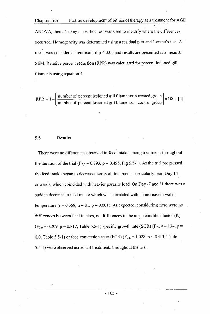
Chapter Five Further development of bithionol therapy as a treatment for AGD
ANOVA, then a Tukey's post hoc test was used to identify where the differences
occurred. Homogeneity was determined using a residual plot and Levene's test. A
result was considered significant if p < 0.05 and results are presented as a mean ±
SEM. Relative percent reduction (RPR) was calculated for percent lesioned gill
filaments using equation 4.
RPR = number of percent lesioned gill filaments in treated group x100 [4] 1 number of percent lesioned gill filaments in control group
5.5 Results
There were no differences observed in feed intake among treatments throughout
the duration of the trial (F2,6 = 0.793, p = 0.495, Fig 5.5-1). As the trial progressed,
the feed intake began to decrease across all treatments particularly from Day 14
onwards, which coincided with heavier parasite load. On Day -7 and 21 there was a
sudden decrease in feed intake which was correlated with an increase in water
temperature (r = 0.359, n = 81, p = 0.001). As expected, considering there were no
differences between feed intakes, no differences in the mean condition factor (K)
(F2,6 = 0.209, p = 0.817, Table 5.5-1) specific growth rate (SGR) (F2,6= 4.134, p =
0.0, Table 5.5-1) or feed conversion ratio (FCR) (F2,6= 1.028, p = 0.413, Table
5.5-1) were observed across all treatments throughout the trial.
- 105 -

Chapter Five Further development of bithionol therapy as a treatment for AGD
0.4 -14 -7 0 '7 14 21 28 35 42 49
Days from Neoparamoeba spp. exposure
Fig 5.5-1. Mean (± SEM) weekly tank feed intake (n = 3) for Atlantic salmon
exposed to Neoparamoeba spp. when fed either control feed (dotted line open
circle), prophylactic bithionol at 25 mg kg -I feed (solid line closed circle) or
therapeutic bithionol at 25 mg kg' feed (solid line open square). No error bars
indicate that all replicates within a treatment exhibited the same value. The
vertical broken line indicates administration of a 3 h freshwater bath.
Feed
Inta
ke (
°/0BW
)
- 106-
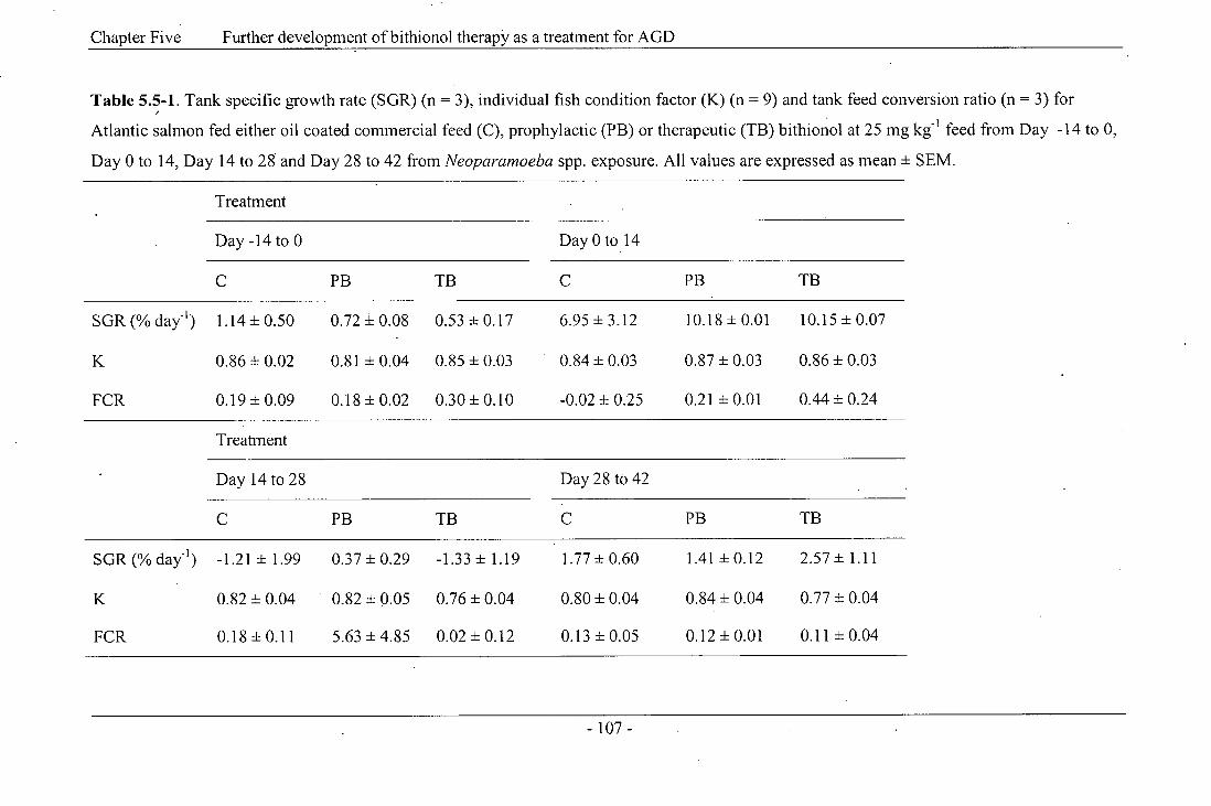
Chapter Five Further development of bithionol therapy as a treatment for AGD
Table 5.5-1. Tank specific growth rate (SGR) (n = 3), individual fish condition factor (K) (n = 9) and tank feed conversion ratio (n = 3) for
Atlantic salmon fed either oil coated commercial feed (C), prophylactic (PB) or therapeutic (TB) bithionol at 25 mg kg -I feed from Day -14 to 0,
Day 0 to 14, Day 14 to 28 and Day 28 to 42 from Neoparamoeba spp. exposure. All values are expressed as mean ± SEM.
Treatment
Day -14 to 0 Day 0 to 14
PB TB PB TB
SGR (% day-I )
FCR
1.14 ± 0.50
0.86 ± 0.02
0.19 ± 0.09
0.72 ± 0.08
0.81 ± 0.04
0.18 ± 0.02
0.53 ± 0.17
0.85 ± 0.03
0.30± 0.10
6.95 ± 3.12
0.84 ± 0.03
-0.02 ± 0.25
10.18 ± 0.01
0.87 ± 0.03
0.21 ± 0.01
10.15 ± 0.07
0.86 ± 0.03
0.44 ± 0.24
Treatment
Day 14 to 28 Day 28 to 42
PB TB PB TB
SGR (% day-1 )
FCR
-1.21 ± 1.99
0.82 ± 0.04
0.18 ± 0.11
0.37 ± 0.29
0.82 ± 0.05
5.63 ± 4.85
-1.33 ± 1.19
0.76 ± 0.04
0.02 ± 0.12
1.77 ± 0.60
0.80 ± 0.04
0.13 ± 0.05
1.41 ± 0.12
0.84 ± 0.04
0.12± 0.01
2.57 ± 1.11
0.77 ± 0.04
0.11 ± 0.04

Chapter Five Further development of bithionol therapy as a treatment for AGD
At the culmination of the trial all fish had exhibited signs of AGD. There
appeared to be no problems associated with palatability as feed was eaten across all
treatments and mortalities were observed across all treatments. No host toxicity was
observed based on gross examination of internal organs and histological
examination of the kidney, liver and muscle (histology not shown).
Fish administered bithionol at 25 mg kg ' either prophylactically (14 days
prior to Neoparamoeba spp. exposure) or therapeutically (upon clinical signs of
AGD), exhibited significantly reduced clinical signs of AGD including gross gill
score (F6,36 = 263.628, p < 0.001, Fig 5.5-2a) and percent lesioned gill filaments
over the entire trial, when compared to control fish (F6,36= 276.011, p <0.001, Fig
5.5-2b). 'Gross gill score and percent lesioned gill filaments were first observed on
Day 7 in all treatment groups. From Day 14 onwards both the gross gill score and
percent lesioned gill filaments increased at a greater rate in the control group
compared to both bithionol treatments. Significant reductions in percent lesioned
gill filaments were first observed between bithionol treated fish and control fish on
Day 14 and continued throughout the duration of the trial (F2,6= 73.035, p <0.001,
Fig 5.5-2b). Bithionol, when administered either prophylactically or therapeutically,
was effective in reducing the proportion of lesioned gill filaments by approximately
30% when compared to the control treatment prior to the freshwater bath
administered on Day 28 PE. At the culmination of the trial (Day 49 PE) the
difference observed between both bithionol and control treatments was
approximately 40% reduction in percent lesions. A similar result was seen with the
gross gill score, where on Day 28 controls had a mean gross gill score of four. This
- 108 -

Chapter Five Further development of bithionol therapy as a treatment for AGD
was significantly higher compared to bithionol treated fish, which exhibited a mean
gross gill score of 2.8, and this difference continued throughout the trial (F),6=
21.851, p = 0.002, Fig 5.5-2a). As expected, there were differences with respect to
time for both gross gill score (F6,36= 263.628, p < 0.001, Fig 5.5-2a) and percent
lesioned filaments (F6,36 = 276.011, p < 0.001, Fig 5.5-2b) with values increasing
until the freshwater bath was administered (Day 28 PE), after which values dropped
to similar levels as seen on Day 14 PE and slowly began to increase until the
culmination of the trial. Interestingly, the progression of lesion development was
similar among all treatments following the freshwater bath until the culmination of
the trial for both gross gill score and percent lesioned gill filaments (Fig 5.5-2a, b).
- 109-

a) G
ross
Gill S
core
Chapter Five Further development of bithionol therapy as a treatment for AGD
0 7 14 21 28 35 42 49
Days from Neoparamoeba spp. exposure
0 7 14 21 28 35 42 49
Days from Neoparamoeba spp. exposure
Fig 5.5-2. (a) gross gill score and (b) percent lesioned gill filaments for Atlantic
salmon with amoebic gill disease (AGD) when fed either control feed (dotted line
open circle), prophylactic bithionol at 25 mg kg-I feed (solid line closed circle) or
therapeutic bithionol at 25 mg kg -I feed (solid line open square) (n = 12 pre-bath
or 9 post-bath). All values are expressed as mean ± SEM. No error bars indicate
that all replicates within a treatment exhibited the same value. The vertical broken
line indicates administration of a 3 h freshwater bath. Lower and uppercase letters
denote significant differences among days and treatments, respectively (p < 0.05).
- 110 -

Chapter Five Further development ofbithionol therapy as a treatment for AGD
When the size of the lesions was examined, both bithionol treatments had
significantly smaller AGD lesions when compared to the control (R),6= 7.442, p =
0.024, Fig 5.5-3). A -relative percent reduction in lesion size of approximately 20-
30% was observed for both bithionol treatments from Day 21 onwards when
compared to the control treatment. There was a significant difference With respect
to time when examining lesion size (F6,36= 42.798, p <0.001, Fig 5.5-3), with
lesion size increasing from Day 0 to Day 21 post-exposure following which it
reached a plateau for all treatments until the culmination of the trial; interestingly
lesion size did not decrease or increase following the administration of a 3 h
freshwater bath.
Crude amoebae numbers were examined both pre- and post- freshwater bath
administration with the control treatment exhibiting a greater number of amoebae
compared to both bithionol treatments prior to the freshwater bath (F2,24 = 11.130, p
< 0.001,Table 5.5-2). However, following the freshwater bath no difference was
seen between treatments with crude amoebae numbers reduced to similar levels
across all treatments (F224 = 1.328, p = 0.284, Table 5.5-2) which was supported by
the proportional decrease in percent lesioned gill filaments (Fig 5.5-2b).

25 -
20 -
15 -
10 -
5 -
0
0
d i -
d I
7 14 21 28 35 42 49
Lesi
on S
ize
(ILU
)
Chapter Five Further development of bithionol therapy as a treatment for AGD
Days from Neoparamoeba spp. exposure
Fig 5.5-3. Lesion size in inter lamellae units (ILU) for Atlantic salmon with
amoebic gill disease (AGD) when fed either control feed (dotted line open circle),
prophylactic bithionol at 25 mg kg -I feed (solid line closed circle) or therapeutic
bithionol at 25 mg kg' feed (solid line open square) (n = 12 pre-bath or 9 post-
bath). Values are expressed as mean ± SEM. No error bars indicate that all
replicates within a treatment exhibited the same value. The vertical broken line
indicates administration of a 3 h freshwater bath. Lower and uppercase letters
denote significant differences among days and treatments, respectively (p < 0.05).
- 112 -

Chapter Five Further development of bithionol therapy as a treatment for AGD
Table 5.5-2. Mean (± SEM) crude amoeba numbers and relative percent reduction pre- and post- 3 h freshwater bath for Atlantic salmon with
amoebic gill disease (AGD) when fed either control feed, prophylactic bithionol at 25 mg kg -1 feed or therapeutic bithionol at 25 mg kg-1 feed.
Letters superscripts denote significant differences among treatments (p < 0.05) (n = 9).
Treatment
Control
Prophylactic bithionol Therapeutic bithionol
Pre-bath amoeba numbers
Post-bath amoeba numbers
228 588± 14638a
5 259 ± 838c
149 017 ± 11
34.81%
3 733 ±422c
168b 168 629 ± 11
26.23% 4 423 ±623c
144b

Chapter Five Further development of bithionol therapy as a treatment for AGD
5.6 Discussion
Atlantic salmon when fed bithionol at 25 mg kg' feed either as a two week
prophylactic or a therapeutic treatment exhibited significantly delayed and reduced
clinical signs of AGD compared to controls during a laboratory infection.
Following 14 days post-exposure, both groups of treated fish had significantly less
affected filaments than control fish. Prior to the freshwater bath administered on
Day 28 PE this difference had decreased; however, there were still significantly
fewer filaments affected than the control fish. A similar result was also seen with
gross gill score where a difference of approximately one gill score between
medicated fish and control fish from Day 14 PE onwards. Similarly, bithionol was
reported effective as a 14 day prophylactic treatment for AGD in Atlantic salmon
with gross gill score halve and a 53% reduction in percent lesions over 28 days
(Florent et al., 2007b). Bithionol was effective against the gill monogenean parasite
Microcotyle sebastis infestations in marine rockfish, Sebastes schlegeli, resulting in
a 66-93% reduction in parasite numbers when used at 1.25 g kg -I feed for 10-20
days (Kim and Choi, 1998). In contrast, bithionol was reported to be ineffective as a
treatment of S. salmonis, Gyrodactylus sp. and I. necator infestations in rainbow
trout (Tojo and Santamarina, 1998a; b; c); however, in these studies effectiveness
was defined as a complete elimination of the parasites. Bithionol when fed at 40 g
kg I feed for 10 days did not achieve complete elimination of the parasites but
produced an 80% reduction in S. salmonis while Gyrodactylus sp. and I. necator
numbers were reduced from a high to low intensity (Tojo and Santamarina, 1998a;
b; c).
- 114 -

Chapter Five Further development of bithionol therapy as a treatment for AGD
Crude numbers of viable amoeba pre- and post-bath were found to be higher in
the control fish compared to the medicated fish pre-bath; however, following the 3 h
freshwater bath amoeba numbers were reduced to similar levels across all
treatments with all exhibiting at least a 90% reduction which is similar to levels
seen in previous studies (Parsons et al., 2001a; Clark et al., 2003). This was not
seen with gross gill score and percent lesioned filaments which were knocked down
proportionally rather than to a similar level. However, all treatments had a similar
percent reduction in lesioned filaments of 60-70% following a freshwater bath and
gill score was reduced from four to two for the controls and from three to one for
both bithionol treatments.
The frequency of freshwater bathing conducted by the Tasmanian Atlantic salmon
industry is determined by the gross examination of gills. Generally; the trigger for
initiating a freshwater bath is a gross gill score of two or approximately 25%
lesioned filaments. So in this study control fish were ready for their first freshwater
bath around Day 14 PE whereas medicated fish were not at bath level until a week
later. Additionally, freshwater bathing only removed approximately two thirds of
the amoebae (Clark etal., 2003), leaving one third of the parasites as a chronic
persistent infection, which eventually triggers another freshwater bath and the cycle
is repeated. Nowak et al. (2007) reported that re-infection following a freshwater
bath occurred although lesions examined histologically immediately post-bath
exhibited no trophozoites of Neoparamoeba spp. However, several studies have
shown that reinfection is possible post bath without adding fresh amoebae to the
seawater (Adams and Nowak, 2001; 2004a; Gross et al., 2004). Hence, following
- 115 -

Chapter Five Further development of bithionol therapy as a treatment for AGD
the freshwater bath at Day 28 PE control fish were ready for a second bath at Day
35 PE compared to medicated feed where bath level was not reached until Day 49
PE. This indicated that it may be advantageous to use bithionol as a combination
therapy with the current freshwater mitigation to achieve the best results for
reduction in clinical signs of AGD.
Not only did medicated fish exhibit reduced clinical signs of AGD throughout the
study but the size of AGD lesions was reduced significantly compared to control
fish. Lesion size seemed to increase until Day 21 after which it reached a plateau for
all treatments. Interestingly, even following a freshwater bath, lesion size remained
the same for all treatments. There was a larger variability in lesion size following
the freshwater bath indicating that lesion size could be altered following the bath.
However, this could be due to larger lesions reducing in size slightly, whilst smaller
lesions had a greater rate of reduction. Furthermore, percent lesions decreased,
creating the possibility that the freshwater bath removed smaller lesions whilst the
larger lesions remain. Further investigation is warranted to investigate the
relationship between lesion size and the impact of a freshwater bath. There has been
minimal published information regarding AGD lesion size. Adams and Nowak
(2001) examined lesion size in order to describe the distribution and structure of
AGD lesions and Embar-Gopinath et al. (2005; 2006) examined lesion size with
respect to the presence of different salmonoid gill bacteria. Lesion size in this study
was similar to that in Embar-Gopinath et al. (2006) but larger than in Embar-
Gopinath et al. (2005).
- 116 -

Chapter Five Further development of bithionol therapy as a treatment for AGD
There were no differences in feed intake with fish in all treatment groups eating a
similar amount throughout the duration of the study. As the trial progressed the feed
intake observed across all treatments began to decrease, particularly from Day 14
onwards, which coincides with heavier parasite load. Furthermore, on Day -7 and
21 there was a sudden decrease in feed intake. This difference coincided with a
sudden increase in water temperature, although this was still within the
physiological range of Atlantic salmon. This is in contrast to previous studies where
intake of bithionol-treated feed was significantly higher than of control feeds
(Florent et al., 2007b). This study identified no differences between treatments
when examining SGR, K and FCR which is similar to previous studies with the
exception of condition factor where fish fed bithionol at 25 mg kg -1 feed to satiation
twice daily for 28 days following Neoparamoeba spp. exposure exhibited a higher
condition factor than control groups (Florent et al., 2007b). It is possible that the
difference seen between these two studies was due to the different method of
feeding that is either to satiation twice daily or at 1% BW day -1 . However, fish in
this study did not to reach the 1% limit regularly, suggesting that there could be
another factor involved such as time of year or temperature. Further investigations
would be needed to determine this.
Bithionol exhibited potential as an oral treatment for AGD at 25 mg kg I feed
when administered either for 2 weeks prior to (prophylactic) Neoparamoeba spp.
exposure or at clinical signs of AGD (therapeutic). A reduction in percent lesioned
gill filaments, lesion size, and gross gill score was achieved over the trial period.
Bithionol warrants further investigation as a possible in-feed treatment for AGD in
- 117 -

Chapter Five Further development of bithionol therapy as a treatment for AGD
Atlantic salmon, especially as a combination therapy with the standard freshwater
bath treatment. Also, examining the effect of pulse feeding or conducting trials
under more realistic field conditions with lower exposure doses and freshwater
baths would be of interest.
5.7 Acknowledgements
This research formed part of a project of the Aquafin CRC and received funds
from the Australian Government's CRC Programme, Fisheries R & D Corporation
and other CRC participants. The authors wish to thank T. Louwen-Skovdam and J.
Ransome for their technical support throughout the study.
- 118 -

CHAPTER 6
GENERAL DISCUSSION

Chapter Six
6 General Discussion
General Discussion
Throughout this thesis it has be shown that bithionol is toxic to Neoparamoeba
spp. in vitro and significantly delayed and reduced the intensity of amoebic gill
disease (AGD) pathology when administered as either a bath or oral treatment.
Therefore, revealing the potential of using bithionol as either an alternative
treatment for AGD or as a combination therapy with the current commercial
freshwater treatment. This indicated that bithionol warrants further investigation as
a treatment for AGD. The primary aim of this research described in previous
chapters was to identify an improved method of treatment for AGD caused by
Neoparamoeba spp. The improved method would have an enhanced efficacy,
alleviating clinical signs and associated pathology compared to the current
mitigation strategy of freshwater bathing whilst being economical. This research
focused primarily on development of an oral treatment for AGD, as producing an
in-feed treatment would assist in alleviating the release of large amounts of the
compound into the surrounding water (Findlay et al., 2000) and is less labour
intensive than bathing fish. The research documented in this thesis successfully
identified possible improvements or alternative treatments for AGD, involving the
oral administration of bithionol:
Herein will discuss the major findings related to the improved or alternative AGD
treatments, address the economic viability and touch on some environmental issues
of using such treatments. These past results have been not only necessary for the
formation of this thesis but have greatly advanced the knowledge surrounding the
- 120 -

Chapter Six General Discussion
treatment of AGD caused by Neoparamoeba spp. and the use of bithionol as a
treatment for parasites. Most notably, the development of a three-tiered approach to
identifying drug treatments: Stage I of drug screening would be the development of
single- and multi-day in vitro toxicity assays and host toxicity procedures; Stage II
of testing, whereby animals are offered drug-coated feed and subsequently
experimentally challenged with the parasite to determine fish toxicity and efficacy
for preventing clinical signs of disease; and Stage III testing involving field trials.
The research in this thesis has laid the necessary ground work for bithionol and
bithionol sulphoxide to be progressed to the final stages of treatment investigation
with a clinical trial.
A similar drug model has been used to develop the current in-feed treatment for
sea lice emamectin benzoate as a 0.2 % aquaculture pre-mix identified as Sli ce ® . A
great deal of effort has gone into the development of sea lice treatments, including
bath or dip treatments using dichlorovos, trichlorfon, azamethiphos, cypermethrin,
carbaryl, pyrethroids and hydrogen peroxide with varying degrees of efficacy
(Costello, 1993; Roth et al., 1993). Oral doses of ivermectin (Palmer et al., 1987),
insect growth regulators (Roth et al., 1993) and emamectin benzoate (Stone et al.,
2000) have also been examined. Some of which made it to commercial field trials
whilst others only to the small scale experimental stages.
The efficacy and toxicity of SLICE ® (emamectin benzoate) as a treatment for sea
lice infestations on Atlantic salmon was evaluated through an extensive series of
clinical studies and field trials (reviewed in Johnson and Margolis, 1993; Roth et
- 121 -
Chapter Six General Discussion
al., 1993; MacKinnon, 1997). Initially, emamectin benzoate was used in an
unformulated state and incorporated in an edible oil for application as a coating on
pelleted feed. Dose rate determination and dose rate confirmation studies were
conducted at several locations throughout Scotland. Small field trials were
conducted in Scotland, Norway, Canada, and Chile. In these field trials, fish were
fed a commercial feed treated with SLICE ®, 0.2% emamectin benzoate aquaculture
premix. A daily diet of medicated feed was administered at the recommended dose
rate of 50 lig kg' biomass day' for 7 consecutive days (Roth etal., 1993;
MacKinnon, 1997). Commercial field trials were conducted in Canada, Chile,
Scotland, and Norway. In Norway the efficacy of SLICE ® was compared to that of
another in feed treatment, teflubenzuron, a chitin synthesis inhibitor known
commercially as Ektobann®. Results from four study sites in Norway showed
SLICE® medicated feed provided better sustained efficacy against sea lice when
compared to teflubenzuron-treated diets (MacKinnon, 1997). Extensive studies
have also been conducted examining the effects on the host, the environmental
impacts and the pharmacokinetics of SLICE ® (MacKinnon, 1993; Chukwudebe et
aL, 1996; Mushtaq etal., 1996), this enabled the drug emamectin benzoate to
licensed for use in food fish under the registered trademark of SLICE ®.
Variability in the effectiveness of freshwater bathing is suggested to be attributed
1 - to water chemistry, in particular total hardness (mg L CaCO3) (Parsons et al.,
2001a; Parsons etal., 2001b; Clark, 2002; Powell and Clark, 2003; Roberts and
Powell, 2003a). Freshwater bathing is an environmentally friendly treatment and
• aids in maintaining the 'clean-green' image of the Australian salmon industry.
- 122 -

Chapter Six General Discussion
Roberts and Powell (2003a) reported that in using soft freshwater (19.3 - 37.4 mg I_, -
CaCO3) delays reinfection and subsequent pathology by at least two weeks
compared to hard freshwater. Providing the ability to reduce the annual number of
bath from ten to seven baths per year (Mitchell, 2001; Roberts, 2004) equating to a
saving of approximately $9.9 million annually. It is possible that with the combined
use of soft freshwater and an in-feed treatment that the freshwater bath frequency
could be reduced even further, thus saving more time and money.
A considerable amount of effort has been placed on identifying possible
treatments for AGD with varying success. Attempts to identify potential
chemotherapeutic agents have been limited due to either lack of direct efficacy on
the parasite, target fish toxicity or the cost of treatment (Alexander, 1991; Howard
and Carson, 1994). The above model has allowed for a strategic system to screen
and identify candidate drugs from a large pharmacopeia, whilst maintaining
effective resource management. Toxicity of several compounds to Neoparamoeba
spp. has been examined in vitro including levamisole (Howard and Carson, 1995),
chlorine dioxide, chloramine-T, hydrogen peroxide (Powell et al., 2003; Powell and
Clark, 2003), amprolium, albendazole, toltrazuril and bithionol (Powell et al.,
2003), all with low to moderate success. Howard and Carson (1994) reported that
levamisole at concentrations > 10 ppm in vitro were lethal to N. pemaquidensis, and
chloramine-T concentrations of 25 and 50 ppm effectively reduced amoeba
numbers after 2 h (Powell and Clark, 2003).
- 123 -

Chapter Six General Discussion
Using Stage I of the drug model, it was identified that bithionol was amoebicidal
at 1 and 10 mg U' (Powell etal., 2003); this was examined further using a greater
-1 variety of concentrations ranging from 0.1 to 10 mg L (Chapter 2). Bithionol and
bithionol sulphoxide (a cheaper and supposedly less toxic alternative) were
successful at reducing amoeba numbers relative to seawater controls. Both were
-1 toxic to Neoparamoeba spp. at concentrations ranging from 0.1 to 10 mg L over
72 h. However, freshwater still remained the most toxic with complete mortality of
Neoparamoeba spp. seen at 48 h. Bithionol was reported to be toxic to the
trophozoites of the protozoan parasite Giardia lambia at 0.42 and 0.28 mM over 24
and 72 h, respectively (Takeuchi et al., 1985). Similar results were seen with
respect to the human protozoan parasite Trichomonas vagina/is where bithionol was
toxic at 0.42 mM after 24 h (Takeuchi et al., 1985). The toxicity of bithionol in
vitro was also examined using the human and primate protozoan parasite
Entamoeba histolytica and found to kill virtually all axenic and polyxenic amoeba
in 24 h at both 0.42 and 0.28 mM (Takeuchi etal., 1984).
With regards to Neoparamoeba spp. very few drugs to date have been successful
at Stage I and thus moved onto Stage II, where animals were offered drug-coated
feed and subsequently experimentally challenged with the parasite to determine fish
toxicity and efficacy for preventing clinical signs of disease. Within this thesis,
Atlantic salmon, Salmo salar, and rainbow trout, Oncorhynchus mykiss, were either
administered bithionol as a bath treatment or offered bithionol-coated feed and
subsequently experimentally challenged with Neoparamoeba spp. to determine fish
toxicity and efficacy for preventing clinical signs of AGD. An efficient way to
- 124-

Chapter Six General Discussion
examine initial in vivo efficacy and toxicity is through bath administration of the
compound as it requires less resources and time. For AGD-affected Atlantic salmon
and rainbow trout, it was identified that a 1 h seawater bath using a bithionol
concentration of 1 mg L -1 exhibited no morbidity and reduced amoeba numbers and
percent lesioned gill filaments from the seawater control. Bath administration of
bithionol has been examined in numerous species infected with various parasites.
Bithionol toxicity and efficacy is dependent upon host species and target parasite.
With regard to salmonid parasites, such as Gyrodactylus sp. and khthyobodo
necator, Santamarina etal. (1991) observed limited toxicity and complete in vitro
efficacy against Gyrodactylus sp. in rainbow trout at 12.5 mg L -1 , with a minimum
20 mg L -1 reported as efficacious in vivo. Tojo etal. (1994b) stated that bithionol
was efficacious in vivo against I. necator in rainbow trout at 25 mg 1,1 for a 3 h bath
on two consecutive days; higher concentrations exhibited some mortality. Bithionol
was reported as efficacious against Neoparamoeba spp. at concentrations ranging
from 1 to 35 mg L -1 ; however, at concentrations > 10 mg L i high percentage
mortality was seen within 3 h for both Atlantic salmon and rainbow trout (Florent et
al., 2007a). Finally, Madsen etal. (2000) determined that bithionol at 0.1 mg L -1
was an effective treatment against trichodiniasis in European eels, Anguilla
anguilla, but found bithionol to have a relatively narrow therapeutic index.
Conversely, Buchmann et al. (1992) demonstrated that bithionol at
0.1 mg 1_, -1 was not efficacious as a static bath against gill parasitic monogeneans,
Pseudodactylogyrus sp. and concentrations > 1 mg 1_, -1 were toxic causing 100%
mortality within 24 h in the European eel. Bithionol as a static bath has also been
reported as effective in killing the ciliate Tetrahymena pyriformis at 60 mg L -1 ,
- 125 -

Chapter Six General Discussion
whilst being non-toxic to the minnow golden shiner, Notemigonus crysoleucas
(Griffin, 1989). Nonetheless, with the scale and intensity of salmonid farming
occurring worldwide, a bithionol bath treatment would be impractical, as it is
insoluble and would require a large amount of the compound to treat the bath water.
On the other. hand, with the determination of toxicity to the target animal and its
efficacy with respect to AGD it would be possible to incorporate bithionol in-feed
and assess as an oral treatment for the control of AGD in Atlantic salmon.
Although, Neoparamoeba spp. is an external parasite, similar to the treatment
strategies for sea lice, the desired delivery method, from an industry perspective is
an in-feed drug therapy. Reasons for this included the fact that in-feed treatment
allows medication during adverse weather conditions and on exposed sites and
reduces the chance of cross-infestation that may occur during the several days
necessary to apply bath treatments to all cages on a site because an in-feed
treatment permits simultaneous medication of all cages on a site. Hence the
treatment of sea lice with an emamectin benzoate as a 0.2 % aquaculture pre-mix
identified as SLICE ® is ideal for famers compared to the bath treatments previously
used (Stone et al., 1999). Furthermore, with the increasing need for the use of
chemotherapeutants in aquaculture, it is very important to minimise the
accumulation of chemicals in food for human consumption and the effect on the
environment. Therefore, consideration for residues in the animal and accumulations
and the effect on the environment must be taken when examining any compounds
for use as treatments for disease in aquatic animals.
- 126-

Chapter Six General Discussion
Bithionol has been suggested as an oral medication due to its ability to provide
easy administration and limited fish handling (Tojo and Santamarina, 1998a).
Bithionol has a wide spread use in the treatment of numerous parasites in different
hosts (El-Sayad, 1997). In cattle, bithionol sulphoxide was reported as effective at
reducing Fasciola hepatica in the rumen when administered a single dose at 30 mg
kg' for the first four weeks but numbers returned to pre treatment levels 20 weeks
following treatment (Ueno etal., 1973). However, when administered as a single
dose at 90 mg kg -1 bithionol sulphoxide reduced the natural rumen fluke infestation
of cattle by 70% (Prasittirat etal., 1997). Bithionol was reported as effective in
removing the worms Thysanosoma actinioides (Allen et al., 1962) and
Hymenolepris nana (Maki and Yanagisawa, 1985) from sheep and mice,
respectively. With examining aquatic animal health, bithionol at 40 g kg -I feed was
offered for.10 days at 2% body weight (BW) per day to rainbow trout infected with
Spironucleus salmonis (formerly Hexamita salmonis), Gyrodactylus sp. or I.
necator and exhibited a reduction in parasite load. Bithionol eliminated
approximately 80% of S. salmonis from rainbow trout whilst both Gyrodactylus sp.
and I. necator infections were reduced from a high to low intensity (Tojo and
Santamarina, 1998a; b; c). Kim and Choi (1998) reported bithionol administered in-
feed at 100-200 mg kg -I BW significantly reduced the number of monogeneans
Microcotyle sebastis on the gills of cultured rock fish (Sebastes schlegeli), with a 20
day feeding duration being most effective. Furthermore, bithionol was reported as
efficacious delay and reducing clinical signs of AGD in Atlantic salmon when fed
at 25 mg kg' feed either as a two week prophylactic treatment or a therapeutic
treatment (Florent et al., 2007b).
- 127

Chapter Six General Discussion
Bithionol was successful in delaying and reducing clinical signs of AGD by
approximately two weeks and even reducing the amoebae numbers by half would
theoretically half the number of baths. Bithionol incorporated into feed would cost
approximately $150 tonne -I (Sigma-Aldrich, 2006); if this successfully halved the
bath numbers it has the potential to lessen the cost of treatment compared to
freshwater. When examining the cost of bithionol and bithionol sulphoxide the
price difference is considerable with bithionol eight times more expensive than
bithionol sulphoxide, thus bithionol sulphoxide if as effective as bithionol is a
significantly cheaper alternative.
Bithionol has been identified as successful in stage I and II of the three-tiered
approach mentioned above. In order to continue through to stage III involving field
testing it would be advantageous to examine the pharmacology and residue levels of
bithionol and its metabolites. Previous studies show that bithionol may work more
effectively when high infection pressure is observed such as that in a laboratory trial
as opposed to a low infection pressure seen typically in a farm situation (Chapter 5).
Thus examining the efficacy of both bithionol and bithionol sulphoxide at lower
infections pressure would be beneficial and aid in collating data to apply for an
experimental license.
The next phase of the research would include further investigation of bithionol as
an in-feed treatment for AGD, incorporating examination of the effect of
prophylactic and therapeutic treatments running experiments for a longer period of
time to obtain more growth data, examining the effect of pulse feeding, or
- 128 -

Chapter Six General Discussion
conducting trials under more realistic field conditions with lower exposure doses
and freshwater baths. Furthermore, sophisticated studies on the pharmacology of
possible treatments are often lacking, owing in part to the limited knowledge about
the biochemistry of parasite, which are more difficult to study in vitro than bacteria
(Barrett-Connor, 1982). Therefore, in addition to studying clinical efficacy of
bithionol as a treatment for AGD, pharmacokinetic examination is essential to
establish correct dosage regimes and hence optimal drug usage (Samuelsen and
Ervik, 1999; Haug and Hals, 2000; Stringer, 2001; Birkett, 2002; Katharios et al.,
2002). Pharmacokinetics describes the relationship between the dose and the
unbound drug concentration at a drug receptor (otherwise known as the site of
action), and the drug concentrations time course within the body (Birkett, 2002).
Stringer (2001) relays pharmacokinetics as "the mathematical description of the rate
and extent of uptake, distribution, and elimination of drugs in the body".
The pharmacokinetics and bioavailability of a drug can be affected by several
parameters including fish species, age, water temperature, salinity, and route of
administration (Haug and Hals, 2000). Consequently knowledge of the
pharmacokinetics and bioavailability of the drug used in the intended species is vital
for correct application (Samuelsen and Ervik, 1999; Stringer, 2001; Birkett, 2002;
Hansen et al., 2003). Favourable pharmacokinetic properties include good
absorption after administration, high bioavailability, good tissue penetration and
biotransformation (Stringer, 2001; Birkett, 2002; della Rocca etal., 2004).
Although studies have been conducted on uptake, distribution and excretion in
rodents a comparison with the results from rodents show that the toxicity of
- 129-

Chapter Six General Discussion
antiparasitic drugs to aquatic organisms Islikely not well correlated to mammals
(Yoshimura and Endoh, 2005).
Neoparamoeba perurans has been confirmed a cosmopolitan protozoan parasite
and, therefore, of significance to the global mariculture industry (Young etal.,
2008). Thus, confirmation of N. perurans in regions of significant finfish
production indicates that AGD is of global significance to the mariculture industry
and therefore the development of a cheaper alternative treatment of this disease has
the potential to become a worldwide issue as opposed to a localised Tasmanian
issue.
6.1 Conclusion
This thesis evaluated the in vitro toxicity of bithionol and bithionol sulphoxide to
Neoparamoeba spp. and determined that both bithionol and bithionol sulphoxide
were toxic to Neoparamoeba spp. at concentrations ranging from 0.1 to 10 mg L -1
over 72 .h; however, freshwater still remained the most toxic with complete
mortality seen at 48 h. Following this, the toxicity of bithionol to Atlantic salmon
and rainbow trout in fresh and seawater and the efficacy of bithionol as a 1 h
seawater bath treatment for AGD were examined. It was determined that bithionol
was toxic at and above 25 mg L -1 in freshwater and seawater for both Atlantic
salmon and rainbow trout; whilst concentrations of 1, 5 and 10 mg L -1 produced an
efficacy comparable to the currently used freshwater bath in Atlantic salmon. To
follow on from Stage I to Stage II of drug testing, the efficacy of bithionol as an
oral treatment for Atlantic salmon affected by AGD was examined. It was reported
- 130-
Chapter Six General Discussion
that bithionol at 25 mg kg' feed, when fed as a two week prophylactic treatment for
Neoparamoeba spp. exposure, delayed and reduced the intensity of AGD
pathology. In order to enhance the knowledge of bithionol as an oral treatment for
AGD the effects of prophylactic and therapeutic treatments were examined as well
as the combination with a freshwater bath. It was identified that there was no
difference in gross gill score or percent lesioned gill filaments when Atlantic
salmon Were fed bithionol at 25 mg kg' feed prophylactically or therapeutically.
When a freshwater bath was administered there was a proportional knock down
suggesting that it may be advantageous to use bithionol as a combination therapy
with the current freshwater mitigation to achieve the best results for reduction in
clinical signs of AGD.
The results of this thesis indicate that bithionol warrants, further investigation as a
possible in-feed treatment for AGD in Atlantic salmon, especially as a combination
therapy with the standard freshwater bath treatment. Also, examining the effect of
pulse feeding or conducting trials under more realistic field conditions with lower
exposure, doses and freshwater baths would be of interest. It would also be
advantageous and necessary in order to develop the treatment further to examine the
environmental effects along with the pharmacokinetics of bithionol.
- 131 -
CHAPTER 7
REFERENCES

Chapter Seven
7 References
References
Adams, M.B., Nowak, B.F., 2001. Distribution and structure of lesions in the gills
of Atlantic salmon, Salmo salar L., affected with amoebic gill disease. J.
Fish Dis. 24, 535-542.
Adams, M.B., Nowak, B.F., 2003. Amoebic gill disease: sequential pathology in
cultured Atlantic salmon, Salmo salar L. J. Fish Dis. 26, 601-614.
Adams, M.B., Ellard, K., Nowak, B.F., 2004. Gross pathology and its relationship
with histopathology of amoebic gill disease (AGD) in farmed Atlantic
salmon, Salmo salar L. J. Fish Dis. 27, 151-161.
Adams, M.B., Nowak, B.F., 2004a. Sequential pathology after initial freshwater
bath treatment for amoebic gill disease in cultured Atlantic salmon, Salmo
salar L. J. Fish Dis. 27, 163-173.
Adams, M.B., Nowak, B.F., 2004b. Experimental amoebic gill disease of Atlantic
salmon, Salm° salar L.: further evidence for the primary pathogenic role of
Neopararnoeba sp. (Page, 1987). J. Fish Dis. 27, 105-113.
Alderman, D.J., 2002. Trends in therapy and prophylaxis 1991-2001. Bull. Eur.
Assoc. Fish Pathol. 22, 117-125.
- 133 -

Chapter Seven References
Alexander, J.M., 1991. Treatment of amoebic gill disease. Field trials - 1990/91.
Salmon Enterprises of Tasmania R & D Report, Dover, Tasmania,
Australia, pp. 51-101.
Allen, R.W., Enzie, F.D., Samson, K.S., 1962. The effects of bithionol and other
compounds on the fringed tapeworm, T4sanosoma athnioides, of sheep.
American Journal of Veterinary Research 23, 236-240.
Anonymous, 2002. Guidance for FDA staff and industry - Compliance policy
guides manual.
Anonymous, 2008. Products distributed by the Centers for Disease Control and
Prevention, Atlanta, U.S.A.
Armstrong, R., 1994. Therapeutant availability properties of the Canadian salmon
farming industry. Bulletin of the Aquaculture Association of Canada 98, 8-
19.
Athanassopoulou, F., Karagouni, E., Dotsika, E., Ragias, V., Tavla, J.,
Christofilloyanis, P., Vatsos, I., 2004. Efficacy and toxicity of orally
administrated anti-coccidial drugs for innovative treatments of Myxobo/us
sp infection in Puntazzo puntazzo. Dis. Aquat. Org: 62, 217-226.
- 134-

Chapter Seven References
Bacq, Y., Besnier, J.M., Duong, T.H., Pavie, G., Metman, E.H., Choutet, P., 1991.
Successful treatment of acute fascioliasis with bithionol. Hepatology 14,
1066- .1069.
Bakke, T.A., Harris, P.D., 1998. Diseases and parasites in wild Atlantic salmon
(Salrno salar) populations. Canadian Journal of Fisheries and Aquatic
Sciences 55, 247-266.
Barrett-Connor, E., 1982. Drugs for the treatment of parasitic infection. Med.
Clin. N. Am. 66, 245-255.
Birkett, D.J., 2002. Pharmacokinetics made easy. McGraw-Hill, Sydney., 131 pp.
Bowers, J.M., Mustafa, A., Speare, D.J., Conboy, G.A., Brimacombe, M., Sims,
D.E., Burka, J.F., 2000. The physiological response of Atlantic salmon,
Salmo salar L., to a single experimental challenge with sea lice,
Lepeophtheirus salmonis. J. Fish Dis. 23, 165-172.
Bowman, J.P., Nowak, B., 2004. Salmonid gill bacteria and their relationship to
amoebic gill disease. J. Fish Dis. 27, 483-492.
Briere, J.J., Favief, J., El Ghouzzi, V., Djouadi, F., Benit, P., Gimenez, A.-P.,
Rustin, P., 2005. Succinate dehydrogenase deficiency in human. Cellular
and Molecular Life Sciences 62, 2317-2324.
- 135 -

Chapter Seven References
Buchmann, K., Felsing, A., Slotved, H.C., 1992. Effects of metrifonate, sodium
chloride and bithionol on an European population of the gill parasitic
monogeneans Pseudodadylogyrus spp. and the host Anguilla anguilla. Bull.
Eur. Assoc. Fish Pathol. 12, 57-60.
Buchmann, K., Lindenstrom, T., 2002. Interactions between monogenean
parasites and their fish hosts. International Journal for Parasitology 32, 309-
319.
Burka, J.F., Hammel, K.L., Horsberg, T.E., Johnson, G.R., Rainnie, D.J., Speare,
D.J., 1997. Drugs in salmonid aquaculture - A review. J. vet. Pharmacol.
Therap. 20, 333-349.
Cameron, D.E., 1992. Amoebic gill disease field research 1991/92. In: Valentine,
P. (Ed.), Proceedings of the SALTAS Research and Development Review
Seminar. SALTAS, Hobart, Tasmania, pp. 13-133.
Chukwudebe, A.C., Feely, W.F., Burnett, T.J., Crouch, L.S., Wislocki, P.G.,
1996. Uptake of emamectin benzoate residues from soil by rotational crops.
Journal of Agricultural and Food Chemistry 44, 4015-4124.
Clark, A., Nowak, B.F., 1999. Field investigations of amoebic gill disease in
Atlantic salmon, Salmo salar L., in Tasmania. J. Fish Dis. 22, 433-443.
- 136 -
Chapter Seven References
Clark, G., Powell, M., Nowak, B., 2003. Effects of commercial freshwater bathing
on reinfection of Atlantic salmon, Salmo salar, with Amoebic Gill Disease.
Aquaculture 219, 135-142.
Clark, G.A., 2002. Efficacy and side effects of freshwater bathing as a treatment
for amoebic gill disease: implications of water chemistry and bath duration.,
School of Aquaculture. University of Tasmania, Launceston, Tasmania, pp.
94.
Costello, M.J., 1993. Review of methods to control sea lice (Caligidae: Crustacea)
infestations of salmon (Salmo salar) farms. In: Boxshall, G.A., DeFaye, D.
(Eds.), Pathogens of wild and farmed fish: Sea lice. Ellis Horwood,
Chichester, England, pp. 219-252.
della Rocca, G., Di Salvo, A., Malvisi, J., Sello, M., 2004. The disposition of
enrofloxacin in seabream (Sparus aurata L.) after single intravenous
injection or from medicated feed administration. Aquaculture 232, 53-62.
Dezfuli, B.S., Pironi, F., Giari, L., Domeneghini, C., Bosi, G., 2002. Effect of
Pomphorhynchus laevis (Acanthocephala) on putative neuromodulators in
the intestine of naturally infected Salmo trutta. Dis. Aquat. Org. 51, 27-35.
Dickerson, H., Clark, T., 1998. Ichthyophthirius multifiliis: a model of cutaneous
infection and immunity in fishes. Immunol. Rev. 166, 377-384.
- 137 -
Chapter Seven References
Douglas-Helders, G.M., Handlinger, J., Carson, J., Nowak, B.F., 2003.
Assessment over time of the infectivity of gill-derived and seawater
dispersed Neoparamoeba pemaquidensis (Page, 1987). Bull. Eur. Assoc.
Fish Pathol. 23, 35-39.
Douglas-Helders, M., Nowak, B., Zilberg, D., Carson, J., 2000. Survival of
Paramoeba pemaquidensis on dead salmon: implications for management
of cage hygiene. Bull. Eur. Assoc. Fish Pathol 20, 167-169.
Douglas-Helders, M., Carson, J., Howard, T., Nowak, B., 2001a. Development
and validation of a new dot blot test for the detection of Paramoeba
pemaquidensis (Page) in fish. J. Fish Dis. 24, 273-280.
Douglas-Helders, M., Saksida, S., Raverty, S., Nowak, B.F., 2001b. Temperature
as a risk factor for outbreaks of amoebic gill disease in farmed Atlantic
salmon (Salmo salar). Bull. Eur. Assoc. Fish Pathol. 21, 114-116.
Durbize, E., Vigan, M., Puzenat, E., Girardin, P., Adessi, B., Desprez, P.,
Humbert, P., Laurent, R., Aubin, F., 2003. Spectrum of cross-
photosensitization in 18 consecutive patients with contact photoallergy to
ketoprofen: associated photoallergies to non-benzophenone-containing
molecules. Contact Dermatitis 48, 144-149.
- 138 -

Chapter Seven References
Dykova, I., Figueras, AI, Novoa, B., 1995. Amoebic gill infection of turbot,
Scophthalmus maximus. Folia Parasitol. 42, 91-96.
Dykova, I., Figueras, A., Novoa, B., Casal, J.F., 1998. Paramoeba sp., an agent of
amoebic gill disease of turbot Scophthalmus maximus. Dis. Aquat. Org . 33,
137-141.
Dykova, I., Figueras, A., Peric, Z., 2000. Neoparamoeba Page, 1987: light and
electron microscopic observations on six strains of different origin. Dis.
Aquat. Org. 43, 217-223.
Dykova, I., Novoa, B., 2001. Comments on diagnosis of amoebic gill disease
(AGD) in turbot, Scophthalmus maximus. Bull. Eur. Assoc. Fish Pathol. 21,
40-44.
Dykova, I., Fiala, I., Lom, J., Luke, J., 2003. Perkinsiella amoebae-like
enclosymbionts of Neoparamoeba spp., relatives of the kinetoplastid
Ichthyobodo. European Journal of Protistology 39, 37-52.
Dykova, I., Nowak, B.F., Crosbie, P.B.B., Fiala, I., Peckova, H., Adams, M.B.,
Machatkova, B., Dvotakova, H., 2005. Neoparamoeba branchiphila n. sp.,
and related species of the genus Neoparamoeba Page, 1987: morphological
and molecular charaterization of selected strains. J. Fish Dis. 8, 49-64.
- 139-

Chapter Seven References
El-Sayad, M.H., 1997. Comparative studies on the effect of bithionol, praziqantel
• and triclabendazole in rabbit fascioliasis 1. Parasitological studies. Journal
of the Egyptian Society of Parasitology 27, 131-142.
Elliott, N.G., Carson, J., Wong, F., 2001. Detection and abundance of Paramoeba
species in the environment. CSIRO Marine Research; Tasmanian
Aquaculture & Fisheries Institute University of Tasmania; Fisheries
Research and Development Corporation, Hobart, Tas., 68 pp.
Embar-Gopinath, S., Butler, R., Nowak, B., 2005. Influence of salmonid gill
bacteria on development and severity of amoebic gill disease. Dis. Aquat.
Org. 67, 55-60.
Embar-Gopinath, S., Crosbie, P., Nowak, B.F., 2006. Concentration effects of
Winogradskyella sp on the incidence and severity of amoebic gill disease.
Dis. Aquat. Org . 73, 43-47.
Enzie, F.D., Colglazier, M.L., 1960. Preliminary trials with bithionol against
tapeworm infections in cats, dogs, sheep and chickens. American Journal of
Veterinary Research July, 628-630.
Evans, A.S., 1976. Causation and disease: the Henle—Koch postulates revisited.
Yale J. Biol. Med. 49, 175-195.
- 140 -

Chapter Seven References
Fiala, I., Dykova, I., 2003. Molecular characterisation of Neoparamoeba strains
isolated from gills of Scophthalmus maximus. Dis. Aquat. Org. 55, 11-16.
Findlay, V.L., Zilberg, D., Munday, B.L., 2000. Evaluation of levamisole as a
treatment for amoebic gill disease of Atlantic salmon, Salmo salar L.
Journal of Fish Diseases 23, 193-198.
Fisk, D.M., Powell, M.D., Nowak, B.F., 2002. The effect of amoebic gill disease
and hypoxia on survival and metabolic rate of Atlantic salmon (Salmo
salar). Bull. Eur. Assoc. Fish Pathol. 22, 190-194.
Florent, R.L., Becker, J.A., Powell, M.D., 2007a. Evaluation of bithionol as a bath
treatment for amoebic gill disease caused by Neoparamoeba spp. Vet.
Parasitol. 144, 197-207.
Florent, R.L., Becker, J.A., Powell; M.D., 2007b. Efficacy of bithionol as an oral
treatment for amoebic gill disease in Atlantic salmon Salmo salar (L.).
Aquaculture 270, 15-22.
Foster, C., Percival, S., 1988. Treatment of Paramoebic gill disease in salmon and
trout. Saltas Aquanote, Hobart. No. 14., 1-4.
Griffin, B.R., 1989. Screening of chemicals to control protozoan parasites of fish.
The Progressive Fish-Culturist 51, 127-132.
- 141 -

Chapter Seven References
Grimnes, A., Jakobsen, P.J., 1996. The physiological effects of salmon lice
infection on post-smolt of Atlantic salmon. J. Fish. Bio. 48, 1179-1194.
Gross, K.A., Morrison, R.N., Butler, R., Nowak, B.F., 2004. Atlantic salmon,
Salmo salar L., previously infected with Neoparamoeba sp. are not resistant
to reinfection and have suppressed phagocyte function. J. Fish Dis. 27, 47-
56.
Hamajima, F., 1973. Metabolism of lung fluke genus Paragonimus. VII. Action
of bithionol on glycolytic and oxidative metabolism of adult worms. Exp.
Parasitol. 34, 1-11.
Hamajima, F., Fujinob, T., Yamagamia, K., Eriguchib, N., 1979. Studies on the in
vitro effects of bithionol and menichlopholan on flukes of Clonorchis
sinensis, Metagonimus takahashii and Paragonimus miyazakii. International
Journal of Parasitology 9, 241-249.
Handeland, S.O., Wilkinson, E., Sveinsbo, B., McCormick, S.D., Stefansson,
S.O., 2004. Temperature influence on the development and loss of seawater
tolerance in two fast-growing strains of Atlantic salmon. Aquaculture 233,
513-529.
- 142 -

Chapter Seven References
Hansen, M.K., Nymoen, U., Horsberg, T.E., 2003. Pharmacokinetic and
pharmacodynamic properties of metomidate in turbot (Scophthalmus
maximus) and halibut (1-hppogiossus hippog/ossus). Journal of veterinary
pharmacology and therapeutics 26, 95-103.
Harder, A., 2002. Chemotherapeutic approaches to trematodes (except
schistosomes) and cestodes: current level of knowledge and outlook. ,
Parasitol. Res. 88, 587-590.
Harris, J.0., Powell, M.D., Attard, M., Green, T.J., 2004. Efficacy of chloramine-
T as a treatment for amoebic gill disease (AGD) in marine Atlantic salmon
(Salmo salar L.). Aquae. Res. 35, 1448-1456.
Harris, J.0., Powell, M.D., Attard, M.G., DeHayr, L., 2005. Clinical assessment
of chloramine-T and freshwater as treatments - for the control of gill amoebae
in Atlantic salmon, Salmo salar L. Aquae. Res. 36, 776-784.
Haug, T., Hals, P.A., 2000. Pharmacokinetics of oxytetracyline in Arctic charr
(Salvelinus alpinus L.) in freshwater at low temperature. Aquaculture 186,
175-191.
Hecht, T., Endemann, F., 1998. The impact of parasites, infections and disease on
the development of aquaculture in sub-Saharan Africa. J. Appl. Ichthyol. 14,
213-221.
- 143 -

Chapter Seven References
Hedrick, R.P., 1998. Relationships of the host, pathogen, and environment:
Implications for diseases of cultured and wild fish populations. J. Aquat.
Anim. Health 10, 107-111.
Hill, A.B., 1965. The environment and disease: association or causation? Proc. R.
Soc. Med. 1965, 295-300.
Howard, T., Carson, J., 1993. Are there alternatives to freshwater treatment of
AGD? In: Valentine, P. (Ed.), Proceedings of the SALTAS Research and
Development Review Seminar. SALTAS, Hobart, Tasmania, pp. 81-87.
Howard, T., Carson, J., Lewis, T., 1993. Development of a model of infection for
amoebic gill disease. In: Valentine, P. (Ed.), Proceedings of the Salmon
Enterprises of Tasmania Research and Development Review Seminar.
Salmon Enterprises of Tasmania Marine Operations, Dover Tas., pp. 103-
ill.
Howard, T., Carson, J., 1994. Amoebic gill disease laboratory research
1993/1994, Proceedings of the Saltas Research and Development Review
Seminar. Salmon Enterprises of Tasmania, Hobart, Tasmania, pp. 71-91.
Howard, T., Carson, J., 1995. Amoebic gill disease - In vitro studies of a
Paramoeba species. Research and Development Review Seminar. Salmon
Enterprises of Tasmania, Hobart, Tasmania, Australia.
- 144 -

Chapter Seven References
Howard, T., 2001. Paramoebiasis of sea-farmed salmonids in Tasmania - a study
of the aietology, pathogenicity and control. University of Tasmania, Hobart.
Howe, G.E., Gingerich, W.H., Dawson, V.K., Olson, J.J., 1999. Efficacy of
hydrogen peroxide for treating saprolegniasis in channel catfish. Journal of
Aquatic Animal Health 11, 222-230.
Iglesias, R.., Parama, A., Alvarez, M.F., Leiro, J.,.Sanmartin, M.L., 2002.
Antiprotozoals effective in vitro against the scuticociliate fish pathogen
Philasterides dicentrarchi. Dis. Aquat. Org. 49, 191-197.
Johnson, S.C., Margolis, L., 1993. Efficacy of ivermectin for control of the
salmon louse Lepeophtheirus salmonis on Atlantic salmon. Dis. Aquat. Org .
17, 101-105.
Jones, S.R.M., 2001. The occurrence and mechanisms of innate immunity against
parasites in fish. Dev. Comp. Immunol. 25, 841-852.
Katharios, P., Iliopoulou-Georgudaki, J., Antimisiaris, S., Kantzaris, V., Pavlidis,
M., 2002. Pharmacokinetics of ivermectin in sea bream, Sparus aurata
using a direct competitive ELISA. Fish Physiology and Biochemistry 26,
189-195.
- 145 -

Chapter Seven References
Kent, M.L., Sawyer, T.K., Hedrick, R.P., 1988. Paramoeba pemaquidensis
(Sarcomastigophora: Paramoebidae) infestation of the gills of coho salmon
Oncorhynchus kisutch reared in sea water. Dis. Aquat. Org . 5, 163-169.
Kent, M.L., 2000. Marine netpen- farming leads to infections with some unusual
parasites. International Journal for Parasitology 30, 321-326.
Kim, K.H., Choi, E.S., 1998. Treatment of Microcotyle sebastis (Monogenea) on
the gills of cultured rockfish (Sebastes schelegeli) with oral administration
of mebendazole and bithionol. Aquaculture 167, 115-121.
LaPatra, S.E., 1998. Factors affecting pathogenicity of infectious hematopoietic
necrosis virus (IHNV) for salmonid fish. Journal of Aquatic Animal Health
10, 121-131.
Leef, M.J., Harris, J.0., Hill, J., Powell, M.D., 2005a. Cardiovascular responses of
three salmonid species affected with amoebic gill disease (AGD). Journal of
Comparative Physiology B-Biochemical Systemic and Environmental
Physiology 175, 523-532.
Leef, M.J., Harris, J.0., Powell, M.D., 2005b. Respiratory pathogenesis of
amoebic gill disease (AGD) in experimentally infected Atlantic salmon
Salmo salar. Dis. Aquat. Org. 66, 205-213.
- 146 -

Chapter Seven References
Leef, M.J., Harris, J.0., Powell, M.D., 2007a. Metabolic effects of amoebic gill
disease (AGD) and chloramine-T exposure in seawater-acclimated Atlantic
salmon Salmo salar. Dis. Aquat. Org. 78, 37-44.
Leef, M.J., Hill, J.V., Harris, J.0., Powell, M.D., 2007b. Increased systemic
vascular resistance in Atlantic salmon, Salmo salar L., affected with
amoebic gill disease. J. Fish Dis. 30, 601-613.
MacKinnon, B., 1993. Host response of Atlantic salmon (Salmo salar) to
infection by sea lice (Caligus elongatus). . Canadian Journal of Fisheries
and Aquatic Sciences. 50, 789-792.
MacKinnon, B.M., 1997. Sea Lice: a review. World Aquaculture 28, 5-10.
Madsen, H.C.K., Buchmann, K., Mellergaard, S., 2000. Treatment of
trichodiniasis in eel (Anguilla anguilla) reared in recirculation systems in
Denmark: alternatives to formaldehyde. Aquaculture 186, 221-231.
Maki, J., Yanagisawa, T., 1985. Anthelmintic effects of bithionol, paromomycin
sulphate, flubendazole and mebendazole on mature and immature
Hymenolepis nana in mice. Journal of Helminthology 59, 211-216.
- 147 -
Chapter Seven References
Mallatt, J., 1985. Fish gill structural changes induced by toxicants and other
irritants: A statistical review. Canadian Journal of Fisheries and Aquatic
Sciences 42, 630-648.
Mansell, B., Powell, M.D., Ernst, I., Nowak, B.F., 2005. Effects of the gill
monogenean Zeuxapta seriolae (Meserve, 1938) and treatment with
hydrogen peroxide on pathophysiology of kingfish, Seriola lalandi
Valenciennes, 1833. J. Fish Dis. 28, 253-262.
Martin, S.W., Meek, A.H., Willeberg, P., 1987. Veterinary epidemiology:
principles and methods. Iowa State University Press, Ames„ 343p. pp.
McCormick, S.D., 1993. Methods for nonlethal gill biopsy and measurement of
Nat, Kt-ATPase activity. Canadian Journal of Fisheries and Aquatic
Sciences 50, 656-658.
Meshi, T., Yoshikawa, M., Sato, Y., 1970. Metabolic fate of bis(3,5-dichloro-2-
hydroxypheny1)-sulfoxide (bithionol sulfoxide). Biochemical pharmacology
19, 1351-1361.
Mitchell, D., 2001. HAC experiences of AGD. In: Battaglene, S.C., Cobcroft,
J.M. (Eds.), The first scientific conference of the Atlantic salmon
aquaculture subprogram handbook, CSIRO Marine Laboratories, Hobart,
Tasmania.
- 148 -

Chapter Seven References
Morgan, J.D., Iwama, G.K., 1991. Effects of salinity on growth, metabolism and
ion regulation in juvenile rainbow and steelhead trout (Oncorhynchus
mykiss) and fall chinook salmon (Oncorhynchus tshawytscha). Canadian
Journal of Fisheries and Aquatic Sciences 48, 2083-2094.
Morrison, R.N., Crosbie, P.B.B., Nowak, B.F., 2004. The induction of laboratory-
based amoebic gill disease revisited. Journal of Fish Diseases 27, 445-449.
Morrison, R.N.., Crosbie, P.B.B., Cook, M.T., Adams, M.B., Nowak, B.F., 2005.
Cultured gill-derived Neoparamoeba pemaquidensis fails to elicit amoebic
gill disease (AGD) in Atlantic salmon Salmo salar. Dis. Aquat. Org . 66,
135-144.
Mourot, D., Dagorn, M., Delephine, B., 1987. Liquid chromatographic
determination of depletion of bithionol sulfoxide and its two major
metabolites in bovine milk. Journal of the Association of Official Analytical
Chemists 70, 810-812.
Mullen, T.E., Nevis, K.R., O'Kelly, C.J., Gast, R.J., Frasca, S., 2005. Nuclear
small-subunit ribosomal RNA gene-based characterization, molecular
phylogeny and per detection of the Neoparamoeba from western Long
Island Sound lobster. J. Shellfish Res. 24, 719-731.
- 149 -

Chapter Seven References
Munday, B.L., Foster, C.K., Roubal, F.R., Lester, R.J.G., 1990. Paramoebic gill
infection and associated pathology of Atlantic salmon, Salmo salar, and
rainbow trout, Salmo gairdneri, in Tasmania. Academic Press, San Diego,
CA, 215-222 pp.
Munday, B.L., Lange, K., Foster, C:, Lester, R.J.G., Handlinger, J., 1993.
Amoebic gill disease of sea-caged salmonids in Tasmanian waters.
Tasmanian Fisheries Research 28, 14-19.
Munday, B.L., Zilberg, D., Findlay, V., 2001. Gill disease of marine fish caused
by infection with Neoparamoeba pemaquidensis. J. Fish Dis. 24, 497-507.
Munday, B.L., Zilberg, D., 2003. Efficacy of, and toxicity associated with, the use
of levamisole in seawater to treat amoebic gill disease. Bulletin of the
European Association of Fish Pathologists 23, 3-6.
Murray, A.G., Peeler, E.J., 2005. A framework for understanding the potential for
emerging diseases in aquaculture. Prey. Vet. Med. 67, 223-235.
Mushtaq, M., Syintsakos, L.R., Krieter, P.A., Colletti, A., Arison, B., Crouch,
L.S., Wislocki, P.G., 1996. Absorption, tissue distribution, excretion and
metabolism of 3H- and 14C-labeled emamectin benzoate in rats. Journal of
Agricultural and Food Chemistry. 44, 3342-3349.
- 150 -

Chapter Seven References
Mustafa, A., Speare, D.J., Daley, J., Conboy, G.A., Burka, J.F., 2000. Enhanced
susceptibility of seawater cultured rainbow trout, Oncorhynchus mykiss
(Walbaum), to the microsporidian Loma salmonae during a primary
infection with the sea louse, Lepeophtheirus salmonis. J. Fish Dis. 23, 337-
341.
Noga, E.J., 2000. Fish disease: diagnosis and treatment. Iowa State University,
Ames, United States of America, 367 pp.
Nowak, B., 2001. Qualitative evaluation of risk factors for amoebic gill disease in
cultured Atlantic salmon. In: Rodgers, C.J. (Ed.), Proceedings of the OIE
International conference on risk analysis in aquatic animal health. Office
International des Epizooties, Paris, France, pp. 148-155.
Nowak, B.F., Munday, B.L., 1994. Histology of gills of Atlantic salmon during
the first few months following transfer to sea water. Bull. Eur. Assoc. Fish
Pathol 14, 77-81.
Nowak, B.F., 1999. Significance of environmental factors in aetiology of skin
diseases of teleost fish. Bull. Eur. Assoc. Fish Pathol. 19, 290-292.
Nowak, B.F., Carson, J., Powell, M.D., Dykova, I., 2002. Amoebic gill disease in
the marine environment. Bull. Eur. Assoc. Fish Pathol. 22, 144-151.
- 151 -
Chapter Seven References
• Nowak, B.F., Dawson, D., Basson, L., Deveney, M., Powell, M.D., 2004. Gill
histopathology of wild marine fish in Tasmania: potential interactions with
gill health of cultured Atlantic salmon, Salmo salar L. J. Fish Dis. 27, 709-
717.
Nowak, B.F., Adams, M.B., Vincent, B.N., O'Brien, D., Mitchell, D., 2007..
Aquafin CRC - Atlantic salmon aquaculture subprogram: Effects of
husbandry on amoebic gill disease. Fisheries Research and Development
Corporation final report project No. 2004/214 ISBN 978-1-86295-412-0, pp.
82.
Page, F.C., 1970. Two new species of Paramoeba from Maine. J. Protozool. 17,
421-427.
Page, F.C., 1987. The classification of "naked" amoebae (Phylum Rhizopoda).
Arch. Protistenkd. 133, 199-217.
Palmer, R., Rodger, E., Drinan, C., Dwyer, C., Smith, P.R., 1987. Preliminary
trials on the efficacy of Ivermectin against parasitic copepods of Atlantic
salmon. Bull. Eur. Assoc. Fish Pathol. 7, 47-54.
Palzenberger, M., Pohla, H., 1992. Gill surface area of water-breathing freshwater
fish. Reviews in Fish Biology and Fisheries 2, 187-216.
- 152 -

Chapter Seven References
Parsons, H., Nowak, B., Fisk, D., Powell, M., 20 .01a. Effectiveness of commercial
freshwater bathing as a treatment against amoebic gill disease in Atlantic
salmon. Aquaculture 195, 205-210.
Parsons, H.J., Nowak, B.F., Powell, M.D., Dix, T., 2001b. Changes in water
quality during commercial freshwater bathing of Atlantic salmon (Salmo
salar) in Tasmania - implications for treatment of amoebic gill disease. Bull.
Eur. Assoc. Fish Pathol. 21, 71-76.
Pfeiler, E., Kirschner, L.B., 1972. Studies on gill ATPase of rainbow trout (Salmo
gairdneri). Biochimica et biophysica acta. Amsterdam 282, 301-310.
Powell, M., Green, T., Plaisted, C., 2003. A cell culture assay for the in vitro
efficacy testing of amoebocidal compounds against Neoparamoeba
pemaquidensis. Bulletin of the European Association of Fish Pathologists
23, 291-296.
Powell, M., Harris, J., 2004. Influence of oxygen on the toxicity of chloramine-T
to Atlantic salmon smolts in freshwater and seawater. Journal of Aquatic
Animal Health 16, 83-92.
Powell, M.D., Fisk, D., Nowak, B.F., 2000. Effects of graded hypoxia on Atlantic
salmon infected with amoebic gill disease. J. Fish. Bio. 57, 1047-1057.
- 153 -

Chapter Seven References
Powell, M.D., Parsons, H.J., Nowak, B.F., 2001. Physiological effects of
freshwater bathing of Atlantic salmon (Salmo salar) as a treatment for
amoebic gill disease. Aquaculture 199, 259-266.
Powell, M.D., Forster, M.E., Nowak, B.F., 2002a. Apparent vascular hypertension
associated with amoebic gill disease affected Atlantic salmon (Salmo salar)
in Tasmania. Bull. Eur. Assoc. Fish Pathol. 22, 328-333.
Powell, M.D., Nowak, B.F., Adams, M.B., 2002b. Cardiac morphology in relation
to amoebic gill disease history in Atlantic salmon, Salmo salar L. J. Fish
Dis. 25, 209-216.
Powell, M.D., Clark, G.A., 2003. In vitro survival and the effect of water
chemistry and oxidative chemical treatments on isolated gill amoebae from
AGD-affected Atlantic salmon. Aquaculture 220, 135-144.
Powell, M.D., Clark, G.A., 2004. Efficacy and toxicity of oxidative disinfectants
for the removal of gill amoebae from the gills of amoebic gill disease
affected Atlantic salmon (Salmo salar L.) in freshwater. Aquae. Res. 35,
112-123.
Powell, M.D., Attard, M., Harris, J.0., Roberts, S.D., Leef, M.J., 2005. Atlantic
salmon aquaculture subprogram: Why fish die - the treatment and
- 154 -

' Chapter Seven References
pathophysiology of AGD. Fisheries Research and Development Corporation
final report project 2001/205 ISBN 1 86295 259 0, pp. 112.
Powell, M.D., Ransome, J., Barney, M., Duijf, R.M.M., Flik, G., 2007. Effect of
dietary inclusion of n-acetyl cysteine on mucus viscosity and susceptibility
of rainbow trout, Oncorhynchus mykiss, and Atlantic salmon, Salmo salar,
to amoebic gill disease. J. World Aquacult. Soc. 38, 435-442.
Powell, M.D., Becker, J.A., Ransome, J., Florent, R.L., Jones, M.A., 2008.
Atlantic salmon aquaculture subprogram: Commercial AGD and salmon
health. Fisheries Research and Development Corporation final report project
2004/213 ISBN 978-86295-378-9, pp. 96.
Prasittirat, P., Nithiuthai, S., Ruengsuk, K., Kitwan, P., Bunmatid, C., Roopan, S.,
Itagaki, H., 1997. Efficacy of bithionol sulfoxide, niclosamide and
fenbendazole against natural rumen fluke infection in cattle. Helminthologia
34, 155-157.
Rabago-Castro, J.L., Sanchez, J.G., Perez-Castafieda, R., Gonzalez-Gonzalez, A.,
2006. Effects of the prophylactic use of Romet (R)-30 and copper sulfate on
growth, condition and feeding indices in Channel catfish (Ictalurus
punctatus). Aquaculture 253, 343-349.
- 155-

Chapter Seven References
Ramstad, A., Colquhoun, D.J., Nordmo, R., Sutherland, I.H., Simmons, R., 2002.
Field trials in Norway with SLICE (R) (0.2 % emamectin benzoate) for the
oral treatment of sea lice infestation in farmed Atlantic salmon Salmo salar.
Dis. Aquat. Org. 50, 29-33.
Reid, L., Clothier, R.H., Khammo, N., 2001. Hydrogen peroxide induced stress in
human keratinocytes and its effect on bithionol toxicity. Toxicology in Vitro
15, 441-445.
Rew, R.S., 1978. Mode of action of common anthelmintics. J. Vet. Pharmacol.
Therap. 1, 183-197.
Roberts, S.D., Powell, M.D., 2003a. Reduced total hardness of fresh water
enhances the efficacy of bathing as a treatment for amoebic gill disease in
Atlantic salmon, Salmo salar L. J. Fish Dis. 26, 591-599.
Roberts, S.D., Powell, M.D., 2003b. Comparative ionic flux and gill mucous cell
histochemistry: effects of salinity and disease status in Atlantic salmon
(Salmo salar L.). Comparative Biochemistry and Physiology Part A 134,
525-537.
Roberts, S.D., 2004. Improving the treatment of amobic gill disease in salmonids
with soft freshwater and the mucolytic drug L-cysteine ethyl ester, School
of Aquaculture. University of Tasmania, Launceston, Tasmania, pp. 208.
- 156 -

Chapter Seven References
Roberts, S.D., Powell, M.D., 2005. Oral L-cysteine ethyl ester (LCEE) reduces
amoebic gill disease (AGD) in Atlantic salmon Salmo salar. Dis. Aquat.
Org. 66, 21-28.
Rodger, H.D., McArdle, J.F., 1996. An outbreak of amoebic gill disease in
Ireland. The Veterinary Record. 139, 348-349.
Roth, M., Richards, R.H., Sommerville, C., 1993. Current practices in the
chemotherapeutic control of sea lice infestations in aquaculture: a review. J.
Fish Dis. 16, 1-26.
Roubal, F.R., Lester, R.J.G., Foster, C.K., 1989. Studies on cultured and gill-
attached Paramoeba sp. (Gymnamoebae: Paramoebidae) and the
cytopathology of paramoebic gill disease in Atlantic salmon, Salmo salar
L., from Tasmania. J. Fish Dis. 12, 481-492.
Samuelsen, 0.B., Ervik, A., 1999. A single-dose pharmacokinetic study of
oxolinic acid and vetoquinol, an oxolinic acid ester, in Atlantic halibut,
Hippoglossus hippoglossus L., held in sea water at 9 degree C. Journal of
Fish Diseases 22, 13-23.
Sanmartin Duran, M.L., Femadez Casal, J., Tojo, J.L., Santamarina, M.T.,
Estevez, J., Ubeira, F.M., 1991. Trichodina sp.: Effect on the growth of
- 157 -

Chapter Seven References
farmed turbot (Scophthalmus maximus). Bull. Eur. Ass. Fish Pathol. 11, 89-
91.
Santamarina, M.T., Tojo, J., Ubeira, F.M., Quinteiro, P., Sanmartin, M.L., 1991.
Anthelmintic treatment against Gyrodactylus sp. infecting rainbow trout
Oncorhynchus mykiss. Dis. Aquat. Org . 10, 39-43.
Sawatari, S., Hamajima, F., 1967. Effects of isothiocyanates and bithionol on
Paragonimus westermani (Kerbert, 1878) in vitro. Japanese Journal of
Parasiology 16, 174-178.
Scharsack, J.P., Steinhagen, D., Kleczka, C., Schmidt, J.0., Korting, W., Michael,
R.D., Leibold, W., Schuberth, H.J., 2003. Head kidney neutrophils of carp
(Cyprinus carpio L.) are functionally modulated by the haemoflagellate
Trypanoplasma borreli. Fish Shellfish Immunol. 14, 389-403.
Schmahl, G., Taraschewski, H., Mehlhorn, H., 1989. Chemotherapy of fish
parasites. Parasitol. Res. 75, 503-511.
Scholz, T., 1999. Parasites in cultured and feral fish. Vet. Parasitol. 84, 317-335.
Shao, Z.Z.J., 2001. Aquaculture pharmaceuticals and biologicals: current
perspectives and future possibilities. Advanced Drug Delivery Reviews 50,
229-243:
- 158 -

Chapter Seven References
Sigma-Aldrich, 2006. Bithionol.
Speare, D.J., Arsenault, G., MacNair, N., Powell, M.D., 1997. Branchial lesions
associated with intermittent formalin bath treatment of Atlantic salmon,
Salmo salar L., and rainbow trout, Oncorhynchus mykiss (Walbaum). J. Fish
Dis. 20, 27-33.
Sprague, J.B., 1969. Measurement of pollutant toxcity to fish, I. Bioassay methods
of acute toxcity. Water. Research 3, 793-821.
Sprague, J.B., 1990. Chapter 15: Aquatic Toxicology. In: Schreck, C.B., Moyle,
P.B. (Eds.), Methods for fish biology. American Fisheries Society,
Bethesda, U.S.A., pp. 491-528.
Steinum, T., Kvellestad, A., Ronneberg, L.B., Nilsen, H., Asheim, A., Fjell, K.,
Nygard, S.M.R., Olsen, A.B., Dale, 0.B.; 2008. First cases of amoebic gill
disease (AGD) in Norwegian seawater farmed Atlantic salmon, Salmo salar
L., and phylogeny of the causative amoeba using 18S cDNA sequences. J.
Fish Dis. 31, 205-214.
Stoffregen, D.A., Bowser, P.R., Babish, J.G., 1996. Antibacterial
chemotherapeutants for finfish aquaculture: A synopsis of laboratory and
field efficacy and safety studies. Journal of Aquatic Animal Health 8, 181-
207.
- 159-

Chapter Seven References
Stone, J., Sutherland, I.H., Sommerville, C.S., Richards, R.H., Varma, K.J., 1999.
The efficacy of emamectin benzoate as an oral treatment of sea lice,
Lepeophtheirus salmonis (Kroyer), infestations in Atlantic salmon, Salmo
salar L. J. Fish Dis. 22, 261-270.
Stone, J., Sutherland, I.H., Sommerville, C., Richards, R.H., Varma, K.J., 2000.
Commercial trials using emamectin benzoate to control sea lice
Lepeophtheirus salmonis infestations in Atlantic salmon Salmo salar. Dis.
Aquat. Org. 41, 141-149.
Stringer, J.L., 2001. Basic concepts in pharmacology: a student's survival guide.
McGraw-Hill, London., 285 pp.
Takashima, F., Hibiya, T., 1994. An atlas of fish histology: normal and
pathological features. Kodansha Ltd., Tokyo, Japan, 195 pp.
Takeuchi, T., Kobayashi, S., Kawasaki, H., 1984. Entamoeba histolytica
inhibition in vitro by bithionol of respiratory activity and growth. Exp.
Parasitol. 58, 1-7.
Takeuchi, T., Kobayashi, S., Tanabe, M., Fujiwara, T., 1985. In vitro inhibition of
Giardia lamblia and Trichomonas vaginalis growth by bithionol,
dichlorophene, and hexachlorophene. Anitmicrobial Agents and
Chemotherapy 27, 65-70.
- 160 -

Chapter Seven References
Tan, C.K., Nowak, B.F., Hodson, S.L., 2002. Biofouling as a reservoir of
Neoparamoeba pemaquidensis (Page, 1970), the causative agent of amoebic
gill disease in Atlantic salmon. Aquaculture 210, 49-58.
Thorburn, M.A., Moccia, R.D., 1993. Use of chemotherapeutics on trout farms in
Ontario. Journal of Aquatic Animal Health 5, 85-91.
Thrusfield, 1995. Veterinary epidemiology. Blackwell Scientific, Oxford, xi, 479
PP.
Tojo, J., Santamarina, M.T., Ubeira, F.M., Leiro, J., Sanmartin, M.L., 1993.
Efficacy of anthelmintic drugs against gyrodactilosis in rainbow trout
(Oncorhynchus mykis .$). Bull. Eur. Assoc. Fish Pathol. 13, 45-48.
Tojo, J., Santamarina, M.T., Ubeira, F.M., Leiro, J., Sanmartin, M.L., 1994a.
Attempts to control khthyobodo necator in rainbow trout (Oncorhynchus
mykiss) by bath treatment. Bull. Eur. Assoc. Fish Pathol. 14, 12-15. .
Tojo, J.L., Santamarina, M.T., Leiro, J., Ubeira, F.M., Sanmartin, M.L., 1994b.
Pharmacological treatments against khthyobodo necator (Henneguy, 1883)
in rainbow trout, Oncorhynchus mykiss (Walbaum). J. Fish Dis. 17, 135-
143.
- 161 -
Chapter Seven References
Tojo, J.L., Santamarina, M.T., 1998a. Oral pharmacological treatments for
parasitic diseases of rainbow trout Oncorhynchus mykiss. I: Hexamita
salmonis. Dis. Aquat. Org . 33, 51-56.
Tojo, J.L., Santamarina, M.T., 1998b. Oral pharmacological treatments for
parasitic diseases of rainbow trout Oncorhynchus mykiss. III: khthyobodo
necator. Dis. Aquat. Org. 33, 195-199.
Tojo, J.L., Santamarina, M.T., 1998c. Oral pharmacological treatments for
parasitic diseases of rainbow trout Oncorhynchus mykiss. II: Gyrodactylus
sp. Dis. Aquat. Org . 33, 187-193.
Tonguthai, K., 1997. Control of freshwater parasites: a Southeast Asia
perspective. International Journal for Parasitology 27, 1185-1191.
Treasurer, J.W., Wallace, C., Dear, G., 2002. Control of sea lice on farmed
Atlantic salmon S. salar L. with the oral treatment emamectin benzoate
(SLICE). Bull. Eur. Assoc. Fish Pathol. 22, 375-380.
Ueno, H., Alvarez, J.M., De Mergen, A.M.R., Cabral, A.F., Bodden, H.A.Q.,
1973. Field trials with rafoxanide, bithionol sulfoxide and niclofolan against
Fasciola hepatica in cattle in the Dominican Republic. Nat. Inst. Anim. Hlth
Quart. 13, 69-74.
- 162 -

Chapter Seven References
VentreIla, V., Pagliarani, A., Trombetti, F., Pirini, M., Trigari, G., Borgatti, A.R.,
2001. Response of rainbow trout gill Na+-ATPase to T-3 and NaC1
administration. Physiological and Biochemical Zoology 74, 694-702.
Wong, F.Y.K., Carson, J., Elliott, N.G., 2004. 18S ribosomal DNA-based PCR
identification of Neoparamoeba pemaquidensis, the agent of amoebic gill
disease in sea-farmed salmoriids. Dis. Aquat. Org . 60, 65-76.
Yang, S.-P., Lin, C.-C., 1967. Treatment of paragonimiasis with bithionol and
bithionol sulfoxide. Chest 52, 220-232.
Yokogawa, M., Yoshimura, H., Okura, T., Sano, M., Tsuji, M., Iwasaki, M.,
Hiros.e, H., 1961a. Chemotherapy of paragonimiasis with bithionol II.
Clinical observations on the treatment of bithionol. Japanese Journal of
Parasitology 10, 317-327.
Yokogawa, M., Yoshimura, H., Sano, M., Okura, T., Tsuji, M., Takizawa, A.,
Harada, Y., Kihata, M., 1961b. Chemotherapy of paragonimiasis with
bithionol I. Experimental chemotherapy on the animals infected with
Paragonimus westermani or P. ohirai. Japanese Journal of Parasitology 10,
302-316.
Yoshimura, H., Endoh, Y.S., 2005. Acute toxicity to freshwater organisms of
antiparasitic drugs for veterinary use. Environmental Toxicology 20, 60-66.
- 163 -

Chapter Seven References
Young, N.D., Crosbie, P.B.B., Adams, M.B., Nowak, B.F., Morrison, R.N., 2007.
Neoparamoeba perurans n. sp., an agent of amoebic gill disease of Atlantic
salmon (Salmo salar). International Journal for Parasitology 37, 1469-1481.
Young, N.D., Dykova, I., Snekvik, K., Nowak, B.F., Morrison, R.N., 2008.
Neoparamoeba perurans is a cosmopolitan aetiological agent of amoebic
gill disease. Dis. Aquat. Org . 78, 217-223.
Zall, D.M., Fisher, M.D., Garner, Q.M., 1956. Photometric determination of
chloride in water. Analytical Chemistry 28, 1665-16678.
Zilberg, D., Munday, B.L., 2000. Pathology of experimental amoebic gill disease
in Atlantic salmon, Salmo salar L., and the effect of premaintenance of fish
in sea water on the infection. J. Fish Dis. 23, 401-407.
Zilberg, D., Gross, A., Munday, B.L., 2001. Production of salmonid amoebic gill
disease by exposure to Paramoeba sp. harvested from the gills of infected
fish. J. Fish Dis. 24, 79-82.
- 164-

Appendix I
8 Appendix I
8.1 Trypsin-EDTA solution in Hanks Balanced Salts Contents and
concentrations
Trypsin 0.5 g L-1
Ethylenediaminetetraacetic acid (EDTA) solution 0.2 g L -1
Hanks Balance Salts (HBSS) Modified containing:
KC1 0.4 gL -1
KH2PO4 (anhyd) 0.06 g L -1
NaHCO3 0.35 g
NaC1 8.0 g
Na2HPO4 (anhydrous) 0.04788 g L -1
D-Glucose 1.0 g
Phenol Red•Na 0.011 g
- 165 -