Embed Size (px)
Citation preview

REVIEW ARTICLE PUJVol. 5, No. 2, 2012ISSN: 0258-3216
Personal non-commercial use only. PUJ copyright © 2011. All rights reserved PUJ; 2012, 5(2): 93-104
Primary Amoebic Meningoencephalitis Caused By Naegleria Fowleri
Dalia A. Abo El-Maaty, Rania S. HamzaDepartment of Medical Parasitology, Faculty of Medicine, Zagazig University, Egypt
Received: May, 2012 Accepted: July, 2012
ABBREVIATIONSB103: Brain cell line 103, CDC: Centers for Disease Control and Prevention, CHO: Chinese Hamster Ovary, CMI: Cell mediated immunity, CNS: Central nervous system, CSF: Cerebro-spinal fluid, E6:Vero monkey kidney cell culture, HLF: Human lung fibroblast, HMG: High mobility group protein, HSP70: Heat shock protein 70, IFA: Immunoflurescent assay, ITS: Internal transcribed spacers, kDa: Kilo Dalton, MAC: Membrane attack complex, MDCK: Madine-Darby canine kidney, MIF: Migration inhibitory factor, Nfa1: Non-fimbrial adhesin genes, PMNs: Polymorphonuclear leucocytes, PAM: Primary amoebic meningoencephalitis, 26S proteasome subunit: High molecular mass protease 1.400 kDa, TNF-α: Tumor necrosis factor-α.
INTRODUCTION
Naegleria fowleri is an ameboflagellate, known as "the brain-eating amoeba". It causes an acute, fulminant and rapidly fatal central nervous system (CNS) infection termed primary amoebic meningoencephalitis (PAM). The infection occurs from inhalation of trophozoites(1). It enters the CNS, after insufflation of infected water, by attaching itself to the olfactory nerve, then migrating through the cribriform plate of the ethmoid bone along the fila olfactoria and blood vessels, and into the anterior cerebral fossae. It feeds on nerve tissue and causes extensive inflammation, necrosis and hemorrhage leading to death(2). N. fowleri is a thermophilic amoeba that grows well in tropical and subtropical climates. Raised temperature during hot summer months or warm water from power plants facilitates its growth(3).
Infection occurs in healthy children and young adults with a recent history of exposure to warm fresh water (polluted water in ponds, swimming pools and man-made lakes). The ameba has been also detected in artificially heated industrial water
sources(4), and in domestic water supplies(5). About 310 cases have been reported with a high case fatality rate of approximately 95%(6,7). The number of reported cases of PAM has increased worldwide in recent years. The increased incidence may be due to greater awareness of the disease or due to the development of more rapid, highly sensitive and specific diagnostic assays such as PCR. In addition, changes in environmental conditions, thermal pollution of water from industry and the development of industrialized areas with nuclear power plants and cooling towers that allow for concomitant greater growth of amoeba and their bacterial food source may afford greater opportunities for infection(8). Naegleria species were isolated and identified from various water sources in Lower and Upper Egypt(9,10).
As several cases of PAM may be misdiagnosed as bacterial meningitis or tubercular meningitis due to lack of awareness, this review aimed to highlight the role of N. fowleri as a causative agent of meningitis to draw special attention for its diagnosis and management.
Corresponding author: Dalia A. Abo El-Maaty, [email protected]: Naegleria fowleri, Amebic Meningoencephalitis, Infection, Diagnosis.

94
Primary Amoebic Meningoencephalitis
Epidemiological Features of Naegleria SpeciesHuman disease caused by free-living amoebae was first reported in 1965 by Fowler and Carter, who studied four patients with PAM in South Australia(11). N. fowleri is pathogenic in human and has a tropism for the CNS. It exists in three forms; the first is an invasive, reproductive trophozoite (ameboid-form) that is thermophilic and thrives best in temperatures from 35° to 46°C; the second is a flagellate pear-shaped motile form that is maintained at temperatures 27° to 37°C, while the third form is a spherical cyst that can survive in much lower temperatures and will convert to a trophozoite form in a suitable environment(12). Trophozoites grow fast at around 42°C and proliferate by binary fission. In their free-living state, trophozoites feed on bacteria, while in tissues they phagocytose red and white blood cells and destroy tissue. A biflagellate form occurs when trophozoites are exposed to a change in ionic concentration such as placement in distilled water. Trophozoites encyst due to unfavorable conditions such as food deprivation, crowding, desiccation, accumulation of waste products and cold temperatures below 10°C(13).
More than 30 Naegleria species have been isolated from environmental sources with only N. fowleri known to cause human infections. Two other species, N. australiensis and N. italica, can cause infection in experimental animals but these species have never been identified in human infections; although they too are found in warm, stagnant bodies of water, and even chlorinated swimming pools, making contact with humans inevitable(14).
While a large number of animals also bathe, swim and frolic in thermally polluted waters, only one case of PAM has been described(15) in a South American tapir and another case in Holstein cattle(16) which was diagnosed by immunofluorescence in brain sections. Recently, an amoeba was isolated from the brain of an infected cow and identified as N. fowleri based on morphology(17) and sequencing of the internal transcribed spacers (ITS), including the 5.8S rRNA genes(18).
Until 2012, the 310 human cases reported worldwide were mostly from Australia, United States, Great Britain, Czechoslovakia, Thailand and Mexico(7). These cases typically occurred during the summer months in warm, humid climates presumably because of warmer water coupled with increased swimming activities(12). In North America, 111 human cases have been reported in the USA and nine cases in Mexico until 2008(19). The majority of infections in Mexico occurred after swimming in water naturally heated by the sun, while in California, a few cases occurred after swimming in geothermal waters. In addition, PAM infections occurred in the USA by using tap water originating from warm groundwater and the amoeba was subsequently isolated from the well(20).
In South America, only seven cases were reported in Venezuela and five cases in Brazil. Almost all cases in South America were related with swimming in natural water or untreated pools. N. fowleri was also isolated from a man-made lake in Brazil(21). In Cuba one human PAM case was recorded, and in Guadeloupe one patient died of PAM after swimming in a pool fed by geothermal water(22,23).
In Australia, 19 reported patients were infected from the town water supply, either by swimming in backyard pools fed by it, or washing out the nose(24). Another nine cases were reported in New Zealand and all of them were related to swimming in geothermal water(25,26).
The presence of N. fowleri in swimming pools, pond water and sewage canals has been confirmed in India(27,28). One case of PAM was reported in a 38-year-old man after swimming in a hot spring in China and he survived the infection(29). In Japan, N. fowleri was isolated from both geothermal water and industrial cooling water but only two PAM cases were detected(30). In India, there were 11 reported PAM cases(31-33). Four of these patients survived, and three of them had no contact with water, which makes it again suspicious whether they really were N. fowleri infections. Most other patients in India had been swimming or bathing in well water, ponds or puddles. In Pakistan, 17 cases were reported till 2011, 13 of whom were reported during a period of 17 months probably due to the use of tap water, which was not adequately filtered or chlorinated(34). The authors referred to these reported cases as Muslims who routinely perform ritual ablution, that involves taking water into the nostrils, creating an ideal portal of entry for N. fowleri into the brain(35,36).
From Africa, there are very few reported cases of PAM. In Nigeria, four cases were reported; one was a farmer sniffing water up into the nose and the others were children. In these children, no contact was noted with water, but it was hypothesized that dust in the air introduced the amoeba. Subsequently, N. fowleri was isolated from nasal passages of children, dust from the air and water samples(37). One PAM case was reported in Namibia, where a child became infected by N. fowleri after swimming in stagnant pools originating from hot springs(38). In Madagascar, one patient died after swimming in a warm fresh water lake(39).
In Egypt, two Naegleria species (gruberi and fowleri) were isolated and identified from various water sources in Lower and Upper Egypt(9). Samples from different water sources of Alexandria and from nasal passages of 500 healthy children inhabiting areas nearby these sources were examined for the presence of free-living amoebae. The same two species were isolated from the water of canals and drains. N. gruberi was found in the nasal passages of six healthy children living near the contaminated canals(10). The authors reported that
Abo El-Maaty and Hamza
95
no amoebae were encountered in the drinking water, swimming pools, sea and lake water included in their study.
PathogenesisThe pathogenesis of PAM is poorly understood. Both pathogenic and nonpathogenic species of Naegleria have been isolated from the environment, but the determinants of virulence and pathogenicity are unknown(8). As with many pathogenic organisms, prolonged growth of N. fowleri in axenic culture in vitro attenuates its virulence while serial passage through mice restores and maintains virulence(40). The portal of CNS entry is the olfactory neuroepithelium. It is believed that the sustentacular cells lining the olfactory neuroepithelium phagocytose the amoebae that enter the nasal passages of the victims while indulging in aquatic activities. The amoebic trophozoites pass through the sieve-like cribriform plate and penetrate into the subarachnoid space and continue on to the brain parenchyma. The incubation period of PAM varies depending on the size of the inoculum, and on the virulence of the particular strain of infecting amoebae(41).
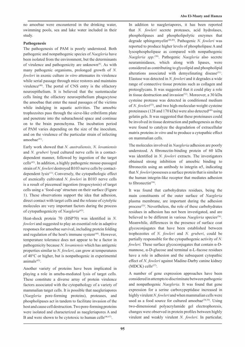
Early work showed that N. australiensis, N. lovaniensis and N. gruberi lysed cultured nerve cells in a contact-dependent manner, followed by ingestion of the target cells(42). In addition, a highly pathogenic mouse-passaged strain of N. fowleri destroyed B103 nerve cells by contact-dependent lysis(13). Conversely, the cytopathologic effect of axenically cultivated N. fowleri in B103 nerve cells is a result of piecemeal ingestion (trogocytosis) of target cells using a ‘food-cup’ structure on their surface (Figure 1). These observations support the idea that adhesion, direct contact with target cells and the release of cytolytic molecules are very important factors during the process of cytopathogenicity of Naegleria(43).
Heat-shock protein 70 (HSP70) was identified in N. fowleri and suggested to play an essential role in adaptive responses for amoebae survival, including protein folding and regulation of the host's immune system(44). However, temperature tolerance does not appear to be a factor in pathogenicity because N. lovaniensis which has antigenic properties similar to N. fowleri, can grow at temperatures of 40°C or higher, but is nonpathogenic in experimental animals(45).
Another variety of proteins have been implicated in playing a role in ameba-mediated lysis of target cells. These constitute a diverse array of protein virulence factors associated with the cytopathology of a variety of mammalian target cells. It is possible that naegleriapores (Naegleria pore-forming proteins), proteases, and phospholipases act in tandem to facilitate invasion of the host and cause cell destruction. Two pore-forming proteins were isolated and characterized as naegleriapores A and B and were shown to be cytotoxic to human cells(46,47).
In addition to naegleriapores, it has been reported that N. fowleri secrete proteases, acid hydrolases, phospholipases and phospholipolytic enzymes that degrade sphingomyelin(48,49). Pathogenic N. fowleri was reported to produce higher levels of phospholipase A and lysosphospholipase as compared with nonpathogenic Naegleria spp.(50). Pathogenic Naegleria also secrete neuraminidases, which along with lipases, were considered as contributing to glycolipid and phospholipid alterations associated with demyelinating disease(51). Elastase was detected in N. fowleri and it degrades a wide range of connective tissue proteins such as collagen and proteoglycans. It was suggested that it could play a role in tissue destruction and invasion(52). Moreover, a 30 kDa cysteine protease was detected in conditioned medium of N. fowleri(53), and two high-molecular-weight cysteine proteinases (128 and 170 kDa) were also detected(49) using gelatin gels. It was suggested that these proteinases could be involved in tissue destruction and pathogenesis as they were found to catalyze the degradation of extracellular matrix proteins in vitro and to produce a cytopathic effect on mammalian cells.
The molecules involved in Naegleria adhesion are poorly understood. A fibronectin-binding protein of 60 kDa was identified in N. fowleri extracts. The investigators obtained strong inhibition of amoebic binding to fibronectin using an antibody to integrin α5, indicating that N. fowleri possesses a surface protein that is similar to the human integrin-like receptor that mediates adhesion to fibronectin(54).
It was found that carbohydrates residues, being the main constituents of the outer surface of Naegleria plasma membrane, are important during the adhesion process(55). Nevertheless, the role of these carbohydrates residues in adhesion has not been investigated, and are believed to be different in various Naegleria species(56). Meanwhile, differences in the presence of surface coat glycoconjugates that have been established between trophozoites of N. fowleri and N. gruberi, could be partially responsible for the cytopathogenic activity of N. fowleri. These surface glycoconjugates that contain α-D-mannose, α-D-glucose and terminal α-L-fucose residues have a role in adhesion and the subsequent cytopathic effect of N. fowleri against Madine-Darby canine kidney (MDCK) cells(57).
A number of gene expression approaches have been considered in attempts to discriminate between pathogenic and nonpathogenic Naegleria. It was found that gene expression for a serine carboxypeptidase increased in highly virulent N. fowleri and when mammalian cells were used as a food source for cultured amoebae(58,59). Using two-dimensional polyacrylamide gel electrophoresis, changes were observed in protein profiles between highly virulent and weakly virulent N. fowleri. In particular,
96
Primary Amoebic Meningoencephalitis
a loss of protein species was observed for the highly virulent strain of Naegleria(58). It was suggested that the loss of proteins may be a critical requisite for conversion of Naegleria to a more virulent state. The differences in the gene expression of highly pathogenic mouse-passaged N. fowleri, weakly pathogenic N. fowleri, and nonpathogenic Naegleria gruberi were examined(60). The authors compared the expression patterns of N. fowleri genes isolated before and after passage through mouse brain by northern blot hybridization analysis, performed on cDNA clones randomly selected from a cDNA library. Demonstrated genes were a ribosomal protein, fumarase, malate dehydrogenase, and ubiquitin. Expression of two genes; high mobility group protein (HMG) and the 26S proteasome subunit, was observed to increase in pathogenic N. fowleri upon continuous mouse passage. The investigators suggested that the identification of expression of genes encoding the 26S proteosome subunit and ubiquitin was indicative of the fact that protein turnover occurs at a higher rate in pathogenic N. fowleri as compared with nonpathogenic N. gruberi. The HMG gene product may serve as a transcription factor, as has been found in other organisms, because two DNA-binding domains were well conserved in the N. fowleri protein. Nevertheless, although the levels of HMG and the 26S proteosome subunit were increased correlative to increased mouse brain passage, a linkage of these gene products to virulence could not be made.
Clones for N. fowleri proteins were examined by immunoscreening. A cDNA clone thus identified and sequenced was designated as non-fimbrial adhesion genes (nfa1) and was found to code for myohemerythrin(61). The authors suggested that, because myohemerythrin is an O2-carrying protein, Nfa1 could play a role in the survival of the amoebae. Using a polyclonal antibody produced to recombinant Nfa1 protein, the protein was localized by immunohistochemistry to the pseudopodia and the area surrounding the food vacuoles of N. fowleri trophozoites(62,63).
N. fowleri co-cultured with Chinese Hamster ovary (CHO) cells in the presence of an anti-Nfa1 polyclonal antibody exhibited reduced cytotoxic activity for the CHO target cells(64). A unique sequence termed Mp2CL5 from a cDNA library was constructed from a highly pathogenic mouse-passaged strain of N. fowleri(65). The sequence was found not to match any currently available in the blast database. Mp2CL5 was found to code for a 17 kDa protein in N. fowleri. Using primers specific to the N. fowleri Mp2CL5 sequence, a PCR assay was developed that identifies N. fowleri in water and soil samples(65). However, it was concluded that although a number of gene products have been identified in N. fowleri using molecular cloning strategies, whether any of these plays a role in virulence or pathogenicity remains to be determined.
Clinical Presentation in Human The time from initial contact (swimming, diving, water skiing, or simply immersing head in water) to onset of illness is usually 5–7 days, and may even be as short as 24 h. Since there is no distinctive clinical features that differentiate PAM from acute pyogenic or bacterial meningoencephalitis, it is imperative to obtain information regarding the patient's contact with fresh water, including hot springs, during the past week(12). The earliest symptoms are sudden onset of bifrontal or bitemporal headaches, high fever, nuchal rigidity, followed by nausea, vomiting, irritability and restlessness. Nuchal rigidity usually occurs with positive Kernig and Brudzinski signs. Alterations in taste and smell may occur initially because of involvement of the olfactory nerve. Photophobia may occur late in the clinical course, followed by neurological abnormalities, including lethargy, seizures, confusion, coma, diplopia or bizarre behaviour, leading to death within a week. Cranial nerve palsies (third, fourth, and sixth cranial nerves) may indicate brain edema and herniation. Cardiac rhythm abnormalities and myocardial necrosis have been found in some cases(12). The acute hemorrhagic necrotizing meningoencephalitis that follows invasion of the CNS generally results in death 7–10 days postinfection(3).
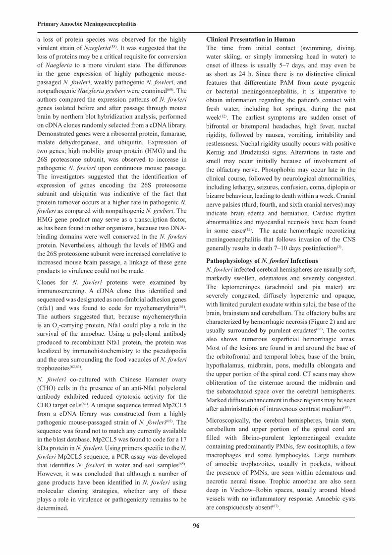
Pathophysiology of N. fowleri InfectionsN. fowleri infected cerebral hemispheres are usually soft, markedly swollen, edematous and severely congested. The leptomeninges (arachnoid and pia mater) are severely congested, diffusely hyperemic and opaque, with limited purulent exudate within sulci, the base of the brain, brainstem and cerebellum. The olfactory bulbs are characterized by hemorrhagic necrosis (Figure 2) and are usually surrounded by purulent exudates(66). The cortex also shows numerous superficial hemorrhagic areas. Most of the lesions are found in and around the base of the orbitofrontal and temporal lobes, base of the brain, hypothalamus, midbrain, pons, medulla oblongata and the upper portion of the spinal cord. CT scans may show obliteration of the cisternae around the midbrain and the subarachnoid space over the cerebral hemispheres. Marked diffuse enhancement in these regions may be seen after administration of intravenous contrast medium(67).
Microscopically, the cerebral hemispheres, brain stem, cerebellum and upper portion of the spinal cord are filled with fibrino-purulent leptomeningeal exudate containing predominantly PMNs, few eosinophils, a few macrophages and some lymphocytes. Large numbers of amoebic trophozoites, usually in pockets, without the presence of PMNs, are seen within edematous and necrotic neural tissue. Trophic amoebae are also seen deep in Virchow–Robin spaces, usually around blood vessels with no inflammatory response. Amoebic cysts are conspicuously absent(67).

Abo El-Maaty and Hamza
97
ImmunologyBecause most PAM patients die soon after infection, there is insufficient time to mount a detectable immune response, hence the limited usefulness of serological tests in the diagnosis of PAM. Previous attempts to detect an antibody response to N. fowleri using immunoflurescent assay (IFA) have therefore been largely unsuccessful(41).
However, a specific antibody response to N. fowleri was documented in a recovered Californian patient. A titre of 1:4096 was demonstrated by IFA in the serum samples collected 7, 10 and 42 days after admission to hospital, and the elevated antibody titres persisted even after 4 years(68).
A previous study used an agglutination test on sera obtained from humans in North Carolina, Pennsylvania and Virginia, that demonstrated antibody specificity for the surfaces of particular Naegleria spp. The study showed that sera from individuals residing in the southeastern United States had significantly greater agglutinating ability than did sera obtained from Pennsylvania. The study showed that the agglutinating antibody is of the IgM class(69). Based on immunoblot studies conducted at Centers for Disease Control and Prevention (CDC), IgM was the principal class of antibody generated by this patient as well as by three others who had contracted PAM. In addition, the CDC study indicated that sera collected from several individuals with a history of extensive swimming in fresh-water lakes in the southeastern United States as well as in California also exhibited IgM antibodies to N. fowleri. These antibodies proved to be particularly well developed to N. fowleri antigens of 190, 66, 30 and 14 kDa. Whether these antibodies have any protective activity is not clear at this time(70).
A survey was conducted to detect IgA antibodies in the serum and saliva of individuals living in Mexico in areas where Naegleria is endemic as compared with those where it is non-endemic. In endemic areas, titers of IgA antibody to N. fowleri were found to be significantly higher in individuals with upper respiratory tract infections as compared with those in healthy individuals(71). The investigators indicated that IgA and IgM present in mucosal secretions could play an important role in preventing infection by blocking adhesion of trophozoites to mucosal epithelium.
The role of cell mediated immunity (CMI) in responsiveness to N. fowleri has been studied in vivo and in vitro using delayed-type hypersensitivity response and macrophage migration assay, respectively(72). Guinea-pigs sensitized with intradermal injections of N. fowleri and other Naegleria species developed a delayed-type hypersensitivity skin reaction when challenged by an intradermal injection with homologous or heterologous Naegleria antigens. A similar study was conducted (73) and the authors suggested that exposure to Naegleria antigens elicited CMI, although exposure to homologous
antigens was more efficient than exposure to heterologous antigens in inhibiting macrophage migration. In contrast, another animal experimental study using mice suggested that CMI does not play a role in protection against Naegleria. Congenitally athymic mice (T cell deficient) and euthymic mice were equally susceptible to infection by N. fowleri(74). In addition, failure to impair host resistance to N. fowleri infection with diethylstilbestrol, which depresses delayed typed hypersensitivity, led authors to conclude that CMI alone does not constitute a major line of defense against N. fowleri(75). Collectively, the role of humoral immunity and CMI in the protection against infection by Naegleria remains unclear.
In addition to the ability to attach to nasal mucosa, to exhibit an increased rate of locomotion, and to destroy target cells by trogocytosis and by release of cytolytic molecules, N. fowleri may have evolved as a successful pathogen because it has developed mechanisms to evade the host immune system. It has been shown that Naegleria are resistant to lysis by host cytolytic molecules such as tumor necrosis factor (TNF)-α, IL-1 and the membrane attack complex (MAC) C5b-C9 of complement(76,77).
Naegleria–Bacteria Interactions In the environment, Naegleria trophozoites are voracious feeders on bacteria, yeast and algae. In seeking these food sources they transform into swimming flagellates that migrate from the soil layer to the water–air interface, dock at that site, and transform back into trophozoites to feed on bacteria present in abundance on the surface microlayer(78). Live Escherichia coli elicit a chemotactic response (directed migration) towards Naegleria, while on the other hand, Naegleria move away from toxin-producing bacteria and encyst. The amoeba growth and survival in the presence of bacteria appear to vary with the density and species of bacteria(79). The interaction of bacteria with amoebae can result in destruction of the bacteria and/or amoebae, or development of a symbiotic relationship. Naegleria ingest and phagocytose bacteria by their ‘food-cups’(80), then lyse and degrade edible bacteria by naegleriapores(43,46). Three strains of Bacillus licheniformis isolated from the same N. fowleri soil habitat were shown to exhibit amoebicidal activity(81). A Naegleria strain was isolated harboring two different populations of bacterial endocytobiont. Microscopic examination revealed different localization and replication sites for the two bacterial endocytobionts, one in the nucleus and the other in the cytoplasm(82).
Naegleria may also serve as natural reservoirs for pathogenic bacteria such as Legionella spp.(83,84) as well as support the growth of both L. pneumophila(85) and Vibrio cholerae(86). These observations are not surprising because in the environment, amoebae and bacteria occupy the same niche including humidifier systems, cooling towers and evaporative condensers that operate at mutual optimal growth temperatures. Human infection

98
Primary Amoebic Meningoencephalitis
with Legionella was acquired not by inhalation of free legionellae, but by inhalation of vesicles released from amebae filled with legionellae or by inhalation of amebae harboring bacteria.
This was confirmed by transmission electron microscopy that revealed the bacteria apparently undergoing binary fission in cytoplasmic vacuoles within the amoebae. Dark-field microscopy also demonstrated that extracellular bacteria were found free in the culture media even after 6 days of co-culture. However, the growth of bacteria within amoebae was observed to be contingent upon experimental culture conditions. Naegleria fowleri supported the growth of Legionella in amoebae growth medium but not in amoeba in saline solution, in which Legionella destroyed the amoebae(84). Bacteria also lysed amoebae when they were cultured on agar plates(87). The researchers added that encysted Naegleria containing Legionella could resist adverse conditions such as chlorination and biocidal compounds, an outcome that could account for their persistence in chemically treated sewage and water systems(88). Thus, it is now recognized that bacteria, either as endosymbionts or ‘passengers,’ are associated with free-living amoebae, including Naegleria. This interaction has potential major human health implications because bacteria residing in trophozoites or amoebic cysts are protected from biocides and therefore may be recalcitrant to antibiotic treatment, and by virtue of passage through amoebae may develop greater pathogenic capability(89).
In an attempt to simulate natural environmental conditions, the uptake and replication of the most pathogenic strain of Legionella (L. pneumophila serogroup 1), in N. lovaniensis was compared with that in another free-living amoeba, Acanthamoeba castellanii(90). It was found that, while other bacterial species commonly found with L. pneumophila in biofilms, did not act as competitors in the uptake of L. pneumophila, they nevertheless influenced the replication of bacteria within the amoebae. Microscopic examination of co-cultures revealed that 100% of Acanthamoeba were infected with L. pneumophila, whereas only 2% of Naegleria were infected with bacteria. However, N. lovaniensis formed cysts containing viable L. pneumophila while A. castellanii did not and was lysed by the bacteria.
DiagnosisNeuroimaging findings of N. fowleri meningitis show nonspecific brain edema with evidence of increased intracranial pressure or cerebral herniation(91). Final diagnostic confirmation is not achieved until trophozoites are isolated and identified from cerebrospinal fluid (CSF) or brain tissue. Lumbar puncture for (CSF) analysis is the primary diagnostic tool. The CSF mimics that of acute bacterial meningitis except that Gram stain is negative. The concentration of erythrocytes in the CSF may correlate with the degree of necrosis and
inflammation present(67). CSF may vary in color from grayish to yellowish-white, and may be tinged red with a few red cells (250/mm−3) in the early stages of disease. However, as the disease progresses the red blood cell number increases to as high as 24,600/mm−3. The white blood cell count, predominantly polymorphonuclear leukocytes (PMN), also may vary from 300 to 26,000 cells/mm−3. The CSF pressure is usually elevated (300–600 mm H2O). The protein concentration may range from 100 to 1000 mg/100 mL, and glucose may be 10 mg/100 mL or lower. A wet-mount of the CSF should be microscopically examined immediately after collection, preferably equipped with phase-contrast optics, for the presence of actively moving trophozoites followed by specific immunofluorescent staining(67). Since Naegleria feed upon bacteria, they can be isolated from clinical specimens such as brain tissue and CSF using non-nutrient agar spread with bacteria. Naegleria grows well on monolayers of Vero monkey kidney cells (E6) and human lung fibroblasts (HLF) cell cultures, destroying the confluent layers within 2-3 days. Like Acanthamoeba and Balamuthia, N. fowleri can be grown in a cell-free axenic medium as well as in a chemically defined medium(92).
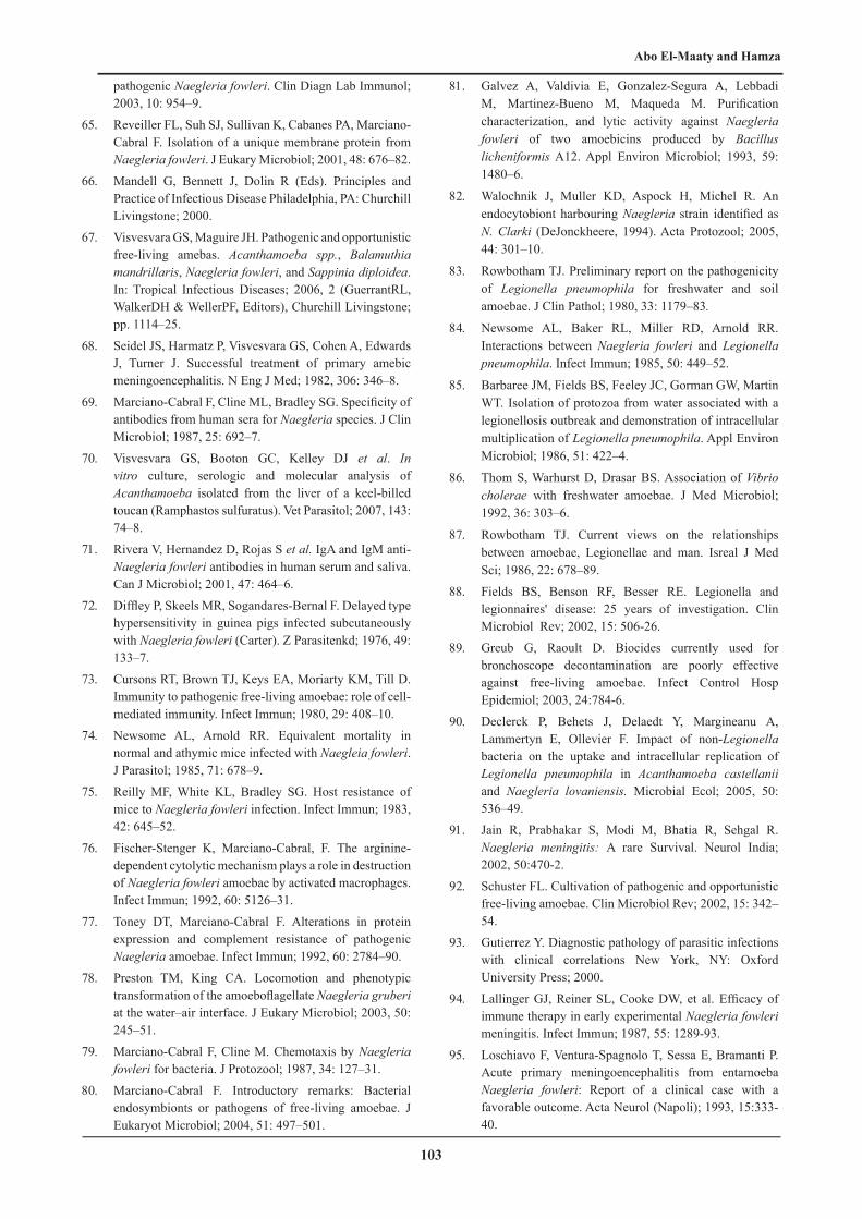
N. fowleri trophozoites vary between 7-15 μm in size when observed in a CSF wet mount. Classically, trophozoite nuclei appear to lack chromatin, are clear with thin membranes, and have a single large, rounded, refringent nucleolus(93). Numerous cytoplasmic vacuoles can be seen aggregated around each organism’s nucleus, and pseudopodia may be identified projecting away from the cells(66) (Figure 3).
Distinguishing the features of amoebic nuclei from those of monocytes is particularly important in diagnosing PAM, as the two cell types can be confused. Monocyte nuclei are significantly larger with higher nuclear to cytoplasmic ratios than those of amoebae. Furthermore, monocyte nuclei do not appear clear but rather have a uniform and slightly ropey chromatin pattern without distinct nucleoli(94). After cytocentrifugation staining specimens with Giemsa or Trichrome stains can enhance N. fowleri trophozoite features(95).
Biochemical techniques such as isoenzyme analysis have been developed for the specific identification of N. fowleri amoebae cultured from the CSF and brain specimens of patients as well as from the environment; water and soil(96). Furthermore, a commercially available enzyme-linked immunosorbent assay based on the use of a monoclonal antibody (5D12), that recognizes a repeated glycosylated epitope present on proteins of N. fowleri, can be used to diagnose infections(97).
More recently, molecular techniques such as PCR and real-time PCR have been developed for the detection of N. fowleri in environmental and clinical samples(98-100). A PCR assay was designed using ribosomal internal

Abo El-Maaty and Hamza
99
transcribed spacers of N. fowleri(101). Sequencing of the 5.8S rRNA gene and the internal transcribed spacers 1 and 2 (ITS1 and ITS2) of N. fowleri showed that specific genotypes can be distinguished. Based on the ITS sequencing of clinical isolates, it was shown that two strains of N. fowleri, isolated from two PAM patients (who visited the same hot spring in California but at different times), belonged to the same type II genotype(102). A recently developed real-time multiplex PCR assay identified N. fowleri DNA in the CSF and brain tissue samples obtained from PAM patients ante-mortem. This test identified all three genotypes known to be present in the United States. The time taken to identify N. fowleri from the time the specimen arrived in the laboratory is ~5 h, and can be cut down to 2 h if the specimen is CSF(103).
A newly developed sensitive, rapid and discriminating technique, that uses a single primer set and the DNA intercalating dye SYTO9 for real-time PCR and melting-curve analysis of the 5.8S rRNA gene flanking the ITS, distinguished several Naegleria species in environmental samples(104). Other more recent reports of a PCR assay detected N. fowleri in fresh brain tissue as well as in formalin-fixed paraffin-embedded brain tissue(105), and a method that involved monitoring the amplification process in real-time with hybridization of fluorescent labeled probes targeting the MpCL5 sequence, which is unique to N. fowleri have also been developed(106).
In summary, N. fowleri CNS disease progression is rapid, and the availability of rapid diagnostic techniques affords an opportunity to apply prompt treatment that is essential for a successful patient outcome(107).
Treatment After identifying amoebae in CSF wet mount, treatment must be initiated immediately. Successful treatments have included high dose administration of systemic and intrathecal amphotericin B with or without miconazole, rifampin, and sulfisoxazole(66). Although N. fowleri is very sensitive to amphotericin B in vitro, only a few patients recovered following intrathecal or intravenous injections of this drug alone or in combination with miconazole(107). Often after the infusion a serious acute reaction was noted consisting of fever, shaking chills, hypotension, anorexia, nausea, vomiting, headache, dyspnoea and tachypnoea(108). Nephrotoxicity is a major issue and can be severe or irreversible(109).
Many other antimicrobial, antifungal and antiparasitic drugs have been screened for therapeutic activity against N. fowleri in vitro and in vivo such as clotrimazole, itraconazole, fluconazole, ketoconazole and chlorpromazine, with varying degrees of efficacy(110,111).
Fluconazole was proven to penetrate the blood-brain barrier remarkably well, showing an unexpected effectiveness for reducing organ dysfunction and mortality in septic
shock, probably by increasing bactericidal activity and the recruitment of neutrophils(112). Azithromycin has been described for the effective treatment of experimental PAM in mice(113). Phenothiazine compounds (chlorpromazine and trifluoperazine), which can accumulate in the CNS, also had inhibitory effects on N. fowleri in vitro(107).
Early diagnosis and treatment with a triple drug regime of intravenous amphotericin B and fluconazole, and oral rifampicin was reported to be successful in treatment of patients of PAM(114) .
Furthermore, miltefosine used as an anticancer drug, and voriconazole used in systemic fungal infections, were found to be effective in in vitro studies(115). Low concentrations of voriconazole (≤ 10 μg ml−1) were amoebastatic, while concentrations ≥10 μg ml−1 were amoebacidal(116).
In an attempt to select other useful therapeutic agents for treating PAM, the amoebicidal activities of antibacterial agents including clarithromycin, erythromycin, hygromycin B, neomycin, rokitamycin, roxithromycin and zeocin were examined in vitro and in vivo. Results showed that amoeba growth was effectively inhibited by treatment with hygromycin B, rokitamycin and roxithromycin in vitro. In the treatment of experimental meningoencephalitis due to N. fowleri, survival rates of mice treated with roxithromycin and rokitamycin were 25% and 80%, respectively, over 1 month. Finally, rokitamycin showed both in vitro and in vivo therapeutic efficacy against N. fowleri and was recommended as a candidate drug for the treatment of PAM(117).
Much work is being done to determine N. fowleri virulence factors that render it pathogenic and can be targeted by drugs(118). One of these factors is Nfa1 gene, whose expression knockdown could potentially be a technique applicable for knockdown of expression of pathogenicity factors in trophozoites(119). A recent study reported chlorpromazine as high potent and rapidly active than amphotericin B and voriconazole against N. fowleri trophozoites(120).
Prevention and ControlNaegleria fowleri is a thermophilic amoeba and hence proliferates in water when the ambient temperature increases above 30°C. With the anticipated temperature increase resulting from global warming, it is possible that cases of N. fowleri PAM may be seen even in countries where it had previously not been recorded(121). Because N. fowleri is susceptible to chlorine in water (one part per million), amoeba proliferation can be controlled by adequate chlorination of heavily used swimming pools, especially during summer months, but sunlight and the presence of organic matter in swimming pools can reduce the efficacy of chlorine. However, it is not possible to chlorinate natural bodies of water such as lakes, ponds and streams, where N. fowleri may proliferate. In high-

100
Primary Amoebic Meningoencephalitis
risk areas, monitoring of recreational waters for N. fowleri amoebae should be considered by local public health authorities and appropriate warnings posted, particularly during the hot summer months. Warning children not to immerse their heads in suspect waters is recommended(41).
Concluding Remarks•Until 2012, about 310 human cases of PAM have been
reported worldwide. The number of reported cases has increased worldwide in recent years. This increase may be due to better awareness of the disease or due to the development of more rapid and specific diagnostic assays such as PCR.
•Not all humans exposed to N. fowleri develop a fatal infection. However, the circumstances under which this free-living ameba gains access to the human host and becomes highly pathogenic remain to be defined.
•Although pathogenic mechanisms remain poorly understood, it is apparent that N. fowleri produce pore-forming proteins that lyse mammalian cells on contact, secrete proteases and phospholipases that degrade mammalian tissue, and synthesize regulatory surface proteins that confer protection to amoebae against complement-mediated-lysis and other cytotoxic substances.
•Pathogenic N. fowleri exhibit a more rapid rate of locomotion in the presence of nerve cells and nerve cell
products, and a more rapid rate of division as compared with nonpathogenic Naegleria.
•IgM is the principal class of antibody generated in PAM, and with IgA, it could play an important role in preventing infection by blocking adhesion of trophozoites to mucosal epithelium.
•No distinctive clinical features differentiate PAM from acute pyogenic or bacterial meningoencephalitis except a history of patient's contact with fresh water during the past week.
•Once amoebae are identified in CSF wet mount, treatment must be initiated immediately with high dose administration of systemic and intrathecal amphotericin B with or without miconazole, rifampin, and sulfisoxazole.
•N. fowleri proliferation can be controlled by adequate chlorination (one part per million), of heavily used swimming pools and in high-risk areas, monitoring of recreational waters for N. fowleri should be considered by local public health authorities particularly during the hot summer months.
•The medical significance of N. fowleri should not be underestimated, not only because they are agents of human disease but also because they can serve as reservoirs for pathogenic bacteria and their capacity to sequester bacteria from biocides in the environment and antibiotics in the host.
Figure (1): Scanning electron microscope shows ‘food-cup’(arrow) structure on the surface of N. fowleri trophozoite attached to lysed brain tissue(8).
Figure (2): Immunohistochemical stain of bovine brain (olfactory bulb) with Naegleria meningoencephalitis. Note numerous brown staining N. fowleri around vessels (arrow) and within neuropil. Bar= 100 um(122).
Figure (3): Wright-Giemsa–stained cytospin of CSF (× 400) reveals copious neutrophils (white arrow), lymphocytes (small arrow), and occasional amoeboid structures (large arrow). Note the numerous cytoplasmic vacuoles, pale staining nuclei (asterisk) with prominent karyosome(12).

Abo El-Maaty and Hamza
101
REFERENCES
1. Gyori E. 19-year old male with febrile illness after jet ski accident. Brain Pathol; 2003, 13: 237–9.
2. Centers for Disease Control and Prevention (CDC). Primary amebic meningoencephalitis-Arizona, Florida, and Texas, 2007. MMWR Morb Mortal Wkly Rep; 2008, 57: 573-7.
3. Martinez AJ, Visvesvara GS. Free-living, amphizoic and opportunistic amoebae. Brain Pathol; 1997, 7: 583–98.
4. Huizinga HW, McLaughlin GL. Thermal ecology of Naegleria fowleri from a power plant cooling reservoir. Appl Environ Microbiol; 1990, 56:2200–5.
5. Marciano-Cabral F, MacLean RC, Mensah AH, La-Pat L. Identification of Naegleria fowleri in domestic water sources by nested PCR. Appl Environ Microbiol; 2003, 69:5864–9.
6. Cervantes-Sandoval I, Serrano-Luna JJ, García-Latorre E, Tsutsumi V, Shibayama M. Characterization of brain inflammation during primary amoebic meningoencephalitis. Parasitol Int; 2008, 57:307–13.
7. Gautam PL, Sharma S, Puri S, Kumar R, Midha V, Bansal R. A rare case of survival from primary amebic meningoencephalitis. Indian J Crit Care Med; 2012, 16: 34-6.
8. Marciano-Cabral F, Cabral GA. The immune response to Naegleria fowleri amebae and pathogenesis of infection. FEMS Immunol Med Microbiol; 2007, 51(2): 243-59.
9. Nashed NN, Youssef FG, Mansour NS. Free-living amoebae in Egypt. 1. Naegleria gruberi and Naegleria fowleri. J Egypt Soc Parasitol; 1991, 21(1):31-42.
10. Sadaka HA, El-Nassery SF, Abou Samra LM, Awadalla HN. Isolation and identification of free-living amoebae from some water sources in Alexandria. J Egypt Soc Parasitol; 1994, 24(2): 247-57.
11. Fowler M, Carter RF. Acute pyogenic meningoencephalitis probably due to Acanthamnoeba spp.: A preliminary report. Br Med J; 1965, 3:740-2.
12. Jones K, Singhatiraj E, MacDougall R, Beaver TR, Nugent K. A 22-year-old man with headache and stiff neck after a water skiing fall. Chest; 2009, 135(1):225-7.
13. Marciano-Cabral F. Biology of Naegleria spp. Microbiol Rev; 1988, 52:114–33.
14. De Jonchheere JF. Molecular definition and the ubiquity of species in the genus Naegleria. Protist; 2004, 155: 89–103.
15. Lozano-Alarcon F, Bradley GA, Houser BS, Visvesvara GS. Primary amebic meningoencephalitis due to Naegleria fowleri in a South American tapir. Vet. Pathol; 1997, 34: 239-43.
16. Daft B, Kinde H, Read D, Visvesvara GS. Naegleria fowleri meningoencephalitis in Holstein cattle associated with drinking surface water. Proceedings of the 42nd Meeting of American Association of Veterinary Laboratory Diagnosticians, San Diego, California; 1999, p. 59.
17. Visvesvara GS, De Jonckheere JF, Sriram R, Daft B. Isolation and molecular typing of Naegleria fowleri from the brain of a cow that died of primary amebic meningoencephalitis. J Clin Microbiol; 2005, 43(8): 4203–4.
18. Morales JA, Chavez AJ, Visvesvara GS, Dubey JP. Naegleria fowleri-associated encephalitis in a cow from Costa Rica. Vet Parasitol; 2006, 139: 221-3.
19. Yoder JS, Eddy BA, Visvesvara GS, Capewell L, Beach MJ. The epidemiology of primary amoebic meningoencephlitis in the USA, 1962–2008. Epidemiol. Infect; 2010, 138: 968–75.
20. Bennett WM, Nespral JF, Rosson MW, McEvoy KM. Use of organs for transplantation from a donor with primary meningoencephalitis due to Naegleria fowleri. Am J Transplant; 2008, 8: 1334–5.
21. Caruzo G, Cardozo J. Primary amoebic meningoencephalitis: A new case from Venezuela. Trop Doct; 2008, 38: 256–7.
22. Cubero-Menéndez O, Cubero-Rego D. Primary amoebic meningoencephalitis: A case report. Rev Neurol; 2004, 38: 336–8.
23. Nicolas M, De Jonckheere JF, Pernin P, Bataille H, Le Bris V, Hermann Storck C. Diagnostic moléculaire d’une méningoencephalite amibienne primitive à l’occasion d’un cas fatal en Guadeloupe. Bull Soc Pat Exo; 2010, 103: 14–8 (English abstract).
24. Dorsch MM, Cameron AS, Robinson BS. The epidemiology and control of primary amoebic meningoencephalitis with particular reference to South Australia. Trans R Soc Trop Med Hyg; 1983, 77: 372–7.
25. Cursons RT, Brown TJ, Keys EA et al. Primary amoebic meningoencephalitis in an indoor heat-exchange swimming pool. NZ Med J; 1979, 90:330–1.
26. Cursons R, Sleigh J, Hood D, Pullon D. A case of primary amoebic meningoencephalitis: North Island, New Zealand. NZ Med J; 2003, 116-20.
27. Bose K, Ghosh DK, Ghosh KN, Bhattacharya A, Das SR. Characterization of potentially pathogenic free-living amoebae in sewage samples of Calcutta, India. Braz J Med Biol Res; 1990, 23: 1271–8.
28. Gupta S. Isolation of Naegleria fowleri from pond water in West Bengal, India. Trans R Soc Trop Med Hyg; 1992, 86: 46-52.
29. Wang A, Kay R, Poon WS, Ng HK. Successful treatment of amoebic meningoencephalitis in a Chinese living in Hong Kong. Clin Neurol Neurosurg; 1993, 95: 249-52.
30. Hara T, Fukuma T. Diagnosis of the primary amoebic meningoencephalitis due to Naegleria fowleri. Parasitol Int; 2005, 54: 219-21.
31. Kaushal V, Chhina DK, Ram S, Singh G, Kaushal RK, Kumar R. Primary amoebic meningoencephalitis due to Naegleria fowleri. J Assoc Phys India; 2008, 56: 459–62.
32. Rai R, Singh DK, Srivastava AK, Bhargava A. Primary amebic meningoencephalitis. Ind Pediat; 2008, 45: 1004-5.

102
Primary Amoebic Meningoencephalitis
33. Gupta N, Bhaskar H, Duggal S, Ghalaut PS, Kundra S, Arora DS. Primary amoebic meningoencephalitis: first reported case from Rohtak, North India. Braz J Inf Dis; 2009, 13: 236–7.
34. Shakoor S, Beg MA, Mahmood SF et al. Primary amoebic meningoencephalitis caused by Naegleria fowleri, Karachi, Pakistan. Emerg Infect Dis; 2011, 17: 258–61.
35. Jamil B, Ilyas A, Zaman V. Primary amoebic meningoencephalitis. Inf Dis J Pakistan; 2008, 17: 66–8.
36. Saleem T, Rabbani M, Jamil B. Primary amoebic meningoencephalitis: Two new cases from Pakistan. Trop Doct; 2009, 39: 242–3.
37. Lawande RV, Macfarlana JT, Weir WR, Awunor-Renner C. A case of primary amoebic meningencephalitis in a Nigerian farmer. Am J Trop Med Hyg; 1980, 29:21–25.
38. Schoeman CJ, van der Vyver AE, Visvesvara GS. Primary amoebic meningoencephalitis in southern Africa. J infect; 1993, 26: 211–4.
39. Jaffar-Bandjee MC, Alessandri JL, Clouzeau J, Samperiz S, Saly JC. Méningo-encéphalite primitive à amibes libres: Premier cas observé à Madagascar. Bull Soc Pathol Exot; 2005, 98: 11–3 (English abstract).
40. Whiteman LY, Marciano-Cabral F. Susceptibility of pathogenic and nonpathogenic Naegleria spp. to complement-mediated lysis. Infect Immun; 1987, 55: 2442–7.
41. Schuster FL, Visvesvara GS. Free-living amoebae as opportunistic and non-opportunistic pathogens of humans and animals. Int J Parasitol; 2004, 34: 1001–27.
42. Marciano-Cabral F, Bradley SG. Cytopathogenicity of Naegleria gruberi for neuroblastoma cell cultures. Infect Immun; 1982, 35:1139–41.
43. Leippe M, Herbst R. Ancient weapons for attack and defense: The pore-forming polypeptides of pathogenic enteric and free-living amoeboid protozoa. J Eukaryot Microbiol; 2004, 51: 516–21.
44. Song KJ, Song KH, Na BK et al. Molecular cloning and characterization of a cytosolic heat shock protein 70 from Naegleria fowleri. Parasitol Res; 2007, 100: 1083–9.
45. Stevens AR, DeJonckheere J, Willaert E. Naegleria lovaniensis new species: Isolation and identification of six thermophilic strains of a new species found in association with Naegleria fowleri. Int J Parasitol; 1980, 10: 51–64.
46. Herbst R, Ott C, Jacobs T, Marti T, Marciano-Cabral F, Leippe M. Pore-forming polypeptides of the pathogenic protozoon Naegleria fowleri. J Biol Chem; 2002, 277: 22353–60.
47. Herbst R, Marciano-Cabral F, Leippe M. Antimicrobial and pore-forming peptides of free-living and potentially highly pathogenic Naegleria fowleri are released from the same precursor molecule. J Biol Chem; 2004, 279: 25955–8.
48. Barbour SE, Marciano-Cabral F. Naegleria fowleri amoebae express a membrane-associated calcium-independent phospholipase A2. Biochem Biophys Acta; 2001, 1530: 123–33.
49. Mat Amin N. Proteinases in Naegleria fowleri strain NF3, a pathogenic amoeba: A preliminary study. Trop Biomed; 2004, 21: 57–60.
50. Cursons RT, Brown TJ, Keys EA. Virulence of pathogenic free-living amoebae. J Parasitol; 1978, 64: 744–5.
51. Eisen D, Franson RC. Acid-active neuraminidases in the growth media from cultures of pathogenic Naegleria fowleri and in sonicates of rabbit alveolar macrophages. Biochem Biophy Acta; 1987, 924: 369–72.
52. Ferrante A, Bates EJ. Elastase in the pathogenic free-living amoebae Naegleria and Acanthamoeba spp. Infect Immun; 1988, 56: 3320–1.
53. Aldape K, Huizinga H, Bouvier J, McKerrow J. Naegleria fowleri: Characterization of a secreted histolytic cysteine protease. Exp Parasitol; 1994, 78: 230–41.
54. Han KL, Lee HJ, Shin MH, Shin HJ, Im KI, Park SJ. The involvement of an integrin-like protein and protein kinase C in amoebic adhesion to fibronectin and amoebic cytotoxicity. Parasitol Res; 2004, 94:53–60.
55. Bonay P, Fresno M. Characterization of carbohydrate binding proteins in Trypanosoma cruzi. J Biol Chem; 1995, 270:11062–70.
56. González-Robles A, Castañón G, Cristóbal-Ramos AR, Hernández-Ramírez VI, Omaña-Molina M, Martínez-Palomo A. Cell surface differences of Naegleria fowleri and Naegleria lovaniensis exposed with surface markers. Exp Parasitol, 2007, 117:399 -404.
57. Cervantes-Sandoval I, Serrano-Luna JJ, Pacheco-Yépez J, Silva-Olivares A, Tsutsumi V, Shibayama M. Differences between Naegleria fowleri and Naegleria gruberi in expression of mannose and fucose glycoconjugates. Parasitol Res; 2010, 106:695–701.
58. Hu WN, Band RN, Kopachik WJ. Virulence related protein synthesis in Naegleria fowleri. Infect Immun; 1991, 59: 4278–82.
59. Hu WN, Kopachik W, Band RN. Cloning and characterization of transcripts showing virulence-related gene expression in Naegleria fowleri. Infect Immun; 1992, 60: 2418–24.
60. Yun HC, Park SJ, Kong HH, Chung DI. Isolation of genes induced in Naegleria fowleri during mouse brain passage. Europ J Protistol; 2002, 38: 105–11.
61. Shin HJ, Cho MS, Jung SY et al. Molecular cloning and characterization of a gene encoding a 13.1 kDa antigenic protein of Naegleria fowleri. J Eukaryot Mirobiol; 2001, 48: 713–17.
62. Oh YH, Jeong SR, Kim JH et al. Cytophatic changes and pro-inflammatory cytokines induced by Naegleria fowleri trophozoites in rat microglial cells and protective effects of an anti-Nfa1 antibody. Parasite Immunol; 2005, 27:453–9.
63. Lee YJ, Kim JH, Jeong SR et al. Production of Nfa1-specific monoclonal antibodies that influences the in vivo cytotoxicity of Naegleria fowleri trophozoites on microglial cells. Parasitol Res; 2007, 101:1191–6.
64. Cho MS, Jung SY, Park S et al. Immunological characterizations of a cloned 13.1-kilodalton protein from
Abo El-Maaty and Hamza
103
pathogenic Naegleria fowleri. Clin Diagn Lab Immunol; 2003, 10: 954–9.
65. Reveiller FL, Suh SJ, Sullivan K, Cabanes PA, Marciano-Cabral F. Isolation of a unique membrane protein from Naegleria fowleri. J Eukary Microbiol; 2001, 48: 676–82.
66. Mandell G, Bennett J, Dolin R (Eds). Principles and Practice of Infectious Disease Philadelphia, PA: Churchill Livingstone; 2000.
67. Visvesvara GS, Maguire JH. Pathogenic and opportunistic free-living amebas. Acanthamoeba spp., Balamuthia mandrillaris, Naegleria fowleri, and Sappinia diploidea. In: Tropical Infectious Diseases; 2006, 2 (GuerrantRL, WalkerDH & WellerPF, Editors), Churchill Livingstone; pp. 1114–25.
68. Seidel JS, Harmatz P, Visvesvara GS, Cohen A, Edwards J, Turner J. Successful treatment of primary amebic meningoencephalitis. N Eng J Med; 1982, 306: 346–8.
69. Marciano-Cabral F, Cline ML, Bradley SG. Specificity of antibodies from human sera for Naegleria species. J Clin Microbiol; 1987, 25: 692–7.
70. Visvesvara GS, Booton GC, Kelley DJ et al. In vitro culture, serologic and molecular analysis of Acanthamoeba isolated from the liver of a keel-billed toucan (Ramphastos sulfuratus). Vet Parasitol; 2007, 143: 74–8.
71. Rivera V, Hernandez D, Rojas S et al. IgA and IgM anti-Naegleria fowleri antibodies in human serum and saliva. Can J Microbiol; 2001, 47: 464–6.
72. Diffley P, Skeels MR, Sogandares-Bernal F. Delayed type hypersensitivity in guinea pigs infected subcutaneously with Naegleria fowleri (Carter). Z Parasitenkd; 1976, 49: 133–7.
73. Cursons RT, Brown TJ, Keys EA, Moriarty KM, Till D. Immunity to pathogenic free-living amoebae: role of cell-mediated immunity. Infect Immun; 1980, 29: 408–10.
74. Newsome AL, Arnold RR. Equivalent mortality in normal and athymic mice infected with Naegleia fowleri. J Parasitol; 1985, 71: 678–9.
75. Reilly MF, White KL, Bradley SG. Host resistance of mice to Naegleria fowleri infection. Infect Immun; 1983, 42: 645–52.
76. Fischer-Stenger K, Marciano-Cabral, F. The arginine-dependent cytolytic mechanism plays a role in destruction of Naegleria fowleri amoebae by activated macrophages. Infect Immun; 1992, 60: 5126–31.
77. Toney DT, Marciano-Cabral F. Alterations in protein expression and complement resistance of pathogenic Naegleria amoebae. Infect Immun; 1992, 60: 2784–90.
78. Preston TM, King CA. Locomotion and phenotypic transformation of the amoeboflagellate Naegleria gruberi at the water–air interface. J Eukary Microbiol; 2003, 50: 245–51.
79. Marciano-Cabral F, Cline M. Chemotaxis by Naegleria fowleri for bacteria. J Protozool; 1987, 34: 127–31.
80. Marciano-Cabral F. Introductory remarks: Bacterial endosymbionts or pathogens of free-living amoebae. J Eukaryot Microbiol; 2004, 51: 497–501.
81. Galvez A, Valdivia E, Gonzalez-Segura A, Lebbadi M, Martinez-Bueno M, Maqueda M. Purification characterization, and lytic activity against Naegleria fowleri of two amoebicins produced by Bacillus licheniformis A12. Appl Environ Microbiol; 1993, 59: 1480–6.
82. Walochnik J, Muller KD, Aspock H, Michel R. An endocytobiont harbouring Naegleria strain identified as N. Clarki (DeJonckheere, 1994). Acta Protozool; 2005, 44: 301–10.
83. Rowbotham TJ. Preliminary report on the pathogenicity of Legionella pneumophila for freshwater and soil amoebae. J Clin Pathol; 1980, 33: 1179–83.
84. Newsome AL, Baker RL, Miller RD, Arnold RR. Interactions between Naegleria fowleri and Legionella pneumophila. Infect Immun; 1985, 50: 449–52.
85. Barbaree JM, Fields BS, Feeley JC, Gorman GW, Martin WT. Isolation of protozoa from water associated with a legionellosis outbreak and demonstration of intracellular multiplication of Legionella pneumophila. Appl Environ Microbiol; 1986, 51: 422–4.
86. Thom S, Warhurst D, Drasar BS. Association of Vibrio cholerae with freshwater amoebae. J Med Microbiol; 1992, 36: 303–6.
87. Rowbotham TJ. Current views on the relationships between amoebae, Legionellae and man. Isreal J Med Sci; 1986, 22: 678–89.
88. Fields BS, Benson RF, Besser RE. Legionella and legionnaires' disease: 25 years of investigation. Clin Microbiol Rev; 2002, 15: 506-26.
89. Greub G, Raoult D. Biocides currently used for bronchoscope decontamination are poorly effective against free-living amoebae. Infect Control Hosp Epidemiol; 2003, 24:784-6.
90. Declerck P, Behets J, Delaedt Y, Margineanu A, Lammertyn E, Ollevier F. Impact of non-Legionella bacteria on the uptake and intracellular replication of Legionella pneumophila in Acanthamoeba castellanii and Naegleria lovaniensis. Microbial Ecol; 2005, 50: 536–49.
91. Jain R, Prabhakar S, Modi M, Bhatia R, Sehgal R. Naegleria meningitis: A rare Survival. Neurol India; 2002, 50:470-2.
92. Schuster FL. Cultivation of pathogenic and opportunistic free-living amoebae. Clin Microbiol Rev; 2002, 15: 342–54.
93. Gutierrez Y. Diagnostic pathology of parasitic infections with clinical correlations New York, NY: Oxford University Press; 2000.
94. Lallinger GJ, Reiner SL, Cooke DW, et al. Efficacy of immune therapy in early experimental Naegleria fowleri meningitis. Infect Immun; 1987, 55: 1289-93.
95. Loschiavo F, Ventura-Spagnolo T, Sessa E, Bramanti P. Acute primary meningoencephalitis from entamoeba Naegleria fowleri: Report of a clinical case with a favorable outcome. Acta Neurol (Napoli); 1993, 15:333-40.

104
Primary Amoebic Meningoencephalitis
96. Moss DM, Brandt FH, Mathews MM, Visvesvara GS. High-resolution polyacrylamide gradient gel electrophoresis (PGGE) of isoenzymes from five Naegleria species. J Protozool; 1988, 38: 26–31.
97. Reveiller FL, Varenne MP, Pougnard C et al. An enzyme-linked immunosorbent assay (ELISA) for the identification of Naegleria fowleri in environmental water samples. J Eukaryot Microbiol; 2003, 50: 109–13.
98. Pelandakis M, Pernin P. Use of multiplex PCR and PCR restriction enzyme analysis for detection and exploration of the variability in the free-living ameba, Naegleria fowleri in the environment. App Environ Microbiol; 2002, 68: 2061–5.
99. Reveiller FL, Cabanes PA, Marciano-Cabral F. Development of a nested PCR assay to detect the pathogenic free-living amoeba Naegleria fowleri. Parasitol Res; 2002, 88: 443–50.
100. Behets J, Declerck P, Delaedt Y, Verlst L, Ollevier F. Quantitative detection and differentiation of free-living amebae species using SYBR green-based real-time PCR melting curve analysis. Curr Microbiol; 2006, 53: 506–9.
101. Pelandakis M, Serre S, Pernin P. Analysis of the 5.8S rRNA gene and the internal transcribed spacers in Naegleria spp. and in N. fowleri. J Eukaryot Microbiol; 2000, 47: 116–21.
102. Zhou L, Sriram R, Visvesvara GS, Xiao L. Genetic variations in the internal transcribed spacer and mitochondrial small subunit rRNA gene of Naegleria spp. J Eukaryot Microbiol; 2003, 50: 522–6.
103. Qvarnstrom Y, Visvesvara GS, Sriram R, Da Silva AJ. A multiplex real-time PCR assay for simultaneous detection of Acanthamoeba spp., Balamuthia mandrillaris and Naegleria fowleri. J Clin Microbiol; 2006, 44: 3589–95.
104. Robinson BS, Monis PT, Dobson PJ. Rapid, sensitive, and discriminating identification of Naegleria spp. by real-time PCR and melting-curve analysis. Appl Environ Microbiol; 2006, 72: 5857–63.
105. Schild M, Gianinazzi C, Gottstein B, Muller N. PCR-based diagnosis of Naegleria spp. infection in formalin-fixed and paraffin-embedded brain sections. J Clin Microbiol; 2007, 45: 564–7.
106. Maďarová L, Trnková K, Feiková S, Klement C, Obernauerová M. A real-time PCR diagnostic method for detection of Naegleria fowleri". Experiment Parasitol; 2010, 126 (1): 37–41.
107. Schuster FL, Visvesvara GS. Opportunistic amoebae: Challenges in prophylaxis and treatment. Drug Resist Updates; 2004, 7: 41–51.
108. Proffitt RT, Satorius A, Chiang SM, Sullivan L, Adler-Moore JP. Pharmacology and toxicology of a liposomal formulation of amphotericin B (AmBisome) in rodents. J Antimicrob Chemother; 1991, 28(Suppl B):49–61.
109. Stevens AR, Shulman ST, Lansen TA, Cichon MJ, Willaert E. Primary amoebic meningoencephalitis: A report of two cases and antibiotic and immunologic studies. J Infect Dis; 1981, 143:193–9.
110. Tiewcharoen S, Junnu V, Suvoutho S. Effect of antifungal drugs on pathogenic Naegleria spp. isolated from natural water sources. J Med Assoc Thai; 2003, 86:876–82.
111. Mattana A, Biancu G, Alberti L, Accardo A, Delogu G, Fiori PL. In vitro evaluation of the effectiveness of the macrolide rokitamycin and chlorpromazine against Acanthamoeba castellanii. Antimicrob Agents Chemother; 2004, 48:4520–7.
112. Jacobs S, Price Evans DA, Tariq M, Al Omar NF. Fluconazole improves survival in septic shock: a randomized double-blind prospective study. Crit Care Med; 2003, 31:1938–46.
113. Goswick SM, Brenner GM. Activities of azithromycin and amphotericin B against Naegleria fowleri in vitro and in a mouse model of primary amebic meningoencephalitis. Antimicrob Agents Chemother; 2003, 17: 524–8.
114. Vargas-Zepeda J, Go´mez-Alcala´AV, Va´zquez-Morales JA, Licea-Amaya L, De Jonckheere JF, Lares-Villa F. Successful treatment of Naegleria fowleri meningoencephalitis by using intravenous amphotericin B, fluconazole and rifampicin. Arch of Med Res; 2005, 36: 83–6.
115. Schuster FL, Guglielmo BJ, Visvesvara GS. In-vitro activity of miltefosine and voriconazole on clinical isolates of free-living amebas Balamuthia mandrillaris, Acanthamoeba spp., and Naegleria fowleri. J Eukaryot Microbiol; 2006a, 53: 121–6.
116. Schuster FL, Honarmand S, Visvesvara GS, Glaser C. Detection of antibodies against free-living amoebae Balamuthia mandrillaris and Acanthamoeba species in a population of patients with encephalitis. Clin Infect Dis; 2006b, 42: 1260–5.
117. Kim JH, Lee YJ, Sohn HJ et al. Therapeutic effect of rokitamycin in vitro and on experimental meningoencephalitis due to Naegleria fowleri. Internat J of Antimicrob Agents; 2008, 32:411–7.
118. Song K, Jeong S, Park S et al. Naegleria fowleri: Functional expression of the Nfa1 protein in transfected Naegleria gruberi by promoter modification. Experiment Parasitol; 2006, 112 (2): 115–20.
119. Jung S, Kim J, Lee Y et al. Naegleria fowleri: nfa1 gene knock-down by double-stranded RNAs. Experiment Parasitol; 2008, 118 (2): 208–13.
120. Tiewcharoen S, Rabablert J, Chetannachan P, Worawirunwong D, Junnu V, Pungsub N. Activity of chlorpromazine on nfa1 and Mp2CL5 genes of Naegleria fowleri trophozoites. Health; 2011, 3: 166-71.
121. Cogo PE, Scaglia M, Gatti S et al. Fatal Naegleria fowleri meningoencephalitis, Italy. Emerg Infect Dis; 2004, 10: 1835–7.
122. Daft BM, Visvesvara GS, Read DH, Kinde H, Uzal FA, Manzer MD. Seasonal meningoencephalitis in Holstein cattle caused by Naegleria fowleri. J Vet Diagn Invest; 2005, 17:605–9.