Embed Size (px)
Citation preview
ORAL CAVITY:
CT/ MR legend:
CT scan/ MR scan of the neck dated:
Oral cavity (Buccal, Lip, alveolus, palate and RMT)
T stage:
Laterality:
Location/ epicenter: Buccal mucosa/ Retromolar trigone/ Alveolus/ Lip
If buccal mucosa: gingivobuccal sulcus (GBS) involvement: upper/ lower/ both
If Retromolar trigone: upper/ lower/ both
If lip: upper/ lower/ angle
Alveolus: upper/ lower
Whether Measurable/ Nonmeasurable.
If measurable
Size: ….X …. X … cm. (<2 cm, 2-4 cm,> 4 cm)
Depth of invasion: (Previous Depth of invasion:)
Primary Disease extent:
Retromolar trigone: Not involved/ Involved
Floor of mouth: Not involved/ Involved
Gingivolingual sulcus: Not involved/ Involved
Tongue: Not involved/ Involved
Masseter muscle involvement: Not involved/ Involved
Masticator space involvement: Not involved/ Involved
Infratemporal fossa: Not involved/ Involved
If yes Extension to High Infratemporal fossa: Present/ Absent
Retroantral space extension: Not involved/ Involved

Medial pterygoid muscles involvement: Not involved/ Involved
Lateral pterygoid muscles involvement: Not involved/ Involved
Pterygoid plates: Not involved/ Involved
Pterygopalatine fossa: Not involved/ Involved
Pterygomaxillary fissure: Not involved/ Involved
Temporalis Muscle: Not involved/ Involved
Condylar fossa: Not involved/ Involved
Maxillary sinus involvement: Not involved/ Involved
Hard palate involvement: Not involved/ Involved
Skin involvement: Not involved/ Involved
Specific comments, if any:
Perineural spread: Absent/ Present/ cannot be commented*
If present:
Nerve involved (V1, V2, V3 etc):
Cranial extent of perineural:
Extension up to skull base: Absent/ Present/ Suspicious or cannot be
commented*
If yes: foramen ovale, foramen rotundum, vidian canal, greater palatine
foramen
Intracranial extension: Present/ Absent/ Suspicious or cannot be commented*
If yes: cavernous sinus involvement: Present/ Absent
Vascular involvement: Absent/ Present (with CCA and ICA)
If present angle of contact: <90, 90 – 179, 180 – 269; >270
IJV status:
Bone status
Dentition: Absent/ Present
Bony Erosion: Absent/ Present: if present: maxillary/ mandibular

If absent:
Height of the mandible free from Para mandibular soft tissue: ….. mm
If present:
Bone invasion absent or limited to cortical bone: Absent/ Present
Medullary/ marrow invasion: Absent/ Present
Mandibular canal (MC) involvement: Absent/ Present
Mandibular foramen (MF) involvement: Absent/ Present
If yes, Superior extent: foramen ovale/ cavernous sinus
The height of the intact mandible at the site of erosion:
N stage
Presence of nodal disease: Metastatic/ Benign (reactive) / Indeterminate
If indeterminate/ suspicious: need for additional imaging
Laterality- Ipsilateral / contralateral / Bilateral
Right levels: Levels IA & IB/II, III, IV, V, VI & retropharyngeal
Left levels: Levels IA & IB/II, III, IV, V, VI & retropharyngeal
Necrosis: Absent/ Present
Perinodal extension/extracapsular spread: Absent/ Present
Vascular involvement:
IJV: involved/ compressed/ cannot be commented upon
CCA abutment: Absent/ Present
ICA abutment: Absent/ Present
ECA abutment: Absent/ Present
If present angle of contact for CCA and ICA: <90, 90 – 179, 180 – 269; >270
Strap muscles involvement: Absent/ Present
Prevertebral fascia invasion: Absent/ Present
Size of the largest node:
Right side: ….. mm and level

Left side: ….. mm and level
M Stage
Lung nodules: Absent / Present
If present:
solitary/ multiple
location:
Size:
suspicious/ TSTC@/ Benign
Any other metastatic lesion (hepatic, adrenal, skeletal): Absent / Present
If yes, specify location and size:
Impression:
T stage
N stage
M stage
Specific comments, if any:
* Needs additional imaging.
# Needs additional imaging/ FNAC correlation.
@ Follow-up/ image guided FNAC correlation.

LARYNX AND HYPOPHARYNX
CT/ MR legend:
CT scan/ MR scan of the neck dated:
Primary:
Laterality:
Larynx/ Hypopharynx:
If Larynx: epicentre of disease: Glottic/ Supraglottic/ Sub glottic
If hypopharynx: epicentre of disease: Pyriform sinus/ post-cricoid
Whether Measurable/ Nonmeasurable. If measurable
Tumor Volume/Transverse dimensions: …………. (AP x transverse x
CC)Volume:…….cc
T stage:
Epiglottis: Not involved/ Involved: If Involved: Free edge (ipsilateral /
both sides)/ Base
Pre-epiglottic space: Not involved/ Involved: If Involved: Less than 25 % /
Less than 50%/ More than 50%
Valleculae: Not involved/ Involved: If Involved: ipsilateral/ both sides
Hyoid bone: Not Involved/ Involved: If Involved: (erosion/ sclerosis)/
cannot be commented
Medial wall of pyriform & AE fold: Not Involved/ Involved: If Involved:
(Ipsilateral/Contralateral)
Lateral wall of pyriform sinus: Not Involved/ Involved
Apex of pyriform sinus: Not Involved/ Involved
Para Glottic Space: Not Involved/ Involved (a) at false cord level b) true cord
level) both
False vocal cord: Not Involved/ Involved
True vocal cord: Not Involved/ Involved
Anterior commissure: Not Involved/ Involved
Posterior commissure: Not Involved/ Involved

Sub-Glottis: Not Involved/ Involved (if involved inferior extent in mm)
Post cricoid: Not Involved/ Involved
Trachea: Not Involved/ Involved
Thyroid gland: Not Involved/ Involved
Pre-vertebral fascia: Not Involved/ Involved/ Indeterminate
Cartilage erosion:
Thyroid cartilage: Not Involved/ Involved: If Involved: (sclerosis/ erosion-lysis/
encased & displaced)
If Eroded: Unilateral/Bilateral laminae, Outer/ Inner cortex/both
Arytenoid cartilage: Not Involved/ Involved: If Involved: (sclerosis/ erosion-
lysis/ encased & displaced).
Cricoid cartilage: Not Involved/ Involved: If Involved: (sclerosis/ erosion/ lysis/
marrow invasion)
Crico-arytenoid joint: Not Involved/ Involved
Exolaryngeal Spread: absent/ Present,
If present mode of spread-through eroded thyroid cartilage/ through
thyrohyoid membrane/ along the posterior aspect of the thyroid cartilage.
N stage:
Presence of nodal disease: Metastatic/ Benign (reactive) / Indeterminate
If indeterminate/ suspicious: need for additional imaging
Laterality- Ipsilateral / contralateral / Bilateral
Right levels: Levels IA & IB/II, III, IV, V, VI & retropharyngeal
Left levels: Levels IA & IB/II, III, IV, V, VI & retropharyngeal
Necrosis: Absent / Present
Perinodal extension/extracapsular spread: Absent / Present
Vascular involvement:
IJV: involved/ compressed/ cannot be commented upon

CCA abutment: Absent / Present
ICA abutment: Absent / Present
ECA abutment: Absent / Present
If present angle of contact for CCA and ICA: <90, 90 – 179, 180 – 269; >270
Strap muscles involvement: Absent / Present
Prevertebral fascia invasion: Absent / Present
Size of the largest node:
Right side: ….. mm and level
Left side: ….. mm and level
M Stage
Lung nodules: Absent / Present
If present:
solitary/ multiple
location:
Size:
suspicious/ TSTC@/ Benign
Any other metastatic lesion (hepatic, adrenal, skeletal): Absent / Present
If yes, specify location and size:
Impression:
T stage
N stage
M stage
Specific comments, if any:
# Needs additional imaging/ FNAC correlation
@ Follow-up/ image guided FNAC correlation
CARCINOMA TONGUE
Laterality:
Tumour size (AP x transverse x CC) MM:
Depth of invasion MM:
T stage:
Crosses the midline: No/ abuts lingual raphe/ yes.
Extrinsic muscles: Not involved/ Involved
Genioglossus: Not involved/ Involved (origin/ insertion)
Hyoglossus: Not involved/ Involved (origin/ insertion)
Geniohyoid: Not involved/ Involved (origin/ insertion)
Lingual neurovascular bundle: Not involved/ Involved (grade:0/I/II/III)
If involved: Unilateral/bilateral
Sublingual space: Not involved/ Involved
Submandibular space: Not involved/ Involved
Mylohyoid: Not involved/ Involved (origin/ insertion)
Floor of mouth: Not involved/ Involved
Masticator space: Not involved/ Involved
ITF: Not involved/ Involved.
If yes Extension to High Infratemporal fossa: Present/ Absent
Posterior one-third of the tongue (BOT):Not involved/ Involved
RMT: Not involved/ Involved
Tonsillo-lingual sulcus: Not involved/ Involved
Tonsil: Not involved/ Involved
Inferior extent: up to vallecular/ epiglottis / PFS
Hyoid: Not involved/ Involved (Distance from hyoid bone)
Valleculae- Not involved/ Involved
Epiglottis: Not involved/ Involved
PFS: Not involved/ Involved
Mandibular involvement:
Cortical breach: Present/ absent
Marrow signal abnormality: Present/ absent
Need for additional imaging: yes (CT bone window)
N stage
Presence of nodal disease: Metastatic/ Benign (reactive) / Indeterminate
If indeterminate/ suspicious: need for additional imaging.
Laterality- Ipsilateral / contralateral / Bilateral.
Right levels: Levels IA & IB/II, III, IV, V, VI & retropharyngeal.
Left levels: Levels IA & IB/II, III, IV, V, VI & retropharyngeal.
Necrosis: Present/ Absent.
Perinodal extension/extracapsular spread: Present/ Absent.
Vascular involvement:
IJV: involved/ compressed/ cannot be commented upon.
CCA: Present/ Absent
ICA: Present/ Absent
ECA: Present/ Absent
If present angle of contact for CCA and ICA: <90, 90 – 179, 180 – 269; >270
Strap muscles involvement: Present/ Absent
Prevertebral fascia invasion: Present/ Absent
Size of the largest node:
Right side: ….. mm and level.
Left side: ….. mm and level.
Impression:
T stage
N stage
Specific comments, if any:
# Needs additional imaging/ FNAC correlation
@ Follow-up/ image guided FNAC correlation

CARCINOMA NASOPHARYNX
CT/ MR legend:
CT scan/ MR scan of the neck dated:
Laterality: Right/ Left/ Both
Crossing midline: No/ Yes
Tumour size (AP x transverse x CC)
Primary tumor extent:
Fossa of Rosenmuller: Not involved/ Involved
Eustachian tube opening: Not involved/ Involved
Pharyngobasillar fascia: Not involved/ Involved
Levator VeliPalatini: Not involved/ Involved
Tensor Velipalatini: Not involved/ Involved
Parapharyngeal space: Not involved/ Involved
Carotid space: Not involved/ Involved
Pterygoid muscles: Not involved/ Involved
If present: medial/ lateral/ both
Infratemporal fossa: Not involved/ Involved
Pterygoid plates: Not involved/ Involved
Pterygopalatine fossa: Not involved/ Involved
Pterygomaxillary fissure: Not involved/ Involved
Masseter muscle: Not involved/ Involved
Masticator space: Not involved/ Involved
Intra-nasal extension: Not involved/ Involved
Pre-vertebral muscles: Not involved/ Involved
Clivus (altered marrow signal): Not involved/ Involved
Intra-cranial extension: absent/ Present
If present: extent
Dural enhancement: Not involved/ Involved
Parenchymal involvement: Not involved/ Involved
Oropharynx: Not involved/ Involved

Perineural spread:
Absent/ Present/ cannot be commented*
If present:
Nerve involved (V1, V2, V3 etc):
Cranial extent of perineural:
Extension up to skull base: Present/ Absent/ Suspicious or cannot be
commented*
If yes: foramen ovale, foramen rotundum, vidian canal, greater palatine
foramen
Intracranial extension: Present/ Absent/ Suspicious or cannot be commented*
If yes: cavernous sinus involvement: Present/ Absent
N stage:
Presence of nodal disease: Metastatic/ Benign (reactive) / Indeterminate
If indeterminate/ suspicious: need for additional imaging
Laterality- Ipsilateral / contralateral / Bilateral
Right levels: Levels IA & IB/II, III, IV, V, VI & retropharyngeal
Left levels: Levels IA & IB/II, III, IV, V, VI & retropharyngeal
Necrosis: Absent / Present
Perinodal extension/extracapsular spread: Absent / Present
Vascular involvement:
IJV: involved/ compressed/ cannot be commented upon
CCA abutment: Absent / Present
ICA abutment: Absent / Present
ECA abutment: Absent / Present
If present angle of contact for CCA and ICA: <90, 90 – 179, 180 – 269; >270
Strap muscles involvement: Absent / Present
Prevertebral fascia invasion: Absent / Present
Suspicious nodes: above cricoid only / above and below cricoid
Size of the largest node:
Right side: ….. mm and level
Left side: ….. mm and level

Impression:
T stage
N stage
Specific comments, if any:
* Needs additional imaging.
# Needs additional imaging/ FNAC correlation.
@ Follow-up/ image guided FNAC correlation.

Neck Imaging Reporting & Data System (NI-RADS)
NIRADS SURVEILLANCE REPORT TEMPLATE
INDICATION: [ ]
Subsite & HPV status: [ ]
Surgery & Chemoradiation: [ ]
TECHNIQUE:
COMPARISON: [<None.>]
FINDINGS:
[<No evidence of recurrent disease is demonstrated at the primary site. >]
[<No pathologically enlarged, necrotic, or otherwise abnormal lymph nodes. >]
Expected post-treatment changes are noted including [<supraglottic mucosal
edema and thickening of the skin and subcutaneous soft tissues.>]
There are no findings to suggest a second primary in the imaged aerodigestive
tract.
Evaluation of the visualized portions of brain, orbits, spine and lungs show no
aggressive lesions suspicious for metastatic involvement.
IMPRESSION:
Primary: [1}. [<Expected post-treatment changes in the neck without evidence
of recurrent disease in the primary site >]
Neck: [1}, [<No evidence of abnormal lymph nodes.>]
CECT or MRI Surveillance Legend:
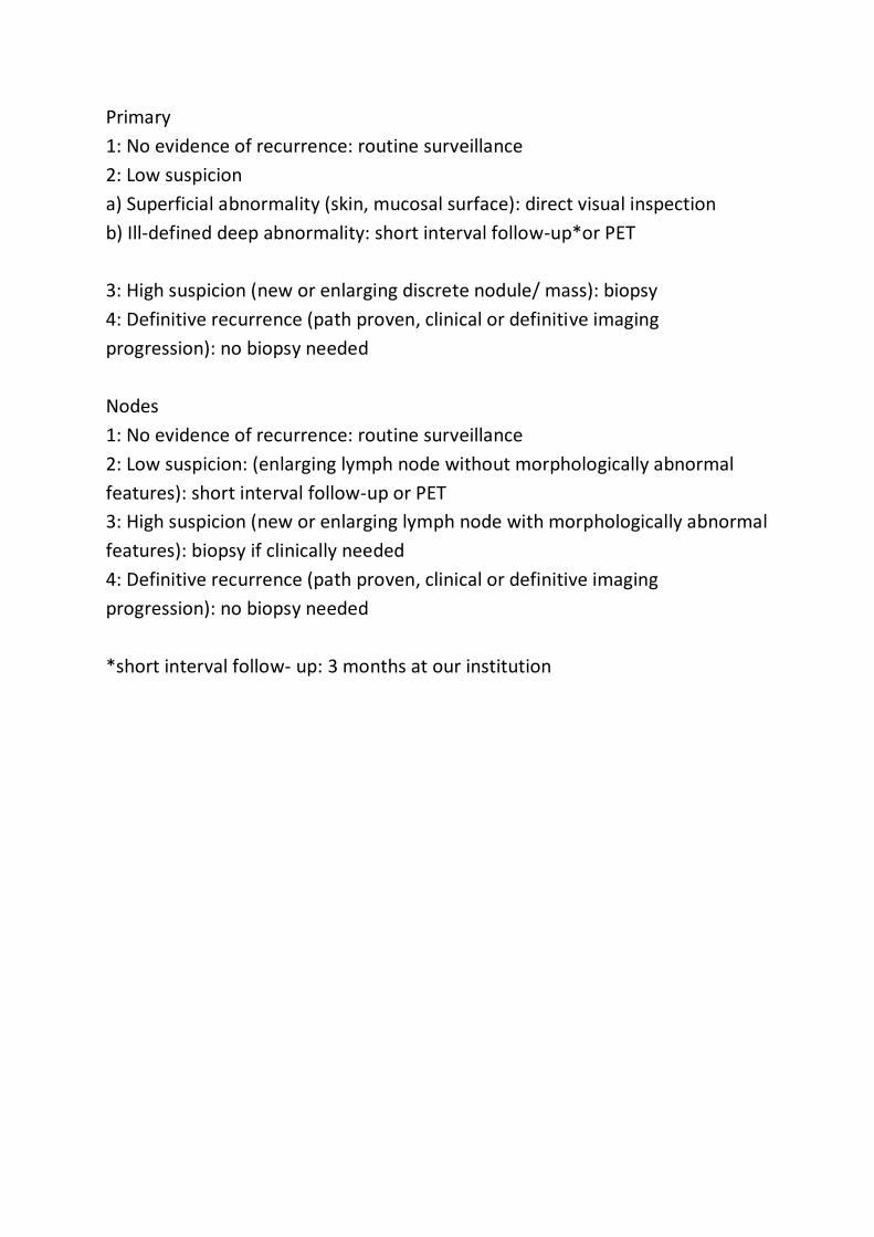
Primary
1: No evidence of recurrence: routine surveillance
2: Low suspicion
a) Superficial abnormality (skin, mucosal surface): direct visual inspection
b) Ill-defined deep abnormality: short interval follow-up*or PET
3: High suspicion (new or enlarging discrete nodule/ mass): biopsy
4: Definitive recurrence (path proven, clinical or definitive imaging
progression): no biopsy needed
Nodes
1: No evidence of recurrence: routine surveillance
2: Low suspicion: (enlarging lymph node without morphologically abnormal
features): short interval follow-up or PET
3: High suspicion (new or enlarging lymph node with morphologically abnormal
features): biopsy if clinically needed
4: Definitive recurrence (path proven, clinical or definitive imaging
progression): no biopsy needed
*short interval follow- up: 3 months at our institution


CACRNIOMA THYROID CT IMAGING
CT legend:
CT scan of the neck dated:
Primary Thyroid nodule:
Location: Right lobe/Left lobe/Isthmus
Size:
Enhancement: Homogeneous/Heterogeneous
Calcifications: Absent/Present
If present: microcalcification/ macrocalcification/ eggshell
Cystic / Necrotic change: Absent/Present
Extra-thyroid extension: Absent/Present
If present CT Grade of ETE*:
Mediastinal extension: Absent/Present
Right aberrant subclavian artery: Absent/Present
T STAGE
Strap muscle involvement: yes/No
T-E groove: Not involved/Involved (Status of vocal cords’ indirect sign of RLN
involvement)
Relationship with trachea(SHIN grade #):
Fat planes with oesophagus: Lost/ maintained. If lost; angle of contact:
Planes with prevertebral fascia: Lost/ maintained
Cricophraynx: Not involved/Involved
Cricoid cartilage: Not involved/Involved
Angle of contact with CCA (<180 / 180-270/>270):
Angle of contact with innominate vessels (<180 / 180-270/>270):
N STAGE:
Laterality- Ipsilateral / contralateral / Bilateral
Compartment: central/ lateral
Node stations:
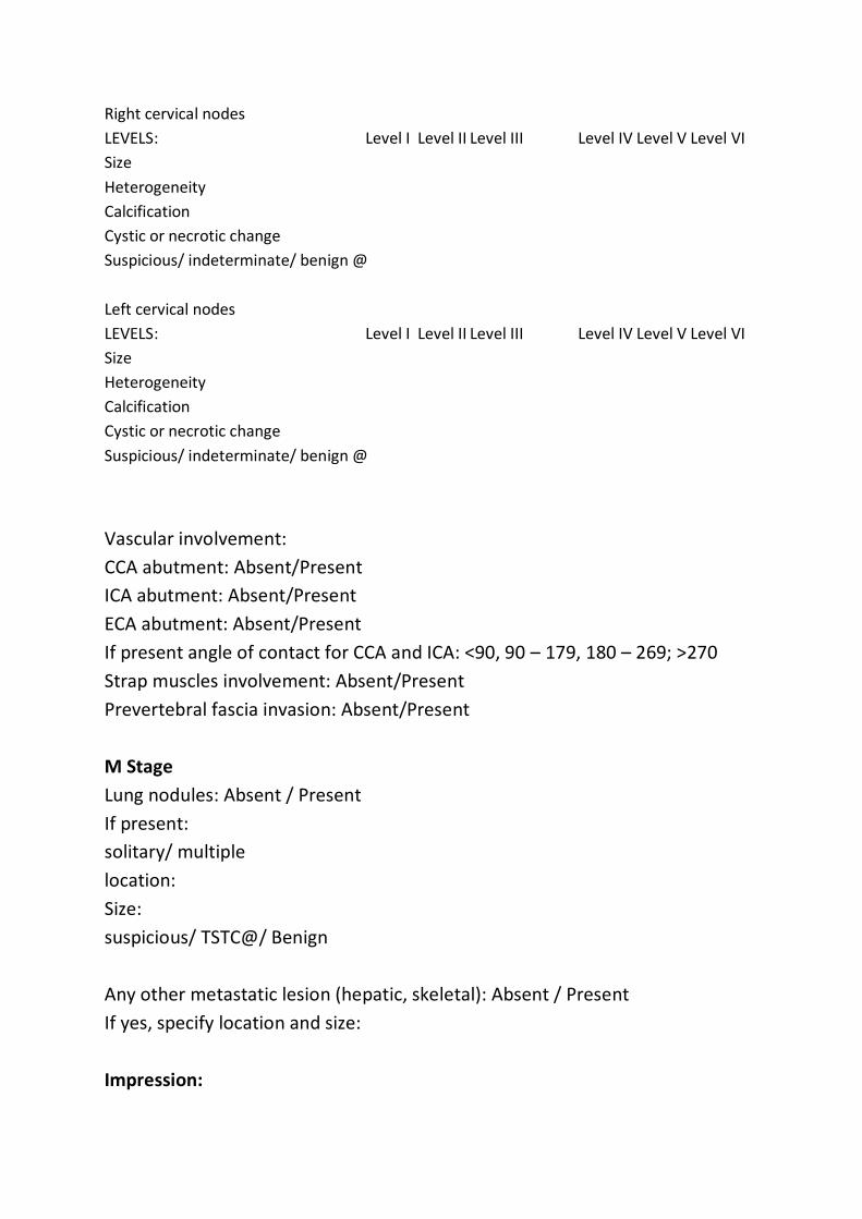
Right cervical nodes
LEVELS: Level I Level II Level III Level IV Level V Level VI
Size
Heterogeneity
Calcification
Cystic or necrotic change
Suspicious/ indeterminate/ benign @
Left cervical nodes
LEVELS: Level I Level II Level III Level IV Level V Level VI
Size
Heterogeneity
Calcification
Cystic or necrotic change
Suspicious/ indeterminate/ benign @
Vascular involvement:
CCA abutment: Absent/Present
ICA abutment: Absent/Present
ECA abutment: Absent/Present
If present angle of contact for CCA and ICA: <90, 90 – 179, 180 – 269; >270
Strap muscles involvement: Absent/Present
Prevertebral fascia invasion: Absent/Present
M Stage
Lung nodules: Absent / Present
If present:
solitary/ multiple
location:
Size:
suspicious/ TSTC@/ Benign
Any other metastatic lesion (hepatic, skeletal): Absent / Present
If yes, specify location and size:
Impression:

T stage
N stage
M stage
Specific comments, if any:
@ Follow-up/ image guided FNAC correlation.
*CT ETE grading:
• I, a tumor which was completely enveloped by thyroid parenchyma;
• II, a tumor in which the percentage of the tumor perimeter in contact
with the thyroid capsule was 1–25%;
• III, a tumor in which the contact with the capsule was 25–50%;
• IV, a tumor in which the contact with the capsule was >50%
# CT Shin grading:
• 0: > 5mm distance between tumor and trachea.
• I: disease abuts external perichondrium.
• II: disease invades into the cartilage +/- destruction.
• III: disease extends into the tracheal mucosa with no
elevation/penetration of mucosa.
• IV: disease is full-thickness invasion with expansion of the tracheal
mucosa with a bulge

USG THYROID DATED
High frequency USG of the thyroid with Doppler and elastography is
performed.
Right thyroid lobe
measures cm.
A well/ill defined solid/cystic/mixed hypoechoic/hyperechoic/isoechoic nodule
is seen in the right lobe of thyroid.
It measures 8 x 9 mm in size.
The nodule is wider than taller.
It shows no/complete/irregular halo.
No/microcalcifications/macrocalcifications are seen
The lesion shows no/central/peripheral/both central and peripheral
vascularity.
It shows no spongiform pattern/ comet tail artifacts.
Extrathyroid extension is not seen.
On elastography it is hard/soft, Asteria ES III.
Left thyroid lobe
measures cm.
A well/ill defined solid/cystic/mixed hypoechoic/hyperechoic/isoechoic nodule
is seen in the left lobe of thyroid.
It measures 8 x 9 mm in size.
The nodule is wider than taller.
It shows no/complete/irregular halo.
No/microcalcifications/macrocalcifications are seen
The lesion shows no/central/peripheral/both central and peripheral
vascularity.
It shows no spongiform pattern/ comet tail artifacts.
Extrathyroid extension is not seen.
On elastography it is hard/soft, Asteria ES III.
Isthmus measures 3 mm.

Few subcm sized reactive appearing nodes are seen in level IB and II region.
There is no suspicious cervical lymphadenopathy.
Bilateral neck vessels are patent.
Impression :-
USG reveals:
Right thyroid nodule appears benign/indeterminate/suspicious on USG with
TIRADS score and TMC RSS Score : low/Intermediate/high risk. FNAC
correlation is suggested
Left thyroid nodule appears benign/indeterminate/suspicious on USG with
TIRADS score and TMC RSS Score : low/Intermediate/high risk. FNAC
correlation is suggested.

USG NECK FOR NODAL MAPPING DATED:
High frequency USG of the neck nodes with Doppler is performed.
Neck nodes:
Right cervical nodes
LEVELS: Level I Level II Level III Level IV Level V Level VI
Short-axis diameter
Long-axis diameter
Loss of hilum
Echogenicity
Microcalcifications
Vascularity at power Doppler US
Suspicious/ indeterminate/ benign
Left cervical nodes
LEVELS: Level I Level II Level III Level IV Level V Level VI
Short-axis diameter
Long-axis diameter
Loss of hilum
Echogenicity
Microcalcifications
Vascularity at power Doppler US
Suspicious/ indeterminate/ benign
Bilateral neck vessels are patent.
IMPRESSION:
USG reveals:
Reactive/ indeterminate/ suspicious right / left side adenopathy is seen.
Comments: Suggested FNAC correlation.