Embed Size (px)
Citation preview
ORAL CAVITY MASS
Rivera, Laila Marie C.Rivere, Djeaune Marie Trissel B.
Robosa, Dean Antonio R. Rodas, Francis Martin F.
Rodriguez, Shereen Reine S.Rogelio, Ma.Graciela A.
Roque, Marianne N.Ruanto, Maria Theresa R.
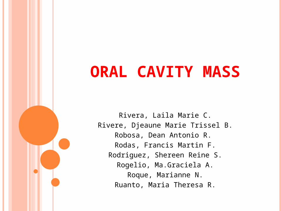
38 Y/O M
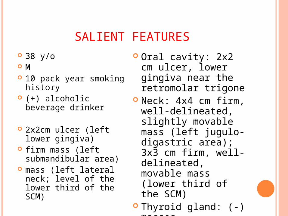
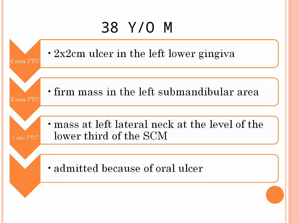
SALIENT FEATURES 38 y/o M 10 pack year smoking
history (+) alcoholic beverage
drinker
2x2cm ulcer (left lower gingiva)
firm mass (left submandibular area)
mass (left lateral neck; level of the lower third of the SCM)
Oral cavity: 2x2 cm ulcer, lower gingiva near the retromolar trigone
Neck: 4x4 cm firm, well-delineated, slightly movable mass (left jugulo-digastric area); 3x3 cm firm, well-delineated, movable mass (lower third of the SCM)
Thyroid gland: (-) masses
1. WHAT IS YOUR CLINICAL IMPRESSION?
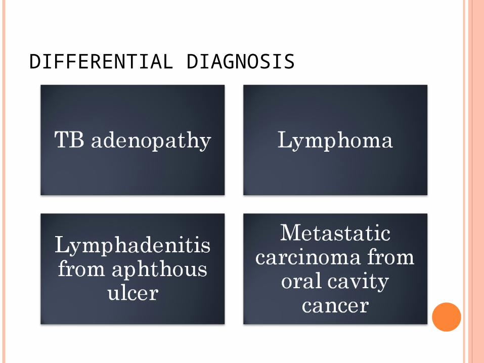
DIFFERENTIAL DIAGNOSIS
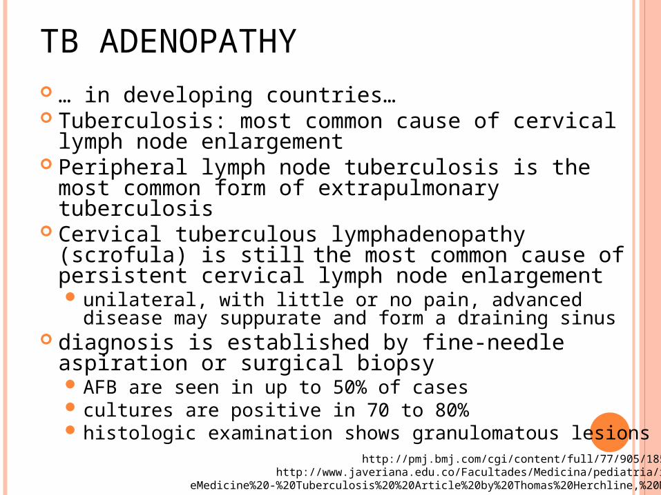
TB ADENOPATHY … in developing countries… Tuberculosis: most common cause of cervical
lymph node enlargement Peripheral lymph node tuberculosis is the most
common form of extrapulmonary tuberculosis Cervical tuberculous lymphadenopathy (scrofula) is
still the most common cause of persistent cervical lymph node enlargement unilateral, with little or no pain, advanced disease may
suppurate and form a draining sinus diagnosis is established by fine-needle aspiration
or surgical biopsy AFB are seen in up to 50% of cases cultures are positive in 70 to 80% histologic examination shows granulomatous lesions
http://pmj.bmj.com/cgi/content/full/77/905/185#SEC3http://www.javeriana.edu.co/Facultades/Medicina/pediatria/revis/
eMedicine%20-%20Tuberculosis%20%20Article%20by%20Thomas%20Herchline,%20MD.htm
LYMPHOMA Lymphoma may be nodal or extranodal A quarter of all extranodal lymphomas occur in
the head and neck Extranodal lymphoma is usually NHL
8% of findings on supraclavicular fine-needle aspirate biopsy yield a diagnosis of lymphoma
Lymphoma is the second most common primary malignancy occurring in the head and neck
Incidence of aggressive non-Hodgkin lymphoma has risen steadily over recent decades
http://emedicine.medscape.com/article/854110-overview
NON-HODGKIN’S LYMPHOMA May manifest in the cervical region and
lymphoid tissue of the Waldeyer ring Appears as a mass in the oropharynx or
nasopharynx Unilateral tonsillar enlargement is highly
suggestive of malignancy. Usually arises in the tongue base In contrast to squamous cell carcinoma, NHL
is bulky, fleshy, and nonulcerating Some patients with indolent NHLs may have
large asymptomatic abdominal massesSplenic or hepatic enlargement
http://emedicine.medscape.com/article/854110-overview
LYMPHADENITIS FROM APHTHOUS ULCER lymphadenitis is an infection of the
lymph nodes; a complication of bacterial infection
swollen glands are usually found near the site of an underlying infection, tumor, or inflammation apthous ulcerApthous ulcer also known as APHTHOUS
STOMATITISpainful open sore inside the mouth, caused
by a break in the mucous membraneEtiology is unknown
Lymphadenitis may occur after skin infections or other bacterial infections, particularly those due to streptococcus or staphylococcus
METASTATIC CARCINOMA FROM ORAL CAVITY CANCER 5% percent of all cancers reported yearly
30% of these cancers occur in the oral cavity squamous cell carcinoma- (most common)
95% of oral cavity cancer Risk Factors:
use of tobacco/ smoking 80% of patients with oral SCC risk of developing malignancy is 5-9 times greater for
smokers than nonsmokers Alcohol- 3-9 times greater risk of developing
cancerof alcohol and tobacco combined may convey a
risk greater than 100 times the general population HPV types 16 and 18 may be found in
approximately 22% and 14% of oropharyngeal tumors
http://www.ahns.info/patienteducation/docs/oralcavity.phphttp://emedicine.medscape.com/article/847678-overview
METASTATIC CARCINOMA FROM ORAL CAVITY CANCER Symptoms most common presentation of cancer of the
floor of the mouth is a painless inflamed superficial ulcer with poorly defined margin
Intermittent bleeding may occur Advanced cases: complaints may include
new or increased pain, pain on swallowing, ear pain, a change in speech, uncoordinated swallowing, or a lump in the neck
sores in the mouth, whether they are related to trauma or to a variation of canker (apthous) sores, should fully heal within three weeks
http://www.ahns.info/patienteducation/docs/oralcavity.php
METASTATIC CARCINOMA FROM ORAL CAVITY CANCER Metastatic neck disease is the most
important factor in the spread of head and neck squamous cell carcinoma (SCC) from primary sites
most commonly involved primary sites larynx, oropharynx, hypopharynx, and oral cavity
Malignant tumors of the oral cavity grow rapidly, with frequent and early metastasis to the surrounding regional lymph nodes
http://emedicine.medscape.com/article/850195-overview
CLINICAL IMPRESSION:
METASTATIC CARCINOMA FROM ORAL CAVITY CANCER

2. WHAT TO DO NEXT
WHAT TO DO NEXT
Perform a thorough head and neck exam under anesthesia
Perform triple endoscopy: (nasopharyngolaryngoscopy, bronchoscopy, esophagoscopy)
Get a biopsy of the oral cavity ulcer
THOROUGH HEAD AND NECK EXAM
Biopsy of primary Fine needle aspiration of possible neck
metastasis Imaging studies:
Chest radiograph: posteroanterior and lateral CT/MRI of primary and neck Panorex or dental x-ray: evaluate mandible
invasion if CT/MRI not performed Barium swallow
THOROUGH HEAD AND NECK EXAM
Laboratory tests Pre anesthesia testing Basic liver function tests
Consutations:-Radiation therapy -for adjuvant or definitive
therapy considerations-Dental: pre radiation dental treatment and for
post therapy
EXAMINATION UNDER ANESTHESIA
Nasopharyngolaryngoscopy and pharyngoscopy
Esophagoscopy Bronchoscopy
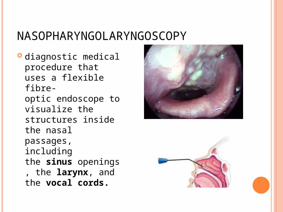
NASOPHARYNGOLARYNGOSCOPY
diagnostic medical procedure that uses a flexible fibre-optic endoscope to visualize the structures inside the nasal passages, including the sinus openings, the larynx, and the vocal cords.
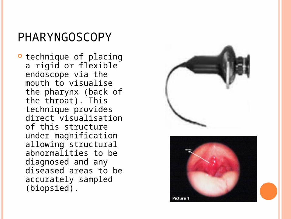
PHARYNGOSCOPY technique of placing a
rigid or flexible endoscope via the mouth to visualise the pharynx (back of the throat). This technique provides direct visualisation of this structure under magnification allowing structural abnormalities to be diagnosed and any diseased areas to be accurately sampled (biopsied).
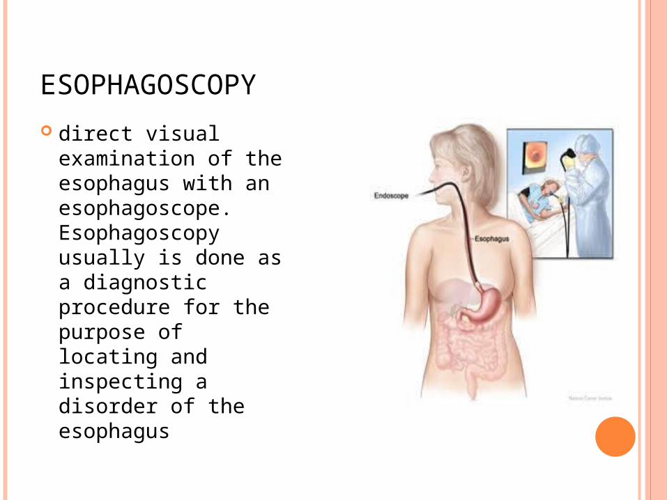
ESOPHAGOSCOPY
direct visual examination of the esophagus with an esophagoscope. Esophagoscopy usually is done as a diagnostic procedure for the purpose of locating and inspecting a disorder of the esophagus
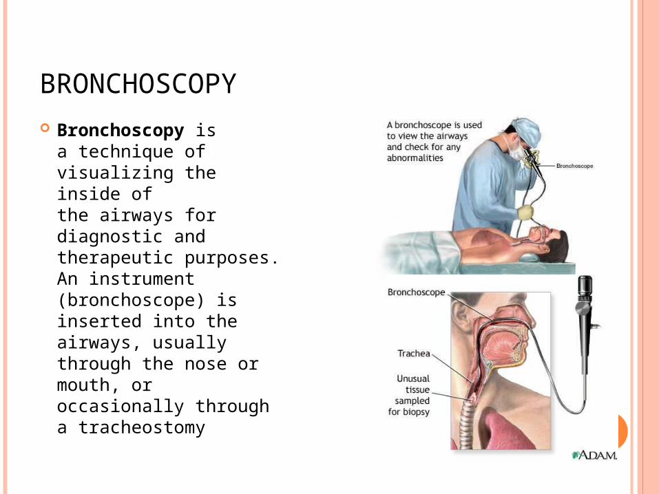
BRONCHOSCOPY
Bronchoscopy is a technique of visualizing the inside of the airways for diagnostic and therapeutic purposes. An instrument (bronchoscope) is inserted into the airways, usually through the nose or mouth, or occasionally through a tracheostomy
FINDINGS: Nasopharyngolaryngoscopy ⊖; Biopsy of ulcer:
well-differentiated squamous cell cancer
Fine needle biopsy of neck mass: Chronic Lymphadenitis

3. WHAT WILL YOU DO NEXT?
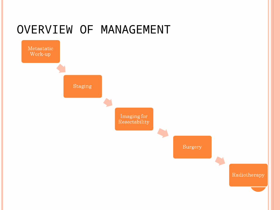
OVERVIEW OF MANAGEMENT
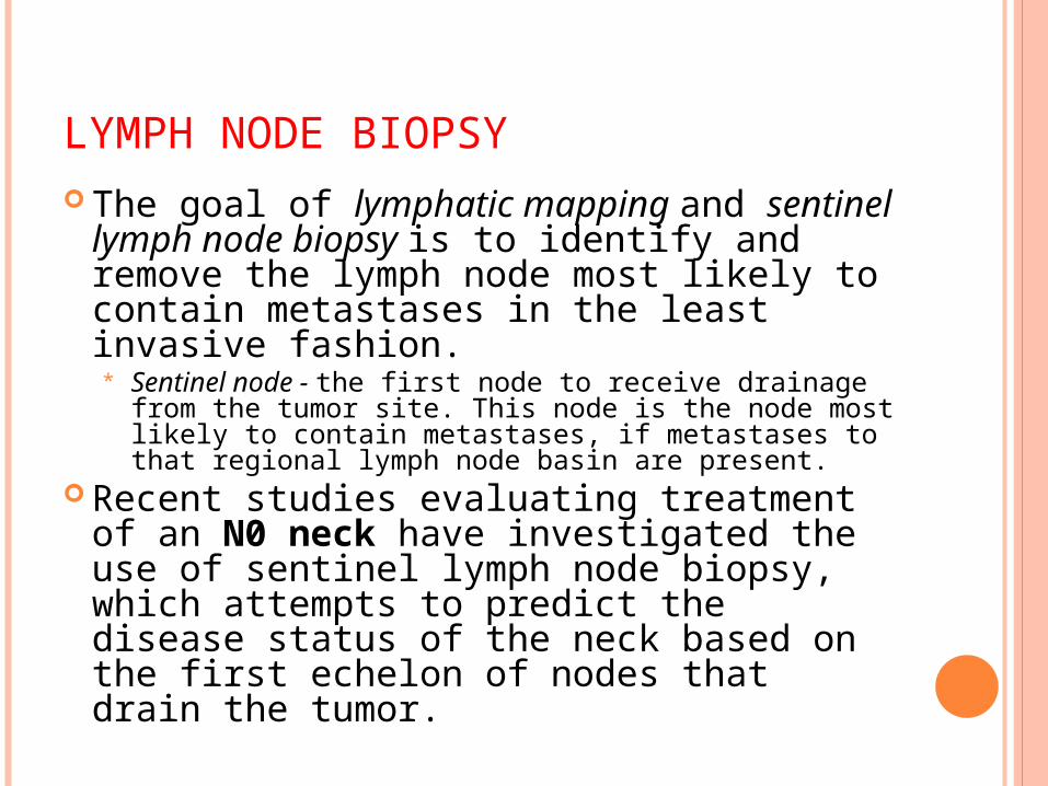
LYMPH NODE BIOPSY The goal of lymphatic mapping and
sentinel lymph node biopsy is to identify and remove the lymph node most likely to contain metastases in the least invasive fashion. * Sentinel node - the first node to receive drainage
from the tumor site. This node is the node most likely to contain metastases, if metastases to that regional lymph node basin are present.
Recent studies evaluating treatment of an N0 neck have investigated the use of sentinel lymph node biopsy, which attempts to predict the disease status of the neck based on the first echelon of nodes that drain the tumor.
METASTATIC WORK-UP
Vigilance for second primary tumors
Patients diagnosed with a head and neck cancer are predisposed to the development of a second tumor within the aerodigestive tract
Patients with a primary malignancy of the oral cavity or pharynx are most likely to develop a second lesion within the cervical esophagus
Once cancer has been proven by biopsy, a CT scan of the chest will be ordered to rule out distant metastasis
Contrast-enhanced CT and MRI of the head and neck may be performed for evaluation of the tumor and detection of occult lymphadenopathy
CT scanning - best at evaluating bony destruction
MRI - determine soft tissue involvement and is excellent at evaluating parotid and parapharyngeal space tumors
Chest radiography or chest CT is performed to rule out synchronous lung lesions
Serum tumor markers such as alkaline phosphatase and calcium may be determined, but such tests are not standard.
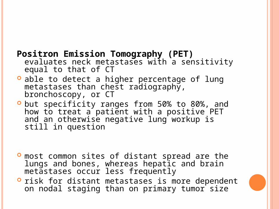
Positron Emission Tomography (PET) evaluates neck metastases with a sensitivity equal to that of CT
able to detect a higher percentage of lung metastases than chest radiography, bronchoscopy, or CT
but specificity ranges from 50% to 80%, and how to treat a patient with a positive PET and an otherwise negative lung workup is still in question
most common sites of distant spread are the lungs and bones, whereas hepatic and brain metastases occur less frequently
risk for distant metastases is more dependent on nodal staging than on primary tumor size
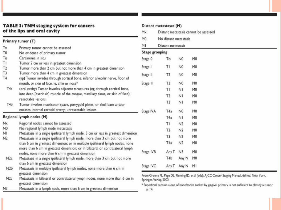
STAGING
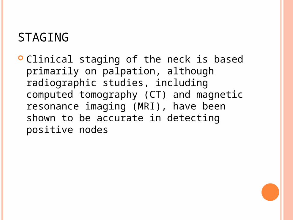
Clinical staging of the neck is based primarily on palpation, although radiographic studies, including computed tomography (CT) and magnetic resonance imaging (MRI), have been shown to be accurate in detecting positive nodes
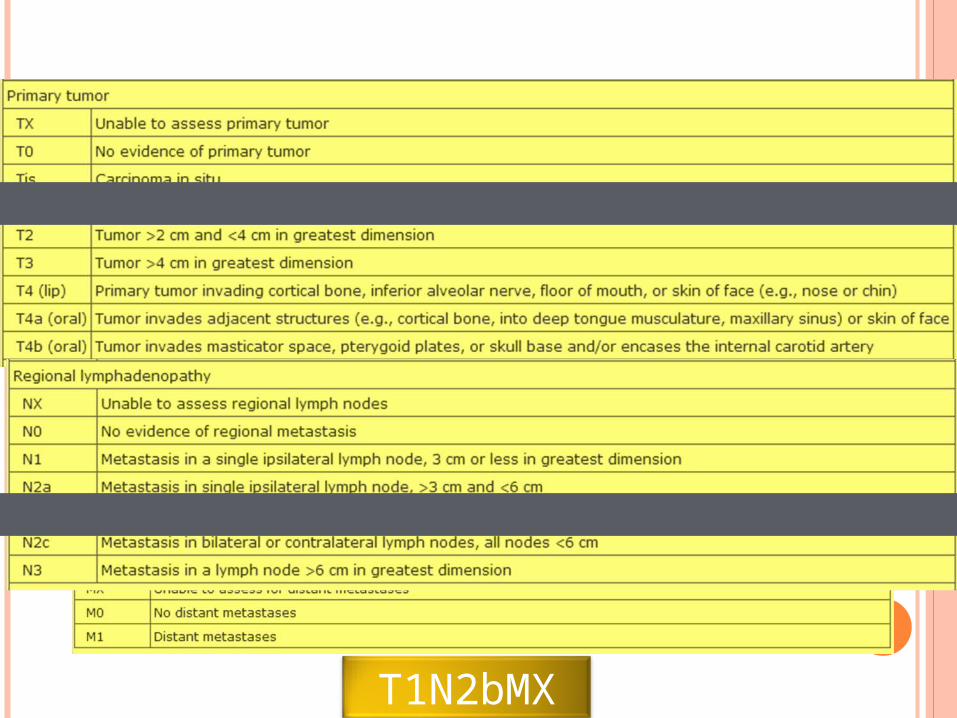
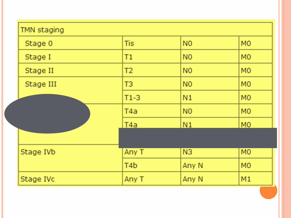
T1N2bMX
IMAGING FOR RESECTABILITY
Panoramic x-ray of the mandible scanning dental X-ray of the upper and lower
jaw shows a two-dimensional view of a half-circle
from ear to ear shows a patient's nasal area, sinuses, jaw
joints, teeth and surrounding bone can reveal cysts, tumors, bone irregularities the mandible can also have an indentation
on its lower border when the patient's masseter has been clenching and grinding
shows the entire mandible, including all of its lower border
Indications for cortical or rim resection of the mandible as determined by physical examination, CT scan, orthopantomogram, and dental films
a. Tumor close to but not involving the periosteum of mandible
b. Tumor involving only mandibular periosteumc. Tumor adjacent to cortical bone of mandible
with no evidence of invasion beyond superficial cortex
d. Tumor adjacent to dentition with no evidence of involvement of periodontal ligament
Indications for segmental resection of the mandible as determined by physical examination, CT scan, orthopantomogram, and dental films
a. Invasion of the medullary space of the mandible b. Tumor fixation to the occlusal surface of the mandible
in the edentulous patientc. Invasion of tumor into the mandible via the
mandibular or mental foramend. Tumor fixed to the mandible following prior
radiotherapy to the mandible, particularly if the tumor is located on the occlusal surface
e. Tumor adjacent to carious dentition with involvement of the periodontal ligament
f. Hypoplastic edentulous mandible with significant loss of vertical height precluding safe performance of a rim resection
Cortical or rim mandibulectomy – if (+) adherence to mandibular periosteum without bony erosion
Segmental resection – if (+) mandible invasion
RESECTION OF RETROMOLAR TRIGONE TUMORS:
usually requires a marginal or segmental mandibulectomy with a soft-tissue and/or osseous reconstruction in order to maximize a patient's postoperative ability for functional speech and swallowing
Ipsilateral elective and therapeutic neck dissection is performed because of the risk of metastasis to the regional lymphatics
RESULTS OF THE PATIENT
Head & neck examinations: ⊖ Chest X-ray: ⊖ Panoramic x-ray of the mandible: lytic lesion of
the body of the mandible near the angle

4. WHAT TYPE OF SURGERY IS INDICATED?
WHAT TYPE OF SURGERY IS INDICATED?
Operative Findings:
3 x 2 cm ulcer of the lower gingiva with invasion into the mandible
5 X 4 cm well-encapsulated firm mass located at the submandibular triangle (level 1 to level 2)
Multiple pinkish-red, firm, grossly enlarged nodes ( 1-2 cm) along the jugular chain (levels 2 to 4)
4 X 3 cm well encapsulated firm mass at the supraclavicular area Operation done:
Wide excision of the ulcer with segmental mandibulectomy with modified radical neck dissection, left; the defect was reconstructed using titanium plates
Final histopath:
Well differentiated squamous cell carcinoma with metastasis to 5/20 lymph nodes, the largest measures 2 cm with extracapsular invasion; margins clear; with bony invasion
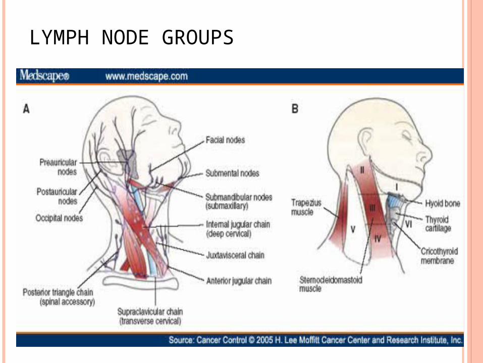
LYMPH NODE GROUPS
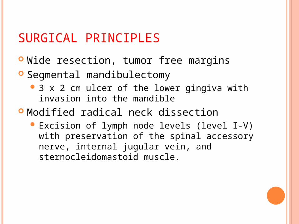
SURGICAL PRINCIPLES
Wide resection, tumor free margins Segmental mandibulectomy
3 x 2 cm ulcer of the lower gingiva with invasion into the mandible
Modified radical neck dissection Excision of lymph node levels (level I-V) with
preservation of the spinal accessory nerve, internal jugular vein, and sternocleidomastoid muscle.
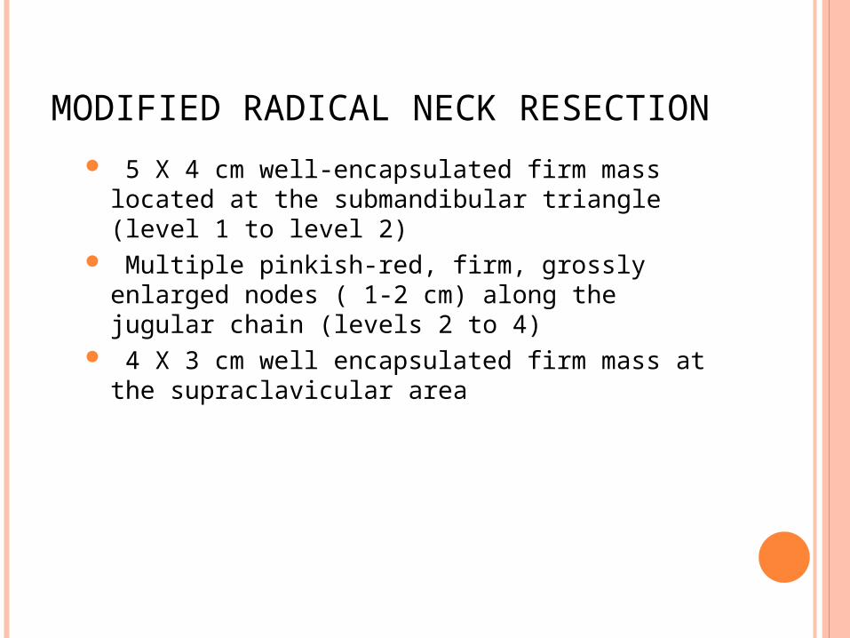
MODIFIED RADICAL NECK RESECTION
5 X 4 cm well-encapsulated firm mass located at the submandibular triangle (level 1 to level 2)
Multiple pinkish-red, firm, grossly enlarged nodes ( 1-2 cm) along the jugular chain (levels 2 to 4)
4 X 3 cm well encapsulated firm mass at the supraclavicular area
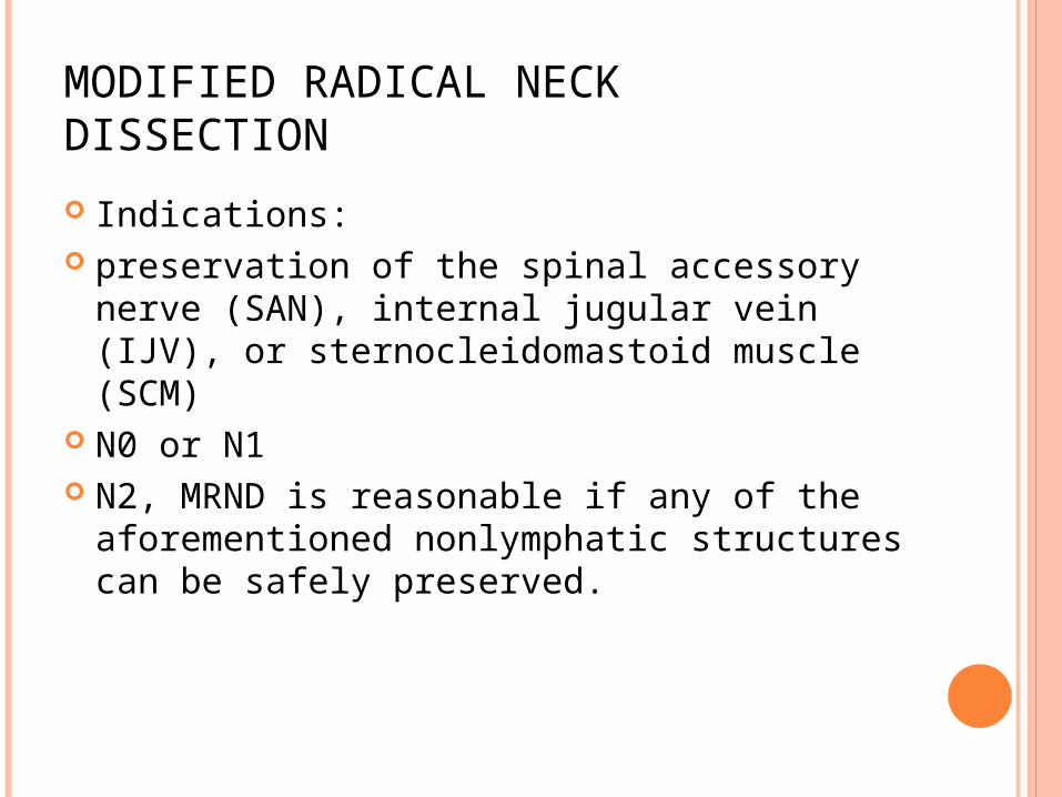
MODIFIED RADICAL NECK DISSECTION
Indications: preservation of the spinal accessory nerve
(SAN), internal jugular vein (IJV), or sternocleidomastoid muscle (SCM)
N0 or N1 N2, MRND is reasonable if any of the
aforementioned nonlymphatic structures can be safely preserved.
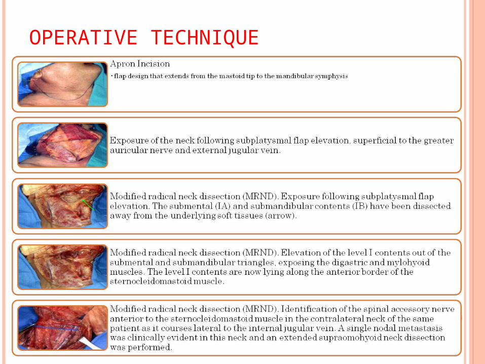
OPERATIVE TECHNIQUE
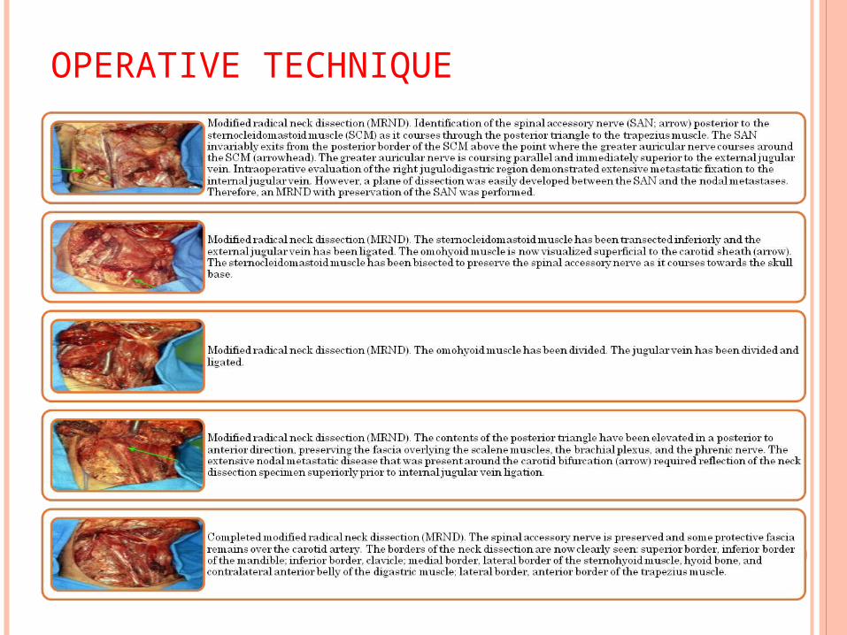
OPERATIVE TECHNIQUE
OTHER SURGICAL PROCEDURES
Radical Neck Dissection Removal of all ipsilateral cervical lymph nodes
(level I-V). Dissection from the inferior border of the mandible to clavicle, posteriorly to the anterior border of the trapezius muscle, and anteriorly to the lateral border of the sternohyoid muscle. Depth is to the fascia overlying the anterior scalene and levator scapulae muscles.
INDICATIONS
Multiple positive neck nodes that are clinically present in an untreated patient or in a patient treated with surgery, irradiation, chemotherapy, or a combination thereof
One or more positive neck nodes that are clinically present and extracapsular extension with involvement of the spinal accessory nerve and internal jugular vein

5. WHAT ADJUVANT TREATMENT IS REQUIRED?
WHAT ADJUVANT TREATMENT IS REQUIRED?
Stage Iva combined modality Surgery + radiation Chemotx + radiation for advanced larynx or
hypopharynx lesions
RADIOTHERAPY
For Stage 3 & 4 Radiation pre or postop Indicators for postoperative adjuvant radiation therapy:
Presence of negative prognostic factors such as extracapsular spread of tumor, perineural invasion, vascular invasion, fixation to surrounding structures, and multiple positive nodes
Preoperative radiation or chemoradiation therapy Given when patients have advanced neck disease that involves
the carotid artery or the deep neck musculature, in the hope that the tumor reduces in size and becomes resectable.
50 to 70 Gy over 5 to 7 weeks Adverse Effect:
Acute: mucositis, skin erythema Late: fibrosis, xerostomia, altered taste, risk of
osteoradionecrosis
CHEMOTHERAPY
No survival advantage for HNSCC compared to surgery and/ or radiation
Role of chemotherapy: as a radiation sensitizer Palliation (symptom control) of recurrent or
unresectable disease, combined with radiation Cisplatin, carboplatin, 5-FU

JOURNAL ON DIAGNOSIS
CYTOLOGIC AND DNA- CYTOMETRIC EARLY DIAGNOSIS
OF ORAL CANCER
Torsten W. Remmerbach, Horst Weidenbach, Natalja Pomjanski, Kristiane Knops, Stefanie Mathes , Alexander Hemprich and
Alfred Böcking
Department of Oral, Maxillofacial and Plastic Surgery, University of Leipzig, Leipzig,Germany
Institute of Pathology, University of Leipzig, Leipzig, Germany
Institute of Cytopathology, Heinrich Heine University, Düsseldorf, Germany
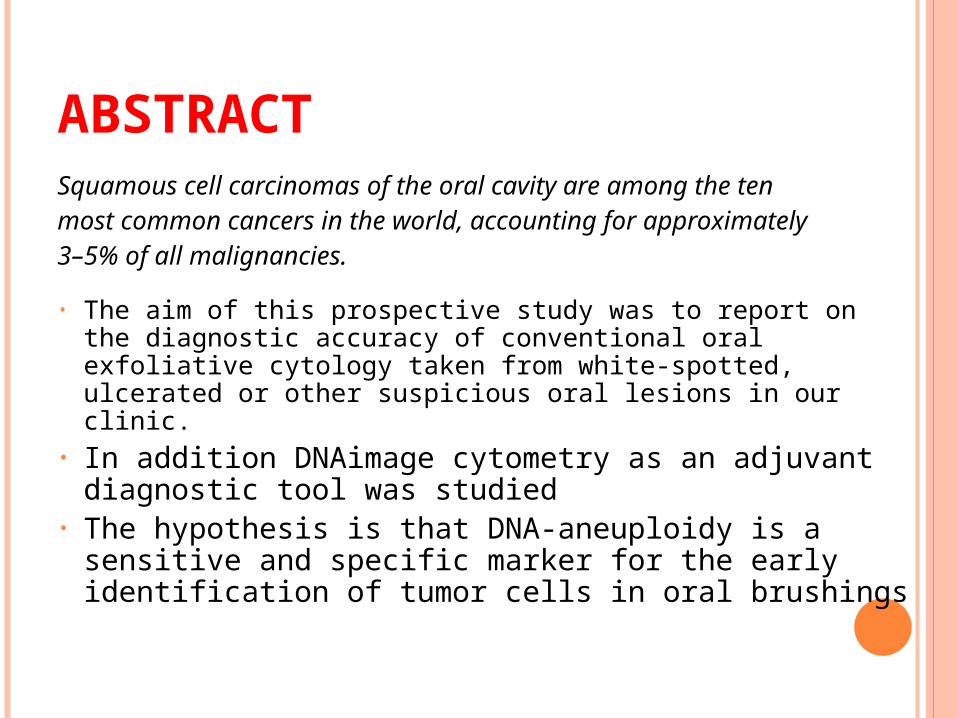
Squamous cell carcinomas of the oral cavity are among the tenmost common cancers in the world, accounting for
approximately3–5% of all malignancies.
• The aim of this prospective study was to report on the diagnostic accuracy of conventional oral exfoliative cytology taken from white-spotted, ulcerated or other suspicious oral lesions in our clinic.
• In addition DNAimage cytometry as an adjuvant diagnostic tool was studied
• The hypothesis is that DNA-aneuploidy is a sensitive and specific marker for the early identification of tumor cells in oral brushings
ABSTRACT
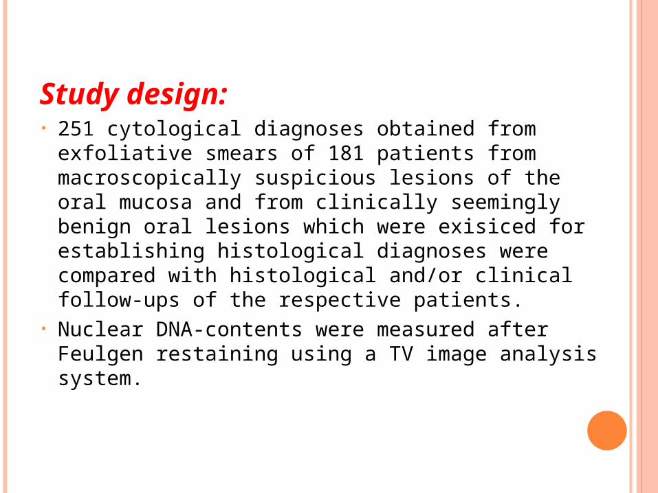
Study design:• 251 cytological diagnoses obtained from exfoliative
smears of 181 patients from macroscopically suspicious lesions of the oral mucosa and from clinically seemingly benign oral lesions which were exisiced for establishing histological diagnoses were compared with histological and/or clinical follow-ups of the respective patients.
• Nuclear DNA-contents were measured after Feulgen restaining using a TV image analysis system.
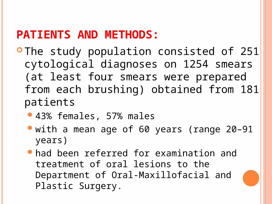
PATIENTS AND METHODS: The study population consisted of 251
cytological diagnoses on 1254 smears (at least four smears were prepared from each brushing) obtained from 181 patients 43% females, 57% maleswith a mean age of 60 years (range 20–91 years)had been referred for examination and treatment
of oral lesions to the Department of Oral-Maxillofacial and Plastic Surgery.
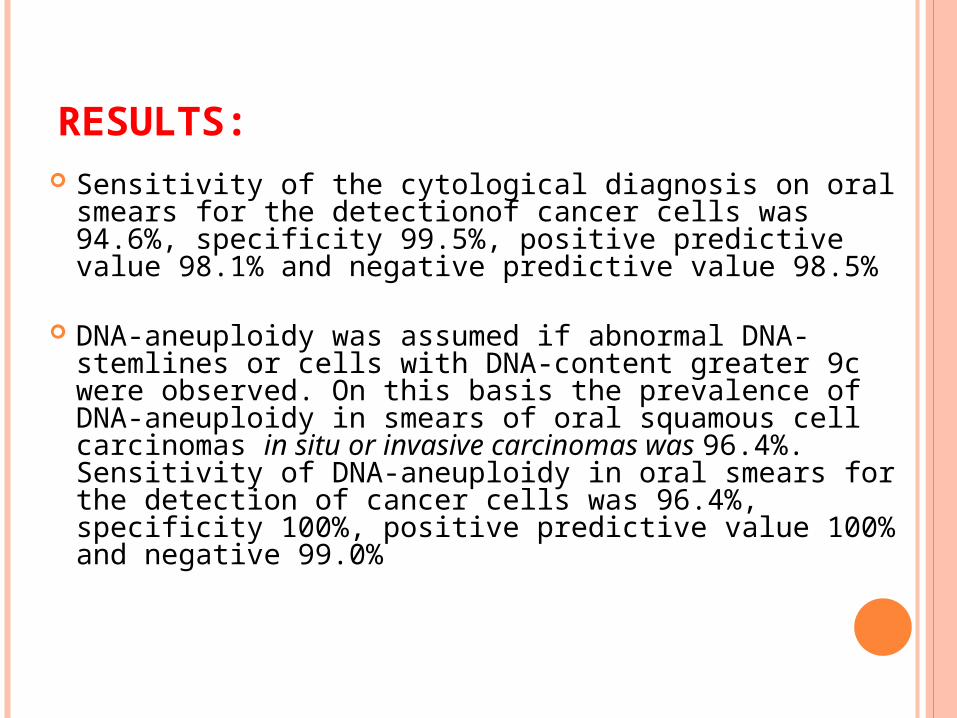
RESULTS: Sensitivity of the cytological diagnosis on oral smears
for the detectionof cancer cells was 94.6%, specificity 99.5%, positive predictive value 98.1% and negative predictive value 98.5%
DNA-aneuploidy was assumed if abnormal DNA-stemlines or cells with DNA-content greater 9c were observed. On this basis the prevalence of DNA-aneuploidy in smears of oral squamous cell carcinomas in situ or invasive carcinomas was 96.4%. Sensitivity of DNA-aneuploidy in oral smears for the detection of cancer cells was 96.4%, specificity 100%, positive predictive value 100% and negative 99.0%
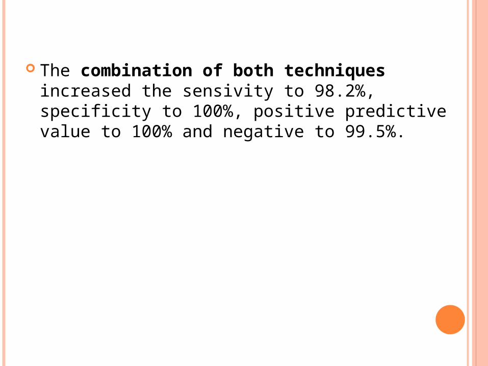
The combination of both techniques increased the sensivity to 98.2%, specificity to 100%, positive predictive value to 100% and negative to 99.5%.
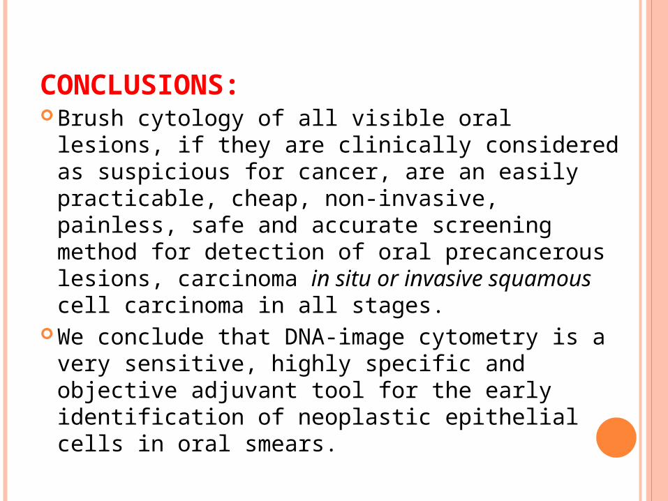
CONCLUSIONS: Brush cytology of all visible oral lesions, if
they are clinically considered as suspicious for cancer, are an easily practicable, cheap, non-invasive, painless, safe and accurate screening method for detection of oral precancerous lesions, carcinoma in situ or invasive squamous cell carcinoma in all stages.
We conclude that DNA-image cytometry is a very sensitive, highly specific and objective adjuvant tool for the early identification of neoplastic epithelial cells in oral smears.

JOURNAL ON TREATMENT
POSTOPERATIVE IRRADIATION WITH OR WITHOUT
CONCOMITANT CHEMOTHERAPY FOR LOCALLY ADVANCED HEAD
AND NECK CANCER
Jacques Bernier, M.D., Ph.D., Christian Domenge, M.D.,
Mahmut Ozsahin, M.D., Ph.D., Katarzyna Matuszewska, M.D.,
Jean-Louis Lefebvre, M.D., Richard H. Greiner, M.D., Jordi Giralt, M.D.,
Philippe Maingon, M.D., Frederic Rolland, M.D., Michel Bolla, M.D.,
Francesco Cognetti, M.D., Jean Bourhis, M.D., Anne Kirkpatrick, M.Sc.,
and Martine van Glabbeke, Ir., M.Sc., for the European Organization for Research
and Treatment of Cancer Trial 22931
INTRODUCTION
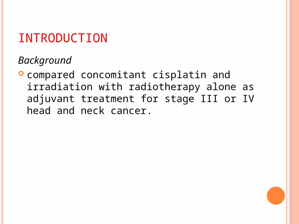
Background compared concomitant cisplatin and
irradiation with radiotherapy alone as adjuvant treatment for stage III or IV head and neck cancer.
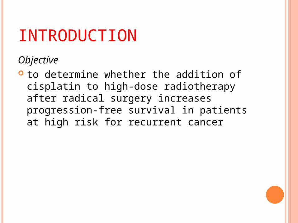
INTRODUCTIONObjective to determine whether the addition of
cisplatin to high-dose radiotherapy after radical surgery increases progression-free survival in patients at high risk for recurrent cancer
METHODOLOGY
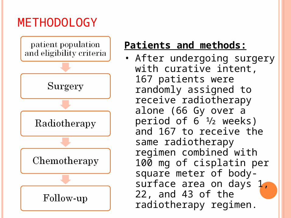
Patients and methods:• After undergoing surgery
with curative intent, 167 patients were randomly assigned to receive radiotherapy alone (66 Gy over a period of 6 1⁄2 weeks) and 167 to receive the same radiotherapy regimen combined with 100 mg of cisplatin per square meter of body-surface area on days 1, 22, and 43 of the radiotherapy regimen.
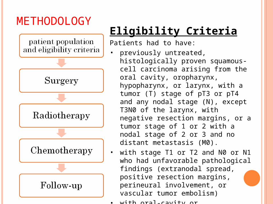
METHODOLOGYEligibility CriteriaPatients had to have:• previously untreated, histologically
proven squamous-cell carcinoma arising from the oral cavity, oropharynx, hypopharynx, or larynx, with a tumor (T) stage of pT3 or pT4 and any nodal stage (N), except T3N0 of the larynx, with negative resection margins, or a tumor stage of 1 or 2 with a nodal stage of 2 or 3 and no distant metastasis (M0).
• with stage T1 or T2 and N0 or N1 who had unfavorable pathological findings (extranodal spread, positive resection margins, perineural involvement, or vascular tumor embolism)
• with oral-cavity or oropharyngeal tumors with involved lymph nodes at level IV or V, according to the anatomical lymph-node distribution
METHODOLOGY
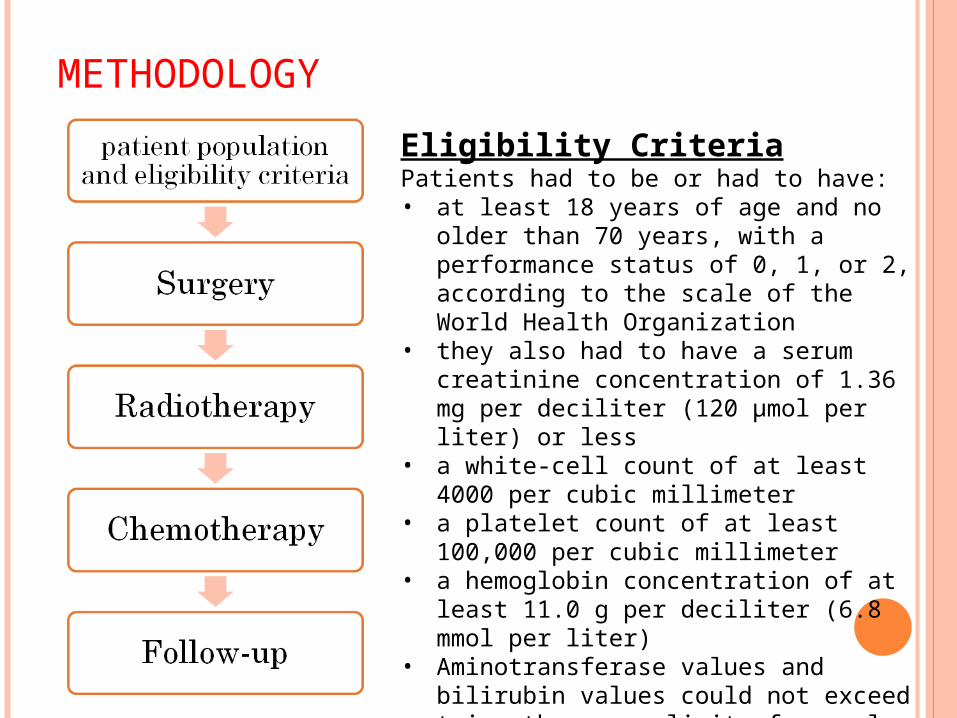
Eligibility CriteriaPatients had to be or had to have:• at least 18 years of age and no older
than 70 years, with a performance status of 0, 1, or 2, according to the scale of the World Health Organization
• they also had to have a serum creatinine concentration of 1.36 mg per deciliter (120 μmol per liter) or less
• a white-cell count of at least 4000 per cubic millimeter
• a platelet count of at least 100,000 per cubic millimeter
• a hemoglobin concentration of at least 11.0 g per deciliter (6.8 mmol per liter)
• Aminotransferase values and bilirubin values could not exceed twice the upper limit of normal were excluded from the study.

METHODOLOGY
Exclusion Criteria• Patients who had a history
of invasive or synchronous cancer (except nonmelanoma skin cancer), had previously received chemotherapy, or had known central nervous system disease
METHODOLOGY
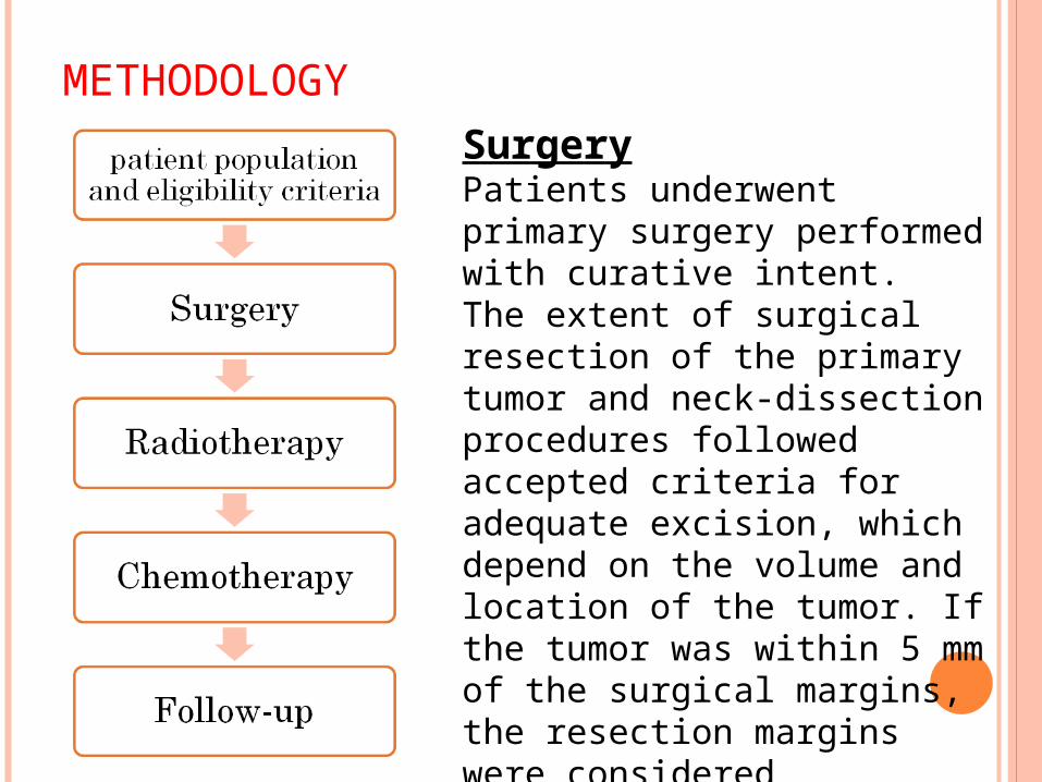
SurgeryPatients underwent primary surgery performed with curative intent.The extent of surgical resection of the primary tumor and neck-dissection procedures followed accepted criteria for adequate excision, which depend on the volume and location of the tumor. If the tumor was within 5 mm of the surgical margins, the resection margins were consideredto be close.
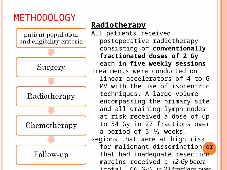
METHODOLOGYRadiotherapyAll patients received postoperative
radiotherapy consisting of conventionally fractionated doses of 2 Gy each in five weekly sessions
Treatments were conducted on linear accelerators of 4 to 6 MV with the use of isocentric techniques. A large volume encompassing the primary site and all draining lymph nodes at risk received a dose of up to 54 Gy in 27 fractions over a period of 5 1⁄2 weeks.
Regions that were at high risk for malignant dissemination or that had inadequate resection margins received a 12-Gy boost (total, 66 Gy) in 33 fractions over a period of 6 1⁄2 weeks.
The dose to the spinal cord was limited to 45 Gy.
METHODOLOGY
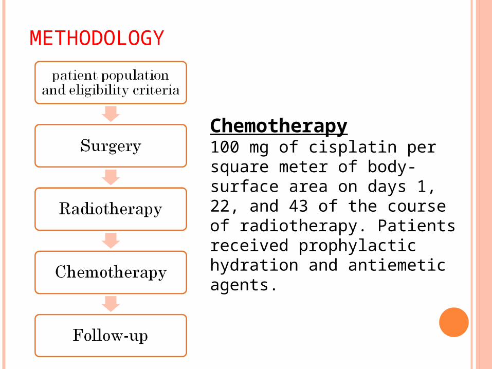
Chemotherapy100 mg of cisplatin per square meter of body-surface area on days 1, 22, and 43 of the course of radiotherapy. Patients received prophylactic hydration and antiemetic agents.
METHODOLOGY
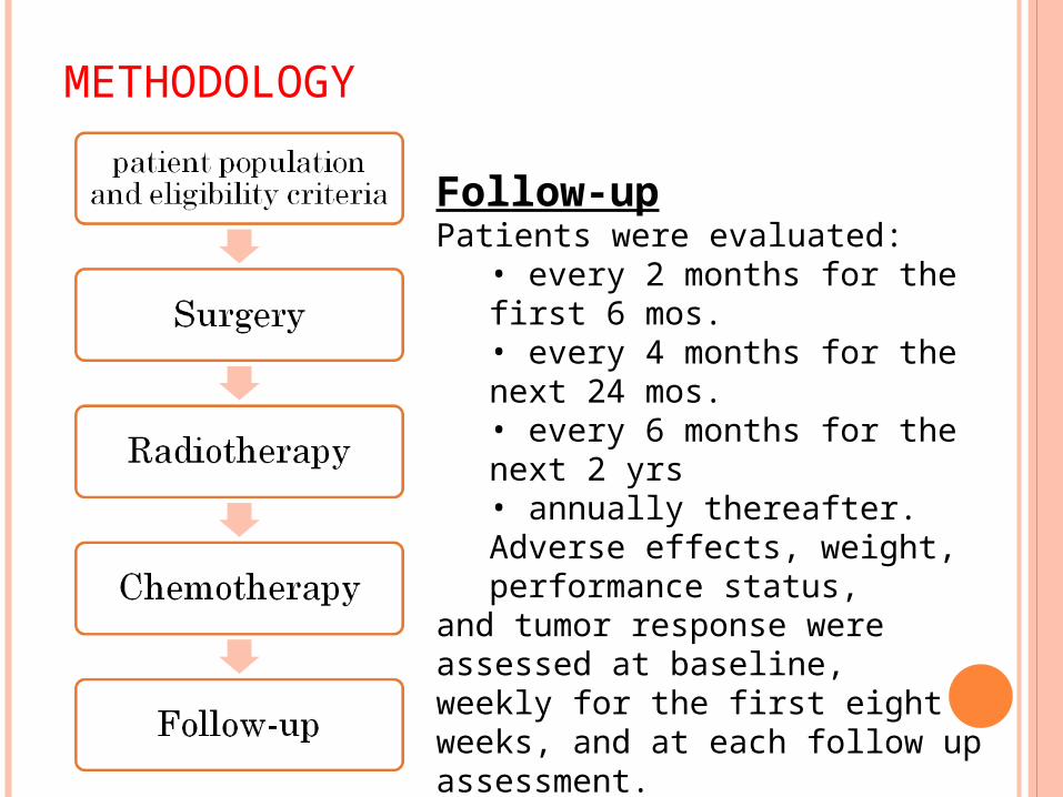
Follow-upPatients were evaluated:
• every 2 months for the first 6 mos.• every 4 months for the next 24 mos. • every 6 months for the next 2 yrs• annually thereafter. Adverse effects, weight, performance status,
and tumor response were assessed at baseline,weekly for the first eight weeks, and at each follow up assessment.
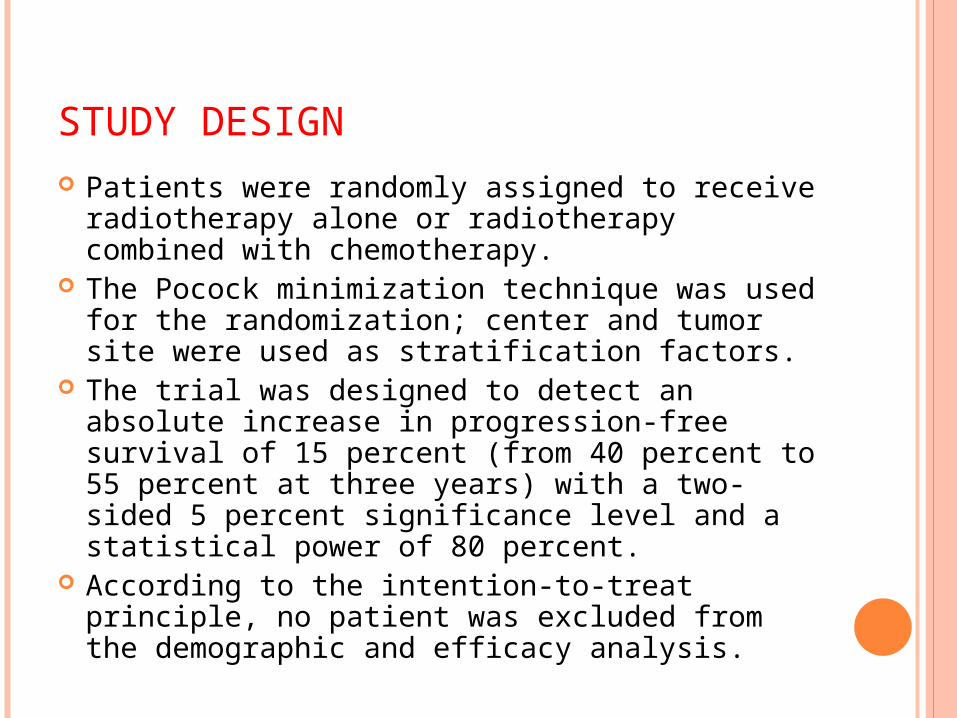
STUDY DESIGN Patients were randomly assigned to receive
radiotherapy alone or radiotherapy combined with chemotherapy.
The Pocock minimization technique was used for the randomization; center and tumor site were used as stratification factors.
The trial was designed to detect an absolute increase in progression-free survival of 15 percent (from 40 percent to 55 percent at three years) with a two-sided 5 percent significance level and a statistical power of 80 percent.
According to the intention-to-treat principle, no patient was excluded from the demographic and efficacy analysis.
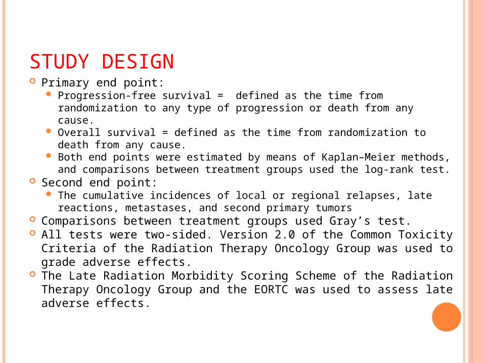
STUDY DESIGN Primary end point:
Progression-free survival = defined as the time from randomization to any type of progression or death from any cause.
Overall survival = defined as the time from randomization to death from any cause.
Both end points were estimated by means of Kaplan–Meier methods, and comparisons between treatment groups used the log-rank test.
Second end point: The cumulative incidences of local or regional relapses, late
reactions, metastases, and second primary tumors Comparisons between treatment groups used Gray’s test. All tests were two-sided. Version 2.0 of the Common Toxicity
Criteria of the Radiation Therapy Oncology Group was used to grade adverse effects.
The Late Radiation Morbidity Scoring Scheme of the Radiation Therapy Oncology Group and the EORTC was used to assess late adverse effects.
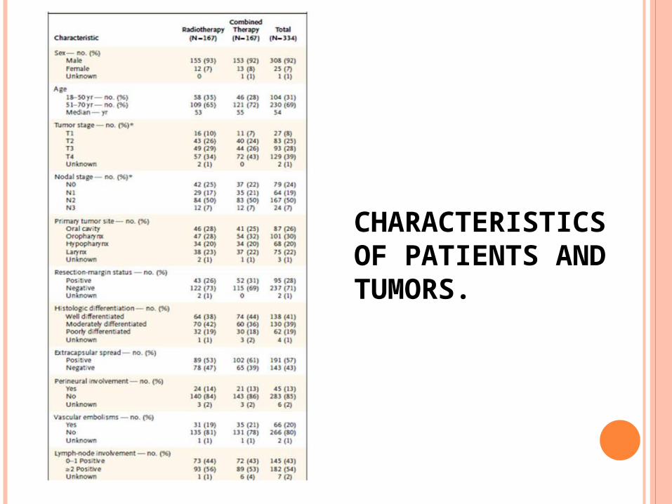
CHARACTERISTICS OF PATIENTS AND TUMORS.
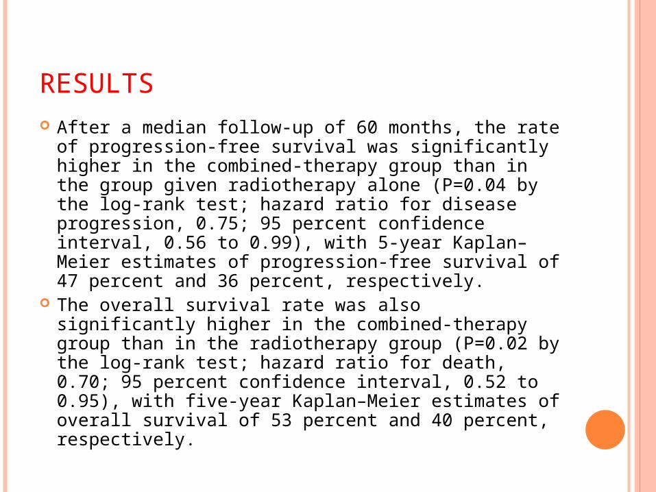
RESULTS After a median follow-up of 60 months, the rate of
progression-free survival was significantly higher in the combined-therapy group than in the group given radiotherapy alone (P=0.04 by the log-rank test; hazard ratio for disease progression, 0.75; 95 percent confidence interval, 0.56 to 0.99), with 5-year Kaplan–Meier estimates of progression-free survival of 47 percent and 36 percent, respectively.
The overall survival rate was also significantly higher in the combined-therapy group than in the radiotherapy group (P=0.02 by the log-rank test; hazard ratio for death, 0.70; 95 percent confidence interval, 0.52 to 0.95), with five-year Kaplan–Meier estimates of overall survival of 53 percent and 40 percent, respectively.
RESULTS The cumulative incidence of local or regional
relapses was significantly lower in the combined-therapy group (P=0.007).
The estimated five-year cumulative incidence of local or regional relapses (considering death from other causes as a competing risk) was 31 percent after radiotherapy and 18 percent after combined therapy. Severe (grade 3 or higher) adverse effects were more frequent after combined therapy (41 percent) than after radiotherapy (21 percent, P=0.001)
the types of severe mucosal adverse effects were similar in the two groups, as was the incidence of late adverse effects.
CONCLUSION
Postoperative concurrent administration of high-dose cisplatin with radiotherapy is more efficacious than radiotherapy alone in patients with locally advanced head and neck cancer and does not cause an undue number of late complications.
References:
Schwartz’s Principles of Surgery, 8th ed. Sabiston Textbook of Surgery, 18th ed. http://www.carleconnect.com/CSP/CSP
%20Fall/7.%20Fall06.Brockenbrough.OralCancers.pdf
http://www.lib.uiowa.edu/commons/oto/iowa/Part3/P3G3.htm
“Postoperative Irradiation with or without Concomitant Chemotherapy for Locally Advanced Head and Neck Cancer,” The new england journal of medicine, 350;19. may 6, 2004. Downloaded from www.nejm.org on November 15, 2009