Embed Size (px)
Citation preview

Megakaryocyte localization in the bone marrow depending on the knock-
out of small Rho GTPases
• • •
Megakaryozytenlokalisation im Knochenmark in Abhängigkeit der Defizienz von kleinen Rho GTPasen
Aus dem Institut für experimentelle Biomedizin, Lehrstuhl I des Rudolf-Virchow-Zentrums und Universitätsklinikums Würzburg
Vorstand: Prof. Dr. rer. nat. Bernhard Nieswandt
Inaugural – Dissertation zur Erlangung der Doktorwürde der
Medizinischen Fakultät der
Julius-Maximilians-Universität Würzburg
vorgelegt von
Philipp Huber
aus Hof
Würzburg, Februar 2019


Mitglieder des Promotionskomittees Referent: Univ-Prof. Dr. rer. nat. Bernhard Nieswandt Korreferent: Priv.-Doz. Dr. Heike Hermanns Berichterstatter: Univ.-Prof. Dr. rer. nat. Philip Tovote Dekan: Prof. Dr. Matthias Frosch Tag der mündlichen Prüfung: 04.03.2020 Der Promovend ist Arzt.


1. Introduction .................................................................................. 11.1 Megakaryocyte development & maturation in the bone marrow and platelet production ......................................................................................... 1
1.1.1 From the hematopoietic stem cell to the mature MK ........................... 2
1.1.2 Proplatelet formation and platelet release ........................................... 4
1.2 The Rho family of small GTPases .......................................................... 61.2.1 RhoA ................................................................................................... 8
1.2.2 Rac1 .................................................................................................... 9
1.2.3 Cdc42 ................................................................................................ 10
1.2.4 G protein coupled receptors G12 and G13 .......................................... 12
1.2.5 RhoF ................................................................................................. 13
1.4 Platelet activation and signaling in thrombus formation ................... 141.4.1 Overview of involved platelet receptors and signaling pathways ...... 14
1.4.2 The role of platelet receptor GPIb-IX-V in hemostasis and thrombosis
................................................................................................................... 15
1.4.3 The role of platelet receptor GPVI in hemostasis and thrombosis .... 16
1.4.4 Platelet receptor Clec-2 and GPCRs in hemostasis and thrombosis 17
1.4.5 Downstream signaling, autocrine self-activation and final stages of
activation .................................................................................................... 19
1.5 Aim of the study ..................................................................................... 22
2. Materials and Methods .............................................................. 232.1 Materials ................................................................................................. 23
2.1.1 Chemicals and reagents ................................................................... 23
2.1.2 Kits .................................................................................................... 25
2.1.3 Antibodies .......................................................................................... 25
2.1.3.1 Purchased primary and secondary antibodies ........................... 26
2.1.3.2 In-lab generated and modified monoclonal antibodies ............... 26
2.1.4 Buffers and solutions ......................................................................... 26
2.2 Methods .................................................................................................. 282.2.1 Creation of ko and dko mouse strains ............................................... 28
2.2.2 Genotyping of mice ........................................................................... 29
2.2.2.1 Mouse DNA sample isolation ...................................................... 29
2.2.2.2 Sample preparation for PCR ...................................................... 29
2.2.2.3 Cdc42 floxed allele detection ...................................................... 30
2.2.2.4 PF4-Cre transgene detection ..................................................... 31
2.2.2.5 G12 and G13 knock-out floxed allele detection ............................. 32
2.2.2.6 RhoF floxed allele detection ....................................................... 33
2.2.2.7 RhoA floxed allele detection ....................................................... 34
Primers ....................................................................................................... 34
2.2.3 Histology ........................................................................................... 35
2.2.3.1 Organ dissection, processing and preservation ......................... 35
2.2.3.2 Hematoxylin and eosin staining, read-out .................................. 35
2.2.4 Measurement of spleen weight ......................................................... 36
2.2.5 Glycoprotein inhibition ....................................................................... 36
2.2.6 Platelet depletion ............................................................................... 36
2.2.7 Determination of platelet count and size ........................................... 36
2.2.8 Platelet preparation and washing ...................................................... 37
2.2.9 Flow cytometry .................................................................................. 37
2.2.10 Platelet spreading assay ................................................................. 38
2.2.10.1 Platelet spreading on fibrinogen ............................................... 38
2.2.10.2 Platelet spreading on von Willebrand Factor (vWF) ................. 39
2.2.11 Data analysis ................................................................................... 39
3. Results ....................................................................................... 413.1 MK localization upon deficiency of different cytoskeletal regulatory proteins ......................................................................................................... 41
3.1.1. Findings from bone marrow and spleen sections of RhoA-/- mice .... 41
3.1.1.1 RhoA-/- mice exhibit an altered distribution of MKs compared to
the wild-type ........................................................................................... 41
3.1.1.2 MK number in the spleen is unaltered in RhoA-/- mice ............... 44
3.1.2 Increased MK counts in the BM and the spleen of RhoA/Cdc42-/- mice
................................................................................................................... 45
3.1.3 Analysis of MK localization in BM of G12/13 double knock-out mice ... 49
3.2. Investigation of signaling pathways involving RhoA in MKs ........... 533.2.1 GPIb-mediated platelet depletion and effects on MK localization in

RhoA-/- mice ............................................................................................... 53
3.2.1.1 Findings one day after platelet depletion in RhoA-/- mice ........... 53
3.2.1.2 Similar recovery of MK counts in RhoA-/- and wt mice at day 10
after platelet depletion ............................................................................ 56
3.2.2 Effect of blockade of important MK surface receptors on MK localization
in RhoA-/- mice ............................................................................................ 59
3.2.2.1 Integrin aIIbb3 blockade does not alter MK compartmentalization
in RhoA-/- mice ........................................................................................ 59
3.2.2.2 Blockade of GPVI does not substantially alter intraluminal
localization of RhoA-/- MKs ..................................................................... 61
3.2.2.3 Blockade of the Clec-2 receptor does not have a major influence
on MK compartmentalization in RhoA-/- mice ......................................... 64
3.2.2.4. Blockade of GPV in RhoA-/- mice does not affect
compartmentalization deficit of MKs in the BM ....................................... 66
3.3 Characterization of the role of RhoF deficient in platelets using conditional knock-out mice ........................................................................ 69
3.3.1 Analysis of platelet activation in RhoF-/- mice by flow cytometry ....... 69
3.3.1.1 Platelet count and size and glycoprotein expression in RhoF-/-
and wt mice ............................................................................................ 69
3.3.1.2 Unaltered integrin aIIbb3 activation in RhoF-/- platelets .............. 70
3.3.1.3 a granule release is not impaired in RhoF-/- platelets ................. 71
3.3.2 Platelet spreading of RhoF-/- mice evaluated by two different agonists
................................................................................................................... 73
3.3.2.1 RhoF-/- platelets spread normally on fibrinogen .......................... 73
3.3.2.2 Adhesion of RhoF-/- platelets on immobilized vWF is similar to the
wt ............................................................................................................ 74
4. Discussion ................................................................................. 774.1 RhoA is a crucial regulator of MK localization and platelet biogenesis ....................................................................................................................... 774.2 RhoF is redundant in filopodia formation ........................................... 804.3 Concluding remarks and outlook ......................................................... 81

5. Summary .................................................................................... 83
6. References ................................................................................. 85
7. Appendix .................................................................................. 1057.1 Abbreviations ....................................................................................... 1057.2 Curriculum Vitae ....................................................................................... I7.3 Publication................................................................................................ II7.4 Acknowledgements ................................................................................ III7.5 Affidavit ................................................................................................... IV

1
1. Introduction
1.1 Megakaryocyte development & maturation in the bone marrow and platelet production
Platelets are small, anucleate, discoid-shaped cell fragments, usually ranging
from 1 to 3 µm in diameter and the essential cellular mediator of hemostasis.
They are released into the circulation from the cytoplasm of megakaryocytes
(MKs), their progenitors, which mature in the bone marrow (BM) and are
produced from hematopoietic stem cells (HSCs). While MKs are the largest cells
encountered in the BM by size (50-100 µm), they represent less than 1% of total
nucleate cells within the BM1. A hallmark event in MK differentiation and
maturation is endomitosis yielding polyploid MKs which is postulated to be
necessary for the assembly of the large quantity of granules and organelles
required in the later platelets.
For platelet formation, the cytoplasm of the MK is reorganized into long processes,
designated proplatelets, and finally the multilobulated nucleus of the MK is
extruded. MKs exhibit roughly 10-20 proplatelets which branch repeatedly and
therefore allow the generation of estimated 103 platelets per MK2. Proplatelets
protrude into the blood stream and via intermediate formation of preplatelets the
mature platelet comes into existence.
The journey from the early MK to the final platelet can also be divided by time
length. MK maturation and migration within the BM compartment takes several
days, whereas the process of proplatelet formation and platelet shedding is
completed within hours.
Given the experimental importance of murine models in hemato- and
megakaryopoiesis it is important to note that platelet production takes
approximately 5 days in humans, while rodents accomplish platelet production
within 2-3 days. Platelet lifetime though is longer in humans with 7-10 days,
whereas it does not surpass 4-5 days in rodents3.

2
1.1.1 From the hematopoietic stem cell to the mature MK
Like all other hematopoietic cells, MKs develop from multipotent HSCs. MK and
erythroid lineages are closely related, sharing a common progenitor the
MK/erythroid-progenitor (ME-P)4. Differentiation from HSCs to MKs is mainly
driven by thrombopoietin (TPO) and its corresponding MK receptor c-Mpl5. It was
further shown that differentiation of the ME-P towards the erythroid or
megakaryocytic lineage downstream of the TPO/Mpl axis is driven by the
balanced antagonism of the transcription factors FLI1 toward megakaryopoiesis
and KLF1 toward erythropoiesis6. Recent work, however, suggests that
megakaryopoiesis can bypass the ME-P stage, when HSCs give rise to MK-
progenitors directly(MK-P)7. In this context, the Bmi1/Runx1 axis is of notable
interest, as Runx1 is responsible for the epigenetic inactivation of KLF18 and
Runx1 activation leads to platelet-producing MKs from MK-Ps in vitro9.
After successful lineage restriction, the MK precursor or promegakaryoblast
initializes the synthesis of platelet proteins and also increases its ploidy by
endomitosis. Usually a response to stress in other nucleated cells, polyploidy is
regularly encountered in MKs with genomic content ranging from 2 to 64N with
the modal being 16N. It has been speculated that polyploidy facilitates production
of the huge quantities of mRNA and protein needed in platelets without mitotic
and cytokinetic stress and helps increase cytoplasm volume more efficiently than
in mitosis10. Theoretically, it also provides a salvage pathway in case of one-allelic
mutation of a MK key gene4.
Although many aspects of endomitosis remain unsolved, it is now believed that
defects in late cytokinesis are responsible3. For mitosis, formation of a complete
cleavage furrow is necessary, forming a contractile ring consisting of nonmuscle
myosin (NM) IIA, NMIIB and F-actin, to effectively separate cells11,12,13. In MKs,
both the required nonmuscle myosins IIA and IIB are expressed. A recent study
from Roy et al. showed that basal RhoA/ROCK activity is required for NMIIB
localization to the cleavage furrow13 and that inhibition of RhoA lead to loss of the
same, promoting polyploidization. In an earlier study on NMIIB, it was already
found out that early occurrences of endomitosis (2N – 4N) rely on inactivation of
the myosin heavy chain (MYH)10 gene-encoded NMIIB in the contractile ring by

3
Runx114, fostering Roy’s results. The authors could also demonstrate that
RhoA/ROCK signaling affects NMIIA/B localization most likely through increased
actin turnover and somewhat independently of the classical RhoA/ROCK
pathway revolving around myosin light chain (MLC) phosphorylation13,15.
Additionally, RhoA activity-related proteins GEF-H1 and ECT2 were identified to
play a role in endomitosis and polyploidization in a third study16. GEF-H1 needs
to be downregulated during the first endomitotic event from 2N to 4N and ECT2
ever thereafter17, probably signifying a close relationship to RhoA/ROCK and
NMIIA/B signaling. These results might possibly constitute the molecular basis of
findings in RhoA-/- mice of increased MK polyploidization,
macrothrombocytopenia and impaired integrin aIIbb3 outside-in signaling18, as
deficiency of NMIIA by MYH9 inactivation yielded comparable results. Firstly,
decreased NMIIA function led to slower MK migration towards the vascular
niche19. Moreover, MYH9 inactivation was accompanied by defective integrin
aIIbb3 outside-in signaling and impaired thrombus formation under flow and lastly,
macrothrombocytopenia as a sign of defective megakaryopoiesis was also
reported20.
Maturation of MKs is also connected to their localization in the BM. MKs form in
the endosteal niche21,22 of the BM and migrate toward the vascular niche23,24,25,
where they release their platelets2,26. Therefore, MK migration and maturation is
linked to one another. Attraction of MKs to the vascular niche is mediated by
chemokines produced by surrounding endothelial and perivascular mesenchymal
cells27. Of major importance in that journey is the cytokine CXCL12 or previously
referred to as SDF-1. The corresponding MK receptor CXCR4 is increasingly
expressed during maturation and administration/stabilization of CXCL12 leads to
a higher percentage of MKs neighboring BM sinusoids28. By expression of high
levels of CXCL12 and FGF-4, even TPO/Mpl axis-deficient MKs could be
recruited to the vascular niche – VCAM-1 and VLA-4-mediated – and shed
functional platelets29. Adult MKs become unresponsive to CXCL12, however, as
overexpression of RGS16 attenuates CXCR4 function, hereby reducing retention
forces and enabling egress of the MK from the BM30,31. It was also shown that
the Wiskott-Aldrich syndrome protein (WASP) is a crucial player in actin

4
polymerization in hematopoietic cells. Lack of WASP leads to premature
proplatelet formation as early as in the endosteal niche and the development of
podosomes which hinder MK migration towards the BM sinus32,33, although this
migration concept has been contested by recent imaging studies34.
Another purpose of endomitosis is the generation of the invaginated membrane
system (IMS), or previously referred to as demarcation membrane system (DMS).
It has been described as an extensive complex of tubules and cisternae
permeating the cytoplasm of the MK and primarily functions as a membrane
reservoir for proplatelet formation3,35,36. Invagination relies on actin-driven
cytoskeletal remodeling via the WASP/WAVE pathway, downstream of PI-4,5-P2
signaling37. Membrane cytoskeleton-bridging proteins like CIP4 are also crucial
in formation of the IMS, as loss of CIP4 results in impaired plasma membrane
stiffening and reduced proplatelet development, because of aberrant IMS
formation33,38,39. A recent study was able to show that a ‘pre-IMS’ structure exists
in immature MKs, thereby proposing a new model for IMS formation which starts
with focal membrane assembly at one peripheral region of the MK, subsequent
invagination and finally expansion through lipid transfer from both the golgi
network and the endoplasmatic reticulum40.
1.1.2 Proplatelet formation and platelet release
Mature MKs extend long branching protrusions called proplatelets through
junctions in the lining of blood sinuses of the BM3,41. Only MKs which are able to
form proplatelets have been shown to successfully produce platelets42,43. The
process of proplatelet development starts with one membrane region of the MK
to erode by giving rise to pseudopodial-like structures which begin to elongate,
branch and taper. Branching is repetitive which helps create the great number of
proplatelet tips required. In the proplatelet tip, a single microtubule forms a loop
and reenters the proplatelet shaft. It will later constitute the microtubule coil
defining the platelet size and its discoid shape41. Studies demonstrated that
microtubules are the main force driving proplatelet elongation, as drugs effecting
disrupting microtubule assembly, e.g. colchicine, vincristine or nocodazole
prevent proplatelet synthesis44. Visualization of microtubule arrangements
5
throughout one MK life cycle showed significant remodeling. Immature MKs
possess a microtubule arrangement in a starburst pattern with microtubules
starting from centrosomes. When proplatelet formation begins, cortical
microtubules are arranged into thick bundles parallel to the plasma membrane
and later form an array lining the entire length of the proplatelet. Tapering takes
place, as microtubule bundles are thick near the proplatelet shaft, whereas only
5-10 microtubules can be found near the proplatelet tip. Further visualization
studies using EB3 – binding to the nucleating plus end of microtubules – found
that proplatelet growth and microtubule assembly rates are not the same,
indicating a certain amount of independency of one another, and that
polymerization occurs in both directions, meaning bundles have mixed
polarity45,46,47. Furthermore, the net plus end oriented motor protein kinesin which
traffics organelles and cargo, was seen to change direction during its migration,
supporting the notion of mixed microtubule polarity41. Additionally, when a certain
degree of proplatelet formation has taken place, blockade of microtubule
assembly does not attenuate proplatelet growth rates, proposing the parallel-
oriented microtubules bundles possess a sliding mechanism by which they
mediate elongation. The minus end-oriented motor protein dynein is
hypothesized to facilitate that sliding, as disruption of its dynactin complex inhibits
proplatelet elongation which can be recovered in permeabilized MKs by addition
of excessive amounts of ATP46. Branching of proplatelet processes, with the
proplatelet usually making a U-turn and the daughter proplatelet emerging from
the vertex, was shown to be driven by the actin cytoskeleton, as inhibition with
cytochalasin or latrunculin leads to a failure of proplatelets to bifurcate and
branch43.
Rho GTPases as general modifiers and effectors of actin-related cytoskeletal
remodeling were shown to be closely connected to proplatelet formation. RhoA
for example is hypothesized to regulate thrombopoiesis in late-stage MKs48.
Knock-out of RhoA produced stiff membranes in the studied MKs and premature
platelet clearance, suggesting involvement also in proplatelet formation, although
no observable defect was perceived49. Other researchers addressed RhoA

6
through one of its effector kinases, PKCe. Downregulation of PKCe was
accompanied by reduced proplatelet length in cultured murine MKs which was
alleviated by RhoA inhibition50. Substantiating this report is a study which showed
that constitutively active RhoA also decreases proplatelet length51. Juxtaposition
of these findings illustrate that RhoA regulation is most likely complex and
intricately regulated from both a temporal and spatial perspective45.
A murine double knock-out of Rac1 and Cdc42, two major contributors in
lamellipodia and filopodia development respectively, showed surprising findings
and potential redundancy of these two Rho GTPases. Unlike previously believed,
double deletion of these proteins had minor impact on actin dynamics, whereas
microtubule formation was severely disrupted, including the marginal platelet
microtubule coil, yielding morphologically altered proplatelets and larger
platelets52.
Research showed that MKs release a heterogeneous cytoplasm mix into the
circulation, most abundantly proplatelet fractions which form dumbbell-like
structures3. Recently added as an intermediary stage of platelet production was
the preplatelet, an immature platelet with dimensions of 3-10 µm in diameter,
probably either the reason of a still higher mRNA content, ‘young (reticulated)
platelet’, or as the result of conditions with few and large platelets, i.e.
macrothrombocytopenia45. Preplatelets were found to interconvert into
aforementioned dumbbell proplatelets and finally convert to mature platelets
through microtubule polymerization and subsequent abscission53,54.
The lung has emerged as a potential organ for the terminal stages of platelet
maturation, as an early study already found the platelet concentration in
postpulmonary vessels to be higher than elsewhere in the circulation55 and rat
models displayed lower platelet counts after lung injury56. Interestingly and
supporting this thesis is that infused MKs from fetal liver-cell and BM cultures
predominantly localize to the lung where they release platelets within 2h57.
1.2 The Rho family of small GTPases
The Rho family of small GTPases was discovered in the wake of the search for

7
Ras-like genes, as Ras had been found to be commonly mutated in human
carcinomas58 and was identified early on in this effort59,60. With additional
discoveries made over time, five distinct subfamilies of the Ras-related small
GTPase superfamily could be established: Ras – constituting its own subfamily
–, Rho, Rab, Ran and Arf61,62.
Regarding the Rho (sub)family, three members came into the spotlight, namely
RhoA, Rac1 and Cdc42, because of their importance for the actin cytoskeleton
and dynamical rearrangement processes63,64. It could further be shown that RhoA,
Rac1 and Cdc42 also take part in signaling events concerning cell cycle entry
and survival, as well as gene expression61,64,65. Not surprisingly, these 3
GTPases are among the most highly conserved across all eukaryotes, including
plants and fungi as well66. Research conducted by Hall, Ridley and Nobes yielded
landmark findings helping understand the roles of the aforementioned proteins67.
The Rho GTPase Rac was shown to be the major contributor in lamellipodia
formation68, whereas Rho is necessary in the development of contractile
actomyosin fibers or so-called stress fibers and focal adhesions69. Cdc42 in turn
takes part in the formation of filopodia, as was found out in cell experiments70.
As of recently, also other members within the Rho family – yet again comprising
8 subgroups with 20 proteins in total71,72 – come into focus. Eight members were
described as ‘atypical’ owing to their natural GTP-bound state, being the result of
nucleotide exchange or amino-acid substitutions73. Most commonly, Rho
GTPases cycle between an active GTP-bound and an inactive GDP-bound form.
Guanine nucleotide exchange factors (GEFs), guanine nucleotide-dissociation
inhibitors (GDIs) and GTPase-activating proteins (GAPs) are the main group of
regulators in this setting74,75,76. Activity of atypical Rho GTPases, however, is
modulated by protein stability, phosphorylation state and gene expression77.
Anucleate platelets serve as a prototype of a minimal cell for the study of the actin
cytoskeleton and microtubule system lacking compensation mechanisms at the
DNA level78,79. Though there is mounting evidence that platelets possess an
intact spliceosome, transcriptional factors working in a non-genomic way and
miRNA indicative of complex post-translational processes which might be
necessary to maintain the platelet proteome during its life time80,81.

8
1.2.1 RhoA
RhoA is the most abundantly expressed isoform of the highly homologous Rho
GTPase proteins RhoA, RhoB and RhoC in human platelets and the sole
representative in murine platelets16,82,83,84. Studies showed that the clostridium
botulinum C3 transferase leads to inactivation of Rho GTPases through ADP
ribosylation which was used to gain insight into their function85,86. RhoA is
activated by the a-subunits of G-protein coupled receptors (GPCRs) G12/13 and
Gq in platelets and mediates e.g. platelet shape change, spreading and clot
retraction18,79,87.
Due to the fact that platelet shape change occurs also in the absence of Gaq, a
Ga13-Rho/Rho-kinase-dependent pathway via myosin light chain (MLC)
phosphorylation was established88.
Upon activation with thrombin, Ga13 binds to p115RhoGEF which leads to
nucleotide exchange from GDP to GTP in RhoA, hence activation of the
GTPase89. The effector protein of RhoA is the Rho-associated protein kinase
(ROCK) which drives MLC phosphorylation and is ultimately responsible for
shape change and granule release90,91,92,93. After initial stages of platelet
activation, the Ga13 subunit associates with integrin aIIbb3 which activates c-Src.
c-Src then activates p190RhoGAP converting RhoA into a GDP-bound inactive
state to allow for platelet spreading. By the time clot retraction occurs, platelet
calcium signaling enhances Calpain protease activity which cleaves the cytosolic
domain of integrin b3, resulting in promotion of the Ga13-RhoGEF association and
activation of RhoA another time for clot retraction79,94,95. These processes
illustrate the complex spatiotemporal regulation of the GTPase.
Murine models deficient of RhoA exhibited that RhoA is indispensable for platelet
adhesion under high shear conditions82. Regarding thrombosis and hemostasis,
RhoA deficiency protects from arterial thrombus formation and ischemic stroke,
whereas hemostasis is defective, as mouse tail-bleeding assays showed. A
moderate aggregation defect downstream of both Ga13 and Gaq was seen, too.
Spreading was not found to be altered on fibrinogen – a substrate of integrin

9
aIIbb318.
In the light of megakaryopoiesis, RhoA-deficient mice display
macrothrombocytopenia which is accompanied by an in increase in BM MKs
stipulating a need for RhoA in later stages of platelet production18,19,51.
1.2.2 Rac1
First discovered as another substrate of the clostridial C3 enzyme96, Rac has
emerged as an important player in actin rearrangement processes. The Rac
family with Rac1, Rac2, Rac3 and RhoG as members, based on homology
analysis97, was shown to drive lamellipodium and membrane-ruffle formation, as
well as membrane extension in phagocytosis in nucleated cells98. Several Rac
GEFs have been shown to mediate Rac activity, including TIAM1, b-PIX and
DOCK18066,99,100, although the most intensively studied Rac GEFs with regard to
platelets are the Vav proteins and P-Rex1. Rac then interacts with its
predominant effector proteins WAVE/Scar, formin protein mDia2 and PAKs which
in turn activate actin-nucleating proteins, e.g. Arp2/366,101,102, or related
regulators ,e.g. cortactin and cofilin103,104. Most interestingly, Rho/ROCK
activation is believed to negatively inhibit lamellipodium formation, as RhoA
inhibition leads to multiple and larger lamellipodia105, demonstrating the need of
a balanced activity of pro-migratory proteins and cellular adhesion mediating
ones66.
In platelets, only Rac1 is expressed in significant amounts106. Activation of Rac1
may occur analogous to Cdc42, i.e. by release of 14-3-3z from the GPIb-IX-V
complex107, when the GPIb receptor is activated by vWF87. Signaling through Gi
coupled platelet receptors such as P2Y1 alone cannot activate Rac1 sufficiently,
whereas their signaling is required for full activation108,109. In case platelet
activation occurs through receptor GPVI or integrin aIIbb3, Rac1 activation is
mediated through the Src family kinase, SFK106,110,111 and Rac1 then activates
PLCg2. Platelet activation through Gaq coupled GPCRs occurs via agonists ADP,
thrombin and TxA2 and has been shown to activate PLCb which also activates
Rac1 112. As PLCs regulate calcium release through IP3 and DAG, it becomes

10
visible that Rac1 modulates calcium-mediated signaling at different stages,
finding itself up- and downstream of PLCs. Exemplary of this observation is that
while PI3K activation is needed for Rac1 to become fully active, it is also a
reported activator of PI3K at the same time113.
In platelet function, Rac1 is required for lamellipodia formation which was learned
from spreading experiments on fibrinogen-, vWF-, laminin- and collagen-coated
surfaces106,114. Aggregation and platelet activation exhibited defects in the setting
of collagen and CRP activation, too, strengthening the importance of Rac1 in the
GPVI-ITAM-Src pathway of platelet activation115. Rac1-deficient mice also lack
stable thrombus formation106,115. It is not clear, however, if this is due to defective
lamellipodia formation or because of a lack of platelet activation and consecutive
scarcity of secondary messengers with the latter explanation being substantiated
by two works so far115,116. This supports a role for Rac1 in platelet granule release
which is backed by studies on nucleated cells which exhibit secretion and
exocytosis deficits, when Rac1 is absent117,118.
Rac1-deficient mice do not exhibit thrombocytopenia, indicating potential
dispensability of the GTPase in light of megakaryopoiesis and platelet
biogenesis115.
1.2.3 Cdc42
The third extensively studied Rho GTPase, Cdc42 was first described by
researchers studying cell polarity in yeast119,120. While several small Rho
GTPases, including RhoD, RhoF, RhoQ and RhoU have been characterized with
regard to filopodia formation58,72,121,122, Cdc42 probably remains the most
important one in nucleated cells70,123,124, as well as platelets125,126. Development
of filopodia can also occur in settings void of Cdc42 which is then attributed to
the small GTPase Rif or RhoF127,128,129.
It might be possible that two distinct entities of filopodia exist101, because cultured
cells expressing Rif display long, thin and flexible filopodia58,130, whereas those
of Cdc42 are shorter and thicker, at the same time being restricted to the cell
periphery131.
Additional roles for Cdc42 have been outlined regarding secretion and

11
exocytosis132,133, too. Cdc42 can be activated through soluble agonists, e.g.
TRAP and ADP, by activation of PARs which leads to relocalization of the
GTPase from the membrane to the cytosol134,135. Another activation pathway is
through 14-3-3z which is released from the GPIb-IX-V complex upon exposure to
vWF107.
No specific GEFs for Cdc42 have been reported so far79. Vav proteins, although
preferentially working through Rac1, also mediate Cdc42 activity79. Noteworthy
effector proteins of the small GTPase include WASP and N-WASP which induce
branching of actin via Arp2/3136,137,138, however, this pathway was later proven
redundant regarding overall filopodia formation ability139. Further well-established
links between Cdc42 and actin rearrangement processes and dynamics are
through effector proteins mDia2 and PAKs140,141. PAK in turn activates LIMK
which phosphorylates cofilin and renders the protein inactive72. Contradicting this
activation pattern are findings that Cdc42-deficient neurons also show increased
cofilin phosphorylation which adds to the complexity of the Cdc42/cofilin
relationship142,143. Further studies established additional effectors, e.g. IRSp53 to
convey filopodia formation144. By now more than 70 Rho GAPs have been
discovered – usually inactivating RhoGTPases – but few have been extensively
characterized unfortunately76. Regarding Cdc42 and Rac1, IQGAP2, OPHN1 and
Nadrin have been shown to regulate GTPase activity145,146,147,148.
Study of Cdc42 in the light of platelet function generated mixed results, maybe
due to differences in methodical approaches125,126,149. Generation of filopodia was
diminished downstream of GPIb, but neither GPVI nor integrin aIIbb3. Granule
release was also upregulated in this study149. Another group found filopodia
formation also to be impaired on fibrinogen and CRP, at the same time reporting
reduced secretion in Cdc42-deficient platelets125. Due to these results, further
studies seem warranted to clarify Cdc42 function in platelet activation.
With regard to MK maturation and HSC differentiation, Cdc42 is not dispensable,
as deletion leads to defective differentiation and homing/lodging of MKs150,151.
Cdc42 deficiency is even accompanied with a reduction of Rac1 activity127,152,
whereas upregulation of Cdc42 leads to faster migration in HSC/progenitor
cells153. Apparently, Cdc42 positively influences Rac activity and therefore

12
lamellipodia formation72. Concerning MK development, early stages of MK
development do not seem to rely on both Cdc42 and Rac1, as ploidy does not
deviate from wild-type (wt) MKs, indicating endomitosis is not impaired52.
However, at later stages when the focus shifts towards the ability of the MK to
produce platelets, it was reported that proplatelet formation is diminished in
Cdc42-deficient mice, as well as structural integrity of MKs is reduced52. This
observation might explain in part, the reported macrothrombocytopenia found in
an earlier study149.
1.2.4 G protein coupled receptors G12 and G13
The G12 and G13 receptor couple to heterotrimeric G proteins on their cytosolic
end154. Both receptors are interaction partners of protease-activated receptors
(PARs) upon stimulation. G12-deficient mice are phenotypically normal. In case
of G13, its effect is mediated through the a-subunit of the G protein which
regulates signaling in the RhoA/ROCK pathway by binding to RhoGEF and
subsequent activation of the GTPase RhoA. Lack of G13, but not G12 is
accompanied by defective shape change and aggregation at low and
intermediate concentrations of PAR4 receptor agonists like thrombin, U46 or
TxA2. This effect can be overcome by use of high agonist concentrations yielding
unaffected platelet shape change. Additionally, in double knock-out G12/13-/- mice,
there is no aggravation of the phenotype observed in G13-deficient mice. At high
levels, TxA2 and thrombin couple to Gq receptors which can also activate the
RhoA/ROCK axis. On the other hand, elaborate platelet functions such as
degranulation or aggregation requiring a higher degree of platelet activation
cannot be mediated by activation of the G13 axis alone, as Gq-deficient platelets
are impaired in these regards79,154,155.
Signaling through both pathways ultimately leads to a net increase of myosin light
chain (MLC) phosphorylation, yet through different mechanisms. G13 signaling
works through the RhoA/ROCK axis and results in reduced MLC phosphatase
activity by phosphorylation. Gq signaling on the other hand elevates intracellular
Ca2+ levels fostering MLC kinase and hence MLC phosphorylation155.

13
1.2.5 RhoF
A more recent addition to the small GTPase family was RhoF or Rif101. It is most
closely related to RhoD within the family, but only shares low overall homology156.
It is not ubiquitously expressed, unlike RhoA, Rac1 and Cdc42, but can be found
in MKs and platelets84,97,157 and has been shown to also drive filopodia
formation58,130. High expression levels have also been found in malignant B-cell
lymphoma and were hypothesized to promote malignant transformation158.
To date, no specific GEFs, GAPs or GDIs for RhoF have been established, when
compared to a list of the most prevalent aforementioned proteins, compiled by
Goggs et al101. The same group is in the process of identifying potential
interaction partners, but their observations have not been published yet.
Regarding potential effector proteins of RhoF, observations confirmed interaction
with mDia1, mDia2 and mDia3, proposing a function similar to those of RhoA
(mDia1) and Rac1 and Cdc42 (mDia2, mDia3)159.
Filopodia development triggered by RhoF lead to longer and thinner protrusions,
than those produced by Cdc42, as mentioned before131. This might be due to the
influence of mDia1 which is not known to activate Cdc42 and therefore might
explain the discrepancy between the observed phenotypes128,129. Supporting its
link to mDia1 activity is stress fibre formation – usually the domain of RhoA – in
HeLa cells160.
Generation of RhoF-deficient mice was helpful to assess potential effects of a
knock-out of the GTPase in platelet function and consequently hemostasis and
thrombosis161. Hematologic indices, activation patterns, integrin signaling and,
most importantly, adhesion assays were unaltered though, in line with the
redundant activation patterns of the so far established interaction partners, i.e.
mDia proteins. It seems likely more effector pathways for RhoF exist, as mDia1-
deficient mice were comparable to wt, especially regarding filopodia formation162,
indicating the differences in RhoF- and Cdc42-induced filopodia might be
explained by other pathways than mDia1.

14
1.4 Platelet activation and signaling in thrombus formation
Platelets are the cellular mediators of hemostasis and essential in forming a
primary plug at sites of vascular injury to prevent further blood loss of the
organism163. Dysregulation of this intricate balance of pro- and anti-coagulant
factors including platelets may result in arterial or venous thrombosis and can
lead to cardiovascular diseases, e.g. myocardial infarction and stroke.
Upon contact with components of the extracellular matrix (ECM), platelet
receptors GPIb-IX-V and GPVI are activated, leading to primary cellular
responses which are upheld by subsequent signaling of GPCRs and autocrine
self-activation by platelet-released pro-coagulatory molecules. These events lead
to transformation of the fragile initial platelet plug to a firmly-adhered blood clot,
allowing appropriate thrombus growth87.
1.4.1 Overview of involved platelet receptors and signaling pathways
After binding of circulating von Willebrand factor (vWF) to the exposed ECM,
platelet deceleration and tethering takes place by binding of platelet glycoprotein
(GP) receptor GPIb-IX-V to the vWF multimers. These rolling platelets are able
to interact with collagen via platelet receptor GPVI, initiating shape change from
a discoid to a spherical shape and cellular activation. One part of the following
intracellular events comprises the exocytosis of preformed cytoplasmic vesicles,
so-called a- and dense granules which serve as potentiators of the platelet
coagulant response and lead to the generation of thromboxane A2 (TxA2). With
respect to a-granules, pro-coagulatory molecules vWF and fibrinogen are
released which recruit additional platelets to the injured site, whereas in dense
granules, e.g. ADP, ATP, TxA2 and serotonin can be found which are termed
second wave mediators because of their autocrine signaling effects which further
sustain platelet activation. These signaling pathways converge in transformation
of platelet integrins from an inactive low-affinity to a functional high-affinity state
securing firm adhesion of platelets to the ECM and platelet-platelet binding for a
stable thrombus87,90,155,163, see also Fig. 1.
15
Fig. 1. Platelet activation model. Damage to the endothelial lining results in exposure of subendothelial ECM proteins with binding of vWF and exposure of collagen. Platelet tethering is then mediated by platelet vWF receptor GPIb which in turn allows binding of the platelet GPVI receptor to collagen, effectuating platelet activation, exocytosis of stored granule content, thromboxane A2 generation and integrin transformation. Likewise exposed tissue factor initiates the plasmatic coagulation cascade resulting in thrombin generation, additionally serving as a strong platelet activator. Finally, integrins a2b1 and aIIbb3 secure the platelet plug at the injured site and allow for thrombus growth by interconnecting platelets. Taken from Stegner et al.87.
Two major activation pathways in platelets exist: one involves signaling through
platelet GPCRs with ADP, thrombin and TxA2 as agonists, while the other is
based on tyrosine phosphorylation of respective receptors GPVI or C-type lectin
receptor 2 (Clec-2) via the associated immunoreceptor tyrosine activation motif
(ITAM) or (hem)ITAM87.
Soluble agonists activate either Gq proteins and hence PLCb, yielding inositol-
1.4.5-triphosphate (IP3) and diacylglycerol (DAG)112 which helps elevate
intracellular calcium levels required for full platelet activation and granule release
or G12/13 proteins which signal through the RhoA/ROCK axis, leading to platelet
shape change89.
1.4.2 The role of platelet receptor GPIb-IX-V in hemostasis and thrombosis
Platelets rapidly decelerate upon exposure and binding of vWF to their GPIb-IX-
V receptor complex, enabling a first contact with the ECM which is mandatory for
any further platelet activation to occur164,165.
Recent models suggest that additionally, a high shear force-driven GPIb
16
activation pathway, e.g. in stenosed arteries exists166,167, but not contradicting the
GPIb-mediated adhesion model.
GPIb signaling in general leads to downstream activation of 14-3-3z which
induces mainly PI3K- and Src-related signaling and PLCg163 activity, leading to
mild direct activation of the main platelet integrin aIIbb387, see also Fig. 2.
However, amplification pathways through TxA2, ADP and PLD are further
initiated163. Most interestingly, mice deficient of PLD isoform D1 displayed an
activation defect on vWF at high shear rates, but not in tail bleeding assays,
making it an interesting antithrombotic target 168. Furthermore, mouse models
deficient of either the GPIba or GPIbb subunit reproduced a congenital bleeding
disorder seen in humans named Bernard-Soulier syndrome, suggesting an
essential role for the receptor in thrombus formation169,170. At the same time,
blockade of GPIba has been proposed as an antithrombotic target, as skin
bleeding time in baboons was not significantly prolonged171 and mice were
profoundly protected from secondary infarct growth in a stroke model172,173.
1.4.3 The role of platelet receptor GPVI in hemostasis and thrombosis
The collagen receptor GPVI contains an ITAM domain, itself bearing Fc receptor
(FcR)g-chain dimers and is exclusive to MKs and platelets174. Once activated, Src
family kinases (SFK) mediate further phosphorylation with the help of adaptor
proteins linker of activated T-cells (LAT) and SLP-76, leading to calcium elevation,
integrin activation and granule release via PLCg2 and PI3K pathways174,175, see
also Fig. 2. GPVI is responsible to mediate firm adhesion to ECM components,
but requires the help of integrins aIIbb3 and a2b1 which undergo a
conformational change upon GPVI-induced signaling176, resulting in stable and
shear-independent adhesion. Studies using GPVI/FcRg-chain-depleted mice
showed marked protection from arterial thrombosis in ischemic stroke
models172,177,178. Moreover, a knock-out model of integrin a2b1 alone did not find
observable defects in hemostasis, whereas combined integrin a2b1 and GPVI
deficiency resulted in drastically impaired hemostatic function179,180.

17
Fig. 2. Overview of platelet receptors and major signaling pathways. Soluble agonists and ECM components couple to a multitude of platelet receptors which are linked on their cytoplasmic ends to either G-proteins or ITAM or (hem)ITAM domains. Downstream, phospholipase signaling and small molecules lead to increases in intracellular calcium levels for farther platelet activation or directly influence the platelet cytoskeleton to induce shape change, aggregation, secretion and integrin activation. Phospholipase (PL) Cγ2 and b. TF tissue factor, TxA2 thromboxane A2, TP TxA2 receptor, PAR protease-activated receptor, RhoGEF Rho-specific guanine nucleotide exchange factor, PI3K phosphoinositide-3- kinase, AC adenylyl cyclase, PIP2 phosphatidylinositol-4,5- bisphosphate, PIP3 phosphatidylinositol-3,4,5- trisphosphate, IP3 inositol-1,4,5-trisphosphate, DAG diacylglycerol. Taken from Stegner et al.87.
1.4.4 Platelet receptor Clec-2 and GPCRs in hemostasis and thrombosis
Similarly functioning receptor Clec-2 was a more recent addition to the canon of
platelet receptors. Originally discovered as a receptor for the snake venom
rhodocytin181, the endogenous ligand remains unknown, although shown to be
present on activated platelets182. Observations of both antibody-treated and Clec-
2 knock-out mice showed reduced thrombus formation in arterial and venous

18
models, indicative of its involvement and importance in hemostatic processes. So
far, it is known that regulation and inhibition of Clec-2 signaling is subject to
immunoreceptor tyrosine-based inhibition motif (ITIM) bearing receptors such as
PECAM-1 or G6b-B, but their role in hemostasis and thrombosis remains poorly
understood up to now87,183,184.
GPCR-mediated signaling is the other main pillar of platelet activation. Most
notable receptors in this regard are P2Y1 and P2Y12 which are activated by ADP
and signal through Gq and Gi2/3 respectively185. Main effect of their signaling is
intracellular calcium release which is in turn required for shape change and
aggregation 186. P2Y1 knock-out leads to ubiquitous response defects upon
activation with all major platelet agonists and to increased bleeding times in
vivo187. Another receptor in this context is P2X1 which is an ATP-gated cation
channel, guiding mainly calcium into the platelet, thereby also facilitating calcium-
dependent shape change. Deficiency of P2X1 results in partial protection against
thromboembolism, whereas overexpression promotes thromboembolism188,189.
TxA2 is a so-called second wave mediator, being released from dense granules190
and activates thromboxane-prostanoid (TP) receptors TPa and TPb87. Both
receptors are coupled to G12/13 and Gq, but signaling is considerably weaker than
through thrombin receptors which share the same effector pathway155. This is
also illustrated by the limited activation potential of the TxA2 analogue U46619
(U46) itself and mainly enhances effects of other agonists163. Patients with
mutated TP receptors exhibit a mild bleeding phenotype32 which is mirrored in
mouse TP receptor knock-out studies191.
The most potent soluble platelet activator, however, is thrombin, a serine
protease which is the final end point of both the plasmatic extrinsic and intrinsic
coagulation pathway and next to platelet activation also converts fibrinogen into
fibrin87. Isoform expression of thrombin receptors varies among mammals with
humans expressing protease-activated receptor (PAR)1 and PAR4, whereas
mice express PAR3 and PAR4. In both species, studies revealed that PAR1/3
function as a co-activator of PAR4 which is usually activated only by high
concentrations of thrombin192,193,194,195. Deficiency of PAR4 abolished thrombin

19
signaling in platelets and protects mice from thrombosis, at the cost of increased
bleeding propensity196,197.
1.4.5 Downstream signaling, autocrine self-activation and final stages of activation
Activation pathways in platelets are numerous and comprise a vast variety of
kinases, nucleotides and effector proteins. Among them, PI3K, cAMP, cGMP,
MAPK and PLA2 are comprehensively described in the literature198,199,200.
Ultimately, platelet activation and signaling leads to an increase of intracellular
calcium which is the prerequisite to many subsequent events87. As this work will
later focus on attachment of platelets and spreading on immobilized surfaces,
platelet glycoprotein and integrin signaling will be discussed in more detail.
Four isoforms of type I phosphoinositide 3-kinases (PI3K) exist, i.e. p110a,
p110b, p110d and p110g198. p110b and p110g appear as promising antithrombotic
targets, as their deficiency is not paired with a bleeding propensity201,202. Deletion
of the catalytic p85 subunit leads to a combined reduction of expression levels of
p110a, p110b and p110g at the same time which makes it a useful tool to
investigate the overall function of PI3Ks198. Despite being responsive to basically
all common platelet activators, p85-deficient platelets are defective in platelet
aggregation, spreading and in response to collagen, establishing PI3Ks as
effector molecules in GPVI signaling. Furthermore p110d deficiency studies
showed impaired lamellipodia formation on CRP and collagen suggesting their
involvement in cytoskeleton processes as well203,204.
Cyclic nucleotides (cAMP, cGMP) on the other hand have been shown to
negatively regulate platelet activation. Platelet antagonists like PGE1 or PGI2
couple to stimulatory G proteins on the platelet membrane and sustain adenylyl
and guanylyl cyclase function, thereby increasing available cAMP and cGMP
levels leading to stabilization of the actin cytoskeleton and inhibition of the
fibrinogen receptor activation. cAMP and cGMP effectors include mainly their
respective protein kinases cAMP-PK and cGMP-PK200. Vasodilator-activated
phosphoprotein (VASP) finds itself downstream of both cAMP-PK and cGMP-PK.

20
Its phosphorylation goes in hand with diminished actin polymerization and
inhibition of the fibrinogen receptor aIIbb3205,206. Phosphorylation of ABP, a
downstream protein of cGMP has also been linked to impaired fibrinogen binding
in a ADP-induced setting of activation207 and increased cytoskeletal stability208.
Additionally, phosphorylation of GPIb of the GPIb-IX-V complex by PGE1 was
accompanied with reduced collagen-mediated actin polymerization209. Together
these studies illustrate how rises in cAMP and cGMP levels negatively influence
platelet adherence or spreading upon contact with components of the ECM like
fibrinogen or vWF.
Also at the level of transcription factors, signaling through collagen and vWF
receptors GPVI and GPIb can be impaired as deficiency of GATA1 shows32,210.
Integrin adhesion receptors or integrins mediate shear-resistant and sustained
adhesion to the ECM and various isoforms are expressed in platelets. Integrins
are heterodimers composed of a transmembranous a- and b-chain respectively.
Three b1-isoforms can be found in platelets with a5b1 constituting a fibronectin
receptor, a6b1 a laminin receptor and a2b1 a collagen receptor87. b1-isoforms
are believed to play only a supportive role in platelet adhesion, as knock-out
models of integrin a2b1 failed to exhibit major hemostatic deficiencies179,211,212.
This is most likely due to the fact that the most abundant platelet integrin aIIbb3
is compensating for the loss, enabling the platelet to attach to the ECM also under
high shear stress212. The other b3-isoform is integrin aVb3, binding partner of
fibronectin, osteopontin and vitronectin. Low expression levels, however,
precluded it from extensive scientific interest which is why its function is not well
characterized87. On the other hand, aIIbb3 has always been in the focus of
platelet research. Lack of integrin aIIbb3 in humans leads to Glanzmann
thrombasthenia, exhibiting severe activation defects upon stimulation with major
agonists and resulting in mucocutaneous bleeding213. In mouse models, similar
results can be gained by deletion of the aIIb- or b3-subunit with absence of
platelet aggregation, lack of thrombus formation and even spontaneous
bleeding87. Integrin aIIbb3 has several ligands, the most important being
fibrinogen by bridging platelets to one another in the developing clot. Its protease-

21
cleaved form fibrin secures the clot at the site of the vascular injury. Fibrinogen
deficiency analogously leads to spontaneous bleeding and a failure of platelets
to aggregate214. Certain mutations of integrin aIIbb3 involving Arg995 and
Asp723 of the respective subunits were shown to correlate with an increased
activation state of the integrin resulting in down-regulation of RhoA and impaired
megakaryopoiesis215.
The multimer vWF is an additional ligand of integrin aIIbb3 and required for
platelet bridges at high shear rates216. Another interaction partner is CD40 ligand
whose role has been addressed in knock-out studies. Lack of CD40 ligand is
accompanied with impaired integrin aIIbb3 outside-in signaling in arterial flow and
therefore high shear settings. Once integration of the thrombus is mediated by
integrin aIIbb3, other proteins, including ephrin kinases, Gas6 and ESAM take
over and affect thrombus stability217.
Upon activation, integrins undergo conformational changes from a low- to a high-
affinity state which coined the term inside-out signaling in platelet research. This
remodeling is necessary for integrins to bind to their ligands and mediate their
effects. As soon as ligands have bound, integrins activate cellular response
pathways which was in turn named outside-in signaling218.
Important interaction partners of integrins comprise talin1 and kindlin3. Actin-
binding protein talin1 associates with Rap1-interacting adaptor molecule
(RIAM)219 to mediate its effect on integrin aIIbb3. Studies on knock-out mice
lacking talin1 found abrogation of inside-out integrin activation, spreading defects
on fibrinogen – substantiating also impaired outside-in signaling – and total
insufficiency of primary hemostasis in vivo220,221. Talin1 relies on phospholipid
PIP2222 for integrin binding which might be the link to how PLD1 modulates
integrin activity, as it enhances PIP2 concentrations through respective kinases.
Kindlin3, sometimes termed Fermt3, also influences integrin b-chain activity, as
deficiency of the integrin adaptor molecule mimics the above described
phenotype of talin1-deficient mice and talin1 cannot operate alone in integrin
activation, either, indicative of the existence of other regulators like kindlin3223.
Recently, mutations of kindlin3 were first observed in humans224,225 with patients
suffering from recurrent clinical bleeding, hence exhibiting features already seen

22
in kindlin3-deficient mice226.
1.5 Aim of the study
With the establishment of MK and platelet-specific knock-out mouse models for
the small Rho GTPases RhoA, Rac1 and Cdc42 insight into their function in
platelet biology could be gained. RhoA and Cdc42 deficiency were shown to be
accompanied by defects and alterations in granule secretion, as well as more and
less pronounced macrothrombocytopenia respectively. With regard to Cdc42
deficiency, impaired filopodia formation downstream of GPIb signaling was
observed, too18,149.
This study wants to address the functions of predominantly RhoA and Cdc42 in
megakaryopoiesis and involved signaling pathways in formation, maturation and
migration of MKs in the BM to supplement our understanding of these GTPases
in the field of platelet and MK biology.
Therefore, appropriate knock-outs and double knock-outs of RhoA and Cdc42
were generated and studied with respect to MK numbers in the BM, as well as
their localization. In a second step, pathways potentially involved in migration and
MK localization in the bone marrow of RhoA knock-out mice were examined by
antibody treatment of mice.
A smaller part of this work is directed at a more recent addition to the small
GTPase family, namely RhoF and its role in filopodia formation. RhoF, next to
Cdc42 has been hypothesized to play a role in filopodia development and for this
reason a MK and platelet-specific knock-out mouse was generated in order to
study the effects of RhoF loss in the setting of platelet activation, degranulation
and most importantly spreading on immobilized surfaces downstream of GPIb
and fibrinogen signaling129,160,161.

23
2. Materials and Methods
2.1 Materials
2.1.1 Chemicals and reagents
acetic acid Roth (Karlsruhe, Germany)
ADP Sigma (Schnelldorf, Germany)
agarose Roth (Karlsruhe, Germany)
agarose, low melting Euromedex (Souffelweyersheim,
France)
ammonium peroxodisulfate (APS) Roth (Karlsruhe, Germany)
apyrase (grade III) Sigma (Schnelldorf, Germany)
Aquatex Merck (Darmstadt, Germany)
bovine serum albumin (BSA) AppliChem (Darmstadt, Germany)
calcium chloride Roth (Karlsruhe, Germany)
Complete mini protease inhibitors
(+EDTA)
Roche Diagnostics (Mannheim,
Germany)
convulxin Alexis Biochemicals (San Diego, USA)
dNTP mix Fermentas (St. Leon-Rot, Germany)
EDTA AppliChem (Darmstadt, Germany)
eosin Roth (Karlsruhe, Germany)
epinephrine Sigma (Schnelldorf, Germany)
ethanol Roth (Karlsruhe, Germany)
Eukitt mounting medium Sigma (Schnelldorf, Germany)
fluorescein-isothiocyanate (FITC) Molecular Probes (Oregon, USA)
Forene® (isoflurane) Abott (Wiesbaden, Germany)

24
Fura-2 acetoxymethyl ester (AM) Molecular Probes (Oregon, USA)
gelatine capsules Agar scientific (Stansted, England)
GeneRuler 1kb DNA Ladder Fermentas (St. Leon-Rot, Germany)
glucose Roth (Karlsruhe, Germany)
hematoxylin Sigma (Schnelldorf, Germany)
HEPES Roth (Karlsruhe, Germany)
high molecular weight heparin Sigma (Schnelldorf, Germany)
human fibrinogen Sigma (Schnelldorf, Germany)
human vWF CSL Behring (Hattersheim, Germany)
igepal CA-630 Sigma (Schnelldorf, Germany)
integrilin GlaxoSmithKline (Germany)
isopropanol Roth (Karlsruhe, Germany)
6x Loading Dye Solution Fermentas (St. Leon-Rot, Germany)
magnesium chloride Roth (Karlsruhe, Germany)
paraformaldehyde Roth (Karlsruhe, Germany)
phenol/chloroform/isoamylalcohol AppliChem (Darmstadt, Germany)
potassium acetate Roth (Karlsruhe, Germany)
prostacyclin Calbiochem (Bad Soden, Germany)
R-phycoerythrin (PE) EUROPA (Cambridge, UK)
Rotiphorese Gel 30 (PAA) Roth (Karlsruhe, Germany)
sodium azide Roth (Karlsruhe, Germany)
sodium chloride AppliChem (Darmstadt, Germany)
sodium cacodylate Roth (Karlsruhe, Germany)
tannic acid Merck (Darmstadt, Germany)
Taq polymerase Fermentas (St. Leon-Rot, Germany)

25
Taq polymerase buffer (10x) Fermentas (St. Leon-Rot, Germany)
TEMED Roth (Karlsruhe, Germany)
3,3,5,5-tetramethylbenzidine (TMB) EUROPA (Cambridge, UK)
thrombin Roche Diagnostics (Mannheim,
Germany)
U-46619 Alexis Biochemicals (San Diego, USA)
Botrocetin was procured from Francoi Lanza (EFS Alsace, Strasbourg, France).
S.P. Watson (University of Birmingham, UK) generously provided collagen-
related peptide (CRP). Rhodocytin was kindly donated by Johannes Eble
(University Hospital Frankfurt, Germany). All other non-listed chemicals were
obtained from either AppliChem (Darmstadt, Germany), Sigma (Schnelldorf,
Germany) or Roth (Karlsruhe, Germany).
2.1.2 Kits
PCR
PCR Extender System 5 PRIME (Hamburg, Germany)
GeneAmp XL PCR Kit Applied Biosystems (New
Jersey, US)
Triple Master PCR System Eppendorf (Hamburg,
Germany
Immunohistochemistry
Peroxidase Labeling Kit Roche (Mannheim, Germany)
2.1.3 Antibodies
Polyclonal and monoclonal antibodies that were used in this thesis can be found
in the following subsections.

26
2.1.3.1 Purchased primary and secondary antibodies
rabbit anti-human vWF antibody DAKO (Hamburg, Germany)
anti-integrin b1chain (CD29) 9EG7 BD Pharmingen
rat anti-mouse GPIba antibody Emfret Analytics (Wuerzburg,
Germany)
rat anti-mouse IgG-HRP DAKO (Hamburg, Germany)
2.1.3.2 In-lab generated and modified monoclonal antibodies
antibody name clone isotype antigen description
JAQ1 98A3 IgG2a GPVI 227
DOM1 89F12 IgG2a GPV 228
DOM2 89H11 IgG1 GPV 228
JON/A 4H5 IgG2b GPIIb/IIIa 229
LEN1 12C6 IgG2b α2 integrin 230
WUG1.9 5C8 IgG1 P-Selectin unpublished
ULF1 97H1 IgG2a CD9 unpublished
p0p4 15E2 IgG2b GPIba 228
p0p6 56F8 IgG2b GPIX 168
JON6 14A3 IgG2b α2bb3
integrin
unpublished
INU1 11E9 IgG1 Clec-2 182
p0p5 13G12 IgG1 GPIba 228
p0p3 7A9 IgG2a GPIba 231
2.1.4 Buffers and solutions
Buffers were prepared and diluted according to protocol using aqua ad iniectabilia
(DeltaSelect Pfullingen, Germany) or deionized water obtained from a MilliQ
Water Purification System (Millipore, Schwalbach, Germany). pH adjustment was

27
performed with HCl or NaOH.
Acid-citrate-dextrose (ACD) buffer, pH 4.5 citric acid anhydrous 65 mM
glucose anhydrous 110 mM
trisodium citrate dehydrate 85 mM
Blocking solution for immunohistochemistry BSA 3%
rat serum 0,3%
TBS-T
Decalcification buffer
EDTA 10%
PBS
FACS buffer
BSA 0,1%
NaN3 0,02%
PBS
Lysis buffer for DNA sampling from mouse tissue EDTA (0,5 M) 5 mM
NaCl 200 mM
SDS 0,2%
TRIS base 100 mM
add Proteinase K (20 mg/ml) 100 µg/ml
Phosphate buffered saline (PBS), pH 7.14 KCl 2,7 mM
KH2PO4 1,5 mM
NaCl 137 mM

28
Na2HPO4x2H2O 8 mM
Tris-buffered saline (TBS), pH 7.3 NaCl 137 mM
TRIS/HCl 20 mM
TE buffer, pH 8 EDTA 1 mM
TRIS base 10 mM
Tyrode’s buffer, pH 7.3 BSA 0,35%
CaCl2 1 mM
glucose 0,1%
HEPES 5 mM
KCl 2.7 mM
MgCl2 1 mM
NaCl 137 mM
NaHCO3 12 mM
NaH2PO4 0.34 mM
2.2 Methods
2.2.1 Creation of ko and dko mouse strains
All knock-out (ko) and double-knock-out (dko) mouse strains studied in this thesis,
i.e. RhoA, RhoA/Cdc42, G12/13 and RhoF were created using a loxP/PF-4 Cre
gene deletion approach. Hereby, the gene in question is introduced to flanking
loxP sites and mice were accordingly labeled, e.g. RhoFfl/fl 232. Floxed (fl) mice
were crossed with mice containing the MK- and platelet specific promoter platelet
factor 4 (PF4) Cre recombinase. This Cre recombinase is located downstream of
PF-4 which gets activated during MK maturation and hence provides a lineage
specific knock-out233 with combined labeling being, e.g. RhoAfl/fl,PF-4Cre +/-. The Cre

29
recombinase excises the loxP flanked regions, leading to loss of the gene product
and resulting in the respective knock-out.
RhoA and Cdc42 floxed mice were generously provided by Cord Brakebusch
(Copenhagen, Denmark). Mice containing the PF-4 Cre recombinase were a gift
from Radek Skoda (Basel, Switzerland). RhoF floxed mice were generated by
ordering targeted ES cells from KOMP.
Mice were maintained on a SV/129/C57/Bl-6 background. 8- to 13-week-old mice
of mixed gender were used for all experiments, if not stated otherwise.
2.2.2 Genotyping of mice
2.2.2.1 Mouse DNA sample isolation
For DNA sampling, a 5mm2 area of ear tissue was obtained. Dissolved in 500 μL
DNA lysis buffer, samples were incubated overnight at 56°C and shaken at 900
rpm. After addition and mixing of 500 μL phenol/chloroform, samples were
centrifuged at 14000 rpm for 10 min at room temperature (RT). The upper phase
was transferred into a new tube containing isopropanol (1:1 vol). Samples were
subsequently centrifuged at 14000 rpm for 10 min at 4°C. The resulting DNA
pellet was then resuspended in 70% ethanol and centrifuged as before. This
washing step was repeated and the resulting DNA pellet was ultimately
resuspended in 100 μl TE buffer. Genotyping by PCR was performed using 2 μl
DNA solution. Following the PCR reaction, separation on agarose gels for ko/wt
analysis was conducted. Sampling was kindly performed by Sebastian Dütting,
Deya Cherpokova, Michael Popp or Ina Thielmann depending on the respective
mouse strain.
2.2.2.2 Sample preparation for PCR
The following pipetting scheme was used for all knock-out and double knock-out
mouse strains described in this thesis for determination of genotype by PCR,

30
5µl 10x Taq buffer
2µl DNA solution
2µl dNTP solution (10µM)
31.5µl H2O
5µl MgCl2 (25mM)
2µl primer 1 1:10 (stock: 1µg/µl)
2µl primer 2 1:10 (stock: 1µg/µl)
0.5µl Taq polymerase
thus creating a final volume of 50µl for 1 sample. Genotyping was again kindly
performed by Sebastian Dütting, Deya Cherpokova, Michael Popp, Ina
Thielmann and lab technician Sylvia Hengst.
2.2.2.3 Cdc42 floxed allele detection
Primers Cdc42_for Cdc42_rev
5’ ATG TAG TGT CTG TCC ATT GG 3’ 5’ TCT GCC ATC TAC ACA TAC AC 3’
PCR program
95°C 2:00 min
95°C 0:30 min
10x 63°C 0:30 min
(-1°C each cycle)
72°C 0:45 min
95°C 0:30 min 35x 53°C 0:30 min

31
72°C 0:45 min
72°C 4:00 min
4°C storage
Band sizes
wt allele : 200 bp
floxed allele : 300 bp
2.2.2.4 PF4-Cre transgene detection
Primers PF4-Cre_for PF4-Cre_rev
5’ CCC ATA CAG CAC ACC TTT TG 3’ 5’ TGC ACA GTC AGC AGG TT 3’
PCR program
96°C 3:00 min
94°C 0:30 min
35x 58°C 0:30 min
72°C 0:45 min
72°C 3:00 min
4°C storage
Band sizes
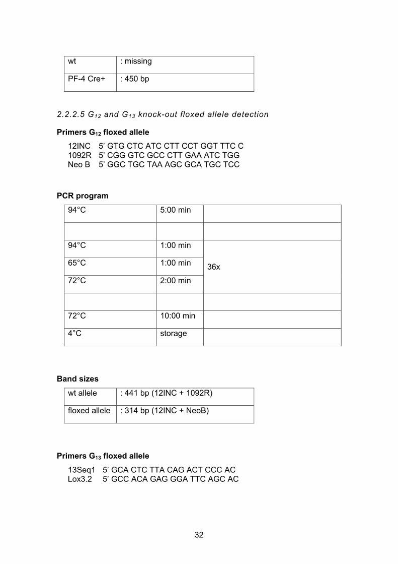
32
wt : missing
PF-4 Cre+ : 450 bp
2.2.2.5 G12 and G13 knock-out floxed allele detection
Primers G12 floxed allele 12INC 1092R Neo B
5’ GTG CTC ATC CTT CCT GGT TTC C 5’ CGG GTC GCC CTT GAA ATC TGG 5’ GGC TGC TAA AGC GCA TGC TCC
PCR program
94°C 5:00 min
94°C 1:00 min
36x 65°C 1:00 min
72°C 2:00 min
72°C 10:00 min
4°C storage
Band sizes
wt allele : 441 bp (12INC + 1092R)
floxed allele : 314 bp (12INC + NeoB)
Primers G13 floxed allele
13Seq1 Lox3.2
5’ GCA CTC TTA CAG ACT CCC AC 5’ GCC ACA GAG GGA TTC AGC AC

33
PCR program 95°C 5:00 min
95°C 0:15 min
39x 56°C 0:15 min
72°C 2:30 min
72°C 10:00 min
4°C storage
Band sizes
wt allele : 400 bp
floxed allele : 470 bp
2.2.2.6 RhoF floxed allele detection
Primers RhoF_for RhoF_rev
5’ CGC GAT CCT CGA ACA TCT AT 3’ 5’ GCC CTG GAA CTC ACT TTG TC 3’
PCR program
95°C 5:00 min
95°C 0:30 min
35x 58°C 0:30 min
72°C 0:45 min

34
72°C 5:00 min
4°C storage
Band sizes
wt allele : 199 bp
floxed allele : 242 bp
2.2.2.7 RhoA floxed allele detection
Primers
JVH11_for JVH15_rev
5’ AGC CAG CCT CTT GAC CGA TTT A 5’ TGT GGG ATA CCG TTT GAG CAT
PCR program
94°C 2:00 min
94°C 0:30 min
35x 55°C 0:30 min
72°C 0:30 min
72°C 10:00 min
4°C storage
Band sizes
wt allele : 297 bp
floxed allele : 393 bp

35
2.2.3 Histology
2.2.3.1 Organ dissection, processing and preservation
Mice were quickly and deeply anesthetized with maximum concentrations of
isoflurane under high flow and humanely euthanized by prompt cervical
dislocation. Dead mice were pinned onto a polystyrene board in supine position
for sectioning and femura were retrieved by longitudinal incision below the iliac
crest. Femura were cleaned from soft tissue and immersed in 4%
paraformaldehyde (PFA) in phosphate buffered saline (PBS) overnight. Femura
were then put into a decalcifying buffer of 10% ethylenediaminetetraacetic acid
(EDTA) in PBS for 1 week with the buffer being changed 3 times. Mice spleens
were obtained by lateral incision below the edge of the left ribcage (Kocher’s
incision). Spleens were left to incubate at 4% PFA in PBS as well. Subsequently,
all organs were embedded in paraffin and cutting was performed using a Microm
cool cut microtome (Thermo Scientific, Braunschweig, Germany), creating 3 µm
thin sections. Sections were left to dry overnight and stored in histology boxes.
2.2.3.2 Hematoxylin and eosin staining, read-out
Dried sections were immersed in Xylol for 10 min twice, before being rehydrated
in a descending alcohol dilution series of 100%, 90%, 80% and 70% for 30 sec
respectively. Sections rested in deionized water for 2 min and were subsequently
put in hematoxylin (Sigma, Schnelldorf, Germany) solution for 15 sec and left to
stain under running tab water for 7 min. Eosin was prepared using a 1:10 dilution
of Eosin G 0.5% (Roth, Karlsruhe, Germany) in ddH2O and one drop of 100%
acetic acid. Sections were counterstained in the eosin solution for 90 sec and
washed shortly in deionized water. Rehydration was carried out in an ascending
alcohol series, compare above for concentrations and time length. Two final steps
of Xylol immersion for 10 min followed and glass cover slips were mounted using
Eukitt. Stained sections were left to dry overnight and stored appropriately. Two
bone marrow/spleen sections and 20 visual fields per section were analyzed at

36
40x magnification, if not stated otherwise.
2.2.4 Measurement of spleen weight
Mice were anesthetized and put on a precision balance for measurement of total
body weight. After dissection of spleens described in 2.2.3.1, spleens were
carefully examined to exclude artificial tissue damage and loss by sectioning.
Remaining adjoining tissue was removed by tweezers and spleens were likewise
measured on a precision balance and related to respective body weight.
2.2.5 Glycoprotein inhibition
Antibodies used for GP blockade experiments were kindly adjusted to 1 mg/ml
antibody in PBS with 0.2% BSA by lab technicians.
For GPIIb/IIIa and GPV blockade, RhoA deficient mice were injected into the
retroorbital plexus with 100 µg 4H5-Fab-fragments or 100 µg 89F12 antibody
respectively on three consecutive days with femura being retrieved on day 5.
Regarding Clec-2 and GPVI blockade, 100 µg 11E9 antibody was used for Clec-
2 and 100 µg of 98A3 antibody for GPVI inhibition. Injection was performed twice
on day 1 and 3; dissection of bones followed on day 5.
2.2.6 Platelet depletion
Platelet depletion was performed by one-time injection of 100µg rat anti-mouse
GPIba into the retroorbital plexus of mice. Circulating platelet counts were
monitored by consecutive blood drawing (vol 50µl) for 10 days after treatment by
a Sysmex KX-21N automated hematology analyzer (Sysmex Corp., Kobe,
Japan). Bones were explanted 1 day or 10 days after antibody injection.
2.2.7 Determination of platelet count and size
Anesthetized mice were bled from the retroorbital plexus using microcapillaries
(vol 50µl) and blood was collected into an Eppendorf tube containing 300µl

37
heparin (20 U/ml) in Tris-buffered saline (TBS). Platelet counts and sizes were
established using a Sysmex KX-21N.
2.2.8 Platelet preparation and washing
Blood was drawn under isoflurane anesthesia by insertion of a heparinized
microcapillary into the retroorbital plexus. At least 700µl of blood was collected
into a tube containing 300µl heparin in TBS. Platelet rich plasma (prp) was
obtained by twice centrifugation at 800 rpm for 5 min with the buffy coat and
supernatant being transferred to a new tube each time. Washing of prp was
performed by centrifugation at 2,800 rpm for 5 min in the presence of apyrase
(0.02 U/ml) and prostacyclin (PGI2, 0.01 µg/ml). The platelet pellet was
resuspended in Tyrode’s buffer without Ca2+ and washed a second time under
equal conditions. After incubation of platelets at 37°C for 5 min, another washing
step concluded platelet preparation and the final platelet pellet was resuspended
in Tyrode’s buffer with Ca2+ and left to incubate at 37°C for 30 min before
fluorescence activated cell sorting (FACS) analysis.
2.2.9 Flow cytometry
Basal surface glycoprotein (GP) expression levels were determined by staining
samples whole blood with fluorophore-conjugated antibodies (see section 2.1.3.)
for 15 min at room temperature (RT). The reaction was stopped by addition of
500µl PBS. For GPIIb/IIIa and P-Selectin activation studies, the respective
agonists were added to the washed-platelet suspension and left to incubate for 6
min at 37°C and 6 min at RT, before halting the reaction by addition of 500µl PBS.
FACS measurement was performed directly after using a FACSCalibur (Becton
Dickinson, Heidelberg, Germany). For two-color stainings, the following listing
provides an overview of settings:
Detectors/Amps
Parameter Detector Voltage P1 FSC E01

38
P2 SSC 380 P3 Fl1 650 P4 Fl2 580 P5 Fl3 150
Threshold Value Parameter 253 FSC-H 52 SSC-H 52 Fl1-H 52 Fl2-H 52 Fl3-H
Compensation Fl1 2.4% of Fl2 Fl2 7.0% of Fl1 Fl2 0% of Fl3 Fl3 0% of Fl2
2.2.10 Platelet spreading assay
2.2.10.1 Platelet spreading on fibrinogen
100 µl of human fibrinogen (Sigma) diluted in PBS (100µg/ml) was pipetted onto
glass cover slips and slides were left to incubate in a damp chamber overnight at
4°C. Cover slips were rinsed with sterile PBS and blocked with PBS 1% BSA at
37°C for 2h. Washed platelets were obtained as described in 2.2.8 and aliquots
of 0,3x10⁶ plts/µl were produced. Platelets were left to incubate at 37°C for 30
min.
Glass cover slips were rinsed with sterile PBS multiple times before being
ultimately washed with Ca2+-containing Tyrode's buffer. Platelets were then
activated by addition of Thrombin (0.01 U/ml) and pipetted onto the designated
glass cover slips to allow for spreading. After the respective time points (5 min,
15 min and 30 min) were reached, spreading was halted by adding PBS 4% PFA.
Glass slides were mounted onto the cover slips and platelet spreading was
analyzed by a Zeiss Axiovert 200 inverted microscope (x100) using differential
interference contrast (DIC) microscopy. Images were captured by a CoolSNAP-
39
EZ camera (Visitron, Munich, Germany) using MetaVue online software and
readout was conducted with the ImageJ cell counter plugin.
2.2.10.2 Platelet spreading on von Willebrand Factor (vWF)
Glass cover slips were coated with 200µl of polyclonal rabbit-α-human vWF-
antibody (A0082, DAKO) diluted in 50mM carbonate-bicarbonate buffer (1:500)
and were incubated in a damp chamber at 4°C overnight. Cover slips were rinsed
with PBS and blocking was conducted with PBS 3% BSA at 37°C for 2h.
To obtain mouse plasma, wild-type mice were bled from the retroorbital plexus
and blood was collected as described in 2.2.8. Blood was centrifuged at 2800
rpm for 10 min, the supernatant transferred to a new tube and centrifuged at
14000 rpm. Incubation of slides with mouse plasma for 2h at 37°C followed.
Washed platelets were obtained as described in 2.2.8 and aliquots of 0,3x10⁶
plts/µl produced. Platelets were left to incubate at 37°C for 30 min before addition
of integrilin (60 µg/ml) to the suspension and renewed incubation for 10 min.
Glass cover slips were rinsed after incubation with sterile PBS multiple times and
as a last step Tyrode's buffer with Ca2+. Platelets were then activated by addition
of Ca2+-containing Tyrode's buffer and high-dose Botrocetin (50 U/ml) and
pipetted onto the designated glass cover slips to allow for spreading. In case of
the vWF spreading assay, time spans analyzed were 10 min, 20 min and 30 min.
The reaction was stopped and platelets were analyzed as described in 2.2.10.1.
2.2.11 Data analysis
All data were explored for normality by Shapiro-Wilk test, including homogenous
variances by Levene-test. Student’s t-test was performed when parametric
conditions were fulfilled and variance was homogenous. The Welch-test
(modified t-test) was used when unequal variances occurred. In case of GP
blockade experiments, in-group comparisons were performed using the t-test for
paired samples. Under non-parametric conditions, the Mann Whitney U-test was
applied to test for statistical significant difference between two groups and exact
significance was calculated (n<30 fulfilled). For comparison of more than two

40
groups, one-way analysis of variance (ANOVA) was used, followed by post-hoc
Tukey-test. SPSS 23 (IBM SPSS Statistics, New York, USA) was used for
statistical analysis. Results are displayed as mean ± SD and usually represent
three individual experiments, if not stated otherwise. Knock-out and littermate
wild-type mice were used for experiments, if not stated otherwise.

41
3. Results
3.1 MK localization upon deficiency of different cytoskeletal regulatory proteins
3.1.1. Findings from bone marrow and spleen sections of RhoA-/- mice
3.1.1.1 RhoA-/- mice exhibit an altered distribution of MKs compared
to the wild-type
Upon platelet activation, the small GTPase RhoA plays a pivotal role in mediating
shape change, the release of a- and dense granules and outside-in signaling
through integrin aIIbb3. RhoA has also been identified as an important actor in
the formation of focal adhesions during cell migration79. Platelet studies revealed
a macrothrombocytopenia with upregulated megakaryopoiesis in the bone
marrow (BM) in RhoA deficient mice18, but RhoA had not been studied in the
highlight of MK migration or localization in the BM.
Mice were created by crossing animals carrying a RhoA gene flanked by loxP
sites – so-called floxed (fl) mice (RhoAfl/fl)232 – with transgenic mice expressing
Cre recombinase downstream of the MK- and platelet-specific platelet factor 4
(PF4) promoter233, leading to loss of the gene product. Floxed, PF4-Cre positive
(RhoAfl/fl,PF4-Cre+/-) RhoA deficient mice are further referred to as RhoA-/- and
RhoAfl/fl,PF4-Cre-/- as wild-type (wt) mice.
Michael Popp kindly provided and genotyped RhoA-/- mice. Mice femura were
harvested, decalcified, embedded in paraffin and then cut by microtome.
Hematoxylin and eosin staining (HE staining) was performed and a microscopic
readout of the localization of MKs of 20 visual fields per bone section conducted
(see Materials and Methods chapter). MKs were assigned to the three groups
intraluminal, adjacent to vessel wall (vessel wall) and stroma.
Strikingly, RhoA-/- mice exhibited a significantly higher number of MKs inside of
BM sinusoids and adjacent to the blood vessel as compared to wt MKs (Fig. 1).

42
In contrast, MKs were found only very rarely intraluminally in wt mice. Fig. 2
depicts representative images of this so far unreported observation in RhoA-/-
mice. The divergence in the percentage of intrasinusoidal MKs in the BM could
potentially be explained by an increase in MK numbers in the BM of RhoA-/- mice,
however, as Fig. 3 shows, no statistically significant difference was found in
comparison to the wt.
Fig. 1. Alterations of MK localization in the BM of RhoA-/- mice. (A) The table depicts the mean numbers of MKs per visual field resting intraluminally, adjacent to the vessel wall (vessel wall) and in the stroma of the BM for RhoA-/- and wt mice in total numbers. (B) Presentation of MK numbers as percentages for RhoA-/- and wt. Representative experiment with n=7 (RhoA-/-) and n=8 (wt) mice. ** p < 0.01.

43
Fig. 2. Intraluminal localization of MKs in RhoA-/- mice. (A) Overview at 5x magnification: the left panel demonstrates preservation quality of the analyzed BM in both the RhoA-/- and the wt mice. Scale bar: 480 µm. (B) Close-up view at 40x magnification: the lower picture of the right panel displays representative pictures of RhoA-/- MKs within the lumen of well-preserved BM sinusoids. The upper picture provides a detailed view of comparable wt BM in which MK transmigration occurs much less frequently. Arrows depict MKs. Scale bar: 60 µm.
Fig. 3. MK counts in the BM of RhoA-/- and wt mice are similar. Total counts of MKs correspond to the experiment depicted in Fig. 1, n=7 (RhoA-/-) and n=8 (wt) mice.
This indicates a distinct and potentially defective compartmentalization of MKs in
the BM in RhoA-/- mice which suggests RhoA is needed in physiological MK

44
localization or migration and might account at least in part for the markedly
reduced platelet count observed in RhoA-/- mice18.
3.1.1.2 MK number in the spleen is unaltered in RhoA-/- mice
Another natural site of hematopoiesis and consequently megakaryopoiesis in
mice is the spleen which is why investigation of potential distinct knock-out
features in this organ was performed. Only the total number of MKs was
determined, as an assignment of MK localization to the aforementioned three
groups remained elusive by conventional microscopy owing to the fact of much
smaller sinusoids and a very densely populated red pulp in the spleen.
Fig. 4 shows that, while a trend towards increased MK numbers was observed in
RhoA-/- mice, it did not reach a statistically significant difference. Notably, the
overall spleen architecture and MK morphology was similar in RhoA-/- and
corresponding wt mice (Fig. 5).
Fig. 4. A trend towards increased MK numbers in spleens from RhoA-/- mice. The number of MKs per visual field in RhoA-/- mice is moderately increased, but not significantly different compared to the wt. Representative experiment with n=8 mice per group.

45
Fig. 5. Similar MK counts in RhoA-/- and wt spleens. (A) Left panel, overview at 5x magnification: RhoA-/- and wt mice exhibit no obvious morphological differences regarding configuration of the organ as a whole and the red pulp. Scale bar: 480 µm. (B) Right panel, close-up view at 40x magnification: observed MKs were similar in size and morphology. Arrows depict MKs. Scale bar: 60 µm.
These findings point out that MK localization in the spleen is not obviously
affected by the knock-out of RhoA. They also indicate that there is no major shift
of megakaryopoiesis from the BM to the spleen as a secondary site of
hematopoiesis, owing to the macrothrombocytopenia reported in RhoA-/- mice.
3.1.2 Increased MK counts in the BM and the spleen of RhoA/Cdc42-/- mice
Cdc42-/- single knock-out mice display mild macrothrombocytopenia which is
associated with a decreased platelet life span149. Circulating platelet numbers
and megakaryocytic replenishment are linked by a negative feedback loop to be
kept in a steady-state234. Cdc42-/- mice also exhibit higher MK numbers in the BM.
In addition, Cdc42-/- MKs derived from murine fetal liver cells showed impaired
proplatelet formation which was even more pronounced upon Rac1/Cdc42
double-deficiency, revealing redundant functions of these two GTPases in
platelet production52. To investigate whether also RhoA and Cdc42 might have

46
overlapping function in MK localization or migration, MK numbers were analyzed
in BM and spleen of RhoA/Cdc42-/- mice. Deya Cherpokova generously provided
and genotyped mice for the following experiments.
Due to insufficient preservation of BM sinusoids in this mouse strain,
compartmental read-out was not possible to conduct and thus only overall MK
counts were established. RhoA/Cdc42-/- mice exhibited significantly higher
numbers of MKs in the BM than wt mice, in line with results of the Cdc42-/- single
knock-out (Fig. 6). This may be the result of an upregulation in megakaryopoiesis
in response to the observed thrombocytopenia. The overall morphology of
RhoA/Cdc42-/- and wt MKs was similar (Fig. 7).
Fig. 6. RhoA/Cdc42-/- mice have increased MK numbers in the BM. The BM of RhoA/Cdc42-/- mice contains more MKs compared to the wt. Representative experiment with n=4 mice per group. * p < 0.05.

47
Fig. 7. BM comparison of RhoA/Cdc42-/- with wt mice. (A) The left panel displays an overview at 5x magnification of both RhoA/Cdc42-/- and wt BM. Scale bar: 480 µm. (B) The right panel with a closer view at 40x magnification of both RhoA/Cdc42-/- and wt MKs. Sinusoid preservation was not sufficient for compartmental read-out. RhoA/Cdc42-/- MKs do not show an altered morphology compared to their wt counterparts. Arrows depict MKs. Scale bar: 60 µm.
Interestingly, splenic MK numbers were strongly increased in RhoA/Cdc42-/- mice
and exceeded wt counts threefold (Fig. 8). No observable changes neither
concerning spleen architecture nor MK morphology could be observed compared
to the wt. Representative images of spleen sections are depicted in Fig. 9.
Fig. 8. RhoA/Cdc42-/- mice display drastically increased counts of MKs in the spleen. The

48
sum of MKs in RhoA/Cdc42-/- mice is greatly increased related to wt mice. Representative experiment with n=4 mice per group. * p < 0.05.
Fig. 9. Spleen architecture and MK morphology are similar in RhoA/Cdc42-/- compared to wt mice. (A) Overview of spleens at 5x magnification does not see a major difference in setup regarding, e.g. organ shape or the red pulp/ white pulp ratio between RhoA/Cdc42-/- and wt mice. Scale bar: 480µm. (B) The right panel at 40x magnification shows a similar phenotype of RhoA/Cdc42-/- and wt MKs. Arrows depict some MKs. Scale bar: 60 µm.
Having observed drastically elevated MK counts in RhoA/Cdc42-/- spleens, total
spleen weight was measured to check for splenomegaly as a sign of a shift of
megakaryopoiesis from the bone marrow to this site of secondary hematopoiesis.
Indeed, spleen weight of RhoA/Cdc42-/- mice was increased by 30% compared
to wt counterparts, compare Fig. 10, indicating a mild splenomegaly.
Although this study lacks the inclusion of Cdc42-/- mice to be compared with
RhoA-/- and RhoA/Cdc42-/- mice, together these findings support the observation
that the defect thrombopoiesis is considerably aggravated upon RhoA/Cdc42
double-deficiency compared to single-deficiency of either RhoA or Cdc42.

49
Fig. 10. RhoA/Cdc42-/- mice show increased spleen weight. RhoA/Cdc42-/- mice compared to wt mice display a significantly increased her spleen weight, indicating a shift of megakaryopoiesis to this secondary hematopoietic organ. Results are given as spleen weight/ body weight of the respective mice. Representative experiment with n=4 mice per group. ** p < 0.01.
3.1.3 Analysis of MK localization in BM of G12/13 double knock-out mice
Signaling through the G12 and more importantly G13 protein is one of the two major
axes of RhoA activation. RhoA can also be activated downstream of Gq protein-
mediated signaling, especially in the setting of high agonist doses155. In platelets,
G12 and G13 of the stimulating type Gs are activated downstream of protease
activated receptors (PARs) and the thromboxane-prostanoid (TP) receptor upon
stimulation with agonists, e.g. thrombin, thromboxane A2 (TxA2) and the TxA2
mimetic U-46619 (U-46)154,155.
A mouse strain double deficient of G12 and G13 (encoded by the genes Gna12
and Gna13) was used to investigate whether RhoA activation/signaling might
also involve these G proteins in MKs. Mice were generously provided and
genotyped by Ina Thielmann. Notably, however, the MK phenotype of RhoA-/-
mice was not observed in G12/13-/- mice resulting in unaltered MK distributions in
the BM compared to the wt (Fig. 11).

50
Fig. 11. G12/13
-/- mice and their wt counterparts show a similar MK distribution in the BM. Representative experiment with n=3 (G12/13
-/-) and n=2 (wt) per group.
Studies of the total numbers of MKs in the BM were performed as well and
showed a similar number of MKs in mice from both genotypes (Fig. 12). It has to
be noted though that the genetic background and the knock-out strategy of mice
were different in G12/13-/- mice, compared to RhoA-/- mice with creation of a Gna13
allele, Gna13ta which contains three loxP sites, along with a cassette of the
neomycin resistance (neor) and thymidine kinase gene (tk). After conversion into
a floxed allele (Gna13flox), mice were crossed with the Cre-deleter mouse strain
EIIa-Cre to allow generation of a Gna13- null allele154.
Fig. 12. Total MK numbers in the BM are similar in G12/13
-/- and wt. Representative experiment with n=3 (G12/13
-/-) and n=2 (wt) mice per group.
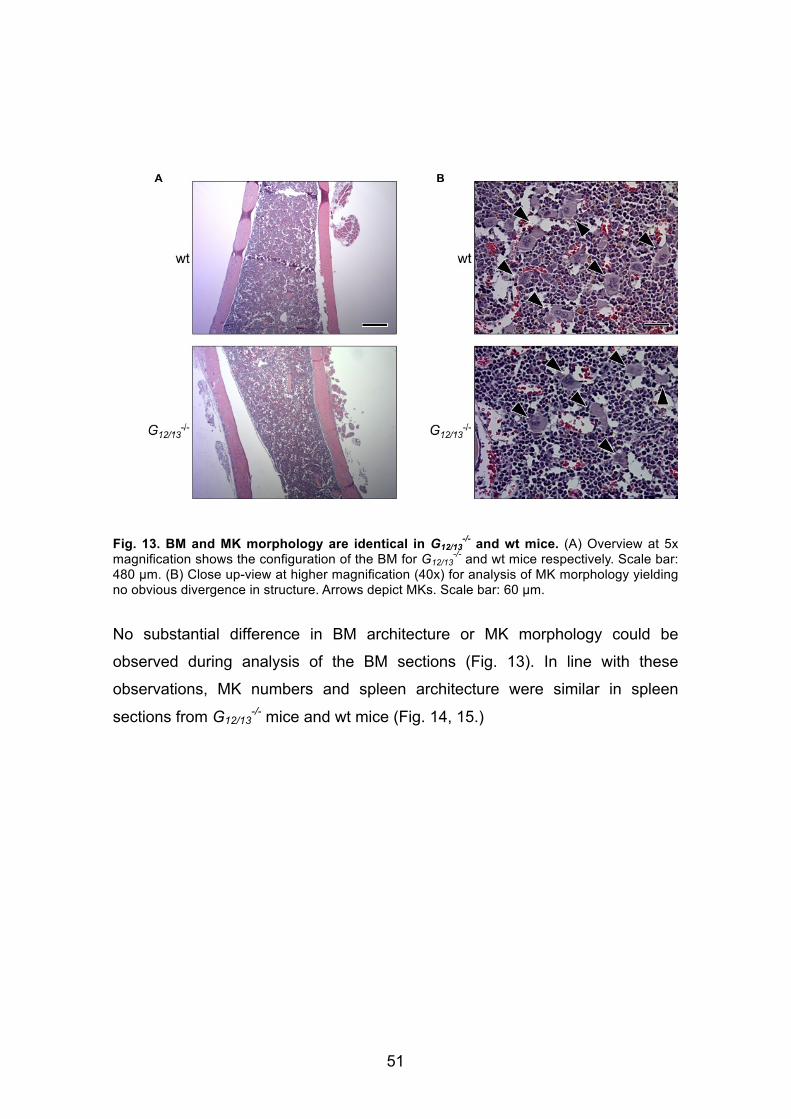
51
Fig. 13. BM and MK morphology are identical in G12/13
-/- and wt mice. (A) Overview at 5x magnification shows the configuration of the BM for G12/13
-/- and wt mice respectively. Scale bar: 480 µm. (B) Close up-view at higher magnification (40x) for analysis of MK morphology yielding no obvious divergence in structure. Arrows depict MKs. Scale bar: 60 µm.
No substantial difference in BM architecture or MK morphology could be
observed during analysis of the BM sections (Fig. 13). In line with these
observations, MK numbers and spleen architecture were similar in spleen
sections from G12/13-/- mice and wt mice (Fig. 14, 15.)

52
Fig. 14. Spleens of G12/13
-/- and wt mice display similar MK numbers. Representative experiment with n=3 mice per group.
Fig. 15. G12/13
-/- mice do not show an altered spleen morphology. (A) Upper panel at 5x magnification demonstrates comparable organ morphology of G12/13
-/- and wt mice. Scale bar: 480 µm. (B) Lower panel depicts a close-up view at 40x magnification of the spleen architecture. No MKs visible on this picture. Scale bar: 60 µm.
Together, these results show that G12/13-/- mice do not reproduce the MK
phenotype observed in the RhoA knock-out, suggesting RhoA-controlled MK

53
compartmentalization takes place independently of signaling through G12 and G13
proteins.
3.2. Investigation of signaling pathways involving RhoA in MKs
3.2.1 GPIb-mediated platelet depletion and effects on MK localization in RhoA-/- mice
3.2.1.1 Findings one day after platelet depletion in RhoA-/- mice
The finding that the RhoA-/- phenotype shows a significantly higher percentage of
intraluminal MKs entails the question which signaling events might contribute to
that phenomenon. First, investigation of megakaryopoiesis under stress was
conducted by antibody mediated platelet depletion in order to assess possible
influences. Michael Popp kindly provided, genotyped and performed antibody
injection on the mice. Depletion was achieved by injection of polyclonal rat anti-
mouse GPIb antibody binding to circulating platelets and leading to consecutive
clearance from the blood stream. Preservation and analysis of bone marrow
sections was carried out as laid out before.
Compartmentalization readout had to be omitted due to technical difficulties
which is why only total numbers of MKs were established. As Fig. 16 shows,
femura of RhoA-/- mice harvested one day after (t=1d) platelet depletion showed
no alteration in their MK counts compared to the wt. Likewise, MK morphology
was similar as compared to BM from the respective non-treated genotype (Fig.
17).

54
Fig. 16. GPIb antibody injected RhoA-/- mice show comparable MK numbers on t=1d. BM of RhoA-/- mice one day after antibody injection (t=1d) exhibit similar MK numbers compared to the wt. Single experiment with n=4 mice per group.
Fig. 17. Unaltered MK morphology in RhoA-/- mice one day after antibody. (A) Left and (B) right panels from different animals with close-up views at 40x magnification detail similar morphology of MKs of RhoA-/- and wt mice one day after GPIb antibody injection and platelet depletion. Arrows depict MKs. Scale bar: 60 µm.
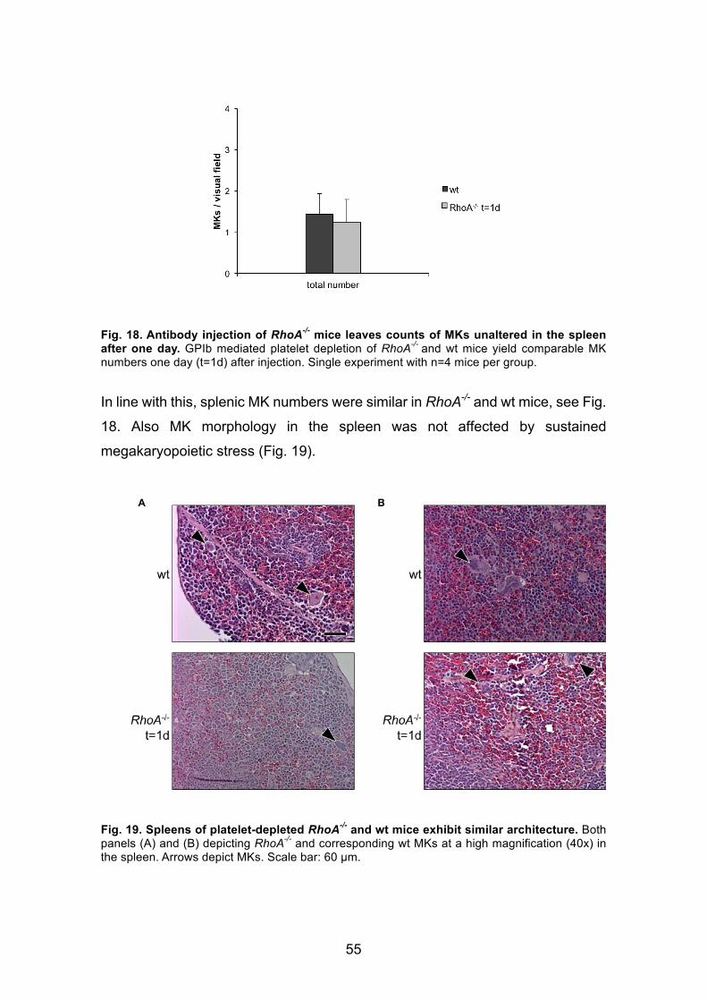
55
Fig. 18. Antibody injection of RhoA-/- mice leaves counts of MKs unaltered in the spleen after one day. GPIb mediated platelet depletion of RhoA-/- and wt mice yield comparable MK numbers one day (t=1d) after injection. Single experiment with n=4 mice per group.
In line with this, splenic MK numbers were similar in RhoA-/- and wt mice, see Fig.
18. Also MK morphology in the spleen was not affected by sustained
megakaryopoietic stress (Fig. 19).
Fig. 19. Spleens of platelet-depleted RhoA-/- and wt mice exhibit similar architecture. Both panels (A) and (B) depicting RhoA-/- and corresponding wt MKs at a high magnification (40x) in the spleen. Arrows depict MKs. Scale bar: 60 µm.

56
These findings illustrate that short-term stress on megakaryopoiesis does not
become visible in a change of MK numbers in the bone in either RhoA-/- or wt
mice.
3.2.1.2 Similar recovery of MK counts in RhoA-/- and wt mice at day
10 after platelet depletion
Analysis of BM sections from RhoA-/- mice 10 days after platelet depletion was
performed in order to investigate longer term effects on MKs. At this time point,
experiments by Michael Popp showed that the platelet numbers in both RhoA-/-
and wt mice had returned to their original levels, respectively (data not shown).
In this experiment, RhoA-/- mice exhibited results similar to the phenotype of wt
mice 10 days after platelet depletion with regard to total MK numbers in the BM
(Fig. 20). Studied sections of bone marrow did not show an altered architecture
of the bone marrow itself, nor at the cellular level regarding MKs (Fig. 21).
Fig. 20. RhoA-/- and wt mice do not show increased totals of MKs at t=10d after platelet depletion. (A) MK numbers in platelet-depleted RhoA-/- and wt mice are comparable to one another. (B) Comparison of RhoA-/- and wt mice at t=10d with untreated mice exhibits similar results. Data for control group taken from experiment depicted in Fig. 2. Single experiment with n=6 mice per group.

57
Fig. 21. Bone marrow sections of RhoA-/- and wt mice obtained at day 10 after platelet depletion show similar morphology. (A) An overview at 5x magnification does not reveal major differences between RhoA-/- (t=10d) and the wt (t=10d) regarding BM structure. Scale bar: 480µm. (B) Detailed view at 40x magnification: RhoA-/- and wt MKs exhibit similar morphologies. Arrows depict MKs. Scale bar: 60 µm.
Examination of the spleen was additionally performed in order to investigate
potential changes elsewhere in the murine hematopoietic system. Comparison of
antibody-injected RhoA-/- and wt mice revealed no divergence in MK numbers in
this organ, neither regarding organ architecture, nor MK appearance (Fig. 22, 23).
Together, these findings indicate that RhoA does not play a major role for the
recovery of megakaryopoiesis under acute stress.

58
Fig. 22. MK numbers are on an equal level 10 days after antibody injection in RhoA-/- and wt mice spleens. RhoA-/- and wt spleens exhibit similar counts of MKs in the spleen (t=10d) after GPIb antibody injection. Single experiment with n=6 mice per group.
Fig. 23. Comparable spleen morphology 10 days after platelet depletion in RhoA-/- and wt mice. (A) Left panel depicting spleen morphologies at a lower magnification (5x) of both RhoA-/- (t=10d) and wt (t=10d) mice. Scale bar: 480 µm. (B) Right panel with a detailed view (40x magnification) of MK morphology in the spleen. Arrows depict MKs. Scale bar: 60 µm.

59
3.2.2 Effect of blockade of important MK surface receptors on MK localization in RhoA-/- mice
3.2.2.1 Integrin aIIbb3 blockade does not alter MK
compartmentalization in RhoA-/- mice
Integrin aIIbb3 is instrumental in platelet activation and mediates a great variety
of effects required for physiological hemostasis such as firm adhesion to the ECM
and cross-linking of platelets to allow a growing platelet plug. Functioning as
adhesion molecules32,87, MKs might likely depend on their function during BM
egress from the stroma towards the lumen of BM sinusoids.
Therefore, investigation of the effect of in vivo blockade of integrin aIIbb3 was
conducted with special focus on the intraluminal compartment, being best
accessible and susceptible to antibody injection.
RhoA-/- and wt mice were injected intravenously (i.v.) into the retroorbital plexus
three times with 100µl of 4H5 F(ab)2 fragments (IgG2b) on consecutive days and
bones were harvested on day 5. Michael Popp kindly provided, genotyped and
injected mice according to protocol.
Analysis of integrin aIIbb3 blocked-RhoA-/- (RhoA-/- 4H5 F(ab)2) mice showed that
the RhoA knock-out phenotype was preserved and a significantly higher number
of MKs were located intraluminally and adjacent to the endothelial lining of BM
sinusoids compared to likewise treated wt (wt 4H5 F(ab)2), see Fig. 24.
Stimulating effects on megakaryopoiesis could not be observed, as total numbers
of MKs remained constant compared with non-injected RhoA-/- and wt mice (Fig.
25). Consistently, histological study of the BM of the dissected femura did not
reveal an alteration in BM architecture (Fig. 26).

60
Fig. 24. MKs of RhoA-/- 4H5 F(ab)2 mice show similar compartmentalization as compared to RhoA-/- mice. (A) MKs of RhoA-/- 4H5 F(ab)2 mice rest intraluminally and adjacent to the vessel wall more often than in the stroma related to wt 4H5 F(ab)2 mice. (B) Percentagewise depiction of MKs for better comparison. Single experiment with n=4 mice per group (RhoA-/-/wt 4H5 F(ab)2). *** p < 0.001, ** p < 0.01.
Fig. 25. Total numbers of MKs of 4H5 F(ab)2-injected RhoA-/- and wt mice are comparable to non-injected mice. (A) Depiction of RhoA-/- and wt mice receiving antibody-mediated integrin aIIbb3 blockade. Similar MK counts were found in both groups. (B) Comparison of 4H5 F(ab)2-injected mice to non-injected RhoA-/-
and wt mice. Data for RhoA-/- mice are taken from Fig. 2. n=4
mice per group (RhoA-/-/wt 4H5 F(ab)2).

61
Fig 26. BM architecture and MK morphology is similar in RhoA-/- 4H5 F(ab)2 and treated wt mice. (A) 5x magnified overview of BM sections does not reveal differences in morphology of integrin aIIbb3-attenuated RhoA-/- and wt mice. Scale bar: 480µm. (B) Close-up view at 40x magnification shows unaltered MK structure for injected RhoA-/- and wt mice. Arrows depict some MKs. Scale bar: 60 µm.
Although of major importance in platelet signaling, blockade of integrin aIIbb3 did
not lead to changes in the phenotype of RhoA-/- mice with regard to MK
localization in the BM. Thus integrin aIIbb does not seem to be required for
neither the transmigration of deficient MKs into BM sinusoids, nor the adhesion
to the vessel wall once inside.
3.2.2.2 Blockade of GPVI does not substantially alter intraluminal
localization of RhoA-/- MKs
Glycoprotein VI (GPVI) is the central platelet collagen receptor235 which might
also indicate a role for GPVI in MK interaction with elements of the extracellular
matrix. To address the effects of a GPVI blockade, RhoA-/- and littermate wt mice
were injected with 100µl of JAQ-1 IgG2a antibody on day 1 and 3. Mice were
generously provided, genotyped and injected by Michael Popp.
Although no statistical significant difference in intraluminal MKs could be
62
observed in RhoA-/- GPVI blocked (RhoA-/- JAQ-1) mice compared to likewise
treated wt JAQ-1 mice, the phenotype of an increased number of MKs inside of
BM sinusoids persisted. Taking into account the stromal and vessel-wall adjacent
distribution of MKs, it can be concluded that GPVI blockade does not influence
the ability of RhoA-/- to transmigrate into BM sinusoids (Fig. 27). An effect of GPVI
blockade on total MK numbers in the BM could not be found (Fig. 28).
Furthermore, BM examination showed no obvious alterations in MK morphology
(Fig. 29).
Fig. 27. JAQ-1-treated RhoA-/- and wt mice display similar levels of intraluminal MKs. (A) BM sections of JAQ-1-injected RhoA-/- and wt mice show a similar distribution of MKs, compared to non-injected RhoA-/- and wt mice with intraluminal and vessel-adjacent populations being increased under RhoA-/- conditions. (B) Percentagewise depiction of MKs for better comparison. Data for RhoA-/-
mice are taken from Fig.2. Single experiment with n=3 (RhoA-/- JAQ-1) and n=4 (wt JAQ-1) per group. ** p < 0.01, * p < 0.05.

63
Fig. 28. Total MK numbers in BM of RhoA-/- JAQ-1 and injected wt mice are similar. (A) RhoA-/- JAQ-1 and wt JAQ-1 mice exhibit no significant difference in MK numbers. (B) The BM of RhoA-/- JAQ-1 and comparable wt mice contains a similar number of MKs compared to non-injected mice. Data for RhoA-/-
mice are taken from Fig. 2. n=3 (RhoA-/- JAQ-1) and n=4 (wt JAQ-1) mice per group.
Fig. 29. Histological evaluation of RhoA-/- JAQ-1 and injected wt mice exhibit physiological phenotypes. (A) Overview at 5x magnification displays BM morphology for both RhoA-/- JAQ-1 and wt JAQ-1 mice. Scale bar: 480 µm. (B) Detailed view at 40x magnification showing regularly configured MKs in GPVI blocked mice. Arrows depict MKs. Scale bar: 60 µm.
These findings suggest that transmigration and adherence to the vessel wall of
MKs happens independently of GPVI and might involve signaling through

64
different receptors and ECM proteins.
3.2.2.3 Blockade of the Clec-2 receptor does not have a major
influence on MK compartmentalization in RhoA-/- mice
The C-type lectin-like receptor 2 (Clec-2) is physiologically activated by
podoplanin and has been studied with regard to antithrombotic and antimetastatic
therapy in hematology and cancer medicine236. Signaling occurs through a hemi
immunoreceptor tyrosine-based activation motif (ITAM) and is hereby similar to
GPVI-mediated signaling via the double YxxL ITAM.
Analogous to injection of a GPVI depleting antibody, treatment with the INU1
antibody results in the transient loss of the Clec-2 receptor from the platelet and
MK surface, yielding a knock-out like phenotype.
Both RhoA-/- and wt mice were injected with 100µl INU1 (IgG1) antibody on days
1 and 3 with bone dissection following on day 5. Michael Popp kindly provided,
genotyped and injected mice according to protocol.
RhoA-/- INU1 mice showed slightly non-significantly increased numbers of
intraluminal MKs, while MKs adjacent to the vessel wall were significantly
increased compared to injected wt INU1 mice, reproducing the results of the
RhoA-/- JAQ1 mice (Fig. 30). The total number of MKs in BM sections of RhoA-/-
INU1 mice differed from non-injected RhoA-/- mice, as they exhibited a higher
number of MKs (Fig. 31). Morphology of the analyzed BM and MKs were not
found to diverge in RhoA-/- INU1 and likewise treated wt mice (Fig. 32).

65
Fig. 30. RhoA-/- and wt INU1 mice exhibit similar numbers of intraluminal MKs in the BM. (A) RhoA-/- INU1 and wt INU1 controls do not differ from in intraluminal MK localization from their untreated counterparts. Vessel wall MKs were thus increased in RhoA-/- INU1 mice, whereas the stromal population was elevated in the wt INU1 mice. (B) Depiction as percentages for better comparison. Single experiment with n=5 (RhoA-/- INU1) and n=6 (wt INU1) mice per group. * p < 0.05.
Fig. 31. The total number of MKs of RhoA-/- INU1 and antibody-treated wt mice in the BM is equal, but totals of RhoA-/- INU1 mice were higher compared to non-treated mice. (A) Blockade of the Clec-2 receptor does not lead to different MK levels in RhoA-/- INU1 and wt INU1 mice. (B) Attenuation of Clec-2 leads to an increase of MK totals in the BM of RhoA-/- mice, compared to uninjected RhoA-/-
mice. Data for RhoA-/- mice are taken from Fig. 2. n=5 (RhoA-/-
INU1) and n=6 (wt INU1) mice per group. * p < 0.05.

66
Fig. 32. BM sections reveal similar morphology of RhoA-/- INU1 and injected wt mice. (A) Left panel depicts an overview at 5x magnification exhibiting equal BM structure of RhoA-/- INU1 and corresponding wt INU1 mice. Scale bar: 480µm. (B) Right panel with a higher magnification (40x) highlighting comparable MK morphology between the injected mice. Arrows depict MKs. Scale bar: 60 µm.
In summary, these results show that after blockade of the Clec-2 receptor the
compartmentalization effect of RhoA deficiency is unaltered and therefore
suggests signaling through (hem)ITAM-independent pathways. With respect to
MK numbers in the BM, blockade of Clec-2 has a slight effect in terms of an
increase of total numbers of MKs in RhoA-/- mice, although this needs to be
validated in further experiments with introduction of a non-injected control group.
3.2.2.4. Blockade of GPV in RhoA-/- mice does not affect
compartmentalization deficit of MKs in the BM
GPV is closely associated with the GPIb-IX complex through a transmembrane
domain and takes part in GPIb-mediated signaling in the presence of von
Willebrand factor (vWF), thrombin or factors XI and XII237. The GPIb-IX-V
receptor complex mediates adhesion of platelets during coagulation and
therefore might be of importance already during migration and adhesion of MKs
in the BM. In this regard, defective GPV could constitute signaling changes which

67
is why GPV antibody-injected RhoA-/- (RhoA-/- 89F12) and comparable wt (wt
89F12) mice were investigated accordingly.
Mice were kindly provided, genotyped and injected by Michael Popp. On day 1
through 3, 100µl of DOM1/89F12 (IgG2a) antibody were injected into the
retroorbital plexus and dissection of bones was performed on day 5.
Compartmentalization analysis of RhoA-/- 89F12 resulted in a significant increase
in vessel wall adjacent MKs in RhoA-/- 89F12 mice. In addition, the intraluminal
population was higher in GPV antibody-treated RhoA-/- than in equally injected wt
mice, whereas the stromal population was lower (Fig. 33). Of note, total numbers
of MKs in the BM were increased in RhoA-/- 89F12 compared to treated wt mice,
whereas MK counts were on a comparable level between injected wt and non-
injected mice (Fig. 34). Analysis of BM sections and MKs did not show altered
architecture and MK morphology in RhoA-/- 89F12 or equally injected wt mice (Fig.
35).
Fig. 33. RhoA-/- 89F12 mice and antibody-injected wt controls differ in the totals of MKs adjacent to the vessel wall. (A) The table depicts the number of MKs in the GPV blockade condition for RhoA-/- 89F12 and wt 89F12 mice respectively, according to BM compartment. Counts are significantly different in MKs adjacent to the vessel. (B) Percentagewise depiction for better comparison. Single experiment with n=3 (RhoA-/- 89F12) and n=4 (wt 89F12) mice per group. * p < 0.05.

68
Fig. 34. RhoA-/- 89F12 mice show increased MK numbers compared to equally treated wt mice. (A) Total counts of MKs were found to be higher in RhoA-/- 89F12 mice than equally injected wt 89F12 mice. (B) MK numbers were on a comparable level with untreated mice. Data for RhoA-
/- mice are taken from Fig. 2. n=3 (RhoA-/- 89F12) and n=4 (wt 89F12) mice per group. * p < 0.05.
Fig. 35. BM structure and MK morphology are not affected by injection of GPV antibody in RhoA-/- and wt mice. (A) Overview at 5x magnification displays equivalent BM configuration of both injected RhoA-/- 89F12 and corresponding wt 89F12 mice. Scale bar: 480µm. (B) Close-up view at 40x magnification depicts unaltered MK morphology between RhoA-/- 89F12 and wt 89F12 mice. Arrows depict MKs. Scale bar: 60 µm.
Together RhoA-/- 89F12 mice show a similar phenotype as observed in non-

69
treated RhoA-/- mice, but GPV blockade in RhoA-/- mice might lead to a moderate
increase in megakaryopoiesis. This finding needs to be validated in subsequent
studies including a control group.
3.3 Characterization of the role of RhoF deficient in platelets using conditional knock-out mice
The small GTPase RhoF has been a more recent addition to the Rho family of
small GTPases and it is presumed to play a role in filopodia formation156. There
is evidence that RhoF-generated filopodia are longer and thinner than filopodia
induced by Cdc42, raising the questions if different subtypes of filopodia exist70,123.
Aim of this study was to characterize the role of RhoF in platelets to complement
recently published results161 and to study RhoF-/- platelets extensively in their
behavior regarding filopodia formation. For this purpose, mice containing loxP
sites introduced into the RhoF gene (RhoFfl/fl) were successfully generated and
crossed with PF-4 Cre transgenic mice, creating RhoFfl/fl,PF-4Cre+/- (further referred
to as RhoF-/-) mice, thereby eliminating RhoF expression in MKs and platelets.
Mice were kindly provided and genotyped by Sebastian Dütting and Sylvia
Hengst.
3.3.1 Analysis of platelet activation in RhoF-/- mice by flow cytometry
3.3.1.1 Platelet count and size and glycoprotein expression in RhoF-/-
and wt mice
Flow cytometric and Sysmex® analysis of RhoF-/- platelets showed ko and wt
platelets were similar in count and size (data not shown).
These findings suggest that megakaryopoiesis is not affected in RhoF-/-
deficiency. This was later supported by investigation of the platelet life span in
vivo which was found to be unaltered in RhoF-/ mice compared to the wt161.
Differences in glycoprotein surface expression levels have to be considered in
diverging signaling responses upon agonist stimulation in platelets. To address
this issue, GP expression in RhoF-/- platelets was studied. Components of the

70
GPIb-IX-V complex were similar in quantitative expression in RhoF-/- platelets
compared to the wt. Glycoprotein CD9 also exhibited same expression levels in
both groups. Integrins aIIbb3, a2 and b1 were neither found to differ in the knock-
out nor the wt, constituting regular integrin expression in RhoF-/- mice. The
collagen receptor GPVI and Clec-2 receptor were equally expressed in RhoF-/-
and wt mice (Fig. 36). In summary, RhoF deficiency was not associated with
altered platelet surface receptor expression levels.
Fig 36. Expression of important platelet surface receptors as determined by flow cytometry. No alterations could be found between RhoF-/- and wt mice for all glycoproteins. Results depicted as means ± SD. Representative experiment with n=4 mice per group.
3.3.1.2 Unaltered integrin aIIbb3 activation in RhoF-/- platelets
Measurement of platelet activation in RhoF-/- mice was conducted through
evaluation of levels of active integrin aIIbb3 levels upon agonist stimulation, with
integrin aIIbb3 constituting the most abundant platelet integrin238,239.
Platelet activation assays showed no major difference in RhoF-/- platelets
compared to the wt in integrin aIIbb3 activation. Upon stimulation with the
agonists ADP and the TxA2 mimetic, U-46619 (U-46) RhoF-/- and wt platelets
showed only moderate signs of activation, visible in a slight increase in mean
fluorescence intensity (MFI). ADP and U-46 combined function as a strong
activator of platelets, but no differences in the response of RhoF-/- and wt platelets

71
were observed. Thrombin signaling, irrespective of the used concentration, was
likewise found to be unaltered in both RhoF-/- compared to wt platelets. Taken
together, activation of RhoF-/- platelets through G13 and Gq receptors seems
unaltered. Epinephrine as an agonist of the Gz receptor did not induce different
responses in RhoF-/- and wt platelets either. ITAM and (hem)ITAM mediated
pathways of platelet activation represented by signaling via the agonists collagen-
related peptide (CRP), convulxin (CVX) and rhodocytin (RC) overall elicited a
higher response in RhoF-/- than wt platelets for almost all tested concentrations
(Fig. 37), but this finding could not reproduced in following experiments,
suggesting technical problems in the activation setting with the wt. Therefore, we
conclude that these results indicate that RhoF deficiency has no impact on
platelet activation, at least under in vitro conditions.
Fig. 37. Integrin aIIbb3 activation assay shows comparable results in RhoF-/- and wt mice. Signaling through major platelet receptors is similar and also unaffected by concentration in RhoF-
/- platelets. Binding of JON/A-PE antibody constitutes mean fluorescence intensity (MFI). Results presented as means ± SD. n=4 mice per group.
3.3.1.3 a granule release is not impaired in RhoF-/- platelets
Another component of great significance in platelet activation is the ability to
release stored granules, thereby sustaining platelet activation through autocrine
signaling87. Investigation of a granule release can be studied by assessment of

72
P-Selectin levels, as it can be found on platelet a granules18. Granule release is
mediated by fusion of platelet granules with the platelet surface membrane, thus
making P-Selectin accessible for antibody binding and subsequent analysis by
flow cytometry.
P-Selectin surface exposure as a marker of degranulation effectiveness upon
stimulation was shown to be equal in RhoF-/- and wt platelets, regarding ADP, U-
46 or combined stimulation. Thrombin stimulation yielded comparable results for
knock-out and wt mice at all studied concentrations and precludes major defects
in G13 and Gq signaling. Stimulation with the agonists CRP, CVX and RC through
receptors GPVI and Clec-2 exhibited similar results in both groups. Finally, also
Gz signaling assessed by the weak agonist epinephrine completed analysis of
degranulation potency and was unaltered in both RhoF-/- and wt platelets (Fig.
38).
Fig. 38. Granule release is fully functional in RhoF-/- mice. P-Selectin exposure as a marker for platelet degranulation is equal in knock-out and wt mice, precluding signaling defects in major platelet receptors and is not affected by concentrations of the studied agonists either. Binding of anti P-Selectin-FITC antibody constitutes mean fluorescence intensity (MFI). Results given as means ± SD. n=4 mice per group.
The findings of sections 3.3.1.2 and 3.3.1.3 suggest that RhoF is dispensable in
platelet activation and granule release in response to agonist stimulation,
reproducing results of a similar study161.

73
3.3.2 Platelet spreading of RhoF-/- mice evaluated by two different agonists
3.3.2.1 RhoF-/- platelets spread normally on fibrinogen
The extracellular matrix component and agonist surface fibrinogen is commonly
used to assess platelet spreading. Fibrinogen is recognized by integrin aIIbb3
and induces outside-in signaling leading to cytoskeletal rearrangements
necessary for platelet spreading240,241. RhoF has been proposed to be essential
for filopodia formation in various cell lines.
To investigate a potential involvement of RhoF in platelet spreading, RhoF-/- and
wt platelets were allowed to adhere to and spread on fibrinogen-treated glass
cover slips and studied at three different time points upon thrombin stimulation.
RhoF-/- platelets were found to spread similarly in comparison to their wt
counterparts at all studied time points. Most interestingly, filopodia formation was
not impaired in RhoF-/- platelets (Fig. 39).

74
Fig. 39. Spreading of platelets on fibrinogen is unaffected by knockout of RhoF. (A) RhoF-
/- and wt platelets spread similarly on fibrinogen at 5 min, 15 min and 30 minutes. Upon activation, platelets start forming filopodia, later lamellipodia, before they become fully spread. Representative images of wt and RhoF-/- platelets. Scale bar: 5 µm. (B) Results are displayed as stacked bars representing the mean percentage of platelets in each spreading phase. Notably, filopodia formation is not impaired in RhoF-/- platelets. Phase1: Unspread platelets, phase 2: filopodia formation, phase 3: lamellipodia formation, phase 4: fully spread platelets. Representative experiment with n=3 mice per group.
3.3.2.2 Adhesion of RhoF-/- platelets on immobilized vWF is similar to
the wt
vWF binds to the GPIb-IX-V complex and this interaction helps platelets adhere
at sites of vessel injuries, especially under high-shear conditions239. Platelet

75
spreading on immobilized vWF is attenuated with platelets only reaching the
filopodial stage of spreading242. By use of this method subtle changes in
spreading ability, especially regarding filopodia formation can be assessed.
RhoF-/- platelets were allowed to adhere to vWF immobilized on glass cover slips
upon incubation with botrocetin to induce GPIb-IX-V signaling under static
conditions. Integrilin which inhibits integrin aIIbb3 was added additionally to
prevent integrin activation. Three different time points were studied. In contrast
to fibrinogen-mediated spreading, only three spreading stages were defined.
Phase 1 comprised quiescent discoid platelets, phase 2 platelets forming 1-3
filopodia and phase 3 platelets with more than 3 filopodia. RhoF-/- platelets
demonstrated unaltered filopodia formation compared to the wt at all studied time
points. Thus, RhoF loss can be compensated effectively during platelet spreading
(Fig. 40).

76
Fig. 40. Adhesion and filopodia formation of platelets on immobilized vWF is unaffected by knockout of RhoF. (A) RhoF-/- and wt platelets spread similarly on vWF at 10 min, 20 min and 30 minutes. In vWF-mediated adhesion, only 3 phases of spreading are discriminated. Representative images of wt and RhoF-/- platelets. Scale bar: 5 µm. (B) Results are displayed as stacked bars representing the mean percentage of platelets in each spreading phase. No significant difference in phase abundancy in RhoF-/- and wt platelets can be detected. Representative experiment with n=3 mice per group.
In summary, platelet spreading of RhoF-/- platelets on fibrinogen was not impaired
and filopodia formation upon adhesion on VWF proceeded with no apparent
defect. As mentioned earlier, Cdc42 has been described as another member of
the Rho family significantly contributing to filopodia formation72. However, studies
from our group performed after the completion of this thesis showed that filopodia

77
formation was unaffected in RhoF/Cdc42 double-deficient platelets (unpublished
results). Thus, other protein(s) seem to be required to facilitate filopodia formation
in platelets.
4. Discussion
4.1 RhoA is a crucial regulator of MK localization and platelet biogenesis
The small GTPase RhoA plays an important role in actin cytoskeleton-driven
remodeling processes in platelets including shape change, spreading and clot
retraction18,79,87. This study focused on the role of RhoA in megakaryopoiesis and
MK localization. Strikingly, a significant proportion of RhoA-/- MKs was found
inside BM sinusoids, ascribing a crucial role to RhoA in the transendothelial
migration process. It is known that MKs extend proplatelets through gaps in the
endothelial lining of BM sinusoids which accounts for the biggest proportion of
produced platelets3. Additionally, whole MKs themselves can migrate into the
intraluminal compartment through endothelial cells4, but the cellular machinery
and the physiological conditions under which this might preferentially occur have
not been clarified. Fostering this observation, MKs can be found in the capillary
bed of the lung which has been shown to be the result of migration, not of edaphic
development243,244,245,246.
Interestingly, proplatelet formation was shown to be regulated by RhoA through
nonmuscular myosin II (NMII) activity. Myosin II consists of 2 heavy and 4 light
chains and is the gene product of the MYH9 gene. Inhibition of MLC
phosphorylation or absence of MYH9 increases proplatelet formation from
MKs19,247. RhoA typically enhances MLC phosphorylation through ROCK which
diminishes proplatelet formation in vitro51, indicating that RhoA is a negative
regulator of proplatelet formation. Therefore, the observed
macrothrombocytopenia seems to be linked to the localization defect, rather than
to deficiencies in proplatelet formation.

78
Recently, an additional model of platelet production through MK rupture was
proposed under conditions of stress, such as acute thrombocytopenia or
inflammation248. This research stems from the observation that MK maturation
and platelet production can occur independently from TPO249. Moreover, in
settings of acute hematopoietic/ megakaryocytic stress, e.g. irradiation or acute
thrombocytopenia, HSCs were able to give rise to MKs and platelets directly
without intermediaries248,250,251,252. The finding presented in this thesis that RhoA
is crucial for MK localization at steady state indicated that it might also be an
important regulator of megakaryopoiesis under stress conditions. However, the
number of MKs at both early (1 day) and late (10 days) after experimentally
induced thrombocytopenia was similar in BM of RhoA-/- and wt mice, not fostering
the role of RhoA in this setting. At the same time, studies of time points in between
day 1 and 10 seem warranted, as the platelet trough is most pronounced through
the first few days and changes in MK numbers might occur only then, also with
regard to the rupture model.
It is also relevant to discuss limitations of this part of the study. Conventional
histological analysis of the compartmentalization of MKs in the BM using
hematoxylin eosin stained sections can make the identification of knock-out MKs
difficult, as they may be morphologically altered as for example Cdc42-/- MKs
exhibit reduced invaginations and demarcation from the surrounding BM stromal
cells 149. Or they might undergo transmigration through the endothelial lining
which could result in aberrant morphology as well. This might explain why in this
work no increase of total MK numbers in the BM of RhoA-/- mice was observed,
in contrast to published results18. RhoA/Cdc42-/- MKs exhibited higher numbers
of MKs in the BM (see section 3.1.3), similar to findings from the Cdc42 single
knock-out149. Additionally, MK numbers in the spleen were increased threefold,
likely indicating severe stress and/ or inefficient megakaryopoiesis. Gross
phenotypical assessment of BM structure and MKs themselves yielded
comparable findings in RhoA/Cdc42-/- and wt MKs, but potentially left many
structurally altered MKs unidentified. Due to this fact, MK counts in the
RhoA/Cdc42-/- mice might be underreported by methods used in this work.
Indeed, after the completion of this thesis, analysis of immunofluorescently

79
stained cryo sections of whole femura enabled Sebastian Dütting and other
members of the group253 to show that RhoA/Cdc42-/- MKs were almost completely
clustered around BM sinusoids, while being unable to transmigrate into the vessel
lumen. Dütting et al.253 could demonstrate a regulatory circuit where RhoA
functions as a stop-signal regarding MK transmigration while GPIb/Cdc42
signaling forms a go-signal. The observed macrothrombocytopenia is thus in part
a direct result of the altered MK distribution and subsequently defective platelet
biogenesis in vivo.
In this thesis, several potential signaling pathways regulating RhoA in MKs were
investigated with the G-proteins G12 and G13 being obvious candidates, as they
have been shown to be situated upstream of RhoA signaling in platelets154. While
G12/13-/-
mice do not exhibit macrothrombocytopenia, redundant functions of the
proteins in MK localization cannot not be excluded. However, the MK distribution
was similar in G12/13-/-
mice compared to the wt, making a major contribution to
RhoA signaling in MKs via these G proteins unlikely.
Integrin aIIbb3 is the most abundant platelet surface receptor, essential in platelet
outside-in signaling, mediating stable adhesion and being crucial for platelet
aggregation via binding of vWF and fibrinogen. Surprisingly, integrin aIIbb3 does
not seem to be involved in the regulation of MK localization in the BM since its
blockade in RhoA-/- mice could not revert the MK mislocalization observed in non-
injected RhoA-/- mice. Whether other integrins, such as integrin b1 are involved
in the regulation of MK localization, remains elusive.
The platelet collagen receptor GPVI is a type I transmembrane protein of the Ig
superfamily and transduces signals through its transmembrane region which
interacts with the Fc receptor (FcR) g-chain235. Inhibitory signaling via GPVI was
recently proposed to be involved the spatial regulation of proplatelet formation in
vivo254. However, transiently GPVI-deficient RhoA-/- mice (treated by JAQ1
antibody) exhibited a similar MK mislocalization in the BM as observed in non-
injected RhoA-/- mice.
Similar to findings from JAQ1 treated RhoA-/- mice, the MK mislocalization in
RhoA-/- mice could not be reverted by transient knock-out of Clec-2 by injection

80
of the INU1 antibody. Notably, INU1-treated RhoA-/- mice exhibited a statistically
significant higher number of BM MKs than non-injected RhoA-/- mice. This effect
could be due to the transient thrombocytopenia upon INU1 antibody injection182
and the subsequent stimulation of megakaryopoiesis.
Taken together, the selective inhibition of (hem)-ITAM signaling pathways does
not influence RhoA signaling in terms of MK migration and compartmentalization
in the BM. While these findings are consistent with normal platelet counts and
thus most probably normal thrombopoiesis in GPVI- or Clec-2 knock-out mice,
they do not exclude a redundant function of the pathways in the process of
platelet production.
Finally, also antibody (89F12)-mediated GPV-blockade in RhoA-/- mice was not
able to revert the MK mislocalization observed in untreated mice. Thus, despite
association of GPV with GPIb, GPV does not seem to be directly involved in the
regulation of MK localization in the BM. This stands in contrast to in vivo blockade
of GPIba by treatment of mice with p0p/B Fab fragments, which after the
completion of this thesis was shown to revert intrasinusoidal localization of RhoA-
/- MKs. Nonetheless, the results presented in this part of the thesis were
confirmed later by using immunofluorescently stained cryo sections253,
suggesting downstream signaling of the GPIb subunit of the GPIb-IX-V complex
occurs irrespective of the functional state of GPV.
Limitations of all the conducted antibody blockade experiments include that i.v.
injection regimen guarantees high availability of the antibody in the circulation
and for intraluminal MKs, but tissue penetration capabilities are not easy to
determine and may depend on the respective antibody.
4.2 RhoF is redundant in filopodia formation
A second part of this thesis focused on the investigation of RhoF, as Cdc42-/-
mice were macrothrombocytopenic, while demonstrating regular filopodia
formation149. With RhoF being hypothesized to be an important (independent)
driver of filopodia formation101, also associated changes of megakaryopoiesis
were imaginable. In contrast to this speculation, platelet indices and platelet size

81
were comparable in RhoF-/- and wt mice indicating that there is no major defect
in megakaryopoiesis upon RhoF deletion. Additionally, activation and
degranulation assays showed no overt phenotype for RhoF-deficient mice. Lastly,
upon adherence to ECM proteins, i.e. vWF and fibrinogen RhoF-deficient
platelets adhered and spread similar as compared to the wt. Together with the
published work by Goggs et al.161, these findings indicate a redundant role for
RhoF in platelet biogenesis and function.
4.3 Concluding remarks and outlook
This work was targeted at studying the effects of various small GTPase single
and double knock-outs on megakaryopoiesis, MK migration and subsequent
compartmentalization in the BM. The observation that RhoA deficiency alters MK
localization might be of relevance for other cell types, too which depend on
locomotion in response to stimuli to fulfill their functions, e.g. immune cells.
As Dütting et al.253 showed, the regulation of MK localization and transendothelial
platelet biogenesis involves a Cdc42/RhoA regulatory circuit downstream of GPIb.
Following up on this work, further detailed studies of potential up- and
downstream regulators of the two GTPases in MKs will be required to decipher
the signaling mechanism regulating platelet biogenesis in vivo.
In the future, a three dimensional (3D) matrix in vitro model of MK maturation and
migration in the BM niche could be helpful, too, next to in vivo experiments, to
monitor the localization/migration of MKs in response to various stimuli and help
clarify which further small GTPases are involved in the process and what their
spatiotemporal regulation might be. Notably, a recent study from David Stegner
and colleagues using 3D in vivo imaging revealed that a distinction between an
osteal and vascular niche is not conceivable in vivo. Their results show that MK
progenitors and mature MKs are always in close contact with the BM sinusoids 34 leading to the revised model that thrombopoiesis is spatially regulated by the
BM vasculature.
Regarding studies on RhoF in platelets and MKs, it could prove useful to create

82
a double knock-out of Cdc42 and RhoF in mice to investigate potential redundant
functions of the two GTPases in thrombopoiesis and filopodia formation.

83
5. Summary
Platelets constitute the cellular component in hemostasis and play a crucial role
in the physiological response to injuries the vessel wall to limit potential blood
loss, but at the same time in pathological processes like plaque rupture due to
atherosclerosis, where their activation and aggregation facilitates arterial
thrombosis leading to ischemic stroke or myocardial infarction.
This work focuses on megakaryocyte physiology with a special interest in the
description of the localization of MKs in the bone marrow in mice single-deficient
of the small Rho GTPase RhoA or double-deficient for RhoA and Cdc42 – another
important Rho GTPase in transgenic mice. The importance of Rho GTPases in
platelet and megakaryocyte physiology has already been extensively
investigated with RhoA being responsible for creation of focal adhesions and
actomyosin contractions in platelets, whereas Cdc42 has been shown to be
important for microtubule rearrangements in megakaryocytes in conjunction with
Rac1. RhoA ko mice were generated and studied with regard to
compartmentalization of megakaryocytes in the bone marrow, revealing the
intraluminal presence of megakaryocytes in bone marrow sinusoids. In a next
step, aggravation, confirmation or abolishment of this finding was studied in
related mouse strains, namely a RhoA/Cdc42 and G12/G13 (upstream regulators
of RhoA activity) dko. Finally, RhoA ko mice treated with antibodies that block
different specific surface receptors were studied in regard to MK
compartmentalization.
In the second and smaller part of this thesis the role of RhoF, a Rho GTPase
which has been postulated to be of importance in filopodia formation in addition
to Cdc42, in platelet function was investigated by analyzing a RhoF ko mouse
strain. Receptor expression, platelet activation, granule release and filopodia
formation in response to various stimuli in RhoF-deficient platelets was studied,
showing no significant difference compared to the wild-type.

84
Zusammenfassung
Blutplättchen stellen die zelluläre Komponente der Blutgerinnung und spielen
eine entscheidende Rolle in der physiologischen Antwort auf Verletzungen der
Gefäßwand, um etwaigen Blutverlust zu vermindern, aber gleichzeitig auch in
pathologischen Prozessen wie durch Atherosklerose vermittelter Plaqueruptur
bei denen ihre Aktivierung und Aggregation zu ischämischem Schlaganfall oder
Myokardinfarkt führen kann.
Diese Arbeit beschäftigt sich mit Megakaryozyten, den Vorläuferzellen der
Thrombozyten, mit besonderem Fokus auf der Beschreibung ihrer Verteilung im
Knochenmark in Abhängigkeit von der Defizienz der kleinen Rho GTPase RhoA
und der kombinierten Defizienz von RhoA und Cdc42 – einer anderen
bedeutenden Rho GTPase in transgenen Mauslinien. Die Bedeutung der Rho
GTPasen in der Megakaryopoiese und Thrombozytenphysiologie wurde bereits
ausgiebig untersucht. So ist RhoA verantwortlich für die Schaffung fokaler
Adhäsionen und die Kontraktilität des Aktin-Myosinapparates, wohingegen für
Cdc42 zusammen mit Rac1 eine Bedeutung im Mikrotubuli Re-Arrangement in
Megakaryozyten gezeigt werden konnte. RhoA defiziente Mäuse wurden
generiert und die megakaryozytäre Kompartimentalisierung innerhalb des
Knochenmarkssinusoide analysiert mit dem erfolgreichen Nachweis intraluminal
gelegener Megakaryozyten. In einem nächsten Schritt wurden eine RhoA/Cdc42
doppeldefiziente und eine Mauslinie mit Doppeldefizienz in G12/G13 (in der
Signalkaskade oberhalb gelegene Regulatoren der RhoA Aktivität) auf eine
Verstärkung, Bestätigung oder einen Verlust intraluminal gelegener
Megakaryozyten untersucht. Letztlich wurden RhoA defiziente Mäuse mit
verschiedenen, bestimmte Oberflächenrezeptorproteine blockierenden
Antikörpern behandelt und der Einfluss auf die megakaryozytäre
Kompartimentalisierung untersucht.
Ein zweiter, kleinerer Teil dieser Arbeit beschäftigte sich mit der Funktion von
RhoF, einer kleinen Rho GTPase, von der angenommen wird neben Cdc42 für
die Filopodienausbildung verantwortlich zu sein. Hierzu wurde eine RhoF-
defiziente Mauslinie analysiert. Die Oberflächenrezeptorexpression, Aktivierung,
Granulafreisetzung und Filopodienausbildung RhoF defizienter Blutplättchen in

85
Abhängigkeit verschiedener Stimuli wurde entsprechend untersucht, allerdings
ohne Nachweis signifikanter Unterschiede zum Wildtyp.
6. References
1. Nakeff A, Maat B. Separation of megakaryocytes from mouse bone marrow by velocity sedimentation. Blood 1974;43:591-595.
2. Hartwig J, Italiano J, Jr. The birth of the platelet. J Thromb Haemost
2003;1:1580-1586. 3. Machlus KR, Italiano JE, Jr. The incredible journey: From megakaryocyte
development to platelet formation. The Journal of cell biology 2013;201:785-796.
4. Bluteau D, et al. Regulation of megakaryocyte maturation and platelet
formation. J Thromb Haemost 2009;7 Suppl 1:227-234. 5. Hirata S, et al. Congenital amegakaryocytic thrombocytopenia iPS cells
exhibit defective MPL-mediated signaling. J Clin Invest 2013;123:3802-3814.
6. Dore LC, Crispino JD. Transcription factor networks in erythroid cell and
megakaryocyte development. Blood 2011;118:231-239. 7. Eto K, Kunishima S. Linkage between the mechanisms of
thrombocytopenia and thrombopoiesis. Blood 2016;127:1234-1241. 8. Kuvardina ON, et al. RUNX1 represses the erythroid gene expression
program during megakaryocytic differentiation. Blood 2015;125:3570-3579.
9. Nakamura S, et al. Expandable megakaryocyte cell lines enable clinically
applicable generation of platelets from human induced pluripotent stem cells. Cell Stem Cell 2014;14:535-548.
10. Zimmet J, Ravid K. Polyploidy: occurrence in nature, mechanisms, and
significance for the megakaryocyte-platelet system. Exp Hematol 2000;28:3-16.
11. Geddis AE, Fox NE, Tkachenko E, Kaushansky K. Endomitotic
megakaryocytes that form a bipolar spindle exhibit cleavage furrow ingression followed by furrow regression. Cell Cycle 2007;6:455-460.
12. Lordier L, et al. Megakaryocyte endomitosis is a failure of late cytokinesis

86
related to defects in the contractile ring and Rho/Rock signaling. Blood 2008;112:3164-3174.
13. Roy A, et al. Activity of nonmuscle myosin II isoforms determines
localization at the cleavage furrow of megakaryocytes. Blood 2016;128:3137-3145.
14. Lordier L, et al. RUNX1-induced silencing of non-muscle myosin heavy
chain IIB contributes to megakaryocyte polyploidization. Nat Commun 2012;3:717.
15. Pleines I, Nieswandt B. RhoA/ROCK guides NMII on the way to MK
polyploidy. Blood 2016;128:3025-3026. 16. Melendez J, et al. RhoA GTPase is dispensable for actomyosin regulation
but is essential for mitosis in primary mouse embryonic fibroblasts. The Journal of biological chemistry 2011;286:15132-15137.
17. Gao Y, et al. Role of RhoA-specific guanine exchange factors in regulation
of endomitosis in megakaryocytes. Dev Cell 2012;22:573-584. 18. Pleines I, et al. Megakaryocyte-specific RhoA deficiency causes
macrothrombocytopenia and defective platelet activation in hemostasis and thrombosis. Blood 2012;119:1054-1063.
19. Chen Z, et al. The May-Hegglin anomaly gene MYH9 is a negative
regulator of platelet biogenesis modulated by the Rho-ROCK pathway. Blood 2007;110:171-179.
20. Leon C, et al. Megakaryocyte-restricted MYH9 inactivation dramatically
affects hemostasis while preserving platelet aggregation and secretion. Blood 2007;110:3183-3191.
21. Kiel MJ, Yilmaz OH, Iwashita T, Yilmaz OH, Terhorst C, Morrison SJ. SLAM
family receptors distinguish hematopoietic stem and progenitor cells and reveal endothelial niches for stem cells. Cell 2005;121:1109-1121.
22. Mendez-Ferrer S, et al. Mesenchymal and haematopoietic stem cells form
a unique bone marrow niche. Nature 2010;466:829-834. 23. Calvi LM, et al. Osteoblastic cells regulate the haematopoietic stem cell
niche. Nature 2003;425:841-846. 24. Greenbaum A, et al. CXCL12 in early mesenchymal progenitors is
required for haematopoietic stem-cell maintenance. Nature 2013;495:227-230.
25. Kunisaki Y, et al. Arteriolar niches maintain haematopoietic stem cell

87
quiescence. Nature 2013;502:637-643. 26. Pallotta I, Lovett M, Rice W, Kaplan DL, Balduini A. Bone marrow
osteoblastic niche: a new model to study physiological regulation of megakaryopoiesis. PLoS One 2009;4:e8359.
27. Malara A, Abbonante V, Di Buduo CA, Tozzi L, Currao M, Balduini A. The
secret life of a megakaryocyte: emerging roles in bone marrow homeostasis control. Cellular and molecular life sciences : CMLS 2015;72:1517-1536.
28. Niswander LM, Fegan KH, Kingsley PD, McGrath KE, Palis J. SDF-1
dynamically mediates megakaryocyte niche occupancy and thrombopoiesis at steady state and following radiation injury. Blood 2014;124:277-286.
29. Avecilla ST, et al. Chemokine-mediated interaction of hematopoietic
progenitors with the bone marrow vascular niche is required for thrombopoiesis. Nat Med 2004;10:64-71.
30. Riviere C, et al. Phenotypic and functional evidence for the expression of
CXCR4 receptor during megakaryocytopoiesis. Blood 1999;93:1511-1523. 31. Berthebaud M, et al. RGS16 is a negative regulator of SDF-1-CXCR4
signaling in megakaryocytes. Blood 2005;106:2962-2968. 32. Nurden A, Nurden P. Advances in our understanding of the molecular basis
of disorders of platelet function. J Thromb Haemost 2011;9 Suppl 1:76-91. 33. Sabri S, et al. Deficiency in the Wiskott-Aldrich protein induces premature
proplatelet formation and platelet production in the bone marrow compartment. Blood 2006;108:134-140.
34. Stegner D, et al. Thrombopoiesis is spatially regulated by the bone marrow
vasculature. Nat Commun 2017;8:127. 35. Yamada E. The fine structure of the megakaryocyte in the mouse spleen.
Acta Anat (Basel) 1957;29:267-290. 36. Radley JM, Haller CJ. The demarcation membrane system of the
megakaryocyte: a misnomer? Blood 1982;60:213-219. 37. Schulze H, et al. Characterization of the megakaryocyte demarcation
membrane system and its role in thrombopoiesis. Blood 2006;107:3868-3875.
38. Haddad E, et al. The thrombocytopenia of Wiskott Aldrich syndrome is not
related to a defect in proplatelet formation. Blood 1999;94:509-518.

88
39. Chen Y, et al. Loss of the F-BAR protein CIP4 reduces platelet production
by impairing membrane-cytoskeleton remodeling. Blood 2013;122:1695-1706.
40. Eckly A, et al. Biogenesis of the demarcation membrane system (DMS) in
megakaryocytes. Blood 2014;123:921-930. 41. Italiano JE, Jr., Patel-Hett S, Hartwig JH. Mechanics of proplatelet
elaboration. J Thromb Haemost 2007;5 Suppl 1:18-23. 42. Choi ES, Nichol JL, Hokom MM, Hornkohl AC, Hunt P. Platelets generated
in vitro from proplatelet-displaying human megakaryocytes are functional. Blood 1995;85:402-413.
43. Italiano JE, Jr., Lecine P, Shivdasani RA, Hartwig JH. Blood platelets are
assembled principally at the ends of proplatelet processes produced by differentiated megakaryocytes. The Journal of cell biology 1999;147:1299-1312.
44. Tablin F, Castro M, Leven RM. Blood platelet formation in vitro. The role of
the cytoskeleton in megakaryocyte fragmentation. J Cell Sci 1990;97 ( Pt 1):59-70.
45. Machlus KR, Thon JN, Italiano JE, Jr. Interpreting the developmental
dance of the megakaryocyte: a review of the cellular and molecular processes mediating platelet formation. British journal of haematology 2014;165:227-236.
46. Patel SR, et al. Differential roles of microtubule assembly and sliding in
proplatelet formation by megakaryocytes. Blood 2005;106:4076-4085. 47. Patel-Hett S, et al. Visualization of microtubule growth in living platelets
reveals a dynamic marginal band with multiple microtubules. Blood 2008;111:4605-4616.
48. Kaushansky K. Determinants of platelet number and regulation of
thrombopoiesis. Hematology Am Soc Hematol Educ Program 2009:147-152.
49. Suzuki A, et al. RhoA is essential for maintaining normal megakaryocyte
ploidy and platelet generation. PLoS One 2013;8:e69315. 50. Gobbi G, et al. Proplatelet generation in the mouse requires PKCepsilon-
dependent RhoA inhibition. Blood 2013;122:1305-1311. 51. Chang Y, et al. Proplatelet formation is regulated by the Rho/ROCK
pathway. Blood 2007;109:4229-4236.

89
52. Pleines I, et al. Defective tubulin organization and proplatelet formation in
murine megakaryocytes lacking Rac1 and Cdc42. Blood 2013;122:3178-3187.
53. Schwertz H, et al. Anucleate platelets generate progeny. Blood
2010;115:3801-3809. 54. Thon JN, Devine MT, Jurak Begonja A, Tibbitts J, Italiano JE, Jr. High-
content live-cell imaging assay used to establish mechanism of trastuzumab emtansine (T-DM1)--mediated inhibition of platelet production. Blood 2012;120:1975-1984.
55. Howell WH, Donahue DD. The Production of Blood Platelets in the Lungs.
The Journal of experimental medicine 1937;65:177-203. 56. Xiao da W, Yang M, Yang J, Hon KL, Fok FT. Lung damage may induce
thrombocytopenia. Platelets 2006;17:347-349. 57. Fuentes R, et al. Infusion of mature megakaryocytes into mice yields
functional platelets. J Clin Invest 2010;120:3917-3922. 58. Aspenstrom P, Fransson A, Saras J. Rho GTPases have diverse effects
on the organization of the actin filament system. The Biochemical journal 2004;377:327-337.
59. Barbacid M. ras genes. Annu Rev Biochem 1987;56:779-827. 60. Madaule P, Axel R. A novel ras-related gene family. Cell 1985;41:31-40. 61. Ridley AJ. Rho family proteins: coordinating cell responses. Trends Cell
Biol 2001;11:471-477. 62. Hall A. Frontiers in Molecular Biology: GTPases 2001. Oxford University
Press. 63. Takai Y, Sasaki T, Matozaki T. Small GTP-binding proteins. Physiol Rev
2001;81:153-208. 64. Etienne-Manneville S, Hall A. Rho GTPases in cell biology. Nature
2002;420:629-635. 65. Ridley AJ. Rho GTPases and cell migration. J Cell Sci 2001;114:2713-
2722. 66. Ridley AJ. Rho GTPase signalling in cell migration. Current opinion in cell
biology 2015;36:103-112.

90
67. Sadok A, Marshall CJ. Rho GTPases: masters of cell migration. Small GTPases 2014;5:e29710.
68. Ridley AJ, Paterson HF, Johnston CL, Diekmann D, Hall A. The small GTP-
binding protein rac regulates growth factor-induced membrane ruffling. Cell 1992;70:401-410.
69. Ridley AJ, Hall A. The small GTP-binding protein rho regulates the
assembly of focal adhesions and actin stress fibers in response to growth factors. Cell 1992;70:389-399.
70. Nobes CD, Hall A. Rho, rac, and cdc42 GTPases regulate the assembly
of multimolecular focal complexes associated with actin stress fibers, lamellipodia, and filopodia. Cell 1995;81:53-62.
71. Fransson A, Ruusala A, Aspenstrom P. Atypical Rho GTPases have roles
in mitochondrial homeostasis and apoptosis. The Journal of biological chemistry 2003;278:6495-6502.
72. Heasman SJ, Ridley AJ. Mammalian Rho GTPases: new insights into their
functions from in vivo studies. Nat Rev Mol Cell Biol 2008;9:690-701. 73. Aspenstrom P, Ruusala A, Pacholsky D. Taking Rho GTPases to the next
level: the cellular functions of atypical Rho GTPases. Exp Cell Res 2007;313:3673-3679.
74. Rossman KL, Der CJ, Sondek J. GEF means go: turning on RHO
GTPases with guanine nucleotide-exchange factors. Nat Rev Mol Cell Biol 2005;6:167-180.
75. Dovas A, Couchman JR. RhoGDI: multiple functions in the regulation of
Rho family GTPase activities. The Biochemical journal 2005;390:1-9. 76. Tcherkezian J, Lamarche-Vane N. Current knowledge of the large
RhoGAP family of proteins. Biol Cell 2007;99:67-86. 77. Chardin P. Function and regulation of Rnd proteins. Nat Rev Mol Cell Biol
2006;7:54-62. 78. Sorrentino S, Studt JD, Medalia O, Tanuj Sapra K. Roll, adhere, spread
and contract: structural mechanics of platelet function. Eur J Cell Biol 2015;94:129-138.
79. Aslan JE, McCarty OJ. Rho GTPases in platelet function. J Thromb
Haemost 2013;11:35-46. 80. Lannan KL, et al. Breaking the mold: transcription factors in the anucleate
platelet and platelet-derived microparticles. Front Immunol 2015;6:48.

91
81. Lindsay CR, Edelstein LC. MicroRNAs in Platelet Physiology and Function.
Semin Thromb Hemost 2016;42:215-222. 82. Schoenwaelder SM, et al. RhoA sustains integrin alpha IIbbeta 3 adhesion
contacts under high shear. The Journal of biological chemistry 2002;277:14738-14746.
83. Martens L, et al. The human platelet proteome mapped by peptide-centric
proteomics: a functional protein profile. Proteomics 2005;5:3193-3204. 84. Rowley JW, et al. Genome-wide RNA-seq analysis of human and mouse
platelet transcriptomes. Blood 2011;118:e101-111. 85. Chardin P, Boquet P, Madaule P, Popoff MR, Rubin EJ, Gill DM. The
mammalian G protein rhoC is ADP-ribosylated by Clostridium botulinum exoenzyme C3 and affects actin microfilaments in Vero cells. EMBO J 1989;8:1087-1092.
86. Sekine A, Fujiwara M, Narumiya S. Asparagine residue in the rho gene
product is the modification site for botulinum ADP-ribosyltransferase. The Journal of biological chemistry 1989;264:8602-8605.
87. Stegner D, Nieswandt B. Platelet receptor signaling in thrombus formation.
J Mol Med (Berl) 2011;89:109-121. 88. Klages B, Brandt U, Simon MI, Schultz G, Offermanns S. Activation of
G12/G13 results in shape change and Rho/Rho-kinase-mediated myosin light chain phosphorylation in mouse platelets. The Journal of cell biology 1999;144:745-754.
89. Hart MJ, et al. Direct stimulation of the guanine nucleotide exchange
activity of p115 RhoGEF by Galpha13. Science (New York, NY) 1998;280:2112-2114.
90. Li Z, Delaney MK, O'Brien KA, Du X. Signaling during platelet adhesion
and activation. Arterioscler Thromb Vasc Biol 2010;30:2341-2349. 91. Suzuki Y, et al. Agonist-induced regulation of myosin phosphatase activity
in human platelets through activation of Rho-kinase. Blood 1999;93:3408-3417.
92. Bauer M, et al. Dichotomous regulation of myosin phosphorylation and
shape change by Rho-kinase and calcium in intact human platelets. Blood 1999;94:1665-1672.
93. Bodie SL, Ford I, Greaves M, Nixon GF. Thrombin-induced activation of
RhoA in platelet shape change. Biochem Biophys Res Commun

92
2001;287:71-76. 94. Gong H, et al. G protein subunit Galpha13 binds to integrin alphaIIbbeta3
and mediates integrin "outside-in" signaling. Science (New York, NY) 2010;327:340-343.
95. Calaminus SD, Auger JM, McCarty OJ, Wakelam MJ, Machesky LM,
Watson SP. MyosinIIa contractility is required for maintenance of platelet structure during spreading on collagen and contributes to thrombus stability. J Thromb Haemost 2007;5:2136-2145.
96. Didsbury J, Weber RF, Bokoch GM, Evans T, Snyderman R. rac, a novel
ras-related family of proteins that are botulinum toxin substrates. The Journal of biological chemistry 1989;264:16378-16382.
97. Boureux A, Vignal E, Faure S, Fort P. Evolution of the Rho family of ras-
like GTPases in eukaryotes. Mol Biol Evol 2007;24:203-216. 98. Jaffe AB, Hall A. Rho GTPases: biochemistry and biology. Annual review
of cell and developmental biology 2005;21:247-269. 99. Montell DJ, Yoon WH, Starz-Gaiano M. Group choreography: mechanisms
orchestrating the collective movement of border cells. Nat Rev Mol Cell Biol 2012;13:631-645.
100. Faroudi M, et al. Critical roles for Rac GTPases in T-cell migration to and
within lymph nodes. Blood 2010;116:5536-5547. 101. Goggs R, Williams CM, Mellor H, Poole AW. Platelet Rho GTPases-a
focus on novel players, roles and relationships. The Biochemical journal 2015;466:431-442.
102. Ridley AJ. Historical overview of Rho GTPases. Methods Mol Biol
2012;827:3-12. 103. Vidal C, Geny B, Melle J, Jandrot-Perrus M, Fontenay-Roupie M.
Cdc42/Rac1-dependent activation of the p21-activated kinase (PAK) regulates human platelet lamellipodia spreading: implication of the cortical-actin binding protein cortactin. Blood 2002;100:4462-4469.
104. Bernard O. Lim kinases, regulators of actin dynamics. The international
journal of biochemistry & cell biology 2007;39:1071-1076. 105. Vega FM, Fruhwirth G, Ng T, Ridley AJ. RhoA and RhoC have distinct roles
in migration and invasion by acting through different targets. The Journal of cell biology 2011;193:655-665.
106. McCarty OJ, et al. Rac1 is essential for platelet lamellipodia formation and

93
aggregate stability under flow. The Journal of biological chemistry 2005;280:39474-39484.
107. Bialkowska K, Zaffran Y, Meyer SC, Fox JE. 14-3-3 zeta mediates integrin-
induced activation of Cdc42 and Rac. Platelet glycoprotein Ib-IX regulates integrin-induced signaling by sequestering 14-3-3 zeta. The Journal of biological chemistry 2003;278:33342-33350.
108. Gratacap MP, Payrastre B, Nieswandt B, Offermanns S. Differential
regulation of Rho and Rac through heterotrimeric G-proteins and cyclic nucleotides. The Journal of biological chemistry 2001;276:47906-47913.
109. Soulet C, et al. A differential role of the platelet ADP receptors P2Y1 and
P2Y12 in Rac activation. J Thromb Haemost 2005;3:2296-2306. 110. Akbar H, et al. Genetic and pharmacologic evidence that Rac1 GTPase is
involved in regulation of platelet secretion and aggregation. J Thromb Haemost 2007;5:1747-1755.
111. Aslan JE, Tormoen GW, Loren CP, Pang J, McCarty OJ. S6K1 and mTOR
regulate Rac1-driven platelet activation and aggregation. Blood 2011;118:3129-3136.
112. Offermanns S, Toombs CF, Hu YH, Simon MI. Defective platelet activation
in G alpha(q)-deficient mice. Nature 1997;389:183-186. 113. Welch HC, Coadwell WJ, Stephens LR, Hawkins PT. Phosphoinositide 3-
kinase-dependent activation of Rac. FEBS Lett 2003;546:93-97. 114. McCarty OJ, Calaminus SD, Berndt MC, Machesky LM, Watson SP. von
Willebrand factor mediates platelet spreading through glycoprotein Ib and alpha(IIb)beta3 in the presence of botrocetin and ristocetin, respectively. J Thromb Haemost 2006;4:1367-1378.
115. Pleines I, et al. Rac1 is essential for phospholipase C-gamma2 activation
in platelets. Pflugers Arch 2009;457:1173-1185. 116. Nonne C, et al. Importance of platelet phospholipase Cgamma2 signaling
in arterial thrombosis as a function of lesion severity. Arterioscler Thromb Vasc Biol 2005;25:1293-1298.
117. Li J, Luo R, Kowluru A, Li G. Novel regulation by Rac1 of glucose- and
forskolin-induced insulin secretion in INS-1 beta-cells. Am J Physiol Endocrinol Metab 2004;286:E818-827.
118. Li Q, et al. Facilitation of Ca(2+)-dependent exocytosis by Rac1-GTPase
in bovine chromaffin cells. J Physiol 2003;550:431-445.

94
119. Johnson DI, Pringle JR. Molecular characterization of CDC42, a Saccharomyces cerevisiae gene involved in the development of cell polarity. The Journal of cell biology 1990;111:143-152.
120. Polakis PG, Snyderman R, Evans T. Characterization of G25K, a GTP-
binding protein containing a novel putative nucleotide binding domain. Biochem Biophys Res Commun 1989;160:25-32.
121. Tao W, Pennica D, Xu L, Kalejta RF, Levine AJ. Wrch-1, a novel member
of the Rho gene family that is regulated by Wnt-1. Genes Dev 2001;15:1796-1807.
122. Neudauer CL, Joberty G, Tatsis N, Macara IG. Distinct cellular effects and
interactions of the Rho-family GTPase TC10. Curr Biol 1998;8:1151-1160. 123. Kozma R, Ahmed S, Best A, Lim L. The Ras-related protein Cdc42Hs and
bradykinin promote formation of peripheral actin microspikes and filopodia in Swiss 3T3 fibroblasts. Mol Cell Biol 1995;15:1942-1952.
124. Yang L, Wang L, Zheng Y. Gene targeting of Cdc42 and Cdc42GAP affirms
the critical involvement of Cdc42 in filopodia induction, directed migration, and proliferation in primary mouse embryonic fibroblasts. Mol Biol Cell 2006;17:4675-4685.
125. Akbar H, et al. Gene targeting implicates Cdc42 GTPase in GPVI and non-
GPVI mediated platelet filopodia formation, secretion and aggregation. PLoS One 2011;6:e22117.
126. Pula G, Poole AW. Critical roles for the actin cytoskeleton and cdc42 in
regulating platelet integrin alpha2beta1. Platelets 2008;19:199-210. 127. Czuchra A, et al. Cdc42 is not essential for filopodium formation, directed
migration, cell polarization, and mitosis in fibroblastoid cells. Mol Biol Cell 2005;16:4473-4484.
128. Goh WI, Sudhaharan T, Lim KB, Sem KP, Lau CL, Ahmed S. Rif-mDia1
interaction is involved in filopodium formation independent of Cdc42 and Rac effectors. The Journal of biological chemistry 2011;286:13681-13694.
129. Pellegrin S, Mellor H. The Rho family GTPase Rif induces filopodia
through mDia2. Curr Biol 2005;15:129-133. 130. Gad AK, Aspenstrom P. Rif proteins take to the RhoD: Rho GTPases at
the crossroads of actin dynamics and membrane trafficking. Cellular signalling 2010;22:183-189.
131. Passey S, Pellegrin S, Mellor H. What is in a filopodium? Starfish versus
hedgehogs. Biochem Soc Trans 2004;32:1115-1117.

95
132. Zhang X, et al. Cdc42 interacts with the exocyst and regulates polarized
secretion. The Journal of biological chemistry 2001;276:46745-46750. 133. Hong-Geller E, Cerione RA. Cdc42 and Rac stimulate exocytosis of
secretory granules by activating the IP(3)/calcium pathway in RBL-2H3 mast cells. The Journal of cell biology 2000;148:481-494.
134. Dash D, Aepfelbacher M, Siess W. Integrin alpha IIb beta 3-mediated
translocation of CDC42Hs to the cytoskeleton in stimulated human platelets. The Journal of biological chemistry 1995;270:17321-17326.
135. Azim AC, Barkalow K, Chou J, Hartwig JH. Activation of the small
GTPases, rac and cdc42, after ligation of the platelet PAR-1 receptor. Blood 2000;95:959-964.
136. Higgs HN, Pollard TD. Activation by Cdc42 and PIP(2) of Wiskott-Aldrich
syndrome protein (WASp) stimulates actin nucleation by Arp2/3 complex. The Journal of cell biology 2000;150:1311-1320.
137. Rohatgi R, et al. The interaction between N-WASP and the Arp2/3 complex
links Cdc42-dependent signals to actin assembly. Cell 1999;97:221-231. 138. Tomasevic N, et al. Differential regulation of WASP and N-WASP by Cdc42,
Rac1, Nck, and PI(4,5)P2. Biochemistry 2007;46:3494-3502. 139. Snapper SB, et al. N-WASP deficiency reveals distinct pathways for cell
surface projections and microbial actin-based motility. Nat Cell Biol 2001;3:897-904.
140. Arias-Romero LE, Chernoff J. A tale of two Paks. Biol Cell 2008;100:97-
108. 141. Peng J, Wallar BJ, Flanders A, Swiatek PJ, Alberts AS. Disruption of the
Diaphanous-related formin Drf1 gene encoding mDia1 reveals a role for Drf3 as an effector for Cdc42. Curr Biol 2003;13:534-545.
142. Wang W, Eddy R, Condeelis J. The cofilin pathway in breast cancer
invasion and metastasis. Nat Rev Cancer 2007;7:429-440. 143. Ng J, Luo L. Rho GTPases regulate axon growth through convergent and
divergent signaling pathways. Neuron 2004;44:779-793. 144. Cory GO, Cullen PJ. Membrane curvature: the power of bananas,
zeppelins and boomerangs. Curr Biol 2007;17:R455-457. 145. Bahou WF, Scudder L, Rubenstein D, Jesty J. A shear-restricted pathway
of platelet procoagulant activity is regulated by IQGAP1. The Journal of
96
biological chemistry 2004;279:22571-22577. 146. Beck S, Fotinos A, Lang F, Gawaz M, Elvers M. Isoform-specific roles of
the GTPase activating protein Nadrin in cytoskeletal reorganization of platelets. Cellular signalling 2013;25:236-246.
147. Elvers M, Beck S, Fotinos A, Ziegler M, Gawaz M. The GRAF family
member oligophrenin1 is a RhoGAP with BAR domain and regulates Rho GTPases in platelets. Cardiovasc Res 2012;94:526-536.
148. Malarkannan S, et al. IQGAP1: a regulator of intracellular spacetime
relativity. J Immunol 2012;188:2057-2063. 149. Pleines I, et al. Multiple alterations of platelet functions dominated by
increased secretion in mice lacking Cdc42 in platelets. Blood 2010;115:3364-3373.
150. Yang L, Wang L, Geiger H, Cancelas JA, Mo J, Zheng Y. Rho GTPase
Cdc42 coordinates hematopoietic stem cell quiescence and niche interaction in the bone marrow. Proceedings of the National Academy of Sciences of the United States of America 2007;104:5091-5096.
151. Yang L, et al. Cdc42 critically regulates the balance between myelopoiesis
and erythropoiesis. Blood 2007;110:3853-3861. 152. Wu X, et al. Cdc42 is crucial for the establishment of epithelial polarity
during early mammalian development. Dev Dyn 2007;236:2767-2778. 153. Yang FC, et al. Rac and Cdc42 GTPases control hematopoietic stem cell
shape, adhesion, migration, and mobilization. Proceedings of the National Academy of Sciences of the United States of America 2001;98:5614-5618.
154. Moers A, et al. G13 is an essential mediator of platelet activation in
hemostasis and thrombosis. Nat Med 2003;9:1418-1422. 155. Offermanns S. Activation of platelet function through G protein-coupled
receptors. Circ Res 2006;99:1293-1304. 156. Ellis S, Mellor H. The novel Rho-family GTPase rif regulates coordinated
actin-based membrane rearrangements. Curr Biol 2000;10:1387-1390. 157. Watkins NA, et al. A HaemAtlas: characterizing gene expression in
differentiated human blood cells. Blood 2009;113:e1-9. 158. Gouw LG, Reading NS, Jenson SD, Lim MS, Elenitoba-Johnson KS.
Expression of the Rho-family GTPase gene RHOF in lymphocyte subsets and malignant lymphomas. British journal of haematology 2005;129:531-533.

97
159. Lammers M, Meyer S, Kuhlmann D, Wittinghofer A. Specificity of
interactions between mDia isoforms and Rho proteins. The Journal of biological chemistry 2008;283:35236-35246.
160. Fan L, Pellegrin S, Scott A, Mellor H. The small GTPase Rif is an
alternative trigger for the formation of actin stress fibers in epithelial cells. J Cell Sci 2010;123:1247-1252.
161. Goggs R, Savage JS, Mellor H, Poole AW. The small GTPase Rif is
dispensable for platelet filopodia generation in mice. PLoS One 2013;8:e54663.
162. Thomas SG, Calaminus SD, Machesky LM, Alberts AS, Watson SP. G-
protein coupled and ITAM receptor regulation of the formin FHOD1 through Rho kinase in platelets. J Thromb Haemost 2011;9:1648-1651.
163. Swieringa F, Kuijpers MJ, Heemskerk JW, van der Meijden PE. Targeting
platelet receptor function in thrombus formation: the risk of bleeding. Blood reviews 2014;28:9-21.
164. Savage B, Saldivar E, Ruggeri ZM. Initiation of platelet adhesion by arrest
onto fibrinogen or translocation on von Willebrand factor. Cell 1996;84:289-297.
165. Li R, Emsley J. The organizing principle of the platelet glycoprotein Ib-IX-
V complex. J Thromb Haemost 2013;11:605-614. 166. Ruggeri ZM, Orje JN, Habermann R, Federici AB, Reininger AJ. Activation-
independent platelet adhesion and aggregation under elevated shear stress. Blood 2006;108:1903-1910.
167. Nesbitt WS, et al. A shear gradient-dependent platelet aggregation
mechanism drives thrombus formation. Nat Med 2009;15:665-673. 168. Elvers M, et al. Impaired alpha(IIb)beta(3) integrin activation and shear-
dependent thrombus formation in mice lacking phospholipase D1. Sci Signal 2010;3:ra1.
169. Ware J, Russell S, Ruggeri ZM. Generation and rescue of a murine model
of platelet dysfunction: the Bernard-Soulier syndrome. Proceedings of the National Academy of Sciences of the United States of America 2000;97:2803-2808.
170. Kato K, et al. Genetic deletion of mouse platelet glycoprotein Ibbeta
produces a Bernard-Soulier phenotype with increased alpha-granule size. Blood 2004;104:2339-2344.

98
171. Wu D, et al. Inhibition of the von Willebrand (VWF)-collagen interaction by an antihuman VWF monoclonal antibody results in abolition of in vivo arterial platelet thrombus formation in baboons. Blood 2002;99:3623-3628.
172. Kleinschnitz C, Pozgajova M, Pham M, Bendszus M, Nieswandt B, Stoll
G. Targeting platelets in acute experimental stroke: impact of glycoprotein Ib, VI, and IIb/IIIa blockade on infarct size, functional outcome, and intracranial bleeding. Circulation 2007;115:2323-2330.
173. Kleinschnitz C, et al. Deficiency of von Willebrand factor protects mice
from ischemic stroke. Blood 2009;113:3600-3603. 174. Nieswandt B, Watson SP. Platelet-collagen interaction: is GPVI the central
receptor? Blood 2003;102:449-461. 175. Watson SP, Herbert JM, Pollitt AY. GPVI and CLEC-2 in hemostasis and
vascular integrity. J Thromb Haemost 2010;8:1456-1467. 176. Lecut C, et al. Principal role of glycoprotein VI in alpha2beta1 and
alphaIIbbeta3 activation during collagen-induced thrombus formation. Arterioscler Thromb Vasc Biol 2004;24:1727-1733.
177. Massberg S, et al. A crucial role of glycoprotein VI for platelet recruitment
to the injured arterial wall in vivo. The Journal of experimental medicine 2003;197:41-49.
178. Konishi H, et al. Platelets activated by collagen through immunoreceptor
tyrosine-based activation motif play pivotal role in initiation and generation of neointimal hyperplasia after vascular injury. Circulation 2002;105:912-916.
179. Holtkotter O, et al. Integrin alpha 2-deficient mice develop normally, are
fertile, but display partially defective platelet interaction with collagen. The Journal of biological chemistry 2002;277:10789-10794.
180. Gruner S, et al. Anti-glycoprotein VI treatment severely compromises
hemostasis in mice with reduced alpha2beta1 levels or concomitant aspirin therapy. Circulation 2004;110:2946-2951.
181. Suzuki-Inoue K, et al. A novel Syk-dependent mechanism of platelet
activation by the C-type lectin receptor CLEC-2. Blood 2006;107:542-549. 182. May F, et al. CLEC-2 is an essential platelet-activating receptor in
hemostasis and thrombosis. Blood 2009;114:3464-3472. 183. Lewandrowski U, et al. Platelet membrane proteomics: a novel repository
for functional research. Blood 2009;114:e10-19.

99
184. Wong C, et al. CEACAM1 negatively regulates platelet-collagen interactions and thrombus growth in vitro and in vivo. Blood 2009;113:1818-1828.
185. Murugappa S, Kunapuli SP. The role of ADP receptors in platelet function.
Front Biosci 2006;11:1977-1986. 186. Hechler B, et al. The P2Y1 receptor is necessary for adenosine 5'-
diphosphate-induced platelet aggregation. Blood 1998;92:152-159. 187. Leon C, et al. Defective platelet aggregation and increased resistance to
thrombosis in purinergic P2Y(1) receptor-null mice. J Clin Invest 1999;104:1731-1737.
188. Hechler B, et al. A role of the fast ATP-gated P2X1 cation channel in
thrombosis of small arteries in vivo. The Journal of experimental medicine 2003;198:661-667.
189. Oury C, Toth-Zsamboki E, Vermylen J, Hoylaerts MF. P2X(1)-mediated
activation of extracellular signal-regulated kinase 2 contributes to platelet secretion and aggregation induced by collagen. Blood 2002;100:2499-2505.
190. Holmsen H. Prostaglandin endoperoxide--thromboxane synthesis and
dense granule secretion as positive feedback loops in the propagation of platelet responses during "the basic platelet reaction". Thrombosis and haemostasis 1977;38:1030-1041.
191. Thomas DW, et al. Coagulation defects and altered hemodynamic
responses in mice lacking receptors for thromboxane A2. J Clin Invest 1998;102:1994-2001.
192. Nakanishi-Matsui M, Zheng YW, Sulciner DJ, Weiss EJ, Ludeman MJ,
Coughlin SR. PAR3 is a cofactor for PAR4 activation by thrombin. Nature 2000;404:609-613.
193. Kahn ML, et al. A dual thrombin receptor system for platelet activation.
Nature 1998;394:690-694. 194. Kahn ML, Nakanishi-Matsui M, Shapiro MJ, Ishihara H, Coughlin SR.
Protease-activated receptors 1 and 4 mediate activation of human platelets by thrombin. J Clin Invest 1999;103:879-887.
195. Leger AJ, et al. Blocking the protease-activated receptor 1-4 heterodimer
in platelet-mediated thrombosis. Circulation 2006;113:1244-1254. 196. Sambrano GR, Weiss EJ, Zheng YW, Huang W, Coughlin SR. Role of
thrombin signalling in platelets in haemostasis and thrombosis. Nature

100
2001;413:74-78. 197. Hamilton JR, Cornelissen I, Coughlin SR. Impaired hemostasis and
protection against thrombosis in protease-activated receptor 4-deficient mice is due to lack of thrombin signaling in platelets. J Thromb Haemost 2004;2:1429-1435.
198. Jackson SF, Schoenwaelder SM. Type I phosphoinositide 3-kinases:
potential antithrombotic targets? Cellular and molecular life sciences : CMLS 2006;63:1085-1090.
199. Puri RN. Phospholipase A2: its role in ADP- and thrombin-induced platelet
activation mechanisms. The international journal of biochemistry & cell biology 1998;30:1107-1122.
200. Schwarz UR, Walter U, Eigenthaler M. Taming platelets with cyclic
nucleotides. Biochem Pharmacol 2001;62:1153-1161. 201. Jackson SP, et al. PI 3-kinase p110beta: a new target for antithrombotic
therapy. Nat Med 2005;11:507-514. 202. Hirsch E, et al. Resistance to thromboembolism in PI3Kgamma-deficient
mice. FASEB J 2001;15:2019-2021. 203. Clayton E, et al. A crucial role for the p110delta subunit of
phosphatidylinositol 3-kinase in B cell development and activation. The Journal of experimental medicine 2002;196:753-763.
204. Senis YA, et al. Role of the p110delta PI 3-kinase in integrin and ITAM
receptor signalling in platelets. Platelets 2005;16:191-202. 205. Harbeck B, Huttelmaier S, Schluter K, Jockusch BM, Illenberger S.
Phosphorylation of the vasodilator-stimulated phosphoprotein regulates its interaction with actin. The Journal of biological chemistry 2000;275:30817-30825.
206. Horstrup K, Jablonka B, Honig-Liedl P, Just M, Kochsiek K, Walter U.
Phosphorylation of focal adhesion vasodilator-stimulated phosphoprotein at Ser157 in intact human platelets correlates with fibrinogen receptor inhibition. Eur J Biochem 1994;225:21-27.
207. Bennett JS, Zigmond S, Vilaire G, Cunningham ME, Bednar B. The platelet
cytoskeleton regulates the affinity of the integrin alpha(IIb)beta(3) for fibrinogen. The Journal of biological chemistry 1999;274:25301-25307.
208. Chen M, Stracher A. In situ phosphorylation of platelet actin-binding
protein by cAMP-dependent protein kinase stabilizes it against proteolysis by calpain. The Journal of biological chemistry 1989;264:14282-14289.

101
209. Fox JE, Berndt MC. Cyclic AMP-dependent phosphorylation of
glycoprotein Ib inhibits collagen-induced polymerization of actin in platelets. The Journal of biological chemistry 1989;264:9520-9526.
210. Hughan SC, et al. Selective impairment of platelet activation to collagen in
the absence of GATA1. Blood 2005;105:4369-4376. 211. Chen H, Locke D, Liu Y, Liu C, Kahn ML. The platelet receptor GPVI
mediates both adhesion and signaling responses to collagen in a receptor density-dependent fashion. The Journal of biological chemistry 2002;277:3011-3019.
212. Nieswandt B, et al. Glycoprotein VI but not alpha2beta1 integrin is
essential for platelet interaction with collagen. EMBO J 2001;20:2120-2130.
213. Nurden AT. Glanzmann thrombasthenia. Orphanet J Rare Dis 2006;1:10. 214. Suh TT, et al. Resolution of spontaneous bleeding events but failure of
pregnancy in fibrinogen-deficient mice. Genes Dev 1995;9:2020-2033. 215. Nurden AT, Pillois X, Fiore M, Heilig R, Nurden P. Glanzmann
thrombasthenia-like syndromes associated with Macrothrombocytopenias and mutations in the genes encoding the alphaIIbbeta3 integrin. Semin Thromb Hemost 2011;37:698-706.
216. Ruggeri ZM, Dent JA, Saldivar E. Contribution of distinct adhesive
interactions to platelet aggregation in flowing blood. Blood 1999;94:172-178.
217. Brass LF, Zhu L, Stalker TJ. Minding the gaps to promote thrombus growth
and stability. J Clin Invest 2005;115:3385-3392. 218. Kasirer-Friede A, Ruggeri ZM, Shattil SJ. Role for ADAP in shear flow-
induced platelet mechanotransduction. Blood 2010;115:2274-2282. 219. Lee HS, Lim CJ, Puzon-McLaughlin W, Shattil SJ, Ginsberg MH. RIAM
activates integrins by linking talin to ras GTPase membrane-targeting sequences. The Journal of biological chemistry 2009;284:5119-5127.
220. Nieswandt B, et al. Loss of talin1 in platelets abrogates integrin activation,
platelet aggregation, and thrombus formation in vitro and in vivo. The Journal of experimental medicine 2007;204:3113-3118.
221. Petrich BG, et al. Talin is required for integrin-mediated platelet function in
hemostasis and thrombosis. The Journal of experimental medicine 2007;204:3103-3111.

102
222. Honda A, et al. Phosphatidylinositol 4-phosphate 5-kinase alpha is a
downstream effector of the small G protein ARF6 in membrane ruffle formation. Cell 1999;99:521-532.
223. Moser M, Nieswandt B, Ussar S, Pozgajova M, Fassler R. Kindlin-3 is
essential for integrin activation and platelet aggregation. Nat Med 2008;14:325-330.
224. Malinin NL, et al. A point mutation in KINDLIN3 ablates activation of three
integrin subfamilies in humans. Nat Med 2009;15:313-318. 225. Svensson L, et al. Leukocyte adhesion deficiency-III is caused by
mutations in KINDLIN3 affecting integrin activation. Nat Med 2009;15:306-312.
226. Moser M, et al. Kindlin-3 is required for beta2 integrin-mediated leukocyte
adhesion to endothelial cells. Nat Med 2009;15:300-305. 227. Nieswandt B, et al. Long-term antithrombotic protection by in vivo
depletion of platelet glycoprotein VI in mice. The Journal of experimental medicine 2001;193:459-469.
228. Nieswandt B, Bergmeier W, Rackebrandt K, Gessner JE, Zirngibl H.
Identification of critical antigen-specific mechanisms in the development of immune thrombocytopenic purpura in mice. Blood 2000;96:2520-2527.
229. Bergmeier W, Schulte V, Brockhoff G, Bier U, Zirngibl H, Nieswandt B.
Flow cytometric detection of activated mouse integrin alphaIIbbeta3 with a novel monoclonal antibody. Cytometry 2002;48:80-86.
230. Gruner S, et al. Multiple integrin-ligand interactions synergize in shear-
resistant platelet adhesion at sites of arterial injury in vivo. Blood 2003;102:4021-4027.
231. Nieswandt B, et al. Acute systemic reaction and lung alterations induced
by an antiplatelet integrin gpIIb/IIIa antibody in mice. Blood 1999;94:684-693.
232. Jackson B, et al. RhoA is dispensable for skin development, but crucial for
contraction and directed migration of keratinocytes. Mol Biol Cell 2011;22:593-605.
233. Tiedt R, Schomber T, Hao-Shen H, Skoda RC. Pf4-Cre transgenic mice
allow the generation of lineage-restricted gene knockouts for studying megakaryocyte and platelet function in vivo. Blood 2007;109:1503-1506.
234. Hirayama Y, et al. Concentrations of thrombopoietin in bone marrow in

103
normal subjects and in patients with idiopathic thrombocytopenic purpura, aplastic anemia, and essential thrombocythemia correlate with its mRNA expression of bone marrow stromal cells. Blood 1998;92:46-52.
235. Dutting S, Bender M, Nieswandt B. Platelet GPVI: a target for
antithrombotic therapy?! Trends Pharmacol Sci 2012;33:583-590. 236. Suzuki-Inoue K, Inoue O, Ozaki Y. Novel platelet activation receptor
CLEC-2: from discovery to prospects. J Thromb Haemost 2011;9 Suppl 1:44-55.
237. De Candia E. Mechanisms of platelet activation by thrombin: a short
history. Thrombosis research 2012;129:250-256. 238. Ni H, Freedman J. Platelets in hemostasis and thrombosis: role of integrins
and their ligands. Transfus Apher Sci 2003;28:257-264. 239. Xu XR, et al. Platelets and platelet adhesion molecules: novel
mechanisms of thrombosis and anti-thrombotic therapies. Thrombosis journal 2016;14:29.
240. Tao L, Zhang Y, Xi X, Kieffer N. Recent advances in the understanding of
the molecular mechanisms regulating platelet integrin alphaIIbbeta3 activation. Protein & cell 2010;1:627-637.
241. Dai B, et al. Integrin-alphaIIbbeta3-mediated outside-in signalling activates
a negative feedback pathway to suppress platelet activation. Thrombosis and haemostasis 2016;116.
242. Yuan Y, et al. The von Willebrand factor-glycoprotein Ib/V/IX interaction
induces actin polymerization and cytoskeletal reorganization in rolling platelets and glycoprotein Ib/V/IX-transfected cells. The Journal of biological chemistry 1999;274:36241-36251.
243. Melamed MR, Cliffton EE, Mercer C, Koss LG. The megakaryocyte blood
count. Am J Med Sci 1966;252:301-309. 244. Kaufman RM, Airo R, Pollack S, Crosby WH. Circulating megakaryocytes
and platelet release in the lung. Blood 1965;26:720-731. 245. Pedersen NT. The pulmonary vessels as a filter for circulating
megakaryocytes in rats. Scand J Haematol 1974;13:225-231. 246. Trowbridge EA, Martin JF, Slater DN. Evidence for a theory of physical
fragmentation of megakaryocytes, implying that all platelets are produced in the pulmonary circulation. Thrombosis research 1982;28:461-475.
247. Eckly A, et al. Abnormal megakaryocyte morphology and proplatelet

104
formation in mice with megakaryocyte-restricted MYH9 inactivation. Blood 2009;113:3182-3189.
248. Nishimura S, et al. IL-1alpha induces thrombopoiesis through
megakaryocyte rupture in response to acute platelet needs. The Journal of cell biology 2015;209:453-466.
249. Ng AP, et al. Mpl expression on megakaryocytes and platelets is
dispensable for thrombopoiesis but essential to prevent myeloproliferation. Proceedings of the National Academy of Sciences of the United States of America 2014;111:5884-5889.
250. Nakamura-Ishizu A, Takubo K, Kobayashi H, Suzuki-Inoue K, Suda T.
CLEC-2 in megakaryocytes is critical for maintenance of hematopoietic stem cells in the bone marrow. The Journal of experimental medicine 2015;212:2133-2146.
251. Sanjuan-Pla A, et al. Platelet-biased stem cells reside at the apex of the
haematopoietic stem-cell hierarchy. Nature 2013;502:232-236. 252. Yamamoto R, et al. Clonal analysis unveils self-renewing lineage-
restricted progenitors generated directly from hematopoietic stem cells. Cell 2013;154:1112-1126.
253. Dutting S, et al. A Cdc42/RhoA regulatory circuit downstream of
glycoprotein Ib guides transendothelial platelet biogenesis. Nat Commun 2017;8:15838.
254. Semeniak D, et al. Proplatelet formation is selectively inhibited by collagen
type I through Syk-independent GPVI signaling. J Cell Sci 2016;129:3473-3484.

105
7. Appendix
7.1 Abbreviations
3D three dimensional
ADP adenosine nucleotide diphosphate
AMR Ashwell-Morrell receptor
Arp actin related protein
ATP adenosine nucleotide triphosphate
BD Becton Dickinson
BSA bovine serum albumin
C Celsius
c-Src cellular Sarcoma kinase
CalDAG-GEF1 Ca2+ and diacylglycerol-regulated
guanine nucleotide exchange factor 1
cAMP cyclic adenosine monophosphate
CAMT congenital amegakaryocytic
thrombocytopenia
Cdc42 cell division cycle protein 42
cGMP cyclic guanosine monophosphate
CIP4 cdc42-interacting protein 4
CRP collagen-related peptide
CVX convulxin
DAG diacylglycerol
ddH2O double-distilled water
DIC differential interference contrast
dko double knock-out
DMS demarcation membrane system
DNA deoxyribonucleic acid
DOCK dedicator of cytokinesis protein

106
e.g. example given
EB3 end-binding protein 3
ECM extracellular matrix
EDTA ethylenediaminetetraacetic acid
et al. et alteri
EtOH ethanol
FACS Fluorescence activated cell sorting
FcR Fc receptor
fg fibrinogen
FGF-4 fibroblast growth factor-4
fig figure
fl flox
FLC fetal liver cells
FLI1 Friend leukemia virus integration 1
FOG Friend of GATA
G6b-B G6b-B receptor of the Immunoglobulin
superfamily
GAP GTPase-activating protein
GATA GATA transcription factor
GDI guanine nucleotide-dissociation
inhibitor
GDP guanine nucleotide diphosphate
GEF guanine nucleotide triphosphate
exchange factor
GP glycoprotein
GPCR G-protein coupled receptor
GPCR G-Protein coupled receptor
GTP guanine nucleotide triphosphate
GTPase guanine nucleotide triphosphate
hydrolase
H2O2 hydrogen peroxide

107
HE hematoxylin and eosin
HRP horse radish peroxidase
HSC hematopoietic stem cell
i.e. id est, that is
i.p. intraperitoneally
i.v. intravenously
IHC immunohistochemistry
IL interleukin
IL interleukin
IP3 inositol 1,4,5-triphosphate
IQGAP1/2 IQ-domain containing GAP 1/2
IRSp53 insulin-receptor substrate p53
ITIM immunoreceptor tyrosine-based
inhibition motif
IVS invaginated membrane system
JAK Janus kinase
KLF1 Kruppel-like factor 1
ko knock-out
LIMK LIM kinase
mab monoclonal antibody
MAPK mitogen activated protein kinase
mDia mammalian diaphanous
ME-P megakaryocyte/erythroid-progenitor
min minute
MK megakaryocyte
MK-P megakaryocyte-progenitor
MMP matrix metalloprotease
Mpl Myeloproliferative Leukemia Protein
MVS multivesicular bodies
MYH myosin heavy chain
N-WASP neuronal-Wiskott-Aldrich syndrome

108
protein
nonmuscle myosin NM
OPHN1 oligophrenin
P-Rex phosphatidylinositol 3,4,5-
trisphosphate-dependent Rac
exchanger
PAK p21-activated kinase
PAR protease-activated receptor
PBS phosphate buffered saline
PCR polymerase chain reaction
PECAM-1 Platelet Endothelial Cell Adhesion
Molecule-1
PF-4 platelet factor-4
PFA paraformaldehyde
PGE1 prostaglandin E1
PGI2 prostacyclin
PI3K phosphatidyl inositol 3 kinase
PKC protein kinase C
plt platelet
prp platelet rich plasma
Rac1 Ras-related C3 botulinum toxin
substrate 1
Ras rat sarcoma
RC rhodocytin
RhoA Ras homologue A
RIAM Rap1-interacting adaptor molecule
ROCK Rho-associated protein kinase
rpm rounds per minute
RT room temperature
Runx runt-related transcription factor
S1P sphingosine 1-phosphate
109
Scar suppressor of cyclic AMP receptor
SCF stem cell factor
SDF-1 stromal cell-derived factor 1
sec second
SFK Src family kinase
Shp SH2 domain-containing protein
phosphatase
STAT Signal Transducer and Activator of
Transcription
TBS Tris buffered saline
TEM transmission electron microscopy
TIAM T-cell Lymphoma Invasion and
Metastasis protein
TP thromboxane-prostanoid
TPO thrombopoietin
TRAP thrombin receptor-activating peptide
TxA2 thromboxane A2
U-46 U-46619
VASP vasodilator-stimulated
phosphoprotein
Vav onc F proto-oncogene
VCAM vascular cell adhesion molecule
VEGFR vascular endothelial growth factor
receptor
VLA very late antigen
vol volume
vWF von Willebrand Factor
WASP Wiskott-Aldrich syndrome protein
WAVE WASP-family Verprolin homologous
wt wild-type
b-PIX Rho guanine nucleotide exchange

110
factor (GEF) 7b

7.2 Curriculum Vitae
Name Philipp Huber
Geburtsdatum 17.05.1989
Geburtsort Hof
Schulische Bildung 1995 - 1999 Besuch der Christian-Wolfrum Grundschule in Hof
1999 - 2008 Besuch des Schiller-Gymnasiums in Hof
2008 Allgemeine Hochschulreife, Abitur: Gesamtnote 1,2
Studium 2008 - 2009 Studium der Soziologie und Volkswirtschaftslehre an der
Universität Mannheim
seit 2009 Stipendiat der Studienstiftung des deutschen Volkes
2009 - 2016 Medizinstudium an der Universität Würzburg
2011 - 2012 Medizinischer Doktorand in der Arbeitsgruppe von Prof.
Dr. Bernhard Nieswandt am Rudolf-Virchow-Zentrum der
Universität Würzburg
2014 Forschungsaufenthalt in den Arbeitsgruppen von Prof.
Cheng und Prof. Andronesi an der Havard Medical School,
Boston, USA
2015 Tertial der Inneren Medizin am Nagasaki University
Hospital
2016 Abschluss des Medizinstudiums mit der Dritten ärztlichen
Prüfung, Gesamtnote 1,5
Berufliche Tätigkeit 2016 - aktuell Assistenzarzt an der I. Medizinischen Klinik und Poliklinik
am Universitätsklinikum Hamburg-Eppendorf
Würzburg, Februar 2019 ……………………………………………….
Philipp Huber

7.3 Publication
Kleinschmidt D, Giannou AD, McGee HM, Kempski J, Steglich B, Huber FJ, Ernst
TM, Shiri AM, Wegscheid C, Tasika E, Hübener P, Huber P, Bedke T, Steffens
N, Agalioti T, Fuchs T, Noll J, Lotter H, Tiegs G, Lohse AW, Axelrod JH, Galun E,
Flavell RA, Gagliani N, Huber S. A Protective Function of IL-22BP in Ischemia
Reperfusion and Acetaminophen-Induced Liver Injury. J Immunol. 2017 Dec
15;199(12):4078-4090.

7.4 Acknowledgements
This thesis is the result of the work in the group of principal investigator Prof. Dr.
Bernhard Nieswandt at the Department of Experimental Biomedicine of the
University Hospital Wuerzburg and the Rudolf Virchow Center, DFG Research
Center for Experimental Biomedicine, University of Wuerzburg.
During the time of my work on my medical thesis in the laboratory, mainly during
2011 and 2012 many group members helped and supported me in my research.
I am very grateful for chance I was given to be part of this amazing and driven
scientific team which is why I would like to express my heartfelt thanks to all group
members below and especially to the following people:
• My supervisor Prof. Dr. Bernhard Nieswandt for giving me the opportunity
to work in his laboratory, his enthusiasm, support and encouragement and
the possibility to present my work in our group.
• Priv.-Doz. Dr. Heike Hermanns for honest scientific discussions and for
reviewing my thesis.
• Prof. Dr. Harald Schulze for fruitful scientific discussions and for reviewing
my thesis.
• Dr. Sebastian Dütting for his constant support, methodically and scientific
wise in numerous discussions and for proofreading my thesis.
• Dr. Irina Pleines for her support in the later stages of my work, her insight,
knowledge and remarks in scientific discussions and for proofreading my
thesis.
• My colleagues Dr. Michael Popp, Dr. Deya Cherpokova, Dr. Ina Thielmann
for their close collaboration regarding the mouse experiments and
maintaining the respective mouse strains.
• All present and former members of the group and lab technicians who
have not been mentioned here by name for the relaxed, enjoyable, while
focused working atmosphere.
• All external collaborators who contributed to this work.
• Lastly and most importantly my parents for being able to allow me to
pursue an academic career and being able to work on such a project.
7.5 Affidavit
I hereby confirm that my thesis entitled “Megakaryocyte localization in the bone
marrow depending on the knock-out of small Rho GTPases” is the result of my
own work. I did not receive any help or support from commercial consultants. All
sources and/or materials applied are listed and specified in the thesis.
Furthermore, I confirm that this thesis has not yet been submitted as part of
another examination process neither in identical nor in similar form.
Wuerzburg, February 2019 ……………………………………………….
Philipp Huber
Eidesstattliche Erklärung
Hiermit erkläre ich an Eides statt, die Dissertation „Megakaryozytenlokalisation
im Knochenmark in Abhängigkeit der Defizienz von kleinen Rho
GTPasen“ eigenständig, d.h. insbesondere selbständig und ohne Hilfe eines
kommerziellen Promotionsberaters, angefertigt und keine anderen als die von mir
angegebenen Quellen und Hilfsmittel verwendet zu haben.
Ich erkläre außerdem, dass die Dissertation weder in gleicher noch in ähnlicher
Form bereits in einem anderen Prüfungsverfahren vorgelegen hat.
Würzburg, Februar 2019 ……………………………………………….
Philipp Huber