Embed Size (px)
Citation preview
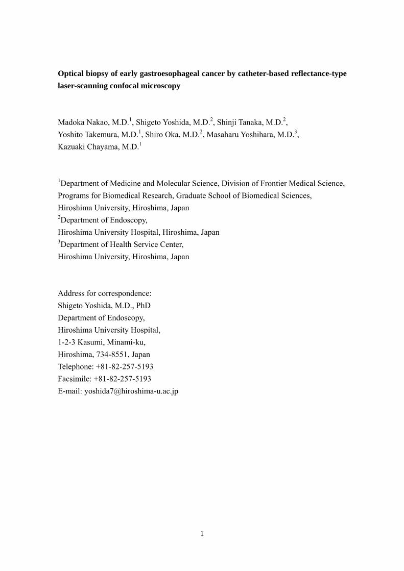
Optical biopsy of early gastroesophageal cancer by catheter-based reflectance-type laser-scanning confocal microscopy Madoka Nakao, M.D.1, Shigeto Yoshida, M.D.2, Shinji Tanaka, M.D.2,
Yoshito Takemura, M.D.1, Shiro Oka, M.D.2, Masaharu Yoshihara, M.D.3, Kazuaki Chayama, M.D.1
1Department of Medicine and Molecular Science, Division of Frontier Medical Science, Programs for Biomedical Research, Graduate School of Biomedical Sciences, Hiroshima University, Hiroshima, Japan 2Department of Endoscopy, Hiroshima University Hospital, Hiroshima, Japan 3Department of Health Service Center, Hiroshima University, Hiroshima, Japan Address for correspondence: Shigeto Yoshida, M.D., PhD Department of Endoscopy, Hiroshima University Hospital, 1-2-3 Kasumi, Minami-ku, Hiroshima, 734-8551, Japan Telephone: +81-82-257-5193 Facsimile: +81-82-257-5193 E-mail: [email protected]
1

Abstract
Magnified endoscopic observation of the gastrointestinal tract has become possible.
However, such observation at the cellular level remains difficult. Laser-scanning
confocal microscopy (LCM) is a novel, noninvasive optical imaging method that
provides instant microscopic images of untreated tissue under endoscopy. We compared
prototype catheter-based reflectance-type LCM images in vivo and histologic images of
early gastroesophageal cancer to assess the usefulness of LCM in diagnosing such
cancer. Twenty sites in the esophagus and 40 sites in the stomach were examined by
LCM under endoscopy prior to endoscopic or surgical resection. A prototype catheter
LCM system, equipped with a semiconductor laser that oscillates at 685 nm and
analyzes reflected light (Mauna Kea Technologies, Paris, France; Fujinon, Saitama,
Japan), was used in vivo without fluorescent agent. In all normal esophageal mucosa
and esophageal cancers, the nuclei were visualized. In 9 of the 10 normal esophageal
mucosa, cell membranes were visualized, and in 5 of the 10 esophageal cancers, cell
membranes were visualized. In all normal gastric mucosa, nuclei and cell membranes
were not visualized, but in 10 of the 20 gastric cancers, nuclei were visualized. This
novel method will aid in immediate diagnosis under endoscopy without the need for
biopsy.
2
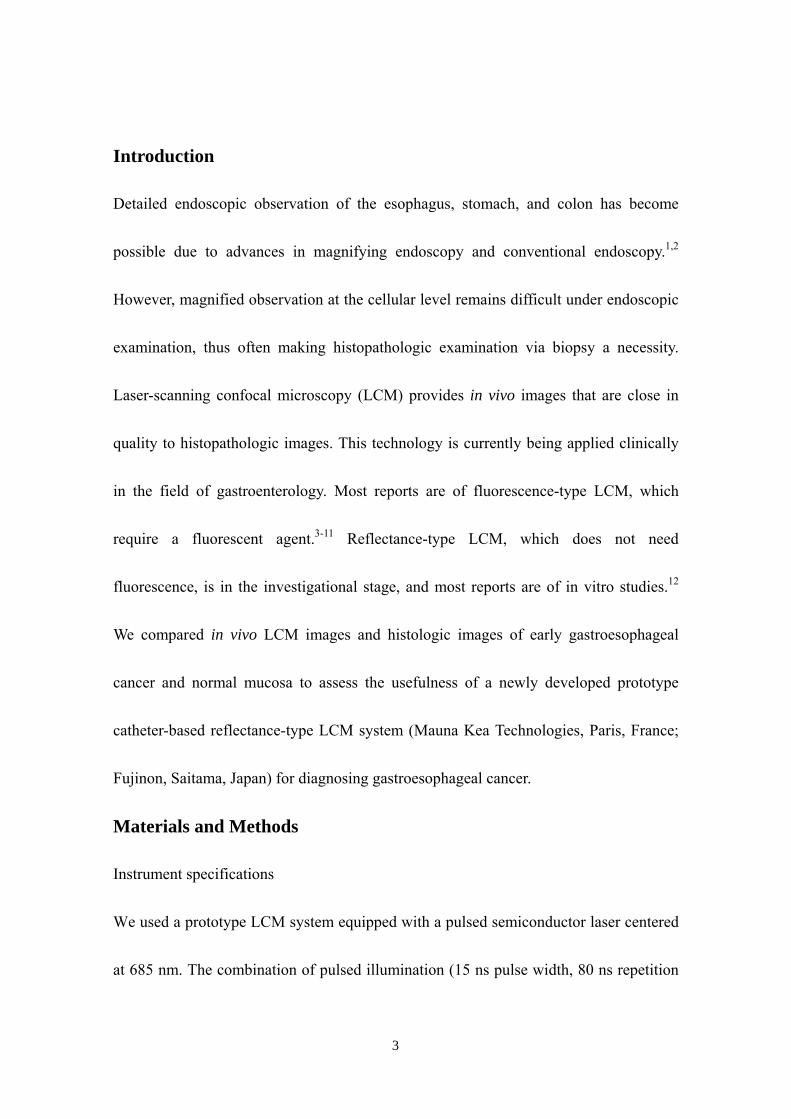
Introduction
Detailed endoscopic observation of the esophagus, stomach, and colon has become
possible due to advances in magnifying endoscopy and conventional endoscopy.1,2
However, magnified observation at the cellular level remains difficult under endoscopic
examination, thus often making histopathologic examination via biopsy a necessity.
Laser-scanning confocal microscopy (LCM) provides in vivo images that are close in
quality to histopathologic images. This technology is currently being applied clinically
in the field of gastroenterology. Most reports are of fluorescence-type LCM, which
require a fluorescent agent.3-11 Reflectance-type LCM, which does not need
fluorescence, is in the investigational stage, and most reports are of in vitro studies.12
We compared in vivo LCM images and histologic images of early gastroesophageal
cancer and normal mucosa to assess the usefulness of a newly developed prototype
catheter-based reflectance-type LCM system (Mauna Kea Technologies, Paris, France;
Fujinon, Saitama, Japan) for diagnosing gastroesophageal cancer.
Materials and Methods
Instrument specifications
We used a prototype LCM system equipped with a pulsed semiconductor laser centered
at 685 nm. The combination of pulsed illumination (15 ns pulse width, 80 ns repetition
3
period) with a time-gated detection of the reflected light (15 ns detection window, 40 ns
delay) permits to overcome the back-reflections onto the proximal optics by means of
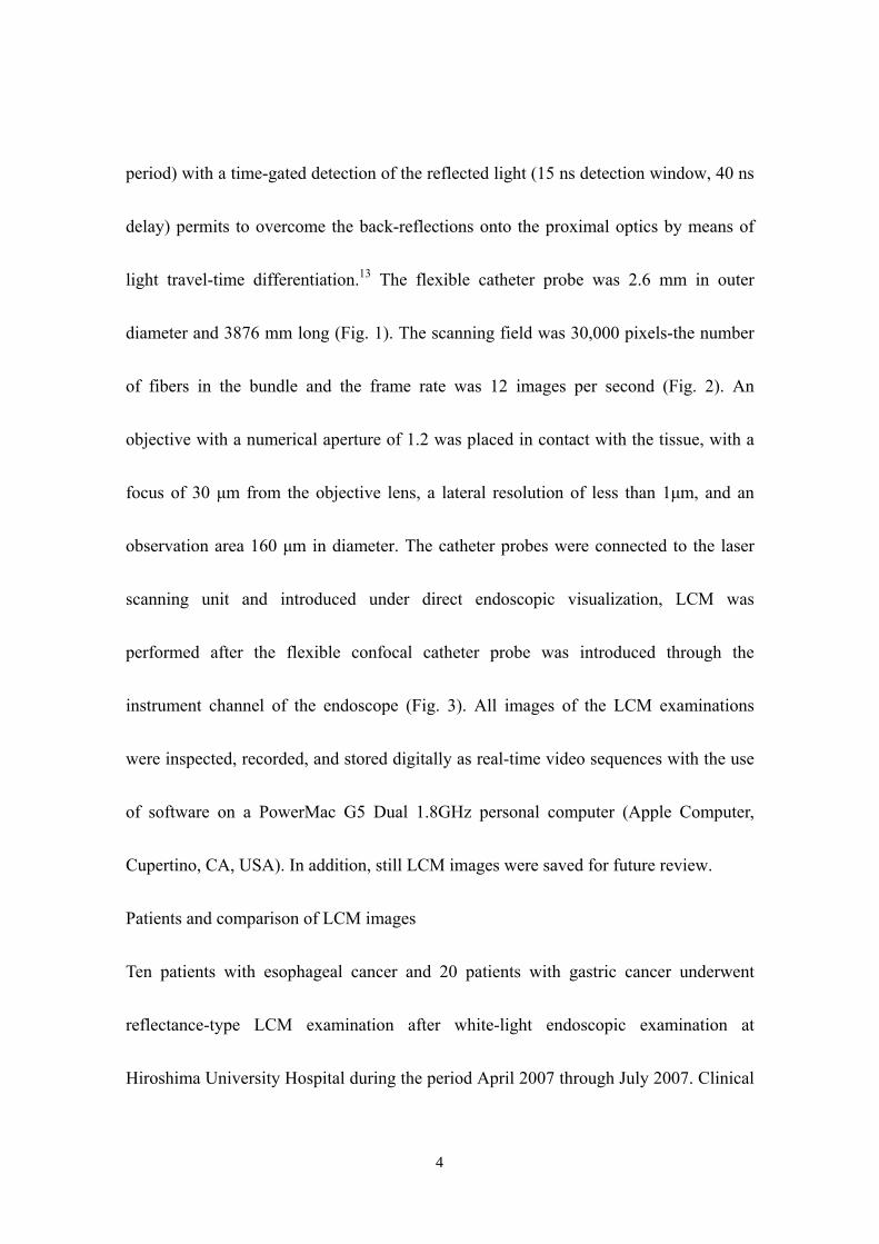
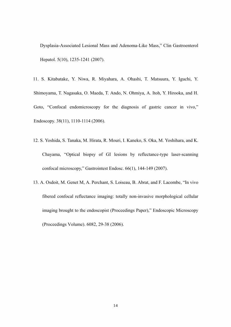
light travel-time differentiation.13 The flexible catheter probe was 2.6 mm in outer
diameter and 3876 mm long (Fig. 1). The scanning field was 30,000 pixels-the number
of fibers in the bundle and the frame rate was 12 images per second (Fig. 2). An
objective with a numerical aperture of 1.2 was placed in contact with the tissue, with a
focus of 30 μm from the objective lens, a lateral resolution of less than 1μm, and an
observation area 160 μm in diameter. The catheter probes were connected to the laser
scanning unit and introduced under direct endoscopic visualization, LCM was
performed after the flexible confocal catheter probe was introduced through the
instrument channel of the endoscope (Fig. 3). All images of the LCM examinations
were inspected, recorded, and stored digitally as real-time video sequences with the use
of software on a PowerMac G5 Dual 1.8GHz personal computer (Apple Computer,
Cupertino, CA, USA). In addition, still LCM images were saved for future review.
Patients and comparison of LCM images
Ten patients with esophageal cancer and 20 patients with gastric cancer underwent
reflectance-type LCM examination after white-light endoscopic examination at
Hiroshima University Hospital during the period April 2007 through July 2007. Clinical
4
characteristics of the lesions are presented in Table 1. The distal tip of the LCM catheter
was placed gently against the mucosa, and an endoscopist captured LCM images of
normal mucosa near the cancer and images of the cancer in the esophagus (normal
mucosa, n = 10; cancer, n = 10) or stomach (normal mucosa, n = 20; cancer, n = 20).
After endoscopic examination, a gastroenterologist (S.Y., who had analyzed LCM
images previously) judged whether the cell nuclei and membranes were visible on the
LCM images.12 After these procedures, patients underwent endoscopic mucosal
resection (EMR), endoscopic submucosal dissection (ESD), endoscopic aspiration
mucosectomy (EAM), or surgery. The resected specimens were fixed in formalin,
embedded in paraffin, sliced with a microtome, deparaffinized, and stained with
hematoxylin-eosin for light microscopic examination. After these procedures, the
histologic diagnosis was confirmed. The endoscopic system used in this study was a
VP-4400 endoscope processor and an EG-590WR or EG-450D upper gastrointestinal
endoscope (Fujinon). The study protocol conformed to the tenets of the Declaration of
Helsinki and was approved by our institutional ethics committee.
Results
With the distal tip of the catheter placed gently against the mucosa and the cancer,
real-time LCM images of the normal mucosa and of the cancer of the esophagus and
5
stomach were obtained safely and easily, and the influence of slight motion was ignore.
Even few water or blood was existed in the normal mucosa and cancer, the influence of
water or blood was also ignore. The time required for scanning each normal and cancer
site ranged between 16 and 390 seconds.
Normal esophageal mucosa
In LCM images of normal esophageal mucosa, high-reflectivity spots were observed
near the center of honeycomb-like structures of high reflectivity. These high reflectivity
spots and structures in the LCM images appeared to correspond to nuclei and cell
membranes, respectively, in the histologic images of hematoxylin-eosin-stained sections
(Fig. 4).
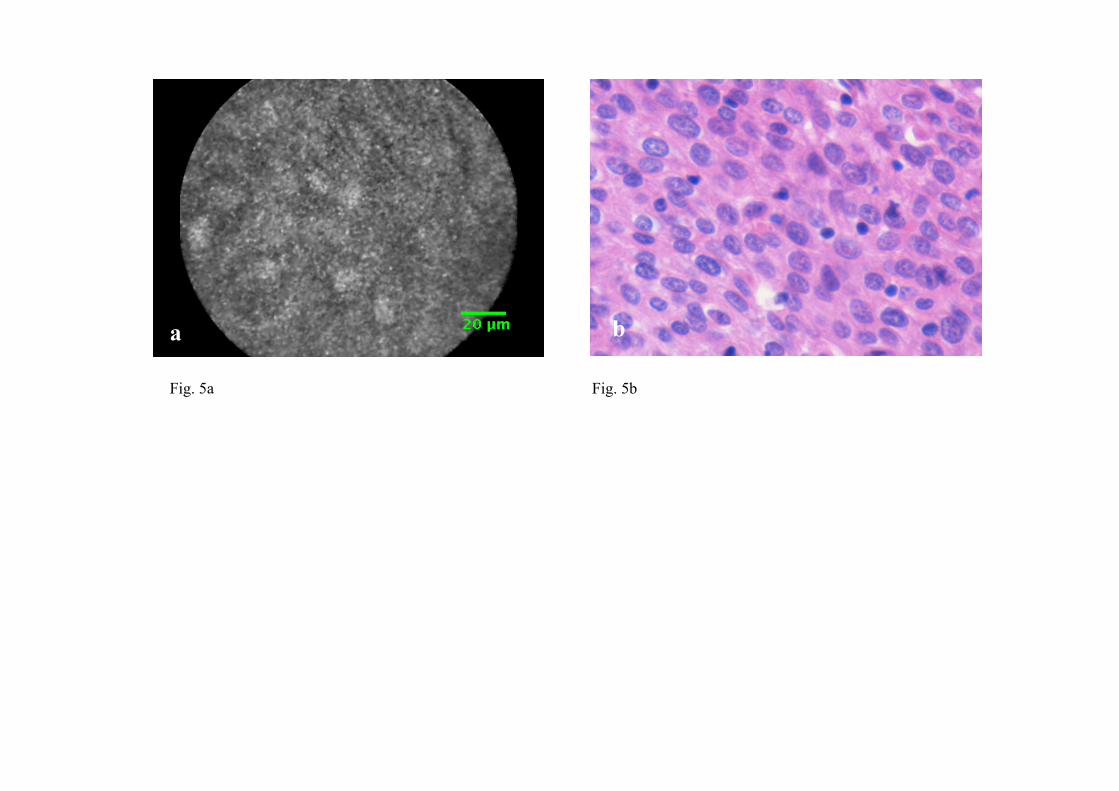
Esophageal cancer
In LCM images of esophageal cancer, high-reflectivity spots that were considered
nuclei were observed. The nucleus-to-cytoplasm (N/C) ratio was much increased, and
honeycomb-like structures of high reflectivity, considered cell membranes, were not
observed (Fig. 5).
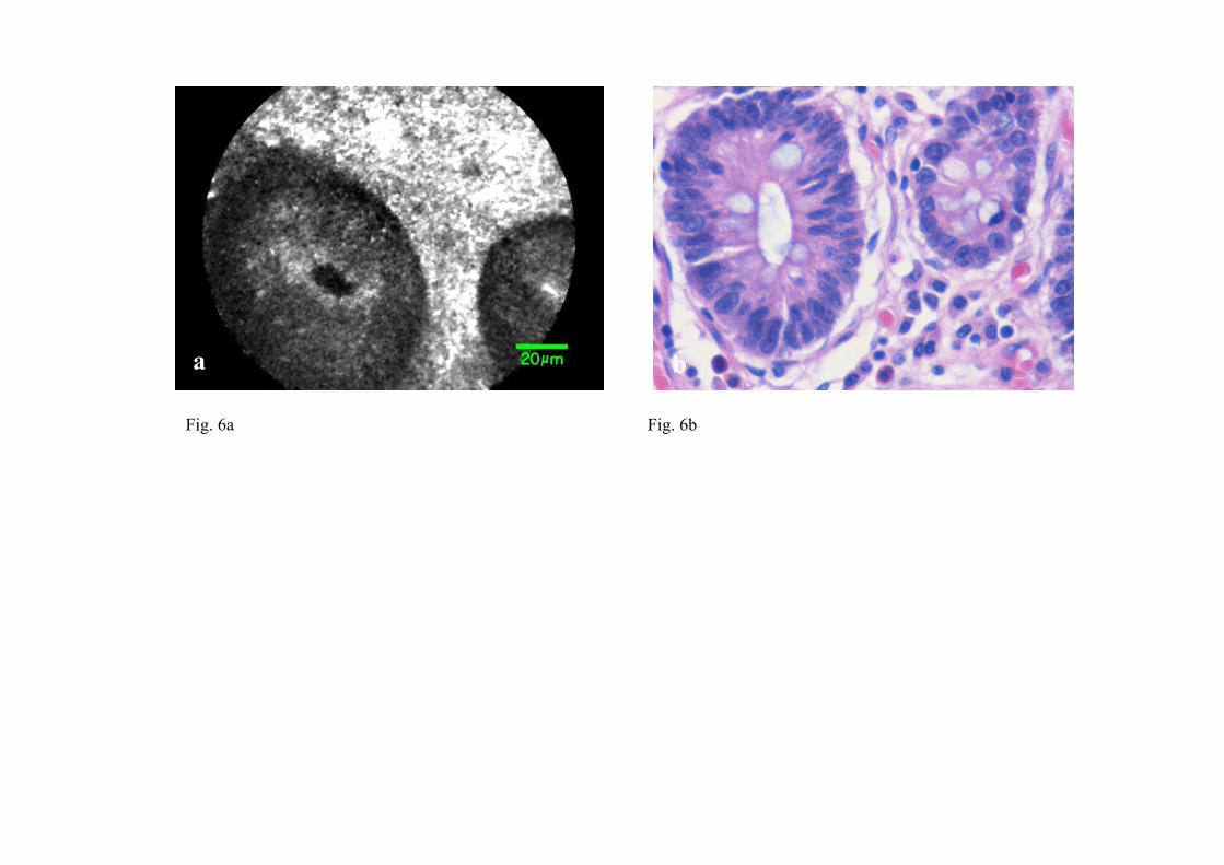
Normal gastric mucosa
In LCM images of normal gastric mucosa, cell membranes and nuclei were not
visualized. However, the crypt cells were arranged like flower petals surrounding the
6

gastric pit (Fig. 6).
Gastric cancer
In LCM images of differentiated adenocarcinoma of the stomach, cell membranes were
not visualized, and a disorganized configuration of glands with high-reflectivity spots
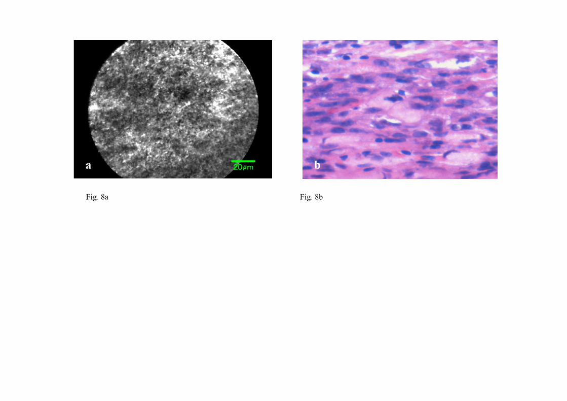
that were considered nuclei was observed (Fig. 7). In LCM images of undifferentiated
adenocarcinoma, no ductal structure was recognized; only a amorphous structure was
seen. Cell membranes and nuclei were not visualized (Fig. 8).
Visualization of nuclei and cell membranes in LCM images in relation to histologic
diagnoses is shown in Table 2. In all normal esophageal mucosa and esophageal cancers,
the nuclei were visualized. In 9 of the 10 (90%) normal esophageal mucosa, cell
membranes were visualized, and in 5 of the 10 (50%) esophageal cancers, cell
membranes were visualized. In all normal gastric mucosa, nuclei and cell membranes
were not visualized, but in 10 of the 20 (50%) gastric cancers, nuclei were visualized.
The storoma was visulized as high reflectivity. In some case, low-reflectivity spots were
also observed in the LCM images which was considered mucin in goblet cells in the
hematoxylin-eosin-stained specimen.
Discussion
Recent advances in endoscopic technology have afforded high-quality, detailed
7
diagnosis of gastrointestinal diseases. To confirm the presence of malignancy, however,
snip biopsy is often performed under endoscopy when endoscopic examination reveals
an abnormality. Thus, biopsy is performed for many lesions that are subsequently
determined not be malignant. Histologic analysis of biopsy material remains the gold
standard for the final diagnosis of a gastrointestinal lesion. Histologic diagnosis via
biopsy involves the following process: formalin fixation of the specimen, cutting the
specimen into small columns, paraffin embedding, ultra-thin slicing, deparaffinization,
staining, glass slide, mounting, and finally light microscopic observation. Moreover, it
takes several days to obtain a diagnosis. Also, snip biopsy is associated with bleeding,
apparent endoscopic disappearance of cancer cells after biopsy, and artificial ulceration,
which make endoscopic treatment, e.g. EMR, ESD, and EAM, difficult. In addition,
because of the bleeding, biopsy cannot be easily performed in patients taking
anticoagulants.
Being able to accurately image a lesion in vivo at the time of endoscopic examination
without biopsy allows for prompt diagnosis and treatment. Fluorescence-type LCM is
reported to be a promising tool for in vivo histopathologic examination during
endoscopy and might overcome the disadvantages associated with conventional
biopsy.3-11 In recent years, there have been several reports describing the ability to
8
obtain an LCM image that corresponds precisely to the histopathologic tissue diagnosis
in cases of gastrointestinal tract disease.3-12 However, many of the reports were based on
observations made on excised specimens or with fluorescence-type LCM. We too have
previously used probe-based reflectance-type LCM to obtain images that are close to
histopathologic specimens in vitro.12 In the present study, however, we conducted
examinations in vivo using catheter-based reflectance-type LCM, which enabled us to
insert the microscope through the instrument channel of endoscope and to capture
images at a single depth of 30 microns below the tissue surface. In our LCM
observations of the esophagus, nuclei were detected at both sites of normal mucosa and
cancer. In our LCM observations of the stomach, nuclei were not recognized in normal
mucosa but were recognized in 50% of cancer sites. Because the slice of LCM was very
thin, we assumed that the nuclei in the normal esophageal mucosa were easily
visualized because the cells were composed of stratified squamous epithelial cells.
Likewise, because the N/C ratio increased, we assumed that the nuclei of the esophageal
cancer and gastric cancer were visualized, whereas the nuclei of the normal gastric
mucosa were not visualized. Although further prospective and large number study, e.g.,
immediate diagnosis of neoplasia versus inflammation, is needed, we have shown that
catheter-based reflectance-type LCM can provide images at the cellular level in vivo,
9
suggesting the possibility of immediate cancer diagnosis under endoscopic observation
without the need for biopsy.
A fluorescence-type LCM system that uses a catheter was recently developed by Mauna
Kea Technologies.3-8 This LCM system has the capability to provide dynamic (12
frames/second) ultrahigh resolution images at the cellular level on a field of view as
wide as 260×260 μm with 1.5 lateral and 10-μm axial resolutions, at 60 µm working
depth. To overcome the limits of the field of view, an image reconstruction algorithm
that uses video mosaicing has been developed.3
One advantage of catheter-based LCM is that it allows the capture of an image during
conventional endoscopic examination without changing to a specialized scope. The
catheter-based reflectance-type LCM used in this study is of a size and flexibility to
pass through the endoscopic instrument channel and to be placed accurately on the
mucosa with guidance from the white-light endoscopic image. Furthermore, there is a
report of in vivo acquisition of real-time and dynamic histologic images of the
peritoneum, liver, and spleen during a novel, minimally invasive transgastric approach
to surgery termed natural orifice transluminal endoscopic surgery (NOTES).4 Compared
to reflectance-type LCM, fluorescence-type LCM can provide images with higher
signal-to-noise ratios (although a fluorescence agent is needed). The fluorescence-type
10

LCM device used for diagnosing cancer,11 visualizing lymphoepithelial lesions in
gastric mucosa-associated lymphoid tissue-type lymphoma,5 detecting angiodysplasia,6
visualizing Helicobacter pylori,9 diagnosing lymphocytic colitis,7 diagnosing a
dysplasia-associated lesional mass or adenoma-like mass in patients with ulcerative
colitis,10 and for functional examinations that provide moving images with visualization
of blood flow through microvessels.8 Unlike fluorescence-type LCM systems,
reflectance-type LCM collects and counts the reflective laser beam and therefore
requires no staining process. The fluorescence-type LCM requires some staining to
obtain clear images, but the acquired image is of high quality, and signal-to-noise ratio
is better than with the reflectance-type LCM. Further comparison of the two systems is
needed but we believe that these two instruments complement each other.
In summary, this feasibility study showed that catheter-based reflectance-type LCM can
be used in clinical practice to provide instant images that correspond well with
hematoxylin-eosin-stained microscopic images. Therefore, we expect that this novel
method will aid in immediate diagnosis under endoscopy without the need for biopsy.
Disclosure
System control software and prototype confocal catheter probes were provided on loan
by Mauna Kea Technologies, Paris, France, and Fujinon, Saitama, Japan at no charge.
11
References
1. S. Nagata, S.Tanaka, K. Haruma, M. Yoshihara, K. Sumii, G. Kajiyama, and F.
Shimamoto, “Pit pattern diagnosis of early colorectal carcinoma by magnifying
colonoscopy: clinical and histological implications,” Int J Oncol. 16(5), 927-934
(2000).
2. M. Hirata, S. Tanaka, S. Oka, I. Kaneko, S. Yoshida, M. Yoshihara, and K.
Chayama, “Magnifying endoscopy with narrow band imaging for diagnosis of
colorectal tumors,” Gassrointest Endosc. 65(7), 988-995 (2007).
3. V. Becker, T. Vercauteren, C. H. von Weyhern, C. Prinz, R. M. Schmid, and A.
Meining, “High-resolution miniprobe-based confocal microscopy in combination
with video mosaicing (with video),” Gastrointest Endosc. 66(5), 1001-1007 (2007).
4. S. von Delius, H. Feussner, D. Wilhelm, A. Karagianni, J. Henke, R. M. Schmid, and
A. Meining, “Transgastric in vivo histology in the peritoneal cavity using
miniprobe-based confocal fluorescence microscopy in an acute porcine model,”
Endoscopy. 39(5), 407-411 (2007).
12
5. A. Morgner, M. Stolte, and S. Miehlke, “Visualization of lymphoepithelial lesions in
gastric mucosa-associated lymphoid tissue-type lymphoma by miniprobe confocal
laser microscopy,” Clin Gastroenterol Hepatol. 5(9), e37 (2007).
6. A. Meining, M. Bajbouj, and R. M. Schmid, “Confocal fluorescence microscopy for
detection of gastric angiodysplasia,” Endoscopy. Epub ahead of print (2007).
7. A. Meining, S. Schwendy, V. Becker, R. M. Schmid, and C. Prinz, “In vivo
histopathology of lymphocytic colitis,” Gastrointest Endosc. 66(2), 398-399 (2007).
8. T. D. Wang, S. Friedland, P. Sahbaie, R. Soetikno, P. L. Hsiung, J. T. Liu, J. M.
Crawford, and C. H. Contag, “Functional imaging of colonic mucosa with a fibered
confocal microscope for real-time in vivo pathology,” Clin Gastroenterol Hepatol.
5(11), 1300-1305 (2007).
9. R. Kiesslich, M. Goetz, J. Burg, M. Stolte, E. Siegel, M. J. Maeurer, S. Thomas, D.
Strand, P. R. Galle, and M. F. Neurath, “Diagnosing Helicobacter pylori in vivo by
confocal laser endoscopy,” Gastroenterology. 128(7), 2119-2123 (2007).
10. D. P. Hurlstone, M. Thomson, S. Brown, N. Tiffin, S. S. Cross, and M. D. Hunter,
“Confocal Endomicroscopy in Ulcerative Colitis: Differentiating
13
Dysplasia-Associated Lesional Mass and Adenoma-Like Mass,” Clin Gastroenterol
Hepatol. 5(10), 1235-1241 (2007).
11. S. Kitabatake, Y. Niwa, R. Miyahara, A. Ohashi, T. Matsuura, Y. Iguchi, Y.
Shimoyama, T. Nagasaka, O. Maeda, T. Ando, N. Ohmiya, A. Itoh, Y. Hirooka, and H.
Goto, “Confocal endomicroscopy for the diagnosis of gastric cancer in vivo,”
Endoscopy. 38(11), 1110-1114 (2006).
12. S. Yoshida, S. Tanaka, M. Hirata, R. Mouri, I. Kaneko, S. Oka, M. Yoshihara, and K.
Chayama, “Optical biopsy of GI lesions by reflectance-type laser-scanning
confocal microscopy,” Gastrointest Endosc. 66(1), 144-149 (2007).
13. A. Osdoit, M. Genet M, A. Perchant, S. Loiseau, B. Abrat, and F. Lacombe, “In vivo
fibered confocal reflectance imaging: totally non-invasive morphological cellular
imaging brought to the endoscopist (Proceedings Paper),” Endoscopic Microscopy
(Proceedings Volume). 6082, 29-38 (2006).
14

Figure legends
Fig. 1. Catheter-based reflectance-type laser-scanning confocal microscope (Mauna Kea
Technologies, Paris, France; Fujinon, Saitama, Japan).
Fig. 2. Schema of the catheter-based reflectance-type laser-scanning confocal
microscopy.
Fig. 3. LCM examination for early gastric cancer under endoscopy.
Fig.4. Images of normal esophageal mucosal. a, Laser-scanning confocal microscopy
image. b, Hematoxylin-eosin-stained tissue from the same specimen.
Fig. 5. Images of esophageal cancer. a, Laser-scanning confocal microscopy image. b,
Hematoxylin-eosin-stained tissue from the same specimen.
Fig. 6. Images of normal gastric mucosa. a, Laser-scanning confocal microscopy image.
b, Hematoxylin-eosin-stained tissue from the same specimen.
Fig. 7. Images of differentiated adenocarcinoma of the stomach. a, Laser-scanning
confocal microscopy image. b, Hematoxylin-eosin-stained tissue from the same
specimen.
Fig. 8. Images of undifferentiated adenocarcinoma of the stomach. a, Laser-scanning
confocal microscopy image. b, Hematoxylin-eosin-stained tissue from the same
specimen.
15
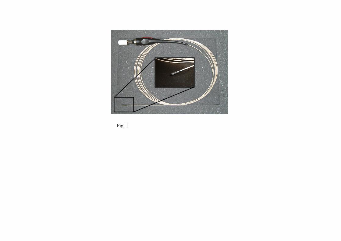
Fig. 1
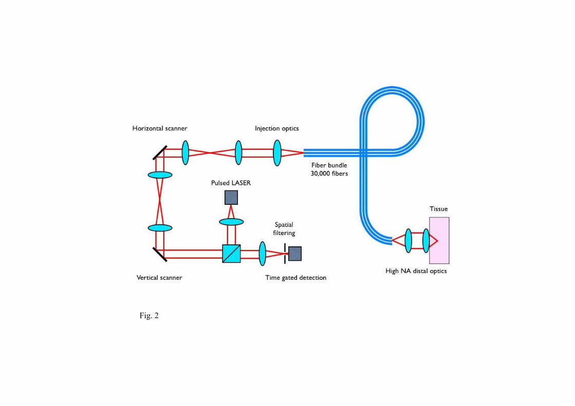
Fig. 2
Fig. 3
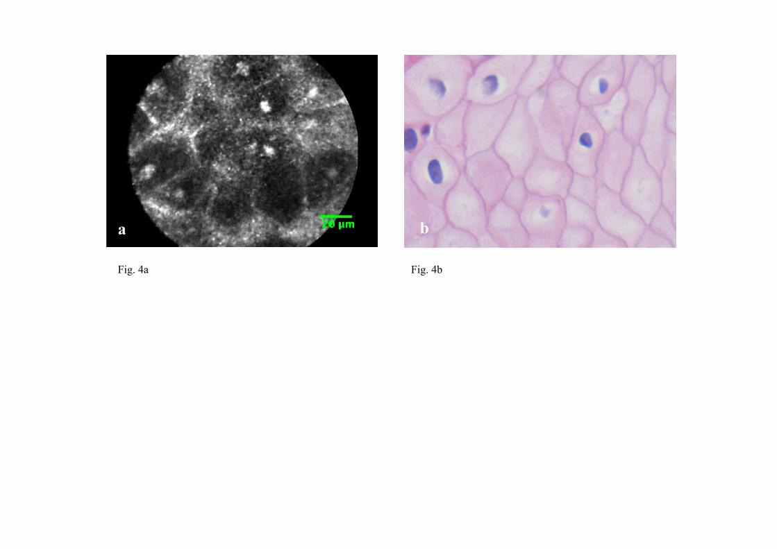
Fig. 4a
a b
Fig. 4b
a a b
Fig. 5a Fig. 5b
A a b
Fig. 6a Fig. 6b

a b
Fig. 7a Fig. 7b
20 µm a b
Fig. 8a Fig. 8b

Table 1. Clinical Characteristics of the lesions
Tumor size (mm) Mean Range Localization of tumor Esophagus
Depth of invasion Mucosa Submucosa Histologic type Squamous cell carcinoma
Esophagus Stomach
22.8±7.5 8-35
10
10 0
10
16.3±10.5 10-35
6 0
12 2
13 7
10 6 1 1 2
Tumor size (mm) Mean Range Localization of tumor Antrum Angle Corpus Cardia Depth of invasion Mucosa Submucosa Histologic type Well Moderately Papillary Poorly Signet ring cell
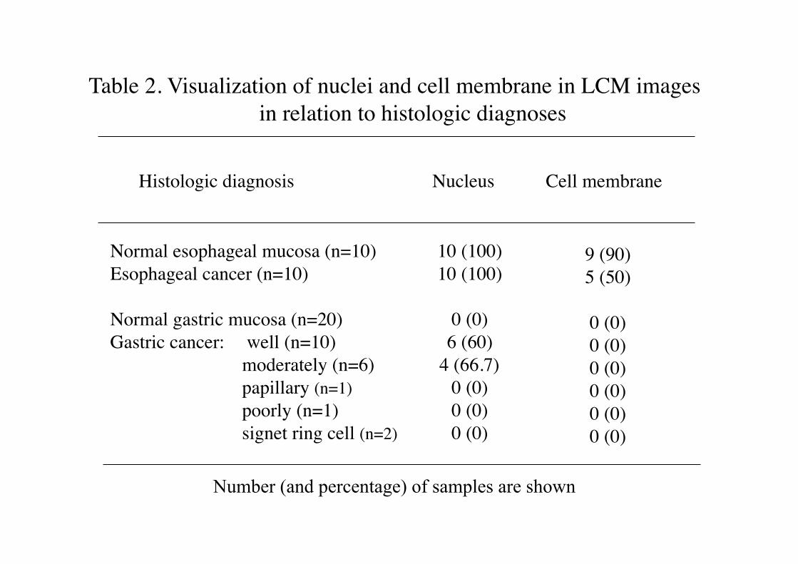
Table 2. Visualization of nuclei and cell membrane in LCM images in relation to histologic diagnoses
Normal esophageal mucosa (n=10) Esophageal cancer (n=10)
Normal gastric mucosa (n=20) Gastric cancer: well (n=10)
moderately (n=6) papillary (n=1) poorly (n=1) signet ring cell (n=2)
Histologic diagnosis Nucleus
10 (100) 10 (100)
0 (0) 6 (60)
4 (66.7) 0 (0) 0 (0) 0 (0)
Cell membrane
9 (90) 5 (50)
0 (0) 0 (0) 0 (0) 0 (0) 0 (0) 0 (0)
Number (and percentage) of samples are shown