Embed Size (px)
Citation preview
G A F N I A N D S T E I N B E R G
Optical Activity of Terbium Ions Bound to Transferrin and Conalbumin Studied by Circular Polarization of Luminescence t A. Gafni and I. Z. Steinberg*
ABSTRACT: The optical activity of terbium ions bound to transferrin and to conalbumin has been studied by the cir- cular polarization of the luminescence of the bound ions. The anisotropy factor is very large (reaching a value of 0.23 at 540 nm). Due to the multitude of electronic transitions re- sponsible for the emission spectrum, the anisotropy factor varies markedly with wavelength and shows fine details. The optical activity has thus been used as a sensitive indicator for probing the asymmetry of the binding sites for the metal ions. The emission spectrum and the spectrum of the anisot- ropy factor are extremely similar for terbium ions bound to
T he storage and transport of iron in living systems are performed by an elaborate system, in which proteins of high affinity for iron play an important role (Aisen, 1970). Trans- ferrin from blood serum and conalbumin from egg white are two examples. These are glycoproteins of about 80,000 molecular weight and apparently comprised of a single poly- peptide chain. There are two binding sites per protein mole- cule which have identical or similar affinity for ferric ions. Two histidine and three tyrosine residues were shown to participate in the metal binding sites of transferrin (Bezkoro- vainy and Grohlich, 1971). The iron ions can be replaced by a variety of other transition metal and rare earth ions. The replaced ions have proved to be very useful as paramagnetic or fluorescent probes for the study of the metal binding sites of transferrin and conalbumin (Aisen et al., 1969; Price and Gibson, 1972; Luk, 1971).
The binding of trivalent terbium ions to transferrin has been recently studied by Luk (1971) and Teuwissen et ai. (1972). Two ions bind per protein molecule, the terbium being highly luminescent in the complex. The emission spectrum is composed of four quite narrow bands (with some fine struc- ture) caused by internal transitions in the 4f shell of the metal. These transitions are essentially forbidden; the corresponding absorption bands are therefore extremely weak and the luminescence lifetime long (about 1.3 msec) (Luk, 1971). The excitation of the terbium ions is affected by energy trans- fer from the tyrosine residues of the protein which are situated near the bound ions (Luk, 1971).
The optical activity of metal absorption bands in metallo- proteins has been extensively used to investigate the binding site of the metal (Vallee and Wacker, 1970). Due to the very weak intensity of the absorption bands of the terbium ions in the visible spectral range, the study of the circular dichroism (CD) of the absorption bands of this ion when bound to pro- teins is impracticable. It has been shown recently, however,
transferrin and to conalbumin, reflecting similarity in structure and conformation of the binding sites of the two proteins. The same spectrum was observed for the anisotropy factor of transferrin-Tba+ complexes of different degrees of satura- tion, and for ternary complexes of the type transferrin- Ho a+-Tb and transferrin-Fe3+-Tb 3+ of various stoichio- metric ratios. These results strongly suggest that under the conditions studied (aqueous solution, pH 8.5, room tem- perature) the two metal binding sites of a transferrin molecule are equivalent in structure and conformation.
that the circular polarization of luminescence (CPL) is also a manifestation of asymmetry of chromophores (Gafni and Steinberg, 1972; Steinberg and Gafni, 1972; Emeis and Ooster- hoff, 1971; Gafni et af., 1973; Steinberg, 1974). More speci- fically, the CPL of a chromophore is related to its asymmetry in the emitting excited state in the same way that its CD is related to the asymmetry in the ground state. For the study of CPL the forbidden nature of the relevant transitions may even turn out to be an advantage since weak transitions may assume very high optical activity (Kuhn, 1958).
In the following we present the behavior of the circular polarization of the luminescence of trivalent terbium ions bound to transferrin and to conalbumin. Since the lumines- cence involves many electronic transitions, the CPL spectrum exhibits an intricate pattern, which renders the CPL a sensi- tive fingerprint of the environment of the ion. A comparison of the metal binding sites of the two proteins and of the two sites in a single protein is thus facilitated.
Experimental Section
Human serum transferrin and avian egg white conalbumin, both iron free, were purchased from Sigma Chemical Co. TbCls and HoClj were obtained from Alfa Inorganics and were of 99.99x purity. FeC13, Analytic Grade, was a product of Fischer Scientific Co. These materials were used without fur- ther purification.
Solutions of tranferrin and conalbumin we= prepared in 0.05 M Tris buffer (pH 8.5) which contained 5 X M
sodium bicarbonate. Protein concentrations were about M.
Complexes of the inorganic salts with the proteins were usually prepared by adding the appropriate volume of metal salt solution in double distilled water (usually 0.025 ml) to the protein solution (usually 0.5 ml) with mild stirring. The con- centration of the salt solution was adjusted to yield the desired ratio of metal-Drotein in the final comdex.
t From the Department of Chemical Physics, The Weizmann Insti- Triple complexes Of the type transferrin-Tb3+-Ho3+ were prepared by adding a solution of HoCI, to a solution of the tute of Science, Rehovot, Israel. ReceiuedJuly 20,1973.
800 B I O C H E M I S T R Y , V O L . 1 3 , N O . 4, 1 9 7 4
O P T I C A L A C T I V I T Y O F T R A N S F E R R I N - T b A N D CONALBUMIN-Tb a +
I I I I I 350 4 0 0 450 500 550 600
Wavelength ( n m )
1 : Absorption spectrum of transferrin-iron prepared by 16 of a solution of FeC13 in water (I@* M) to 1 ml of a
solution of transferrin (8 X M) in Tris buffer containing 5 X l O - 3 ~ sodium bicarbonate(pH 8.5); light path 1.0 cm; room tem- perature (-23 ").
transferrin-Tb3+ complex (1 : 1). The amount of Ho3+ added was fivefold that of Tb3+ present.
Triple complexes of the type transferrin-Tb 3+-Fe 3+ or conalbumin-Tb 3+-Fe3+ of different stoichiometric ratios were prepared in three different ways: (a) a mixture of the two cations in the desired ratio was added to a solution of the apoprotein, the total number of cations of heavy metal being equal to the number of protein binding sites; (b) Fe3+ was added to a transferrin-Tb3+ complex of ratio 1 :2 to replace part of the Tb 3+; (c) Fea+ and transferrin were mixed in a molar ratio of 1: l and allowed to react for 12 hr. TbH was then added to populate a part of the free binding sites.
In view of the complications observed by Bates and Schla- bach (1973) in the binding of ferric ions to transferrin under certain conditions, the spectra of transferrin-Fe3+ complexes of different stoichiometric ratios, prepared as described above, were studied at various intervals after mixing the constituents. Figure 1 shows the spectrum of a transferrin-Fe3+ mixture of molar ratio 1 :2, 20 hr after preparation. Similar spectra, but of proportionately lower intensity, were obtained for lower degrees of saturation. The absorption developed most of its intensity within 2 hr after mixing the constituents and was practically constant after 20 hr. It may be noted that the spectrum of the transferrin-Fe 3+ complex is similar in shape to that reported by Bates and Schlabach for fully saturated trans- ferrin though it is somewhat lower in intensity. Thus, most of the iron (at least 80 %) binds at the specific sites of the protein under the conditions used in this study. As regards the trans- ferrin-Tb complexes, we have observed by fluorimetric titrations that two terbium ions bind per protein molecule, in agreement with the studies reported by Luk (1971).
The solutions were studied on the day of preparation, at least 1 hr after preparation of complexes containing protein and lanthanide ions and 3 hr after preparation of complexes containing iron. Very similar results were, however, obtained 1 week after preparation, the solutions being stored in the meantime at 4".
All measurements were carried out at room temperature (-23").
Measurements of circular polarization of luminescence were performed with an instrument built in our laboratory. The molecules under study are excikd by unpolarized mono- chromated light, and the circularly polarized component of
- ~ . .
0 - 4
-12
- I6
I b I L
520 560 600 W a v e l e n g t h l n m )
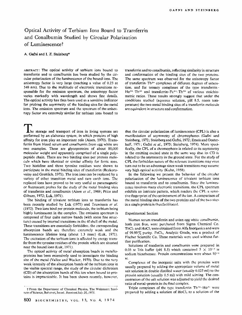
FIGURE 2: Fluorescence spectra and the spectra of the emission anisotropy factor, gem, of the Tb3+ complexes of transferrin and conalbumin. The ratio of meta1:protein is 2; solvent, 0.05 M Tris buffer (pH 8.5) and 5 X M sodium bicarbonate; temperature, -23"; spectral bandwidth of the emission monochromator, 1.6 nm. Upper part: emission spectra (identical for the two metal- protein complexes). Lower part: spectrum of the emission anisot- ropy factor: (-) transferrin-Tba+; (---) conalbumin-Tb3+.
the emitted light is selectively modulated by an elasto-optic light modulator and detected by a photomultiplier. The ac and dc components of the electric signal generated by the mod- ulated beam after monochromatization are amplified and monitored. Full details of the instrument are described else- where (Steinberg and Gafni, 1972). The luminescence was ex- cited by the emission bands at 290-305 nm of a 100-W high- pressure mercury arc (Osram, HBO 100 W/2). The spectral bandwidth of the excitation monochromator was about 30 nm, while the spectral resolution of the emitted light varied from 0.5 to 1.6 nm. The CPL is expressed by the anisotropy factor for emission defined as gem = AF/(F/2), where F is the total luminescence intensity and AF is the intensity of the circular polarized component of the luminescence. AF is as- signed a plus sign for left-handed circular polarization.
Fluorescence spectra were obtained from the dc signal of the above instrument for measuring CPL. These spectra were not corrected for the response of the instrument. This, however, will not distort the shape of the emission bands to any signifi- cant extent because of the narrow spectral range of the bands.
Results and Discussion
Optical Activity of Tba+ Bound to Transferrin and Con- albumin. The luminescence and CPL spectra of the terbium ions in the complexes transferrin-Tb 3+ and conalbumin-Tb 3f
are presented in Figure 2. The metal binding sites were sat- urated with Tb3+ cations. The emissions spectra presented in Figure 2 are composed of several narrow bands each of which
801 B I O C H E M I S T R Y , VOL. 13 , N O . 4, 1 9 7 4
G A F N I A N D S T E I N B E R G
TABLE I : Emission Anisotropy Factor, gem, of Transferrin-Tb3+ of Molar Ratio 1 :2.'
Emission Wavelength (nm)
487 540 540.5 541 544 548 550 580 ~~~~~~~
-9.1 +8 .4 -3.6 -3 .7 0 .0 g e m X 10' +1.47 -23.0 -20.0
a Spectral bandwidth of the emission monochromator, 0.5 nm. Except for the spectral bandwidth, the conditions are the same as those described in Figure 2.
shows fine structure. The bands stem from electronic transi- tions from the 5D4 atomic level to several ' F levels of different J values (Ofelt, 1963; Thomas et al., 1963). These levels are split by the ligand field into components whose number and separation depend on the nature and symmetry of the environ- ment of the cation (Filipescu et a / . , 1964). The various emission lines therefore result from independent electronic transitions. Since each of the electronic transitions is associated with its own electric and magnetic transition dipole moments, the optical activity may markedly change from transition to transition, which is the reason for the pronounced variation of the anisotropy factor with wavelength observed for the protein-terbium complexes. The exceptionally large values obtained for the emission anisotropy factor at a few wavelengths are noteworthy. (Some of them, measured at a high spectral resolution of 0.5 nm, are presented in Table I.) This is made possible by the forbidden nature of the transi- tions. The wealth of detail in the CPL spectra of the protein- Tb3+ complexes, the large signals obtained, and the high sensi- tivity of the optical activity to the nature of the environment of the chromophores involved render CPL a very sensitive tool for probing the metal binding sites.
The emission spectra of the terbium ions in transferrin- Tb3+ and conalbumin-Tb3+ are identical. Still more striking is the close similarity in the CPL spectra of the two protein- Tba+ complexes, which, except for a slight relative shift in wavelength, are of very similar shape and of comparable mag- nitude. It is very unlikely that the similarity between the CPL spectra of the two protein-metal complexes is a coincidence, since each of the spectra is made up of a large number of elec- tronic transitions, each with distinct electric and magnetic transition dipole moments which are very sensitive in the pres- ent case to pertubations by the environment because of the forbidden nature of the transitions. The binding sites of the two proteins thus seem to offer nearly identical asymmetric environments to the bound metal ions. Despite the close sim- ilarity between the CPL spectra of the two proteins, small dif- ferences do occur and possibly reflect differences in sequence and folding of the two proteins in regions removed from the immediate surroundings of the bound metal ions.
Optical Actiaity of Bound Tb3+ on Partial Saturation oj the Protein Binding Sites with Metal Ions. There is a con- troversy as to the equivalence of the two metal binding sites of a molecule in the iron binding proteins. The following ex- periments were performed in relation to this problem.
The CPL of a few complexes of terbium with transferrin of different degrees of saturation was measured at a few wave- lengths. The molar ratios of Tba+ :protein were 0.6,1.1,2, and an excess of 5 . The CPL was measured at 487, 540, and 544 nm. The values of the anisotropy factor obtained for the various Tb3+ :protein ratios at each wavelength were identical.
These results show that one or more of the following cir- cumstances hold. (1) The sites are identical in their asymmetry.
802 B I O C H E M I S T R Y , V O L . 1 3 , N O . 4, 1 9 7 4
(2) The sites have equal affinity for the Tba+; the metal thus populates the two sites equally at any level of saturation. (3) The binding of the terbium to the sites is highly cooperative; the Tba+ ions thus populate the protein in an all-or-none fashion. Here again the observed optical activity will not de- pend on the extent of saturation even if the two sites of a pro- tein molecule are of different asymmetry. As discussed above, equal optical activity for the two sites strongly suggests in our case that the sites are identical in constitution and structure. Alternative 1 above thus suggests that the two binding sites on a protein molecule are identical, while alternatives 2 and 3 may imply nonequivalent sites.
The optical activity of a ternary complex of the type trans- ferrin-Tba+-Ho a+ was investigated. This complex was prepared by adding a fivefold excess of Ho 3+ to a 1 : 1 complex of trans- ferrin and Tb*+. Ho3+ has a higher affinity for transferrin than Tb3+ and is nonfluorescent under the present experimental circumstances (Luk, 1971). Indeed, on adding Ho3+ ions to the transferrin-Tba+ complex the fluorescence dropped in intensity by nearly an order of magnitude, but the anisotropy factor (measured at 540 and 554 inn) remained essentially the same. These results render somewhat unlikely alternative 2 discussed above, namely that the two sites on a transferrin molecule are nonequivalent but happen to have equal affinity for Tba+. The experiments with the ternary complexes will require that the sites also have equal affinity for Ho3+.
In order to test alternative 3 discussed above, namely that the sites are nonequivalent but that the binding of Tba+ at the two sites of transferrin is cooperative, the optical activity of ternary complexes of transferrin with ferric and terbium ions of different molar ratios was studied at a few wavelengths. The complexes of transferrin-Tb a+ with the following molar ratios were tested: l : l : l , 1:0.33:1.67, 1 :0 ,5 :1 , and 1:0.8:1. ' The wavelengths selected were 540 and 544 nm for the first two complexes and 487,540,544, and 550 nm for the last two com- plexes. The complexes were prepared by a variety of methods as described in the Experimental Section. The same anisotropy factor and hence the same optical activity were obtained for the luminescence of the terbium throughout. (Similar experi- ments with conalbumin-metal complexes of the type conalbu-
'Free Tb3- in solution is not excited under the conditions used, while Tb3+ bound at the specific site of transferrin is rendered highly luminescent by transfer of excitation energy from an adjacent tyrosine residue. I t is of interest to note that negligible fluorescence of the terbium ions was observed when added to transferrin which was saturated with iron. The terbium is thus obviously excluded by the iron from the specific binding site of the protein. The possibility cannot be excluded that nonspecific binding occurs at other sites which do not provide a proper chromophore for excitation of the Tb3+ by energy transfer. This possibility does not affect, however, the conclusions drawn below, since nonspecific binding, if it occurs a t all, does not affect the measure- ments.
O P T I C A L A C T I V I T Y O F T R A N S F E R R I N - T b * + A N D CONALBUMIN-Tb"
min-Tba+-Fea+ of molar ratios 1 : 0.5 : 1 and 1 : 0.8 : 1 showed References the same optical activity as saturated conalbumin-Tba+.) The binding of ferric ions to transferrin is known to be non- cooperative (Lane, 1971). It is bound with much higher affinity than terbium (Aisen, 1970; Luk, 1971). Furthermore, once bound the reequilibration of the ferric ions among the sites is negligible even for periods of days (Aisen and Leibman, 1968). Thus, when ferric ions bind to the transferrin below saturation levels, they block randomly and irreversibly (for our purposes) the sites they occupy. A fraction of the protein molecules thus have only a single site available for binding of Tba+ ions. For these sites cooperative binding of Tba+ is of course impossible. Thus, the explanation that cooperativity of binding is the cause of the invariability of the optical activity of the terbium with degree of saturation of the protein may be discarded.
To summarize, the two metal binding sites of a transferrin molecule seem to be equivalent. The possibility that they are nonequivalent but of equal affinity for each of the metal ions tested cannot be discarded but seems unlikely. The binding of terbium ions to the metal binding sites is not coopera- tive.
Electron paramagnetic resonance (epr) studies of iron and chromium complexes of transferrin and conalbumin at very high protein concentrations (10-14 %) and very low tempera- ture (77 and 103'K) have indicated differences between trans- ferrin and conalbumin binding sites and two types of metal complexes with each protein (Price and Gibson, 1972; Aisen et al., 1969). Due to the markedly different conditions under which the epr studies and the present investifation were con- ducted, a comparison between the conclusions drawn in the previous and the present studies seems to be of little mean- ing.
Aisen, P. (1970), Mt. Sinai, J . Med. 37,213. Aisen, P., Aasa, R., and Redfield, A. G. (1969), J. Biol. Chem.
Aisen, P., and Leibman, A. (1968)) Biochem. Biophys. Res.
Bates, G. W., and Schlabach, M. R. (1973)) J . Biol. Chem.
Bezkorovainy, A., and Grohlich, D. (1971), Biochem. J . 123,
Emeis, C. A., and Oosterhoff, L. J. (1971), J. Chem. Phys. 54,
Filipescu, N., Sager, W. F., and Serafin, F. A. (1964), J . Phys.
Gafni, A., Schlessinger, J., and Steinberg, I. Z. (1973), Isr. J .
Gafni, A., and Steinberg, I. Z. (1972), Photochem. Photobiol.
Kuhn, W. (1958), Annu. Rev. Phys. Chem. 9,417. Lane, R. S. (1971)) Biochim. Biophys. Acta 243,193. Luk, C. K. (1971), Biochemistry I O , 2838. Ofelt, G. S. (1963), J . Chem. Phys. 38,2171. Price, E. M., and Gibson, J. F. (1972), J . Biol. Chem. 247,8031. Steinberg, I. Z. (1974), in Trends in Biochemical Fluorescence
Spectroscopy, Chen, R., and Edelhoch, H., Ed., New York, N. Y . , Marcel Dekker.
Steinberg, I. Z., and Gafni, A. (1972)) Rev. Sci. Instrum. 43,409. Teuwissen, B., Masson, P. L., Osinski, P., and Hermans, J. F.
Thomas, K. S., Singh, S., and Dieke, G. H. (1963), J . Chem.
Vallee, B. L., and Wacker, W. E. C. (1970), Proteins5,lM.
244,4628.
Commun. 32,220.
248,3228.
125.
4809.
Chem. 68,3324.
Chem. 11,423.
15,93.
(1972), Eur. J . Biochem. 31,239.
Phys. 38,2180.
B I O C H E M I S T R Y , V O L . 1 3 , N O . 4, 1 9 7 4 803