Embed Size (px)
Citation preview
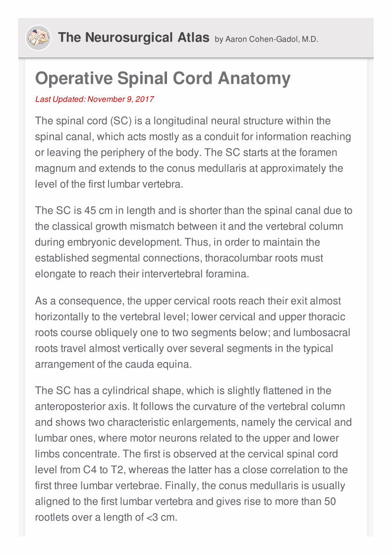
OperativeSpinalCordAnatomyLastUpdated:November9,2017
Thespinalcord(SC)isalongitudinalneuralstructurewithinthespinalcanal,whichactsmostlyasaconduitforinformationreachingorleavingtheperipheryofthebody.TheSCstartsattheforamenmagnumandextendstotheconusmedullarisatapproximatelythelevelofthefirstlumbarvertebra.
TheSCis45cminlengthandisshorterthanthespinalcanalduetotheclassicalgrowthmismatchbetweenitandthevertebralcolumnduringembryonicdevelopment.Thus,inordertomaintaintheestablishedsegmentalconnections,thoracolumbarrootsmustelongatetoreachtheirintervertebralforamina.
Asaconsequence,theuppercervicalrootsreachtheirexitalmosthorizontallytothevertebrallevel;lowercervicalandupperthoracicrootscourseobliquelyonetotwosegmentsbelow;andlumbosacralrootstravelalmostverticallyoverseveralsegmentsinthetypicalarrangementofthecaudaequina.
TheSChasacylindricalshape,whichisslightlyflattenedintheanteroposterioraxis.Itfollowsthecurvatureofthevertebralcolumnandshowstwocharacteristicenlargements,namelythecervicalandlumbarones,wheremotorneuronsrelatedtotheupperandlowerlimbsconcentrate.ThefirstisobservedatthecervicalspinalcordlevelfromC4toT2,whereasthelatterhasaclosecorrelationtothefirstthreelumbarvertebrae.Finally,theconusmedullarisisusuallyalignedtothefirstlumbarvertebraandgivesrisetomorethan50rootletsoveralengthof<3cm.
TheNeurosurgicalAtlas byAaronCohen-Gadol,M.D.
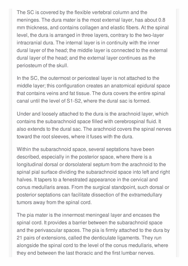
TheSCiscoveredbytheflexiblevertebralcolumnandthemeninges.Theduramateristhemostexternallayer,hasabout0.8mmthickness,andcontainscollagenandelasticfibers.Atthespinallevel,theduraisarrangedinthreelayers,contrarytothetwo-layerintracranialdura.Theinternallayerisincontinuitywiththeinnerdurallayerofthehead;themiddlelayerisconnectedtotheexternaldurallayerofthehead;andtheexternallayercontinuesastheperiosteumoftheskull.
IntheSC,theoutermostorperiosteallayerisnotattachedtothemiddlelayer;thisconfigurationcreatesananatomicalepiduralspacethatcontainsveinsandfattissue.TheduracoverstheentirespinalcanaluntilthelevelofS1-S2,wheretheduralsacisformed.
Underandlooselyattachedtotheduraisthearachnoidlayer,whichcontainsthesubarachnoidspacefilledwithcerebrospinalfluid.Italsoextendstotheduralsac.Thearachnoidcoversthespinalnervestowardtherootsleeves,whereitfuseswiththedura.
Withinthesubarachnoidspace,severalseptationshavebeendescribed,especiallyintheposteriorspace,wherethereisalongitudinaldorsalordorsolateralseptumfromthearachnoidtothespinalpialsurfacedividingthesubarachnoidspaceintoleftandrighthalves.Ittaperstoafenestratedappearanceinthecervicalandconusmedullarisareas.Fromthesurgicalstandpoint,suchdorsalorposteriorseptationscanfacilitatedissectionoftheextramedullarytumorsawayfromthespinalcord.
Thepiamateristheinnermostmeningeallayerandencasesthespinalcord.Itprovidesabarrierbetweenthesubarachnoidspaceandtheperivascularspaces.Thepiaisfirmlyattachedtotheduraby21pairsofextensions,calledthedenticulateligaments.Theyrunalongsidethespinalcordtotheleveloftheconusmedullaris,wheretheyendbetweenthelastthoracicandthefirstlumbarnerves.
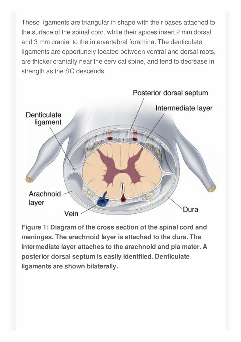
Theseligamentsaretriangularinshapewiththeirbasesattachedtothesurfaceofthespinalcord,whiletheirapicesinsert2mmdorsaland3mmcranialtotheintervertebralforamina.Thedenticulateligamentsareopportunelylocatedbetweenventralanddorsalroots,arethickercraniallynearthecervicalspine,andtendtodecreaseinstrengthastheSCdescends.
Figure1:Diagramofthecrosssectionofthespinalcordandmeninges.Thearachnoidlayerisattachedtothedura.Theintermediatelayerattachestothearachnoidandpiamater.Aposteriordorsalseptumiseasilyidentified.Denticulateligamentsareshownbilaterally.
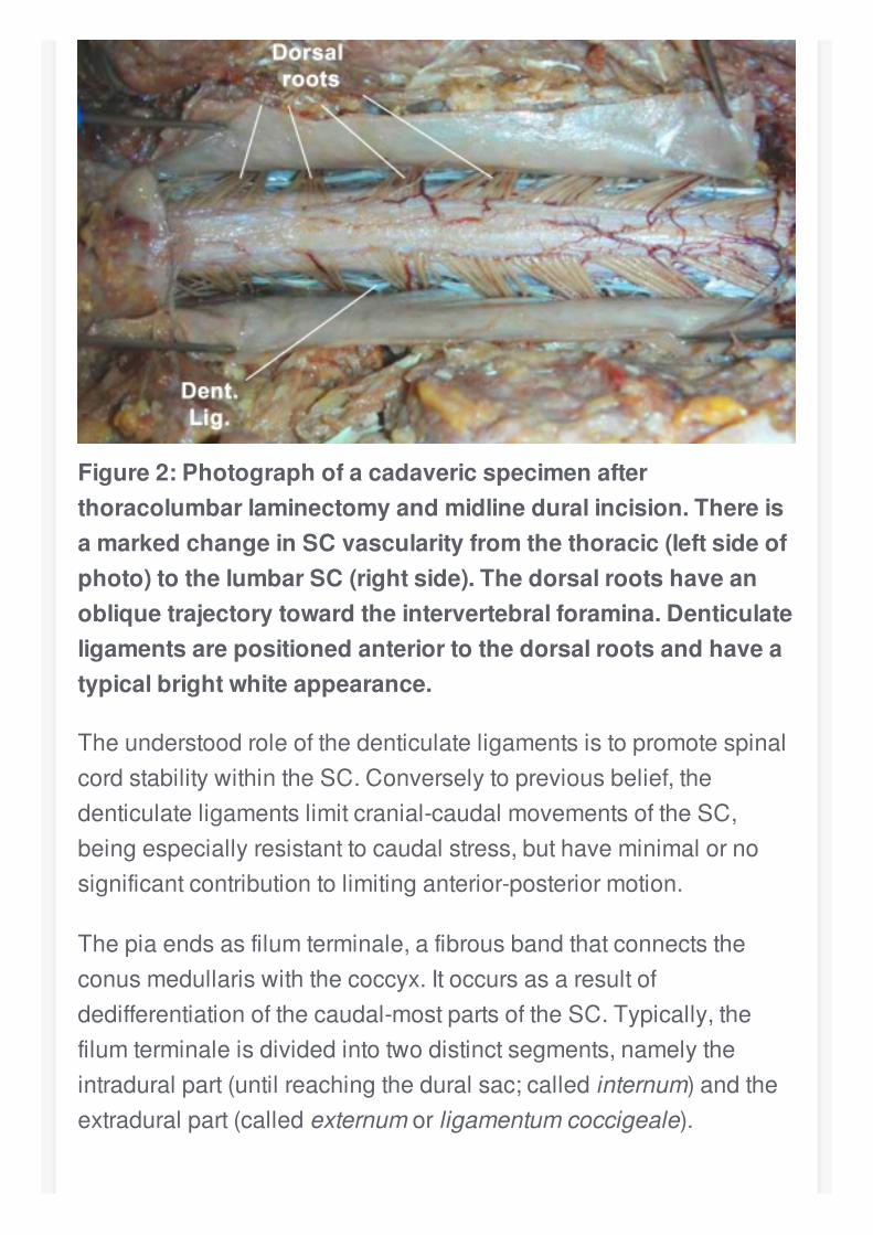
Figure2:Photographofacadavericspecimenafterthoracolumbarlaminectomyandmidlineduralincision.ThereisamarkedchangeinSCvascularityfromthethoracic(leftsideofphoto)tothelumbarSC(rightside).Thedorsalrootshaveanobliquetrajectorytowardtheintervertebralforamina.Denticulateligamentsarepositionedanteriortothedorsalrootsandhaveatypicalbrightwhiteappearance.
TheunderstoodroleofthedenticulateligamentsistopromotespinalcordstabilitywithintheSC.Converselytopreviousbelief,thedenticulateligamentslimitcranial-caudalmovementsoftheSC,beingespeciallyresistanttocaudalstress,buthaveminimalornosignificantcontributiontolimitinganterior-posteriormotion.
Thepiaendsasfilumterminale,afibrousbandthatconnectstheconusmedullariswiththecoccyx.Itoccursasaresultofdedifferentiationofthecaudal-mostpartsoftheSC.Typically,thefilumterminaleisdividedintotwodistinctsegments,namelytheintraduralpart(untilreachingtheduralsac;calledinternum)andtheextraduralpart(calledexternumorligamentumcoccigeale).
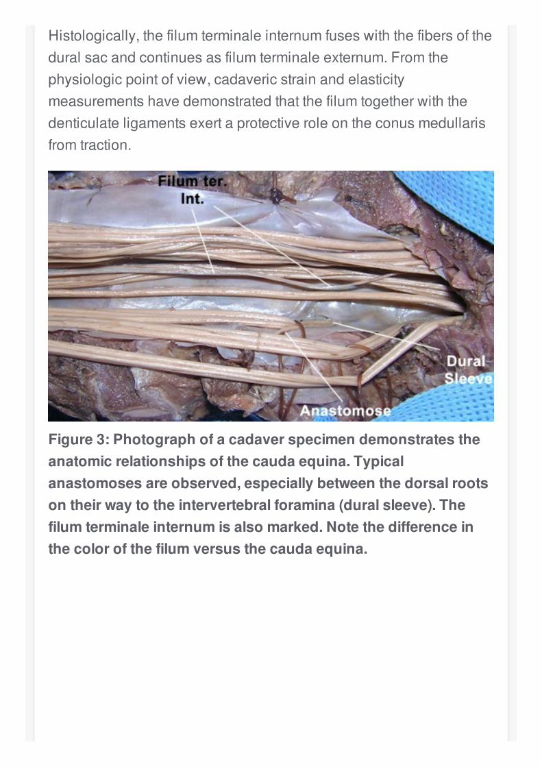
Histologically,thefilumterminaleinternumfuseswiththefibersoftheduralsacandcontinuesasfilumterminaleexternum.Fromthephysiologicpointofview,cadavericstrainandelasticitymeasurementshavedemonstratedthatthefilumtogetherwiththedenticulateligamentsexertaprotectiveroleontheconusmedullarisfromtraction.
Figure3:Photographofacadaverspecimendemonstratestheanatomicrelationshipsofthecaudaequina.Typicalanastomosesareobserved,especiallybetweenthedorsalrootsontheirwaytotheintervertebralforamina(duralsleeve).Thefilumterminaleinternumisalsomarked.Notethedifferenceinthecolorofthefilumversusthecaudaequina.
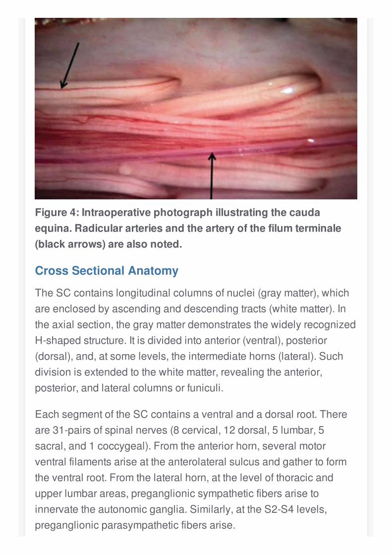
Figure4:Intraoperativephotographillustratingthecaudaequina.Radiculararteriesandthearteryofthefilumterminale(blackarrows)arealsonoted.
CrossSectionalAnatomy
TheSCcontainslongitudinalcolumnsofnuclei(graymatter),whichareenclosedbyascendinganddescendingtracts(whitematter).Intheaxialsection,thegraymatterdemonstratesthewidelyrecognizedH-shapedstructure.Itisdividedintoanterior(ventral),posterior(dorsal),and,atsomelevels,theintermediatehorns(lateral).Suchdivisionisextendedtothewhitematter,revealingtheanterior,posterior,andlateralcolumnsorfuniculi.
EachsegmentoftheSCcontainsaventralandadorsalroot.Thereare31-pairsofspinalnerves(8cervical,12dorsal,5lumbar,5sacral,and1coccygeal).Fromtheanteriorhorn,severalmotorventralfilamentsariseattheanterolateralsulcusandgathertoformtheventralroot.Fromthelateralhorn,atthelevelofthoracicandupperlumbarareas,preganglionicsympatheticfibersarisetoinnervatetheautonomicganglia.Similarly,attheS2-S4levels,preganglionicparasympatheticfibersarise.
Dorsalsensoryafferentsentertheposterolateralsulcusontheirwaytotheposteriorhorn.Deeperfissuresareconsistentlyencounteredattheventral(anteriormedianfissure)anddorsal(posteriormediansulcus)surfaces.Moreover,theposteriorcolumnisdividedbytheposteriorintermediatesulcusatupperthoraciclevels.
InthecenteroftheSCliesthecentralcanal.Itconsistsoftheremnantsoftheneuraltubecentralcavitylinedbyependymalcellsandfilledwithcerebrospinalfluid.Theanteriorandposteriorcommissuresenclosethecentralcanal.
Thegraymatterhornsaresomatotopicallyorganizedandcontaindifferentclassesoffunctionalneurons.Asaresult,motoneuronsthatinnervateaxialmusclesaremediallylocatedintheventralhorn,whereasmotoneuronsthatcontroldistallimbmovementsarelocatedmorelaterally.Finally,motoneuronsresponsibleforcontrollingproximallimbmuscleslieinbetween.
Theposteriorhornhasalayeredneuronalorganization,whichisbasedonsynapticinputsandoutputs.Thesuperficiallayersreceiveexteroceptivesensoryinformationaboutpain,temperature,andlighttouch,andgeneratethecontralateralspinothalamictracts.Thedeeplayersareinvolvedwithproprioceptiveinformationandcontributetotheipsilateralspinocerebellartracts.Theposteriorcervicalhornalsoincludesthespinalnucleusofthetrigeminalnerve.
Thewhitematterisorganizedintractsassociatedwithmajormotororsensoryfunctions.Theposteriorcolumnenlarges,astheSCascends,toincludemoreaxonscarryingfinetouch,vibration,andproprioceptiveinformationfromthelowerlimbsmedially(fasciculusgracilis)andtheupperlimbslaterally(fasciculuscuneatus).Thelateralcolumncontainstwomostprominentascendingtracts,namelythelateralspinothalamicandthespinocerebellarones,andone
descendingtract,thelateralcorticospinaltract.Finally,theanteriorspinothalamicandcorticospinaltractsarefoundintheanteriorcolumn.
Thespinothalamictractscarrycontralateralinformationaboutcrudetouch,pain,andtemperature.Twotopographicaldetailsinthesetractsareworthdiscussing.First,theinitialsynapseoccursattheposteriorhorn,wheresecond-orderneuronsgenerateaxonsthatcourseapproximatelytwotothreelevelsaboveinordertodecussatethroughtheanteriorcommissure.Second,sacralfibersareplacedmorelaterallythancervicalones,therebyjustifyingthephenomenonofsensorysacralsparingincentralSClesions.
Thelateralcorticospinaltractisformedatthecaudalmedulla,wheremostofthefiberscomingfromtheprimarymotorcortexdecussate.Theuncrossedfiberscontinueasanteriorcorticospinaltractanddecussateonlyatthelevelofthesynapsewiththeventralhornneurons.Thistractisinvolvedinthecontrolofproximallimbandaxialmuscles.
Thelateralcorticospinaltractshowsasimilartopographicaldistributiontothespinothalamictract,insuchawaythatsacralfibersaresituatedlaterally.Inclinicalscenarios,compressivecervicalconditionsinitiallyaffectmotorfunctionsofthelowerlimbs.Severalothertractsaredescribed,eventhoughtheyarelessclinicallyrelevant.
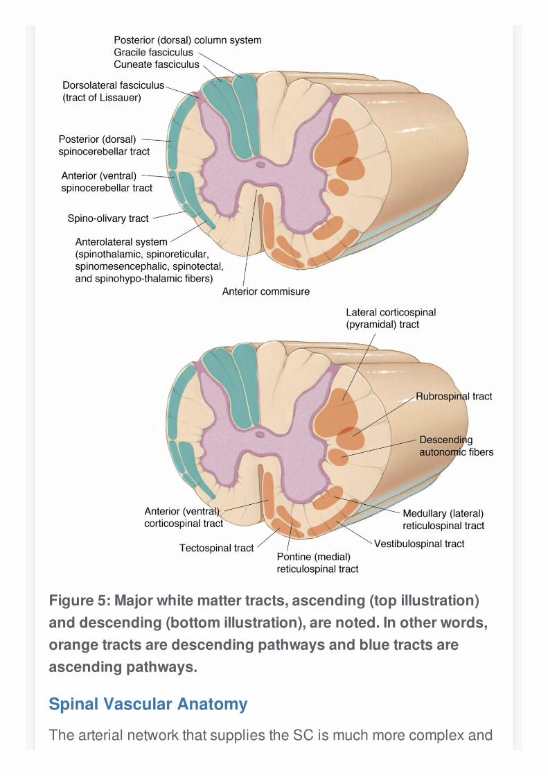
Figure5:Majorwhitemattertracts,ascending(topillustration)anddescending(bottomillustration),arenoted.Inotherwords,orangetractsaredescendingpathwaysandbluetractsareascendingpathways.
SpinalVascularAnatomy
ThearterialnetworkthatsuppliestheSCismuchmorecomplexand
extensivethanthatofthebrain.Thespinalvascularanatomystartsinthesegmentalextraspinalarteries,whichcorrespondtothepathwaysofbloodfromtheaortaandprovidenotonlythearterialsupplytothecord,butalsotothenerveroots,dura,andparaspinalmusculature.
Eachsegmentalarteryhasaventralandadorsalbranch.Thedorsaldivisiongivesoffaspinalbranch,whichsplitsintotheretrocorporeal(anteriorspinalcanal),prelaminar(posteriorspinalcanal),andradiculararteries.Theradiculararteryistermedtheradiculomeningealarterywhenitfeedsthenerverootsandduraateverylevel.
Ontheotherhand,ifthesearteriestakepartinthecordvascularnetwork,theyarebettertermedradiculomedullaryarteriesiftheysupplytheanteriorspinalartery(ASA),andradiculopialorposteriorradiculomedullaryarteries,iftheysupplytheposteriorspinalarteries(PSAs)andsurfacevasocoronaoftheSC.
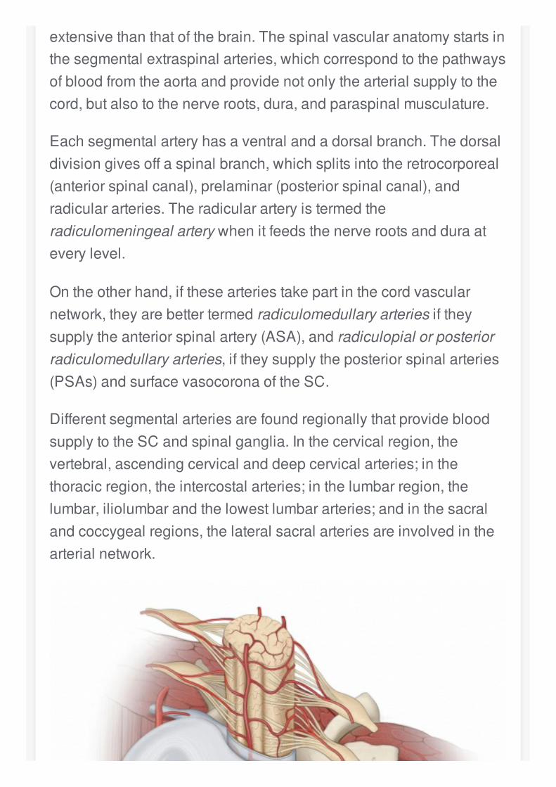
DifferentsegmentalarteriesarefoundregionallythatprovidebloodsupplytotheSCandspinalganglia.Inthecervicalregion,thevertebral,ascendingcervicalanddeepcervicalarteries;inthethoracicregion,theintercostalarteries;inthelumbarregion,thelumbar,iliolumbarandthelowestlumbararteries;andinthesacralandcoccygealregions,thelateralsacralarteriesareinvolvedinthearterialnetwork.
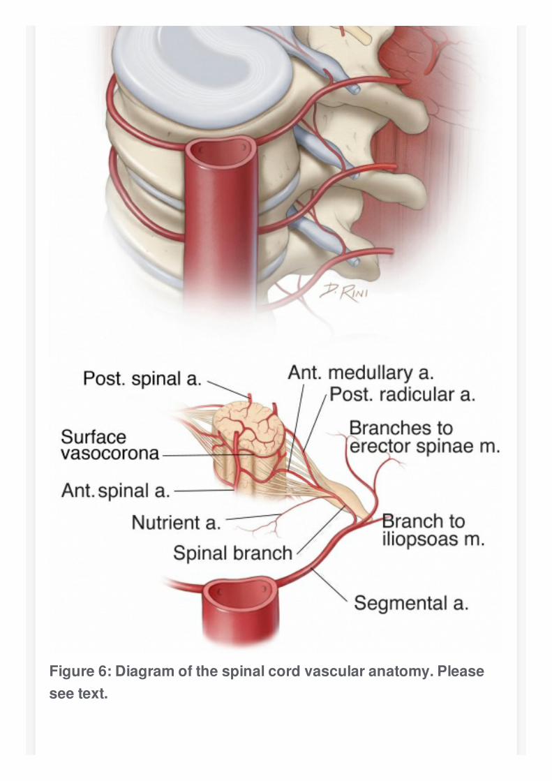
Figure6:Diagramofthespinalcordvascularanatomy.Pleaseseetext.
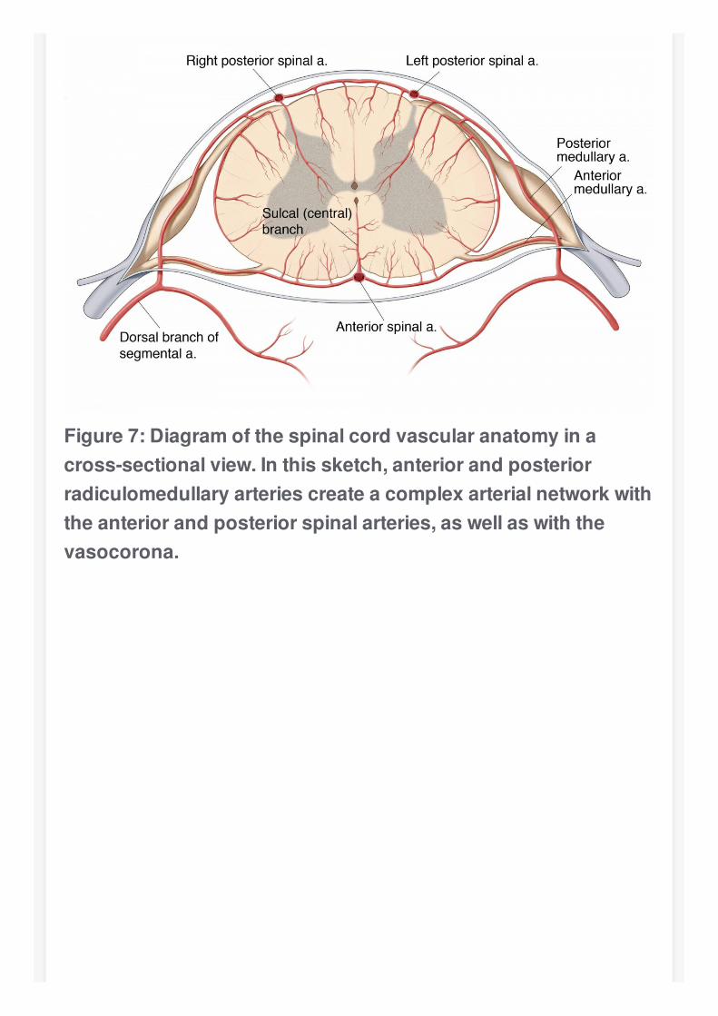
Figure7:Diagramofthespinalcordvascularanatomyinacross-sectionalview.Inthissketch,anteriorandposteriorradiculomedullaryarteriescreateacomplexarterialnetworkwiththeanteriorandposteriorspinalarteries,aswellaswiththevasocorona.
Figure8:DiagramoftheSCsegmentalarterialanatomy.
Inthecervicalregion,8to10unpairedradiculomedullaryarteries,includingatleast1medullaryarterytothecervicalenlargementatthelevelofC6,connectdirectlywithASA.
Thethoracicregionhasanotherwisepoorvascularitywithfrom2to4smallmedullaryarteries.Thelumbarenlargementatthethoracolumbarregionisrichlysuppliedby1singlevessel,calledthearteryofAdamkiewicz.ItcommonlyarisesbetweenT9andT12in75%ofthespecimens,eventhoughhigher(T5-T8)orlower(L1-L2)levelsmayalsocarrythisimportantarteryin15%and10%,respectively.
ThearteryofAdamkiewiczisalsoknownasthegreatradiculararteryorevenasarteriaradiculomedullarismagnaandhasaleft-sidedpredominance.Asthearterypiercesthedura,aslightcaudalturnmayoccasionallybeseen.ThearterythenjoinstheventralrootonitswaytotheventralsurfaceoftheSC,wherethearteryanastomoseswiththeASAatorjustbeforeitstypicalandcharacteristichairpinturn.ThePSAsreceiveapproximately10to28feeders,whichcanalsodemonstrateahairpinconfigurationintheparamedianlocations.
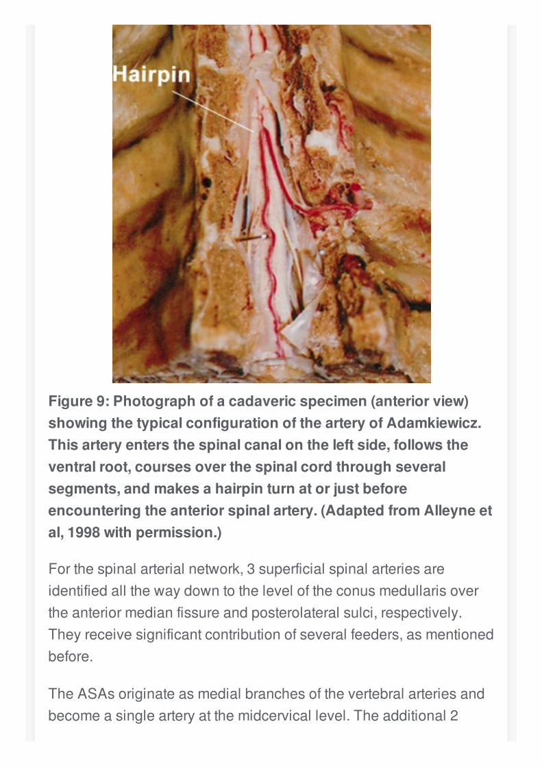
Figure9:Photographofacadavericspecimen(anteriorview)showingthetypicalconfigurationofthearteryofAdamkiewicz.Thisarteryentersthespinalcanalontheleftside,followstheventralroot,coursesoverthespinalcordthroughseveralsegments,andmakesahairpinturnatorjustbeforeencounteringtheanteriorspinalartery.(AdaptedfromAlleyneetal,1998withpermission.)
Forthespinalarterialnetwork,3superficialspinalarteriesareidentifiedallthewaydowntotheleveloftheconusmedullarisovertheanteriormedianfissureandposterolateralsulci,respectively.Theyreceivesignificantcontributionofseveralfeeders,asmentionedbefore.
TheASAsoriginateasmedialbranchesofthevertebralarteriesandbecomeasinglearteryatthemidcervicallevel.Theadditional2
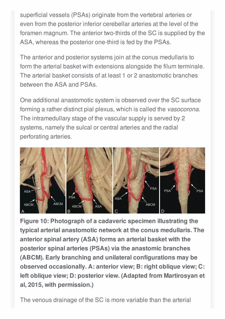
superficialvessels(PSAs)originatefromthevertebralarteriesorevenfromtheposteriorinferiorcerebellararteriesattheleveloftheforamenmagnum.Theanteriortwo-thirdsoftheSCissuppliedbytheASA,whereastheposteriorone-thirdisfedbythePSAs.
Theanteriorandposteriorsystemsjoinattheconusmedullaristoformthearterialbasketwithextensionsalongsidethefilumterminale.Thearterialbasketconsistsofatleast1or2anastomoticbranchesbetweentheASAandPSAs.
OneadditionalanastomoticsystemisobservedovertheSCsurfaceformingaratherdistinctpialplexus,whichiscalledthevasocorona.Theintramedullarystageofthevascularsupplyisservedby2systems,namelythesulcalorcentralarteriesandtheradialperforatingarteries.
Figure10:Photographofacadavericspecimenillustratingthetypicalarterialanastomoticnetworkattheconusmedullaris.Theanteriorspinalartery(ASA)formsanarterialbasketwiththeposteriorspinalarteries(PSAs)viatheanastomicbranches(ABCM).Earlybranchingandunilateralconfigurationsmaybeobservedoccasionally.A:anteriorview;B:rightobliqueview;C:leftobliqueview;D:posteriorview.(AdaptedfromMartirosyanetal,2015,withpermission.)
ThevenousdrainageoftheSCismorevariablethanthearterial
supplyandhasbeendividedintointrinsicandextrinsicsystems.Theintrinsicsystemcontainscentrifugallyorientedperipheralorradialveins,whichemergefromcapillariesatthegray-whitematterjunction.
Sulcalorcentralveinsdraintheanteriorhorns,anteriorcommissure,andassociatedwhitematterandcompletetheintrinsicsystemofvenousdrainage.Theextrinsicsystemofvenousdrainagehasadirectcorrelationtothepatternsofthearterialsystem.Twodominantveinsareusuallyobserved,theanteriorandposteriormedianveins.Thefirstreceivesvenousdrainagefromthesulcalveins,whereasthelatterreceivesvenousdrainagefromtheperipheralveins.
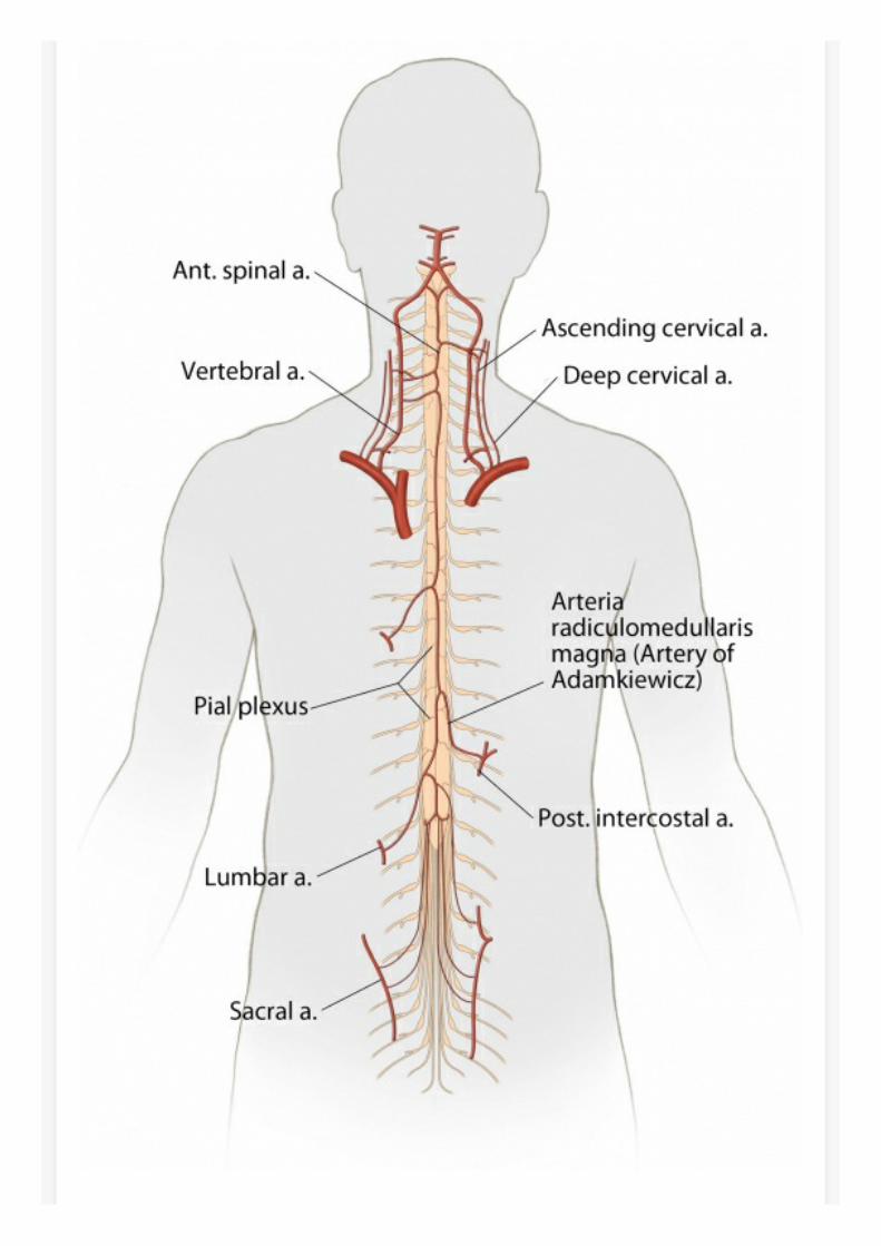
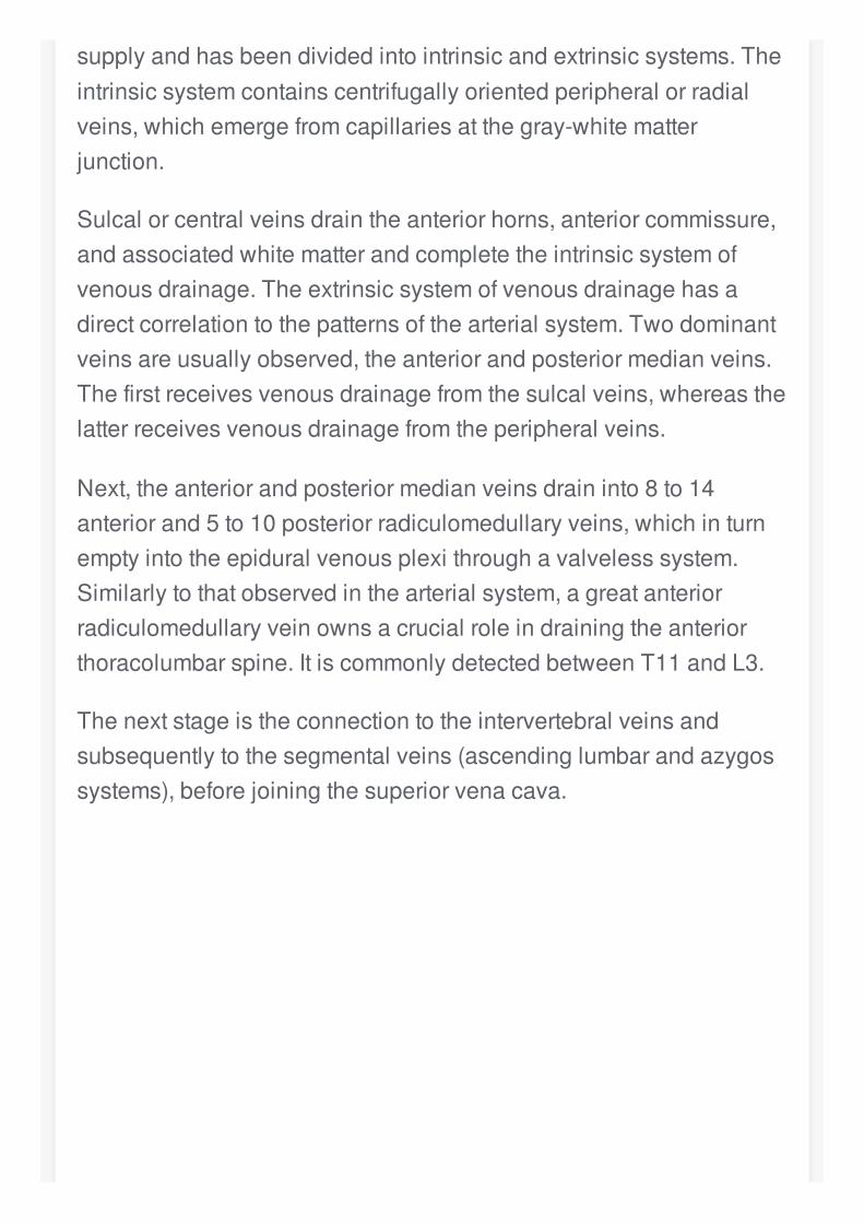
Next,theanteriorandposteriormedianveinsdraininto8to14anteriorand5to10posteriorradiculomedullaryveins,whichinturnemptyintotheepiduralvenousplexithroughavalvelesssystem.Similarlytothatobservedinthearterialsystem,agreatanteriorradiculomedullaryveinownsacrucialroleindrainingtheanteriorthoracolumbarspine.ItiscommonlydetectedbetweenT11andL3.
Thenextstageistheconnectiontotheintervertebralveinsandsubsequentlytothesegmentalveins(ascendinglumbarandazygossystems),beforejoiningthesuperiorvenacava.
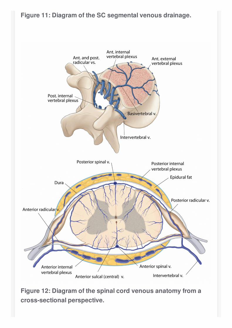
Figure11:DiagramoftheSCsegmentalvenousdrainage.
Figure12:Diagramofthespinalcordvenousanatomyfromacross-sectionalperspective.
PearlsandPitfalls
TheSChasacomplexanatomicalorganizationforreceiving,processing,andtransferringinformation.Longtractsgenerallycrossthemidline,eventhoughdecussationoccursatdifferentlevels.Dependingontheaxialandlongitudinalextentofthelesion,differentmotorandsensorysyndromesmaydevelop.TheexistenceofsulciandfissuresovertheSCsurfaceprovidesrelativelysafeentryroutesforreachingintramedullarytumors.WhenapproachingthelowerthoracicSClevels,thesurgeonshouldinvestigatethelocationofthearteryofAdamkiewicz.
Contributor:MarcusAndréAcioly,MD,PhD
DOI:https://doi.org/10.18791/nsatlas.v9.ch01
References
AlleyneCHJr,CawleyCM,ShengelaiaGG,BarrowDL.MicrosurgicalanatomyofthearteryofAdamkiewiczanditssegmentalartery.JNeurosurg.1998;89:791-795.
BosmiaAN,HoganE,LoukasM,TubbsRS,Cohen-GadolAA.Bloodsupplytothehumanspinalcord:partI.Anatomyandhemodynamics.ClinAnat.2015;28:52-64.
ChoTA.Spinalcordfunctionalanatomy.Continuum(MinneapMinn).2015;21(1SpinalCordDisorders):13-35.
DeVlooP,MoneaAG,SciotR,vanLoonJ,VanCalenberghF.Thefilumterminale:acadaverstudyofanatomy,histology,andelasticproperties.WorldNeurosurg.2016;90:565-573.
HauckEF,WittkowskiW,BotheHW.IntraduralmicroanatomyofthenerverootsS1-S5attheiroriginfromtheconusmedullaris.JNeurosurgSpine.2008;9:207-212.
KlekampJ,SamiiM.SurgeryofSpinalTumors.1stEd.BerlinHeridelberg:Springer,2007.
MartirosyanNL,KalaniMY,LemoleGMJr,SpetzlerRF,PreulMC,TheodoreN.Microsurgicalanatomyofthearterialbasketoftheconusmedullaris.JNeurosurgSpine.2015;22:672-676.
NicholasDS,WellerRO.Thefineanatomyofthehumanspinalmeninges.Alightandscanningelectronmicroscopystudy.JNeurosurg.1988;69:276-282.
RossignolS.Anatomyandphisiologyofthespinalcord,inFehlingsMG,BoakyeM,VaccaroAR,RossignolS,DittunoAJrBurnsAS(eds):EssentialsofSpinalCordSurgery:BasicResearchtoClinicalPractice.1stEd.NewYork-Stuttgart:Thieme,2013,pp.3-17.
SinghP,GobinP.SpinalvascularanatomyandimplicationsfortreatmentofarteriovenousmalformationsinSpetzlerRF,KondziolkaDS,HigashidaRT,KalaniMYS(eds):ComprehensiveManagementofArteriovenousMalformationsoftheBrainandSpine.1stEd.Cambridge:CambridgeUniversityPress,2015,pp.29-36.
TubbsRS,SalterG,GrabbPA,OakesWJ.Thedenticulateligament:anatomyandfunctionalsignificance.JNeurosurg.2001;94(2Suppl):271-275.