Embed Size (px)
Citation preview
11
Unit
Thre
e
Human Anatomy & Physiology
Divisions of the Nervous SystemKaren W. Smith, InstructorDivisions of the Nervous SystemDivisions of the Nervous SystemKaren W. Smith, InstructorKaren W. Smith, Instructor
BRAIN & SPINAL CORD
Refer to the following URLs. Be sure to study these along with your book.
http://www.sirinet.net/~jgjohnso/nervous.html
http://faculty.washington.edu/chudler/ap.html
http://faculty.washington.edu/chudler/synapse.html
http://www.emc.maricopa.edu/faculty/farabee/BIOBK/BioBookNERV.html
The brain includes:cerebrum, diencephalon, brain stem, & cerebellum
The brain lies within the cranial cavity of the skull.
The brain stem includes:midbrain, pons, medulla oblongata
The brain stem connects the brain and spinal cord& allows 2-way communication between them.
The spinal cord occupies the vertebral canal withinthe vertebral column.
I. Introduction
*Organs of the central nervous system are the brain & spinal cord.
*The brain lies within the cranial cavity of the skull.
*The spinal cord continues from the brain & is inside the vertebral canal within the vertebral column.
*Protection of the brain and spinal cord is provided by bone, fluid, & by the membranes called meninges that surround these structures.
*Meninges (membranes – 3 layers) are located between the bone & the soft tissues of the nervous system. They protect the brain & spinal cord.
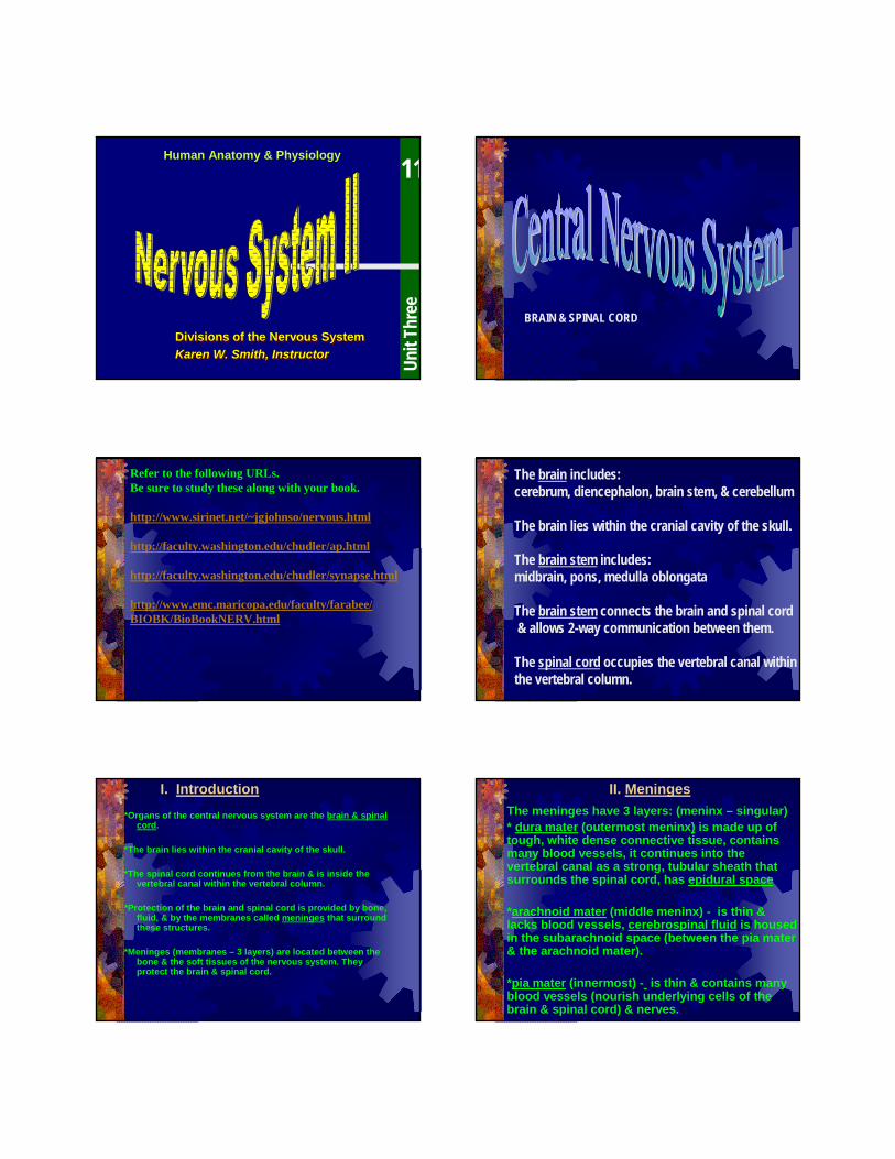
II. MeningesThe meninges have 3 layers: (meninx – singular)* dura mater (outermost meninx) is made up of tough, white dense connective tissue, contains many blood vessels, it continues into the vertebral canal as a strong, tubular sheath that surrounds the spinal cord, has epidural space
*arachnoid mater (middle meninx) - is thin & lacks blood vessels, cerebrospinal fluid is housed in the subarachnoid space (between the pia mater & the arachnoid mater).
*pia mater (innermost) - is thin & contains many blood vessels (nourish underlying cells of the brain & spinal cord) & nerves.
The dura mater is being lifted away from the brain.
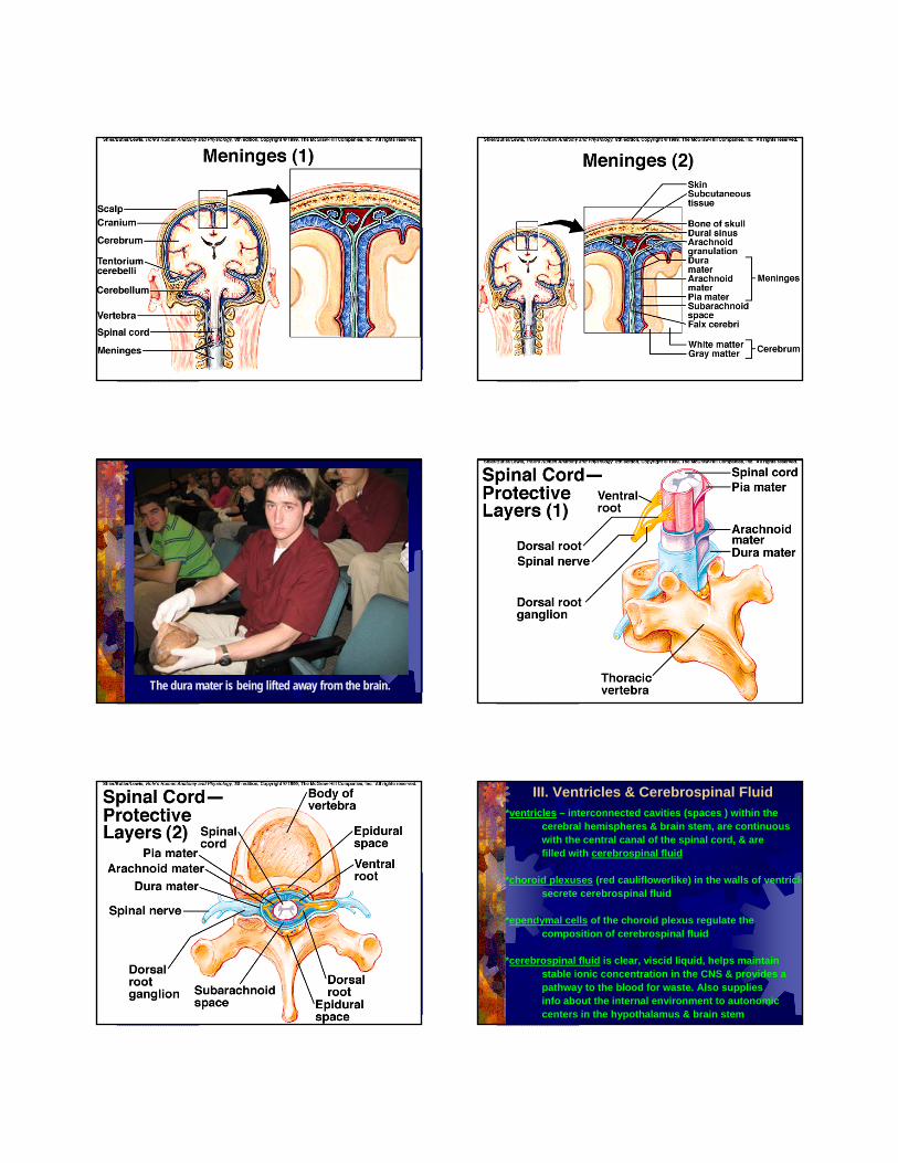
III. Ventricles & Cerebrospinal Fluid*ventricles – interconnected cavities (spaces ) within the
cerebral hemispheres & brain stem, are continuouswith the central canal of the spinal cord, & are filled with cerebrospinal fluid
*choroid plexuses (red cauliflowerlike) in the walls of ventriclesecrete cerebrospinal fluid
*ependymal cells of the choroid plexus regulate the composition of cerebrospinal fluid
*cerebrospinal fluid is clear, viscid liquid, helps maintainstable ionic concentration in the CNS & provides apathway to the blood for waste. Also suppliesinfo about the internal environment to autonomic centers in the hypothalamus & brain stem
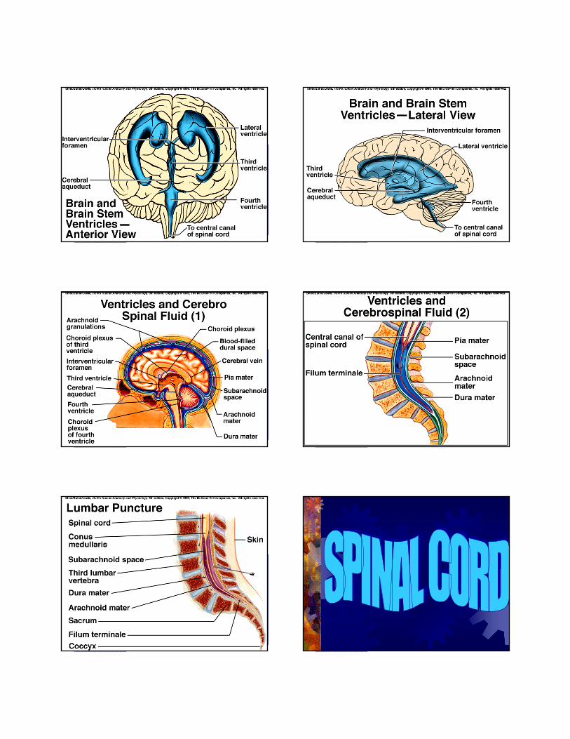
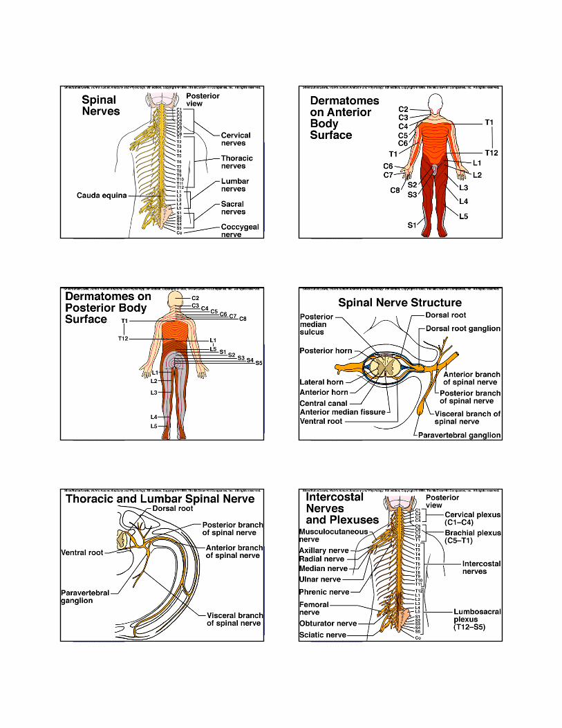
IV. Spinal Cord
A. The spinal cord is a slender column of nerve fibers that begins at the base of the brain & extends down through the vertebral canal. It terminates near the intervertebral disk that separates the first & second lumbar vertebrae.
B. Structure of the Spinal Cord*31 segments, each gives rise to a pair of spinal nerves that branch to reach the central nervous system*cervical enlargement – supplies nerves to the upper limbs*lumbar enlargement – supplies nerves to the lower limbs (continued next slide)
An actual spinal cord
*2 deep longitudinal grooves divide the spinal cord into right & left halves (ant. & post. fissures)
*white matter surrounds a central core of gray matter; white matter is composed of bundles of myelinated nerve fibers
*gray matter produces a pattern that resemblesa butterfly with outspread wings; neurons ingray matter are interneurons
*central canal – continuous with the ventricles of the brain & contains cerebrospinal fluid
*gray matter divides the white matter into 3 regionscalled anterior, lateral, & posterior funiculi
*each funiculi is a column of myelinated nerve fibers that comprise major nerve pathways called nerve tracts
C. Functions of the Spinal Cord2 major functions:#1 – conduit for nerve impulses to & from the brain#2 – center for spinal reflexes
*spinal cord provides a 2-way communication system between the brain & structures outside the nervous system
D. Reflex Arcs – carry out simplest responses
Includes the sensory receptor of a sensory neuron, an interneuron(s) within the CNS, a motor neuron whose fibers pass outward from the CNS to effectors.
Reflexes whose arcs pass through the spinal cord arespinal reflexes.
E. Reflex Behavior*reflexes are automatic, subconscious responses to changes*they help maintain homeostasis (ie.) heart rate, breathing
rate, blood pressure, & digestion
*the knee-jerk reflex employs only 2 neurons
*withdrawal reflexes are protective actions
*crossed extensor reflex – allows sensory impulses arriving at one side of the cord to pass across to the other side andproduce an opposite effect
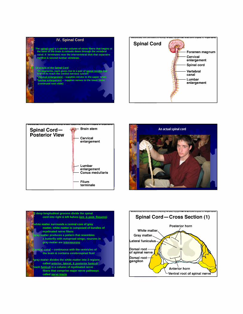
F. Ascending & Descending TractsThe nerve tracts of the spinal cord together with the spinalnerves provide a two-way communication system betweenthe brain and body parts outside the nervous system.
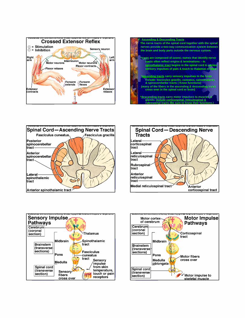
*tracts are composed of axons; names that identify nervetracts often reflect origins & terminations – ie. spinothalamic tract begins in the spinal cord & carriessensory impulses of pain & touch to thalamus of brain
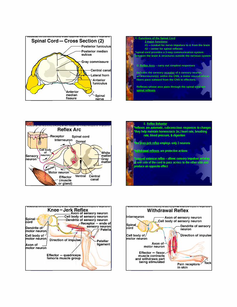
*ascending tracts carry sensory impulses to the braininclude: fasciculus gracilis, cuneatus, spinothalamic, & spinocerebellar tracts ( Know functions)
(many of the fibers in the ascending & descending tracts cross over in the spinal cord or brain)
*descending tracts carry motor impulses to muscles & glands; include corticospinal, reticulospinal &rubrospinal tracts (Be sure to know their functions.)
V. Brain A. The brain is the largest, most complex portion of the nervous
system, containing 100 billion multipolar neurons.B. It is responsible for processing sensory information,
producing sensations, storing memory, integrating information, reasoning, controlling visceral activities, providing personality, generating emotions, and initiating motor activities.
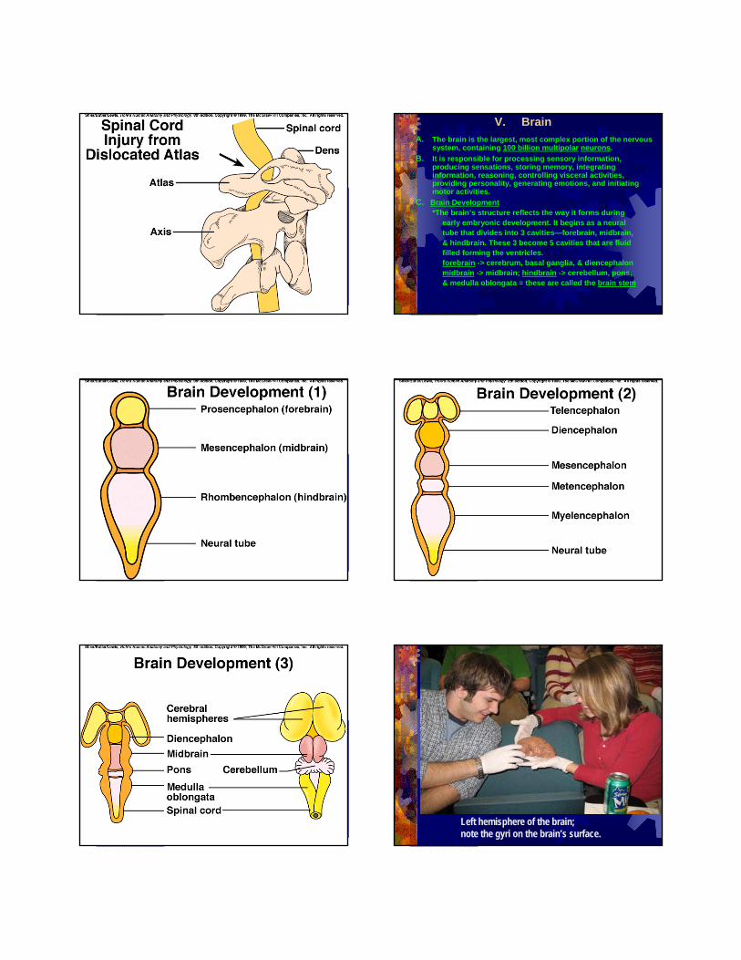
C. Brain Development*The brain’s structure reflects the way it forms during
early embryonic development. It begins as a neuraltube that divides into 3 cavities—forebrain, midbrain, & hindbrain. These 3 become 5 cavities that are fluidfilled forming the ventricles. forebrain -> cerebrum, basal ganglia, & diencephalonmidbrain -> midbrain; hindbrain -> cerebellum, pons, & medulla oblongata = these are called the brain stem
Left hemisphere of the brain; note the gyri on the brain’s surface.
V. Brain D. Structure of the Cerebrum – develops
from the anterior portion of the forebrain (largest part)
*consists of 2 large masses = cerebral hemispheres
*corpus callosum (deep bridge of nerve fibers) connect cerebral hemispheres
*surface marked by ridges & grooves called convolutions, & gyri; sulci divide each hemisphere into lobes
*know 5 lobes of the cerebral hemisphere: frontal, parietal, temporal, occipital, & insula
*cerebral cortex – thin layer of gray matter near surface ofthe brain; beneath cerebral cortex is a mass of white matter that makes up the bulk of the cerebrum
Inside the right hemisphere.
The lobes of the cerebral hemispheres are named after theskull bones that they underlie. (5 lobes)
1. Frontal lobe
2. Parietal lobe
3. Temporal lobe
4. Occipital lobe
5. Insula – located deep within the frontal, parietal, & temporal lobes;(integrates memories & sensations of taste, sound, sight, & touch)
E. Functions of the Cerebrum
*is concerned with higher brain functions, ie. thought, reasoning, interpretation of sensory impulses, control ofvoluntary muscles, & memory storage; intelligence &personality
*cerebral cortex = sensory, motor, & association areas*motor areas – in frontal lobe*sensory areas – in several lobes = parietal, temporal, &
occipital lobes*association areas – analyze & interpret; provide memory,
reasoning, verbalizing, judgment, & emotions*one cerebral hemisphere usually dominates for certain
intellectual functions*short-term memory is probably electrical*long-term memory is probably encoded in patterns of
synaptic connections
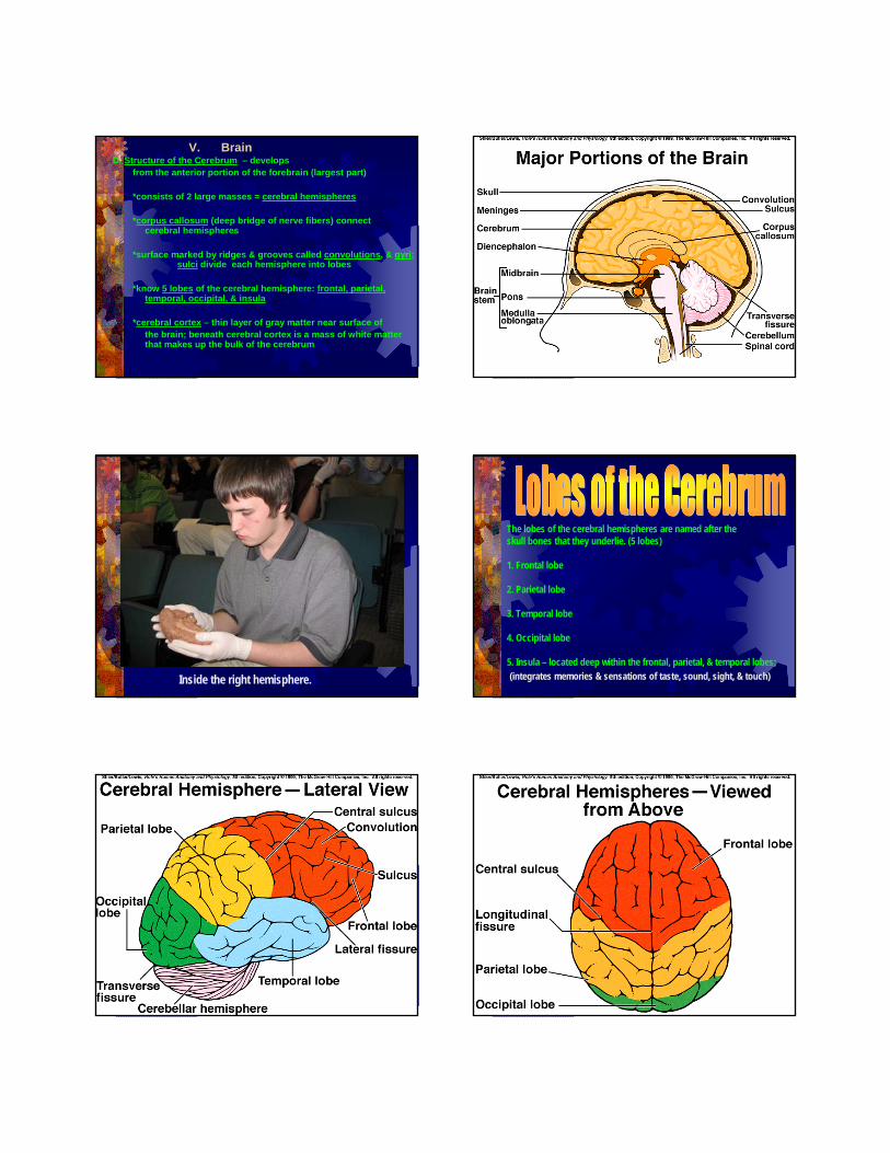
F. Basal Nuclei (basal ganglia)
*masses of gray matter located deep within the cerebral hemispheres (unmyelinated); called thecaudate nucleus, putamen, & globus pallidus
*develop from anterior portion of the forebrain
*they relay motor impulses originating in the cerebralcortex, & aid in controlling motor activities (muscular activities)
*they produce most of the inhibitory neurotransmitterdopamine thus controlling certain muscular activities (lack of causes Parkinson’s Disease)
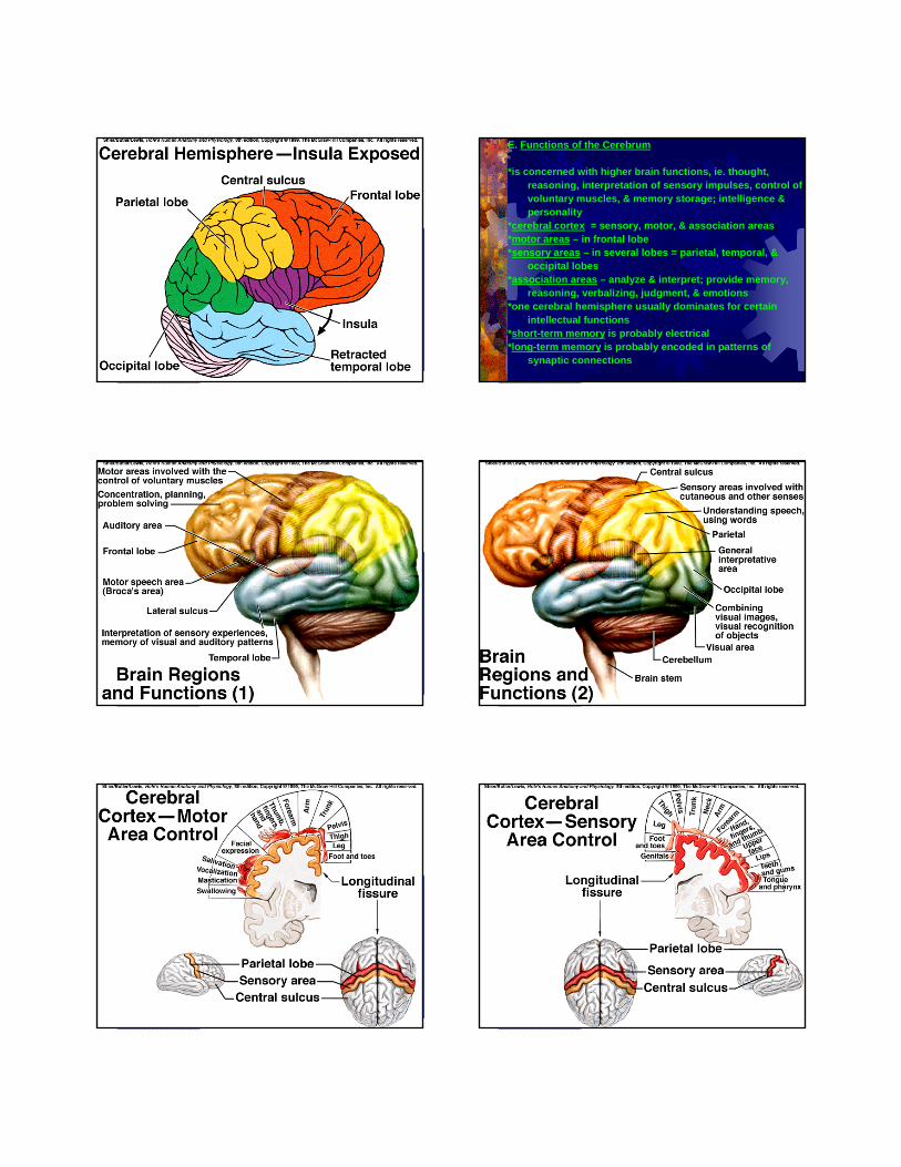
H. Diencephalon
*located between the cerebral hemispheres & above brain stem; *contains thalamus & hypothalamus
*thalamus – bulges into 3rd ventricle; selects incoming sensory impulses & relays them to the cerebral cortex; does not receive sense of smell
*hypothalamus – forms lower walls & floor of the 3rd
ventricle; important in maintaining homeostasis by regulating a variety of visceral activities & by linking the nervous system & endocrine systems
I. Brain Stem*connects the brain to the spinal cord; consists of the
midbrain, pons, & medulla oblongata; these structures include many tracts of nerve fibers & masses of gray matter called nuclei
*midbrain – short section of the brain stem, it contains bundles of myelinated nerve fibers that join lower parts of the brain stem & spinal cord with higher parts of the brain; helps with eye & head movements
*pons – a rounded bulge on the underside of the brain stem, helps regulate rate & depth of breathing
*medulla oblongata – extends from the level of the foramen magnum to the pons; transmits all ascending & descending impulses & contains several vital & nonvital reflex centers
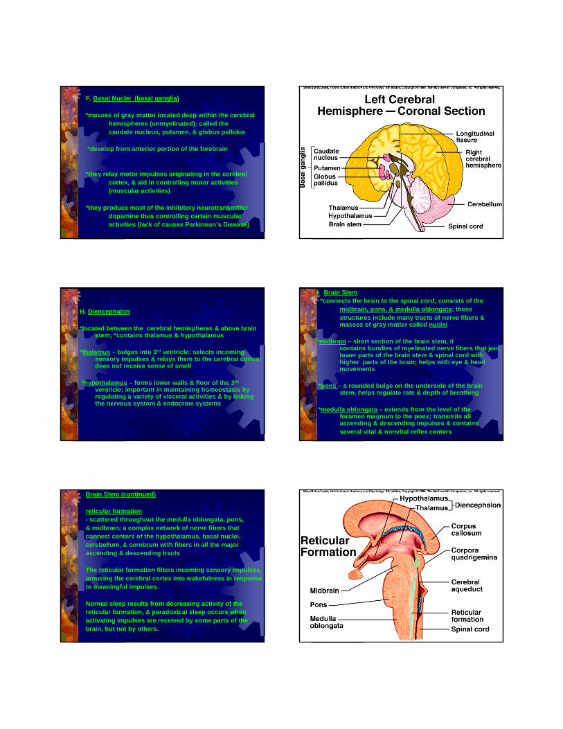
Brain Stem (continued)
reticular formation- scattered throughout the medulla oblongata, pons, & midbrain; a complex network of nerve fibers thatconnect centers of the hypothalamus, basal nuclei, cerebellum, & cerebrum with fibers in all the majorascending & descending tracts
The reticular formation filters incoming sensory impulses,arousing the cerebral cortex into wakefulness in responseto meaningful impulses.
Normal sleep results from decreasing activity of thereticular formation, & paradoxical sleep occurs when activating impulses are received by some parts of thebrain, but not by others.
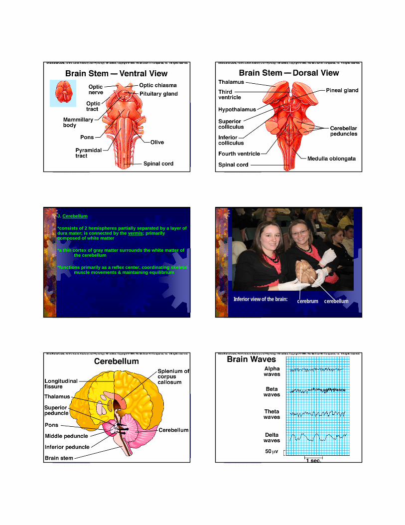
J. Cerebellum
*consists of 2 hemispheres partially separated by a layer of dura mater; is connected by the vermis; primarily composed of white matter
*a thin cortex of gray matter surrounds the white matter of the cerebellum
*functions primarily as a reflex center, coordinating skeletal muscle movements & maintaining equilibrium
Inferior view of the brain: cerebrum cerebellum
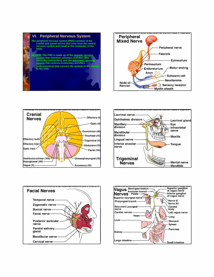
VI. Peripheral Nervous SystemThe peripheral nervous system (PNS) consists of the
cranial and spinal nerves that arise from the central nervous system and travel to the remainder of the body.
REVIEW: The PNS is made up of the somatic nervoussystem that oversees voluntary activities (like muscular contraction), and the autonomic nervous system that controls involuntary activities (subconscious) that connect the actions of the viscera to the CNS.
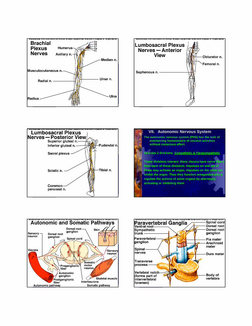
VII.VII. Autonomic Nervous SystemAutonomic Nervous SystemThe autonomic nervous system (PNS) has the task of
maintaining homeostasis of visceral activities without conscious effort.
Includes 2 divisions: Sympathetic & Parasympathetic
These divisions interact: Many viscera have nerve fibersfrom each of these divisions. Impulses on one set of fibers may activate an organ, impulses on the other setinhibit the organ. Thus they function antagonistically ®ulate the actions of some organs by alternately activating or inhibiting them
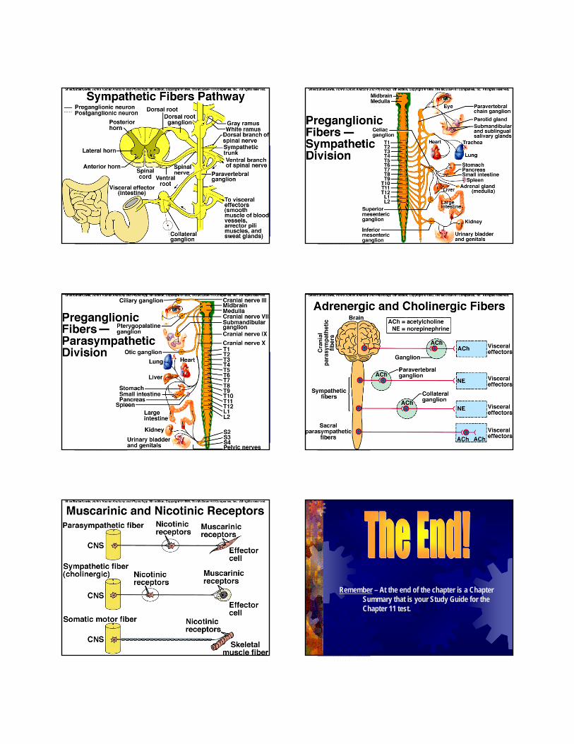
Remember – At the end of the chapter is a Chapter Summary that is your Study Guide for the Chapter 11 test.