Embed Size (px)
Citation preview
CONCEPTS OF BIOLOGYChapter 17 THE IMMUNE SYSTEM AND DISEASE
PowerPoint Image Slideshow
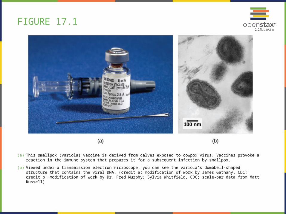
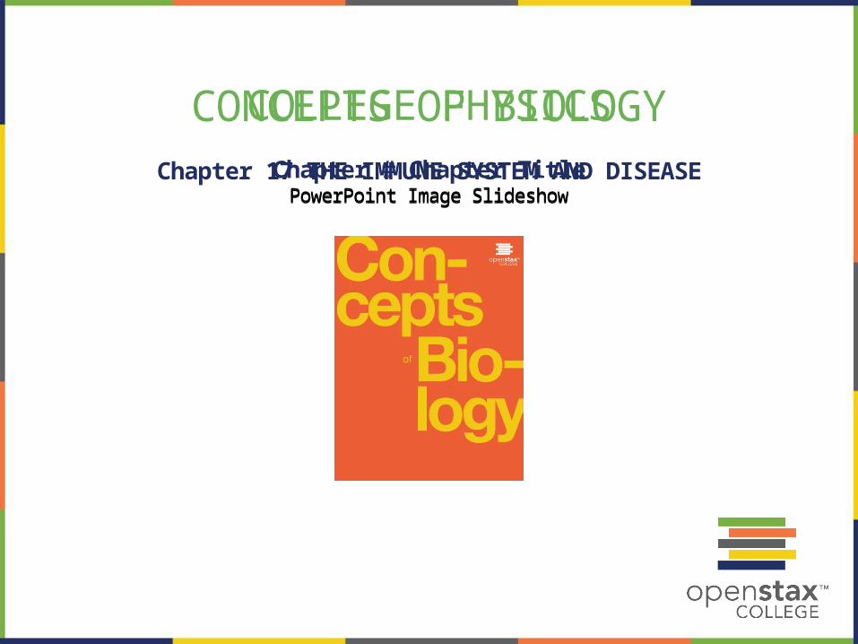
FIGURE 17.1
(a) This smallpox (variola) vaccine is derived from calves exposed to cowpox virus. Vaccines provoke a reaction in the immune system that prepares it for a subsequent infection by smallpox.
(b) Viewed under a transmission electron microscope, you can see the variola’s dumbbell-shaped structure that contains the viral DNA. (credit a: modification of work by James Gathany, CDC; credit b: modification of work by Dr. Fred Murphy; Sylvia Whitfield, CDC; scale-bar data from Matt Russell)
FIGURE 17.2
(a) The tobacco mosaic virus, seen by transmission electron microscopy, was the first virus to be discovered.
(b) The leaves of an infected plant are shown. (credit a: scale-bar data from Matt Russell; credit b: modification of work by USDA, Department of Plant Pathology Archive, North Carolina State University)
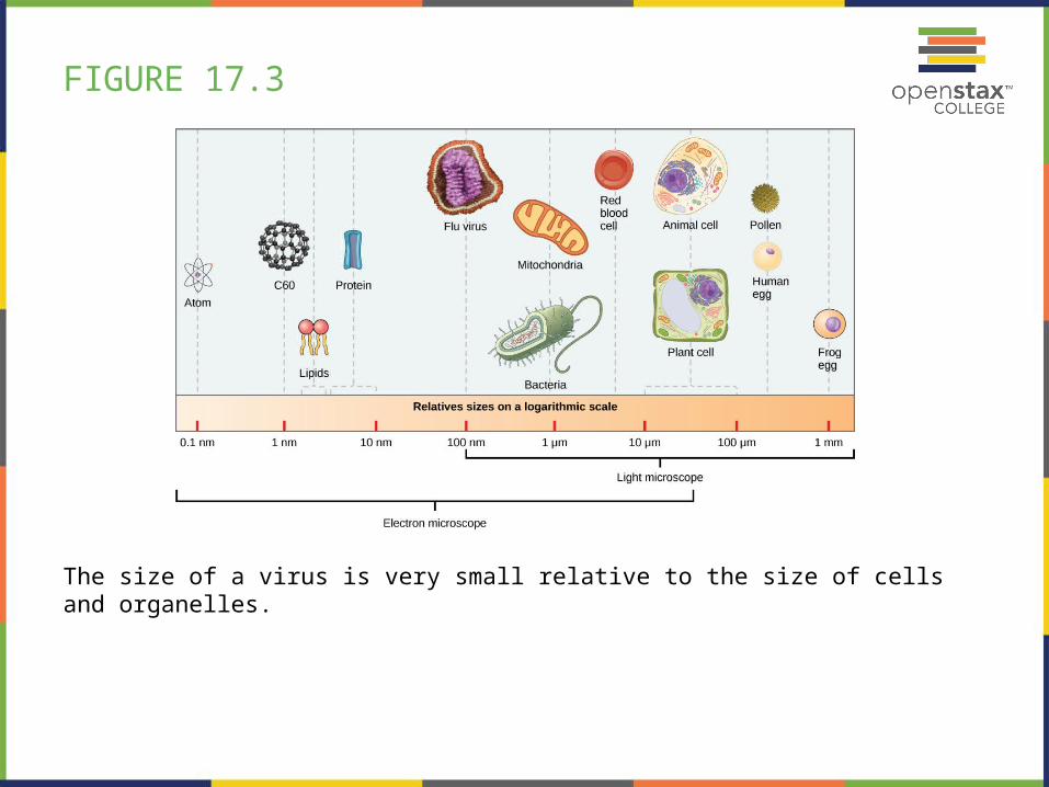
FIGURE 17.3
The size of a virus is very small relative to the size of cells and organelles.
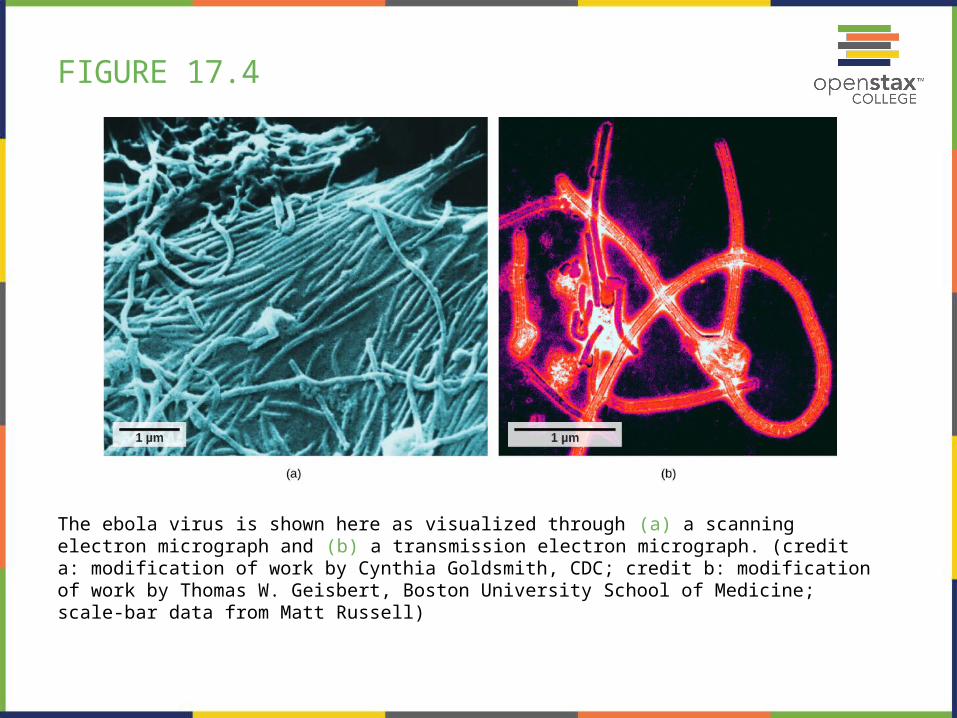
FIGURE 17.4
The ebola virus is shown here as visualized through (a) a scanning electron micrograph and (b) a transmission electron micrograph. (credit a: modification of work by Cynthia Goldsmith, CDC; credit b: modification of work by Thomas W. Geisbert, Boston University School of Medicine; scale-bar data from Matt Russell)
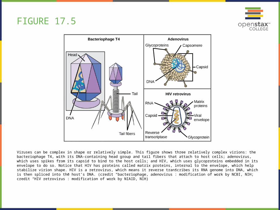
FIGURE 17.5
Viruses can be complex in shape or relatively simple. This figure shows three relatively complex virions: the bacteriophage T4, with its DNA-containing head group and tail fibers that attach to host cells; adenovirus, which uses spikes from its capsid to bind to the host cells; and HIV, which uses glycoproteins embedded in its envelope to do so. Notice that HIV has proteins called matrix proteins, internal to the envelope, which help stabilize virion shape. HIV is a retrovirus, which means it reverse transcribes its RNA genome into DNA, which is then spliced into the host’s DNA. (credit “bacteriophage, adenovirus”: modification of work by NCBI, NIH; credit “HIV retrovirus”: modification of work by NIAID, NIH)
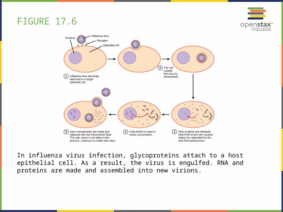
FIGURE 17.6
In influenza virus infection, glycoproteins attach to a host epithelial cell. As a result, the virus is engulfed. RNA and proteins are made and assembled into new virions.
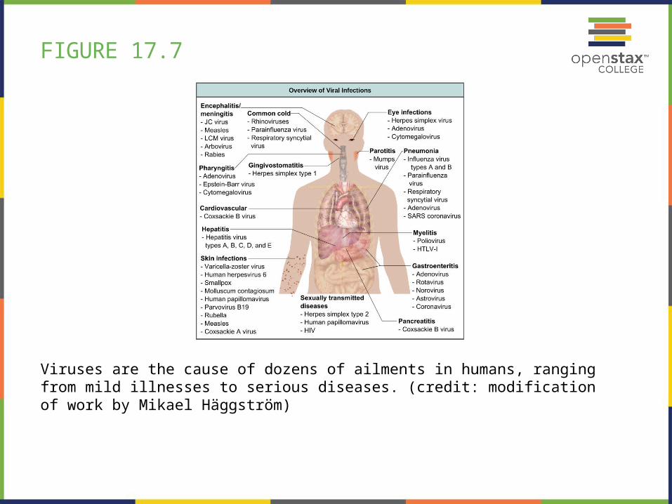
FIGURE 17.7
Viruses are the cause of dozens of ailments in humans, ranging from mild illnesses to serious diseases. (credit: modification of work by Mikael Häggström)
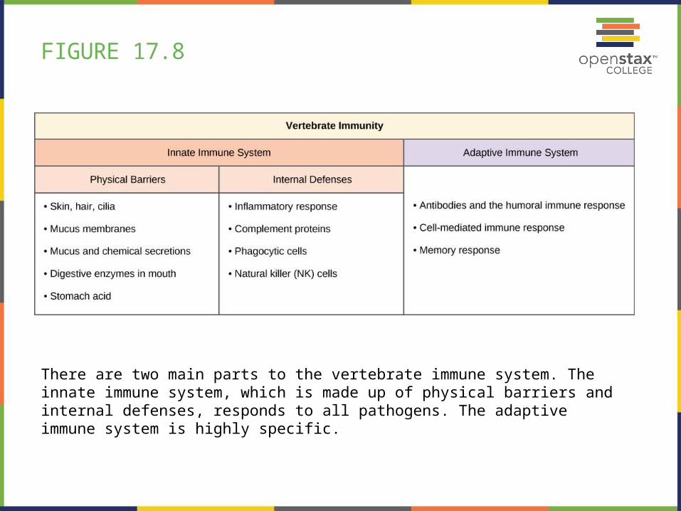
FIGURE 17.8
There are two main parts to the vertebrate immune system. The innate immune system, which is made up of physical barriers and internal defenses, responds to all pathogens. The adaptive immune system is highly specific.
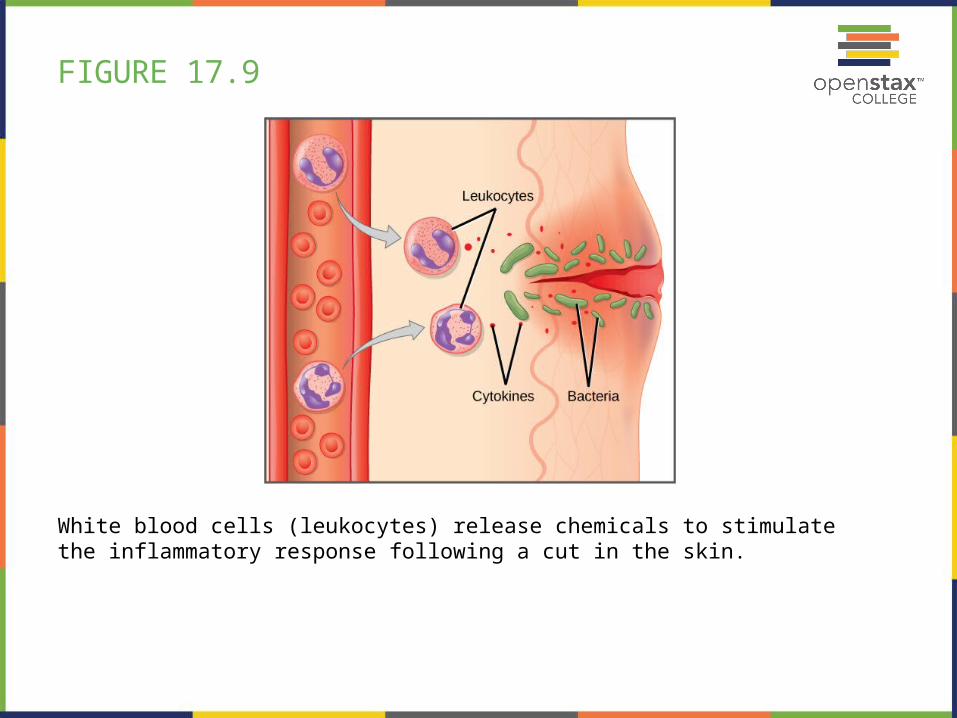
FIGURE 17.9
White blood cells (leukocytes) release chemicals to stimulate the inflammatory response following a cut in the skin.
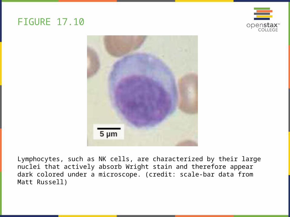
FIGURE 17.10
Lymphocytes, such as NK cells, are characterized by their large nuclei that actively absorb Wright stain and therefore appear dark colored under a microscope. (credit: scale-bar data from Matt Russell)
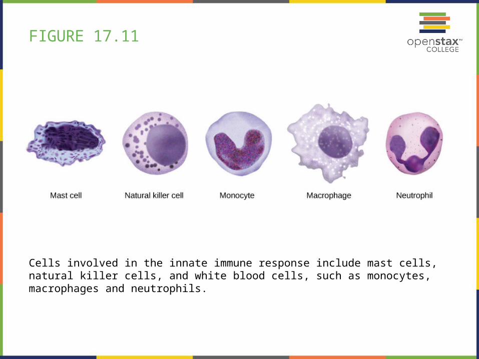
FIGURE 17.11
Cells involved in the innate immune response include mast cells, natural killer cells, and white blood cells, such as monocytes, macrophages and neutrophils.
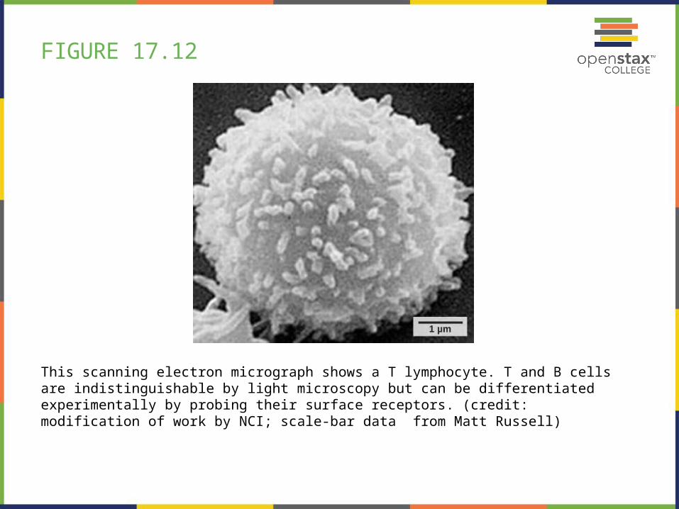
FIGURE 17.12
This scanning electron micrograph shows a T lymphocyte. T and B cells are indistinguishable by light microscopy but can be differentiated experimentally by probing their surface receptors. (credit: modification of work by NCI; scale-bar data from Matt Russell)
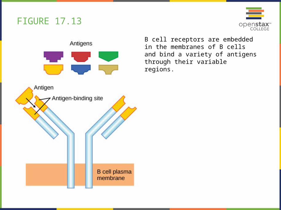
FIGURE 17.13
B cell receptors are embedded in the membranes of B cells and bind a variety of antigens through their variable regions.
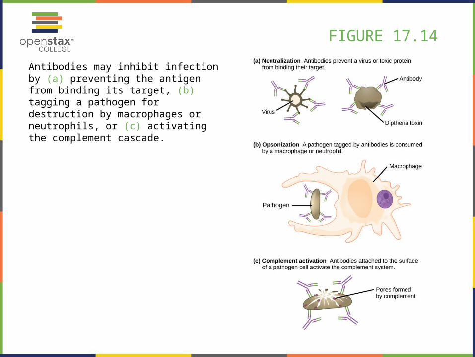
FIGURE 17.14
Antibodies may inhibit infection by (a) preventing the antigen from binding its target, (b) tagging a pathogen for destruction by macrophages or neutrophils, or (c) activating the complement cascade.
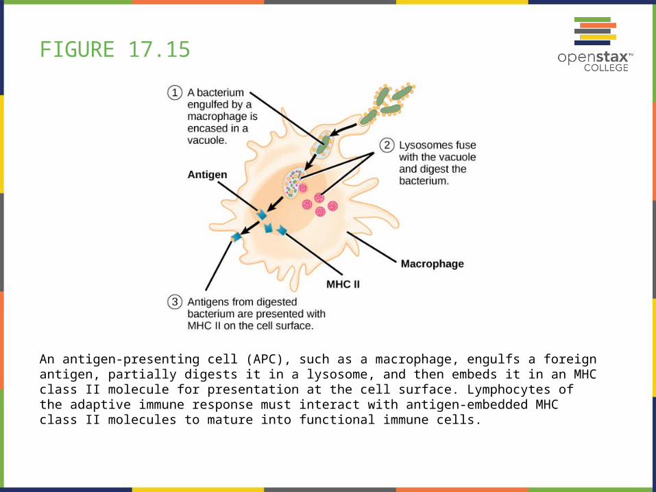
FIGURE 17.15
An antigen-presenting cell (APC), such as a macrophage, engulfs a foreign antigen, partially digests it in a lysosome, and then embeds it in an MHC class II molecule for presentation at the cell surface. Lymphocytes of the adaptive immune response must interact with antigen-embedded MHC class II molecules to mature into functional immune cells.
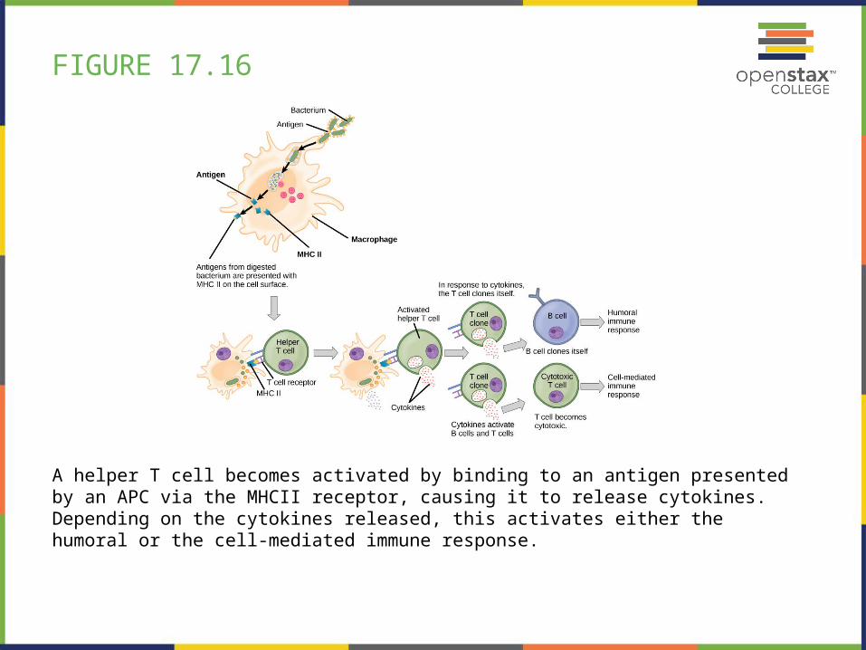
FIGURE 17.16
A helper T cell becomes activated by binding to an antigen presented by an APC via the MHCII receptor, causing it to release cytokines. Depending on the cytokines released, this activates either the humoral or the cell-mediated immune response.
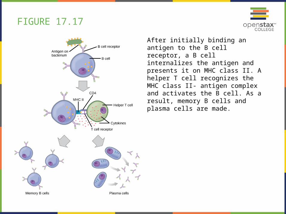
FIGURE 17.17
After initially binding an antigen to the B cell receptor, a B cell internalizes the antigen and presents it on MHC class II. A helper T cell recognizes the MHC class II- antigen complex and activates the B cell. As a result, memory B cells and plasma cells are made.
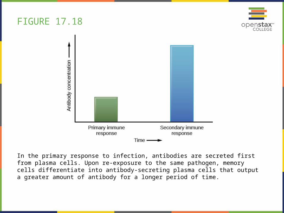
FIGURE 17.18
In the primary response to infection, antibodies are secreted first from plasma cells. Upon re-exposure to the same pathogen, memory cells differentiate into antibody-secreting plasma cells that output a greater amount of antibody for a longer period of time.
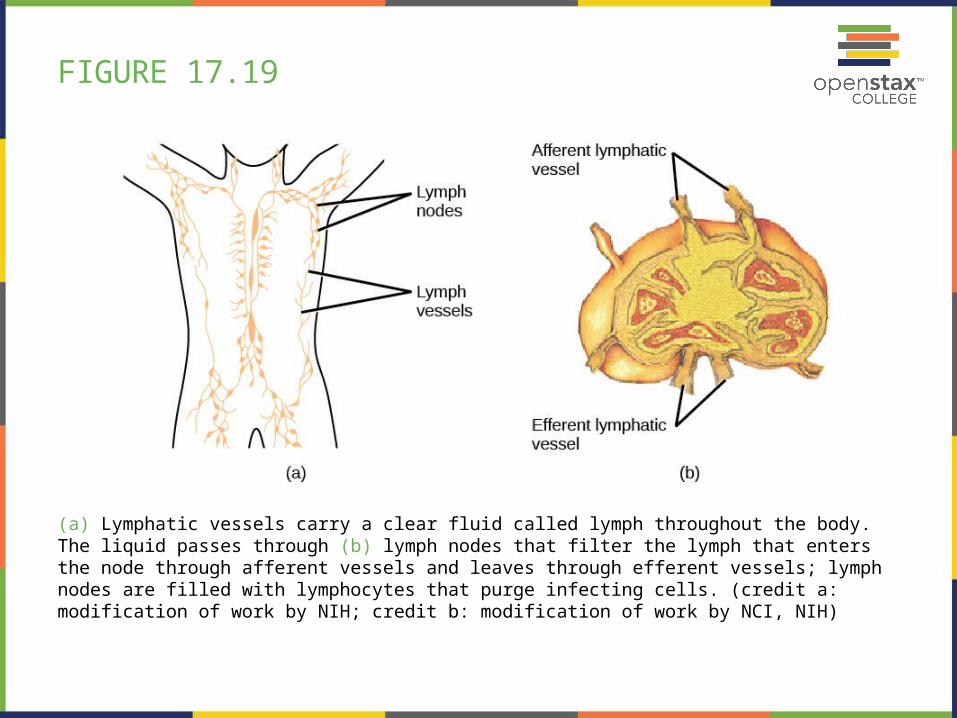
FIGURE 17.19
(a) Lymphatic vessels carry a clear fluid called lymph throughout the body. The liquid passes through (b) lymph nodes that filter the lymph that enters the node through afferent vessels and leaves through efferent vessels; lymph nodes are filled with lymphocytes that purge infecting cells. (credit a: modification of work by NIH; credit b: modification of work by NCI, NIH)
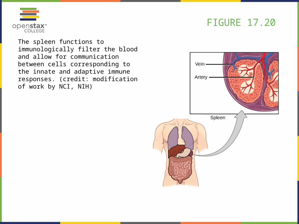
FIGURE 17.20
The spleen functions to immunologically filter the blood and allow for communication between cells corresponding to the innate and adaptive immune responses. (credit: modification of work by NCI, NIH)
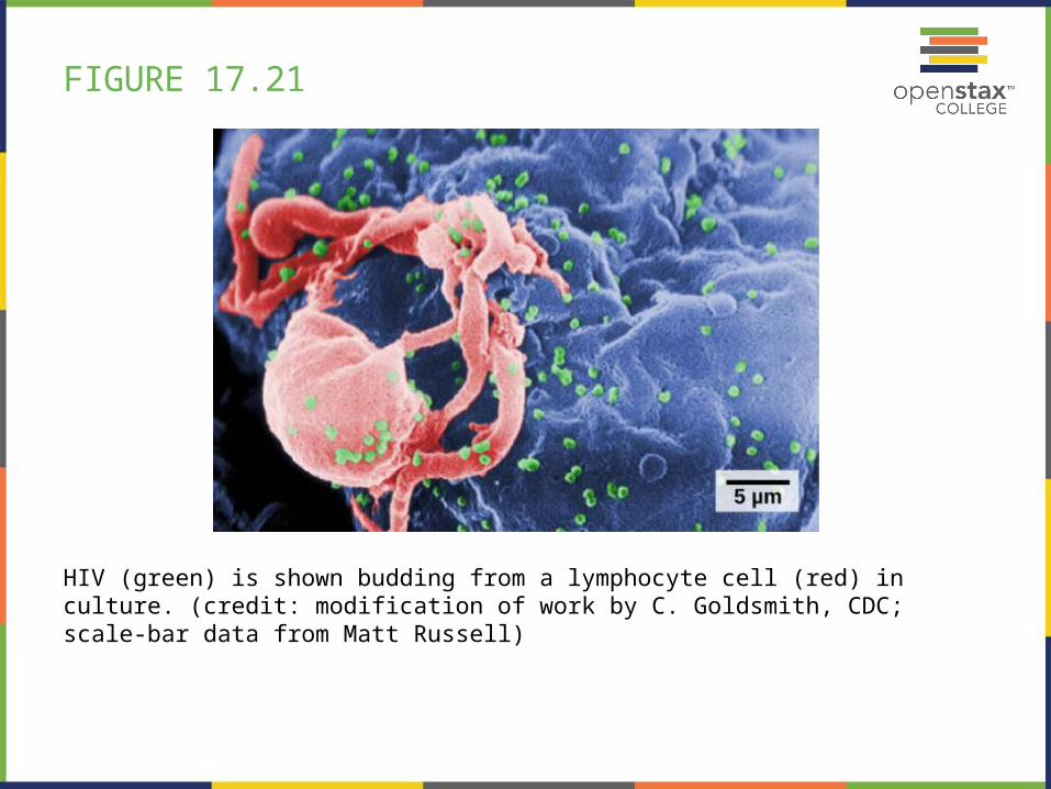
FIGURE 17.21
HIV (green) is shown budding from a lymphocyte cell (red) in culture. (credit: modification of work by C. Goldsmith, CDC; scale-bar data from Matt Russell)
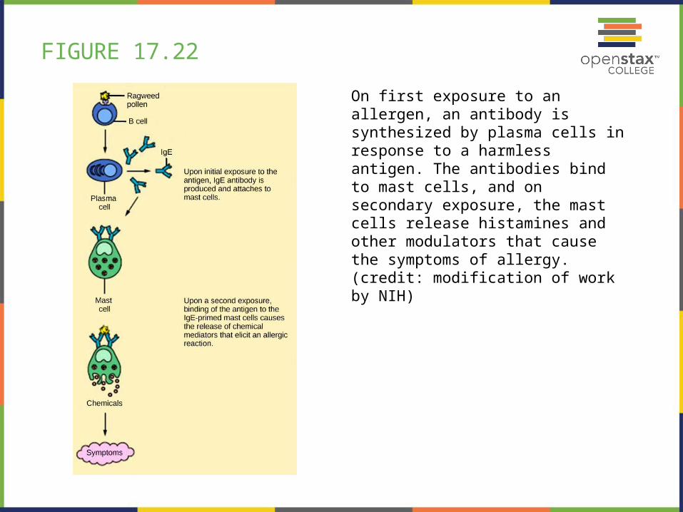
FIGURE 17.22
On first exposure to an allergen, an antibody is synthesized by plasma cells in response to a harmless antigen. The antibodies bind to mast cells, and on secondary exposure, the mast cells release histamines and other modulators that cause the symptoms of allergy. (credit: modification of work by NIH)
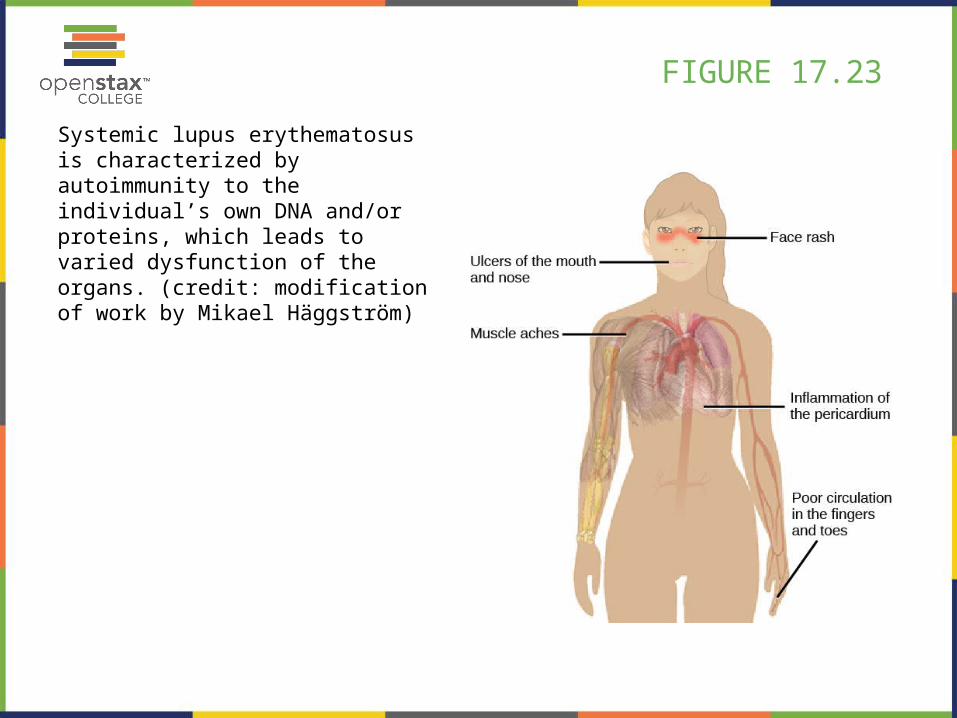
FIGURE 17.23
Systemic lupus erythematosus is characterized by autoimmunity to the individual’s own DNA and/or proteins, which leads to varied dysfunction of the organs. (credit: modification of work by Mikael Häggström)

This PowerPoint file is copyright 2011-2013, Rice University. All Rights Reserved.