Embed Size (px)
Citation preview

INTRODUCTION
Pyogenic granuloma was first reported as botryomycosis humaine in 1897.1 It is a benign inflammatory vascular lesion, mainly found in the skin and oral mucosa. Only a few cases of pyogenic granuloma in the gastrointestinal tract have been re-ported.2 A pyogenic granuloma is usually seen as a protrud-ing polypoid lesion and occasionally as a submucosal tumor-like lesion on endoscopy. Endosonographic findings for pyo-genic granuloma have not yet been reported. In this study, we reported the endosonographic findings obtained and the tr-eatment approaches used for two patients with esophageal pyo-genic granuloma.
CASE REPORTS
Case 1A 58-year-old man underwent upper endoscopy as part of
Clin Endosc 2013;46:81-84
Copyright © 2013 Korean Society of Gastrointestinal Endoscopy 81
a medical check-up. The endoscopic findings showed the presence of a polypoid lesion in the lower esophagus. The pa-tient was asymptomatic, and physical examination showed unremarkable findings. The upper endoscopy showed a 1-cm polypoid mass that was located just above the esophagogas-tric junction (Fig. 1A). The mass had a smooth surface and was covered with white exudates; it did not exhibit cushion sign. Endoscopic ultrasonography (EUS) showed that the le-sion was homogeneously hyperechogenic (Fig. 1B), had clear margins, and was located in the submucosa. Endoscopic and EUS findings suggested possibility of an inflammatory lesion such as inflammatory fibrinoid polyp. The patient wanted to remove the tumor strongly and then endoscopic resection was planned without biopsy. For endoscopic resection, a saline solution was injected to lift the lesion. Next, we performed en-doscopic mucosal resection (EMR). The lesion was comple-tely resected without complications (Fig. 1C-E). Histological examination showed edematous granulation tissue contain-ing numerous capillaries with acute and chronic inflammato-ry cell infiltration (Fig. 1F). These histological findings were indicative of pyogenic granuloma. Recurrence was not ob-served during the follow-up endoscopy performed 6 months later.
Case 2A 54-year-old man visited our hospital because of the inci-
CASE REPORT
Esophageal Pyogenic Granuloma: Endosonographic Findings and Endoscopic Treatments
Hyeog Gyu Seoung, Gwang Ha Kim, Geun Am Song, Ji Hye Kim, Min Young Oh, Jeong Cheon Choi, Jung Hee Koh and Chang Jun ParkDepartment of Internal Medicine, Pusan National University School of Medicine and Biomedical Research Institute, Pusan National University Hospital, Busan, Korea
Pyogenic granuloma is a benign inflammatory vascular lesion, mainly found in the skin and oral mucosa. A few cases of pyogenic gran-uloma in the gastrointestinal tract have been reported, and the esophagus was the main site in these cases. These patients were diagnosed with pyogenic granuloma after they underwent upper endoscopy and biopsy. Endoscopic resection is a favorable treatment option for esophageal pyogenic granuloma. Recently, we observed characteristic endosonographic findings in two cases with esophageal pyogenic granuloma, which were then treated successfully by endoscopic resection.
Key Words: Endosonography; Esophagus; Pyogenic granuloma
Open Access
Received: January 17, 2012 Revised: February 23, 2012Accepted: March 23, 2012Correspondence: Gwang Ha KimDepartment of Internal Medicine, Pusan National University School of Medi-cine and Biomedical Research Institute, Pusan National University Hospital, 179 Gudeok-ro, Seo-gu, Busan 602-739, KoreaTel: +82-51-240-7869, Fax: +82-51-244-8180, E-mail: [email protected] This is an Open Access article distributed under the terms of the Creative Commons Attribution Non-Commercial License (http://creativecommons.org/licenses/by-nc/3.0) which permits unrestricted non-commercial use, distribution, and reproduction in any medium, provided the original work is properly cited.
Print ISSN 2234-2400 / On-line ISSN 2234-2443
http://dx.doi.org/10.5946/ce.2013.46.1.81
82 Clin Endosc 2013;46:81-84
Pyogenic Granuloma of Esophagus
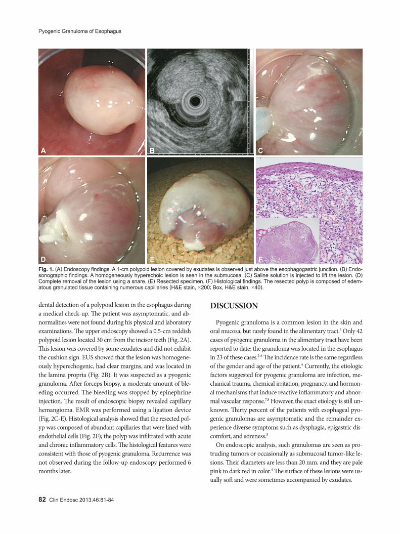
dental detection of a polypoid lesion in the esophagus during a medical check-up. The patient was asymptomatic, and ab-normalities were not found during his physical and laboratory examinations. The upper endoscopy showed a 0.5-cm reddish polypoid lesion located 30 cm from the incisor teeth (Fig. 2A). This lesion was covered by some exudates and did not exhibit the cushion sign. EUS showed that the lesion was homogene-ously hyperechogenic, had clear margins, and was located in the lamina propria (Fig. 2B). It was suspected as a pyogenic granuloma. After forceps biopsy, a moderate amount of ble-eding occurred. The bleeding was stopped by epinephrine injection. The result of endoscopic biopsy revealed capillary hemangioma. EMR was performed using a ligation device (Fig. 2C-E). Histological analysis showed that the resected pol-yp was composed of abundant capillaries that were lined with endothelial cells (Fig. 2F); the polyp was infiltrated with acute and chronic inflammatory cells. The histological features were consistent with those of pyogenic granuloma. Recurrence was not observed during the follow-up endoscopy performed 6 months later.
DISCUSSION
Pyogenic granuloma is a common lesion in the skin and oral mucosa, but rarely found in the alimentary tract.3 Only 42 cases of pyogenic granuloma in the alimentary tract have been reported to date; the granuloma was located in the esophagus in 23 of these cases.2-6 The incidence rate is the same regardless of the gender and age of the patient.4 Currently, the etiologic factors suggested for pyogenic granuloma are infection, me-chanical trauma, chemical irritation, pregnancy, and hormon-al mechanisms that induce reactive inflammatory and abnor-mal vascular response.7,8 However, the exact etiology is still un-known. Thirty percent of the patients with esophageal pyo-genic granulomas are asymptomatic and the remainder ex-perience diverse symptoms such as dysphagia, epigastric dis-comfort, and soreness.5
On endoscopic analysis, such granulomas are seen as pro-truding tumors or occasionally as submucosal tumor-like le-sions. Their diameters are less than 20 mm, and they are pale pink to dark red in color.4 The surface of these lesions were us-ually soft and were sometimes accompanied by exudates.
A
D
B
E
C
F Fig. 1. (A) Endoscopy findings. A 1-cm polypoid lesion covered by exudates is observed just above the esophagogastric junction. (B) Endo-sonographic findings. A homogeneously hyperechoic lesion is seen in the submucosa. (C) Saline solution is injected to lift the lesion. (D) Complete removal of the lesion using a snare. (E) Resected specimen. (F) Histological findings. The resected polyp is composed of edem-atous granulated tissue containing numerous capillaries (H&E stain, ×200; Box, H&E stain, ×40).
Seoung HG et al.
83
Our patients had submucosal tumor-like lesions, and th-erefore, we performed EUS. EUS findings for pyogenic gran-uloma have not yet been reported. EUS showed that the pa-tients had lesions that were homogeneously hyperechogenic, had clear borders, and were located in the lamina propria or submucosa. It is thought that the hyperechogenicity reflects the proliferation of blood vessels in lesions like hemangiomas.9 These EUS features would be helpful in the differential diag-nosis of esophageal submucosal tumor-like lesions, such as gastrointestinal stromal tumor, leiomyoma, lipoma, granular tumor, and duplication cyst. In case 2, bleeding occurred by forceps biopsy and then epinephrine was injected to stop the bleeding. Since pyogenic granuloma is a highly vascular le-sion, massive bleeding is expected after biopsy. Therefore, if there is a possibility of pyogenic granuloma in esophageal sub-mucosal tumor-like lesions by the above mentioned EUS find-ings, we could avoid endoscopic biopsy for histological diag-nosis.
EMR and endoscopic snare polypectomy are the most fa-vorable treatment options for esophageal pyogenic granulo-mas.3,6 In our patients, the lesions were completely resected
using traditional EMR or EMR with a ligation device. Recur-rence of tumors that are resected by surgical or endoscopic me-thods is rare.
Pyogenic granuloma is histologically characterized by cap-illary hemangiomas with lobulated proliferation in the ede-matous stroma. The granuloma is consisted of numerous new-ly formed capillaries of variable sizes, with the infiltration of acute and chronic inflammatory cells.3 Kaposi’s sarcoma must be included in the differential diagnosis in such cases. Kaposi’s sarcoma is suspected more often than pyogenic granuloma when a patient has immunosuppression due to conditions such as AIDS or transplantation; it is also suspected to a gr-eater extent when spindle cells and eosinophilic globules are observed during histological analysis.10
In summary, esophageal pyogenic granuloma is a rare sub-mucosal tumor-like polypoid lesion. On EUS, it appears as a homogeneously hyperechoic lesion that has clear borders and is located in the lamina propria or submucosa. These EUS features would be helpful in distinguishing pyogenic granu-loma from other lesions. Endoscopic resection is an effective treatment for esophageal pyogenic granuloma.
A
D
B
E
C
F Fig. 2. (A) Endoscopy findings. A 0.5-cm pinkish polypoid lesion is seen 30 cm from the incisor teeth. (B) Endosonographic findings. A ho-mogeneously hyperechoic lesion is seen in the lamina propria. (C) Band ligation was performed using a ligation device and then snare re-section was done. (D) Complete resection of the lesion. (E) Resected specimen. (F) Histological findings. The resected polyp is composed of abundant capillaries that are lined with endothelial cells (H&E stain, ×200; Box, H&E stain, ×40).

84 Clin Endosc 2013;46:81-84
Pyogenic Granuloma of Esophagus
Conflicts of InterestThe authors have no financial conflicts of interest.
REFERENCES
1. Poncet A, Dor L. Botryomycose humaine. Rev Chir 1897;18:996-1003.2. Park SY, Park CH, Lee WS, Kim HS, Choi SK, Rew JS. Pyogenic gran-
uloma of the duodenum treated successfully by endoscopic mucosal resection. Gut Liver 2009;3:48-51.
3. Moffatt DC, Warwryko P, Singh H. Pyogenic granuloma: an unusual cause of massive gastrointestinal bleeding from the small bowel. Can J Gastroenterol 2009;23:261-264.
4. Kusakabe A, Kato H, Hayashi K, et al. Pyogenic granuloma of the stomach successfully treated by endoscopic resection after transarterial embolization of the feeding artery. J Gastroenterol 2005;40:530-535.
5. Cho HS, Jung ES, Lee YJ, et al. A case of esophageal pyogenic granulo-
ma. Korean J Gastrointest Endosc 2009;38:210-213.6. Chung IS, Kim SW, Choi MG, et al. A case of pyogenic granuloma in
the terminal ileum treated by endoscopic snare polypectomy. Korean J Gastrointest Endosc 2002;24:176-180.
7. Kerr DA. Granuloma pyogenicum. Oral Surg Oral Med Oral Pathol 1951;4:158-176.
8. Bhaskar SN, Jacoway JR. Pyogenic granuloma: clinical features, inci-dence, histology, and result of treatment: report of 242 cases. J Oral Surg 1966;24:391-398.
9. Paltiel HJ, Burrows PE, Kozakewich HP, Zurakowski D, Mulliken JB. Soft-tissue vascular anomalies: utility of US for diagnosis. Radiology 2000;214:747-754.
10. Reis-Filho JS, Souto-Moura C, Lopes JM. Classic Kaposi’s sarcoma of the tongue: case report with emphasis on the differential diagnosis. J Oral Maxillofac Surg 2002;60:951-954.