Embed Size (px)
Citation preview
OPEN ACCESS ATLAS OF OTOLARYNGOLOGY, HEAD &
NECK OPERATIVE SURGERY
LATERAL TEMPORAL BONE RESECTION SURGICAL TECHNIQUE
Rajeev Mathew, Tashneem Harris, Parag Patel
Lateral temporal bone resection refers to en
bloc resection of the external auditory canal
(EAC) with the tympanic membrane (TM).
It is generally performed for malignancies
involving the EAC, the most common being
primary squamous cell carcinoma. Tumours
may arise from the skin of the external ear,
parotid, glandular adnexa of the ear or may
be metastases to parotid nodes.
History and examination
History and examination are directed at
making a diagnosis and determining the
extent of the tumour. Refractory pain is a
hallmark of advanced malignancy of the ear
canal. Other presenting symptoms include
chronic ear discharge and hearing loss. A
lesion of the ear canal may be visible and
there may be a discharge. The parotid and
neck should be examined for evidence of
metastases. Facial nerve function should be
documented. Assessing sensation of the
face on the affected side as well as the lower
cranial nerves is crucial to detect intracra-
nial and inferomedial extension to the jugu-
lar foramen. Differential diagnoses include
skull base osteomyelitis and inflammatory
conditions e.g. TB.
Special investigations
The tumour is biopsied in the office to
confirm the diagnosis. Imaging is requested
to:
• Determine local extent of the tumour
• Stage the tumour (see below)
• Determine its operability
• Rule out regional nodal metastases to
parotid and cervical nodes
• Plan the surgical approach
High resolution CT scans (0.5mm slices
with and without contrast) of the temporal
bone, brain, parotid and neck allow assess-
ment of the soft tissue extent, bony destruc-
tion, as well as clinical staging. MRI is use-
ful to assess soft tissue involvement particu-
larly if disease appears to have breached the
EAC/TM and there is concern about invol-
vement of dura or brain. Pure tone audiome-
try should be performed. Carotid angiogra-
phy +/- balloon occlusion is considered if
surgery is contemplated and there is con-
cern about carotid artery involvement.
Modified Pittsburgh Staging System
T1: Limited to EAC without bony or soft
tissue extension into mastoid/middle ear
T2: Limited EAC erosion (not full thick-
ness) or radiological findings consistent
with <0.5cm soft tissue involvement
T3: Full thickness erosion of EAC with
<0.5cm soft tissue involvement or facial
nerve paralysis
T4: Eroding cochlea, petrous apex, carotid
canal, jugular foramen, medial wall of mid-
dle ear, dura or with >0.5cm soft tissue
extension
Practical application of staging system
T1: Sleeve resection or lateral temporal
bone resection
T2: Lateral temporal bone resection
T3: May be suited to lateral temporal bone
resection with sacrifice of facial nerve
T4: Not amenable to lateral temporal bone
resection
Surgical Anatomy
The lateral 1/3 of the EAC is composed of
fibroelastic cartilage and contains the
Fissures of Santorini - these communicate
with the parotid anteriorly and the soft tis-
sue overlying the mastoid posteriorly. The
medial 2/3 of the EAC consists mainly of
the tympanic portion of the temporal bone.
2
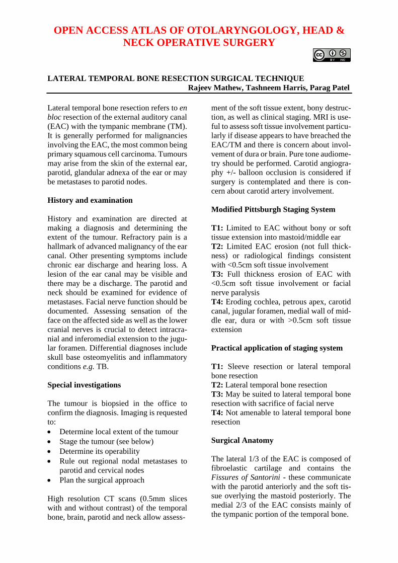
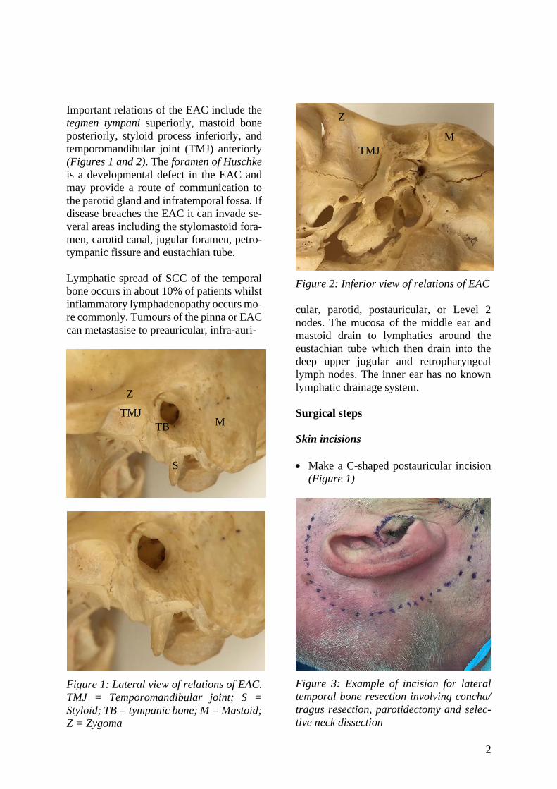
Important relations of the EAC include the
tegmen tympani superiorly, mastoid bone
posteriorly, styloid process inferiorly, and
temporomandibular joint (TMJ) anteriorly
(Figures 1 and 2). The foramen of Huschke
is a developmental defect in the EAC and
may provide a route of communication to
the parotid gland and infratemporal fossa. If
disease breaches the EAC it can invade se-
veral areas including the stylomastoid fora-
men, carotid canal, jugular foramen, petro-
tympanic fissure and eustachian tube.
Lymphatic spread of SCC of the temporal
bone occurs in about 10% of patients whilst
inflammatory lymphadenopathy occurs mo-
re commonly. Tumours of the pinna or EAC
can metastasise to preauricular, infra-auri-
Figure 1: Lateral view of relations of EAC.
TMJ = Temporomandibular joint; S =
Styloid; TB = tympanic bone; M = Mastoid;
Z = Zygoma
Figure 2: Inferior view of relations of EAC
cular, parotid, postauricular, or Level 2
nodes. The mucosa of the middle ear and
mastoid drain to lymphatics around the
eustachian tube which then drain into the
deep upper jugular and retropharyngeal
lymph nodes. The inner ear has no known
lymphatic drainage system.
Surgical steps
Skin incisions
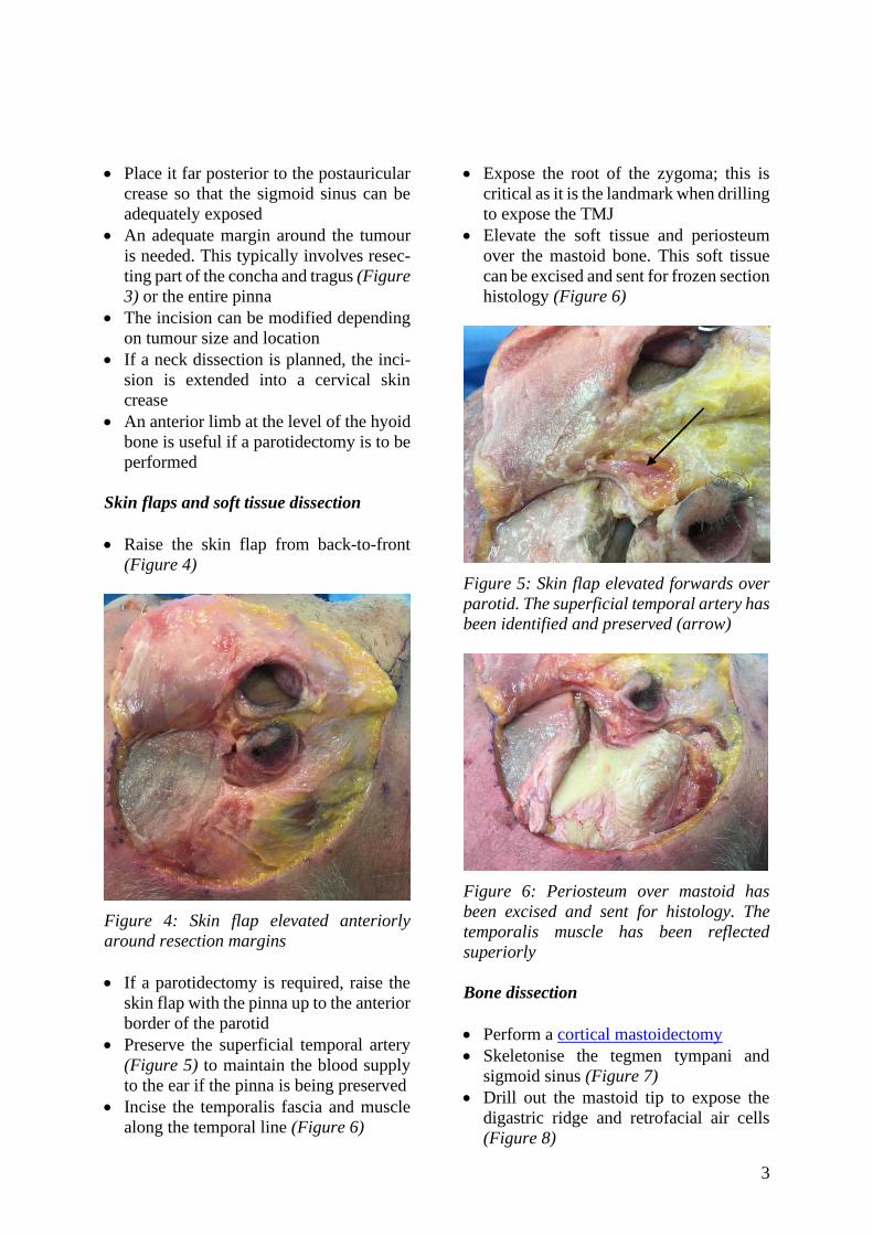
• Make a C-shaped postauricular incision
(Figure 1)
Figure 3: Example of incision for lateral
temporal bone resection involving concha/
tragus resection, parotidectomy and selec-
tive neck dissection
M TB
TMJ
S
TMJ
Z
Z
M
3
• Place it far posterior to the postauricular
crease so that the sigmoid sinus can be
adequately exposed
• An adequate margin around the tumour
is needed. This typically involves resec-
ting part of the concha and tragus (Figure
3) or the entire pinna
• The incision can be modified depending
on tumour size and location
• If a neck dissection is planned, the inci-
sion is extended into a cervical skin
crease
• An anterior limb at the level of the hyoid
bone is useful if a parotidectomy is to be
performed
Skin flaps and soft tissue dissection
• Raise the skin flap from back-to-front
(Figure 4)
Figure 4: Skin flap elevated anteriorly
around resection margins
• If a parotidectomy is required, raise the
skin flap with the pinna up to the anterior
border of the parotid
• Preserve the superficial temporal artery
(Figure 5) to maintain the blood supply
to the ear if the pinna is being preserved
• Incise the temporalis fascia and muscle
along the temporal line (Figure 6)
• Expose the root of the zygoma; this is
critical as it is the landmark when drilling
to expose the TMJ
• Elevate the soft tissue and periosteum
over the mastoid bone. This soft tissue
can be excised and sent for frozen section
histology (Figure 6)
Figure 5: Skin flap elevated forwards over
parotid. The superficial temporal artery has
been identified and preserved (arrow)
Figure 6: Periosteum over mastoid has
been excised and sent for histology. The
temporalis muscle has been reflected
superiorly
Bone dissection
• Perform a cortical mastoidectomy
• Skeletonise the tegmen tympani and
sigmoid sinus (Figure 7)
• Drill out the mastoid tip to expose the
digastric ridge and retrofacial air cells
(Figure 8)
4
• Skeletonise the mastoid segment of the
facial nerve (Figure 9)
• Keep the posterior bony ear canal relati-
vely thick to avoid inadvertent exposure
of cancer
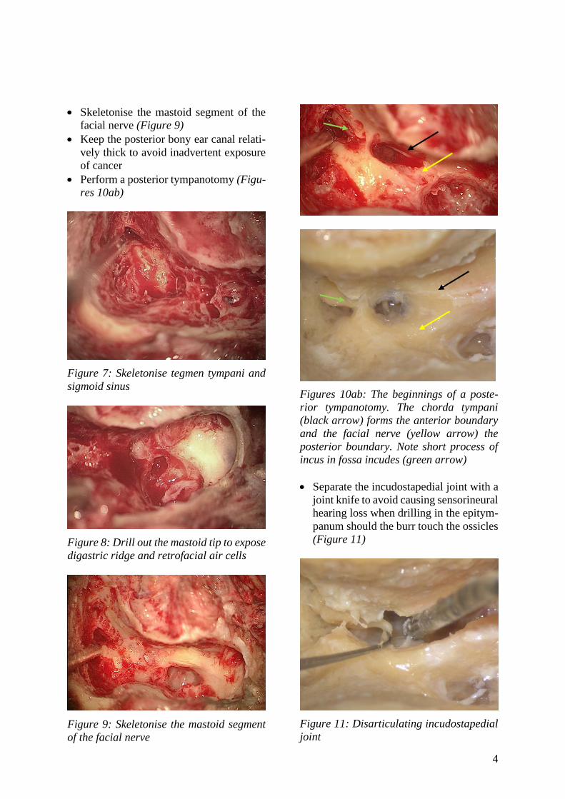
• Perform a posterior tympanotomy (Figu-
res 10ab)
Figure 7: Skeletonise tegmen tympani and
sigmoid sinus
Figure 8: Drill out the mastoid tip to expose
digastric ridge and retrofacial air cells
Figure 9: Skeletonise the mastoid segment
of the facial nerve
Figures 10ab: The beginnings of a poste-
rior tympanotomy. The chorda tympani
(black arrow) forms the anterior boundary
and the facial nerve (yellow arrow) the
posterior boundary. Note short process of
incus in fossa incudes (green arrow)
• Separate the incudostapedial joint with a
joint knife to avoid causing sensorineural
hearing loss when drilling in the epitym-
panum should the burr touch the ossicles
(Figure 11)
Figure 11: Disarticulating incudostapedial
joint
5
• Drill the bone superior to the EAC to-
wards the root of the zygoma in the space
between the tegmen tympani and the
EAC, using progressively smaller burrs
(Figure 12)
Figure 12: The curved arrow shows the
drilling trajectory towards the root of zygo-
ma. Arrowhead points to the TMJ capsule
• When there is limited space between the
tegmen and the ear canal, it may be
necessary to remove the bone of the teg-
men and expose dura rather than to over-
thin the ear canal and expose cancer
• Use a diamond burr to avoid injuring the
dura and causing a CSF leak
• Avoid sharp ledges of bone over the dura
to allow good light into the surgical field
• Once one reaches the 12 o’clock position
of the ear canal, direct the drilling trajec-
tory inferiorly
• Expose the anterior epitympanum
• At this point one can remove the incus
and use a malleus nipper to remove the
malleus head
• Keep drilling anteriorly until the TMJ
capsule becomes apparent. It is soft and
whiter in colour (Figures 12, 13)
• Ensure that the TMJ capsule is exposed
all the way from the ossicular heads me-
dially to the squamous temporal bone lat-
erally
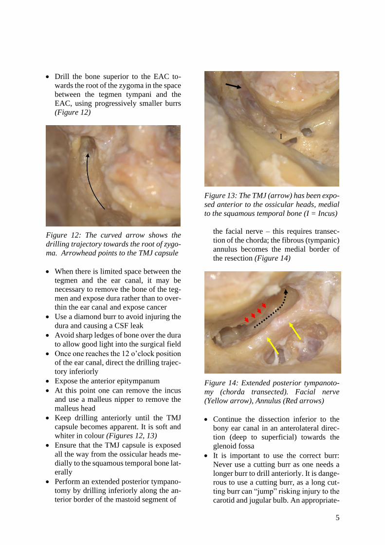
• Perform an extended posterior tympano-
tomy by drilling inferiorly along the an-
terior border of the mastoid segment of
Figure 13: The TMJ (arrow) has been expo-
sed anterior to the ossicular heads, medial
to the squamous temporal bone (I = Incus)
the facial nerve – this requires transec-
tion of the chorda; the fibrous (tympanic)
annulus becomes the medial border of
the resection (Figure 14)
Figure 14: Extended posterior tympanoto-
my (chorda transected). Facial nerve
(Yellow arrow), Annulus (Red arrows)
• Continue the dissection inferior to the
bony ear canal in an anterolateral direc-
tion (deep to superficial) towards the
glenoid fossa
• It is important to use the correct burr:
Never use a cutting burr as one needs a
longer burr to drill anteriorly. It is dange-
rous to use a cutting burr, as a long cut-
ting burr can “jump” risking injury to the
carotid and jugular bulb. An appropriate-
I
6
sized (2-2.5mm) diamond burr should
rather be used
• It is important to check the height of the
jugular bulb on the CT scan to ensure
that the jugular bulb is not inadvertently
injured!
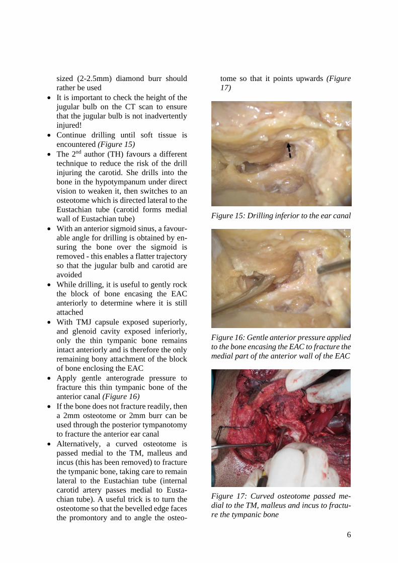
• Continue drilling until soft tissue is
encountered (Figure 15)
• The 2nd author (TH) favours a different
technique to reduce the risk of the drill
injuring the carotid. She drills into the
bone in the hypotympanum under direct
vision to weaken it, then switches to an
osteotome which is directed lateral to the
Eustachian tube (carotid forms medial
wall of Eustachian tube)
• With an anterior sigmoid sinus, a favour-
able angle for drilling is obtained by en-
suring the bone over the sigmoid is
removed - this enables a flatter trajectory
so that the jugular bulb and carotid are
avoided
• While drilling, it is useful to gently rock
the block of bone encasing the EAC
anteriorly to determine where it is still
attached
• With TMJ capsule exposed superiorly,
and glenoid cavity exposed inferiorly,
only the thin tympanic bone remains
intact anteriorly and is therefore the only
remaining bony attachment of the block
of bone enclosing the EAC
• Apply gentle anterograde pressure to
fracture this thin tympanic bone of the
anterior canal (Figure 16)
• If the bone does not fracture readily, then
a 2mm osteotome or 2mm burr can be
used through the posterior tympanotomy
to fracture the anterior ear canal
• Alternatively, a curved osteotome is
passed medial to the TM, malleus and
incus (this has been removed) to fracture
the tympanic bone, taking care to remain
lateral to the Eustachian tube (internal
carotid artery passes medial to Eusta-
chian tube). A useful trick is to turn the
osteotome so that the bevelled edge faces
the promontory and to angle the osteo-
tome so that it points upwards (Figure
17)
Figure 15: Drilling inferior to the ear canal
Figure 16: Gentle anterior pressure applied
to the bone encasing the EAC to fracture the
medial part of the anterior wall of the EAC
Figure 17: Curved osteotome passed me-
dial to the TM, malleus and incus to fractu-
re the tympanic bone
7
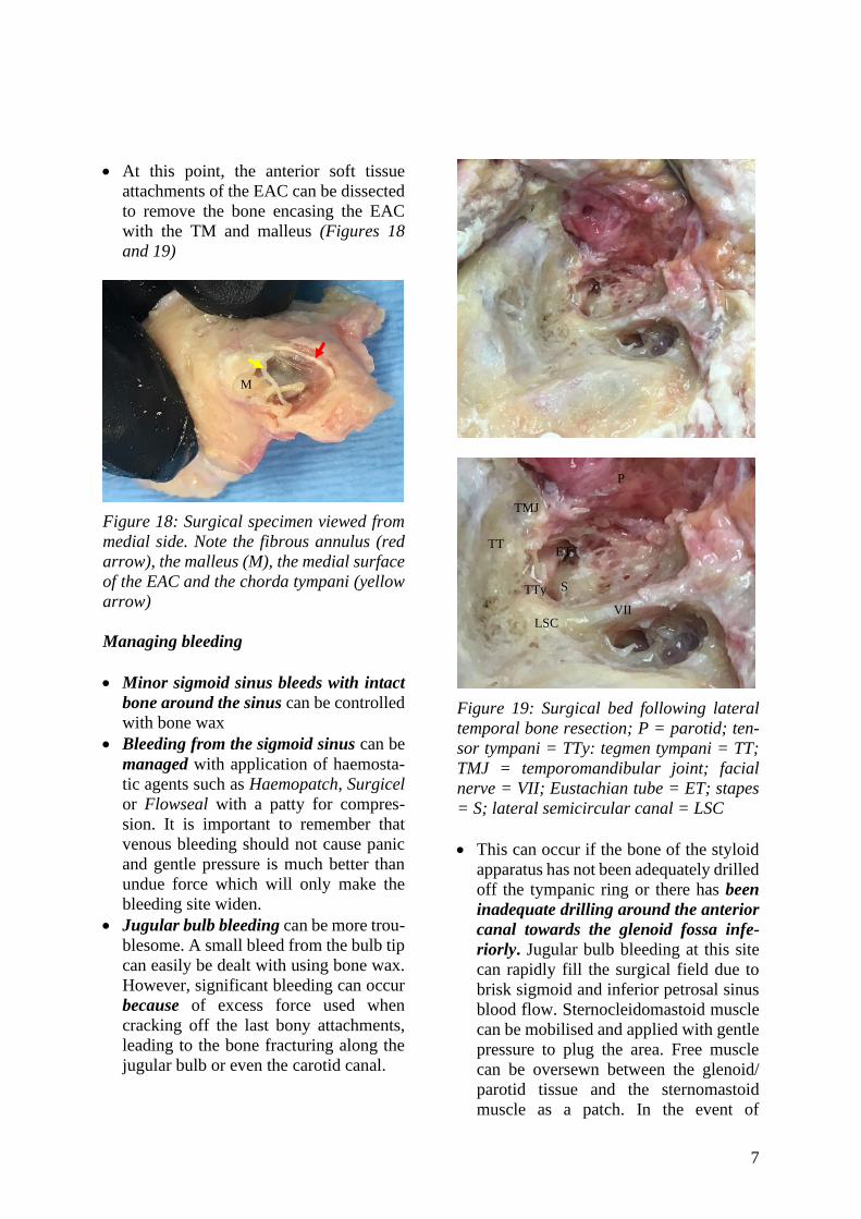
• At this point, the anterior soft tissue
attachments of the EAC can be dissected
to remove the bone encasing the EAC
with the TM and malleus (Figures 18
and 19)
Figure 18: Surgical specimen viewed from
medial side. Note the fibrous annulus (red
arrow), the malleus (M), the medial surface
of the EAC and the chorda tympani (yellow
arrow)
Managing bleeding
• Minor sigmoid sinus bleeds with intact
bone around the sinus can be controlled
with bone wax
• Bleeding from the sigmoid sinus can be
managed with application of haemosta-
tic agents such as Haemopatch, Surgicel
or Flowseal with a patty for compres-
sion. It is important to remember that
venous bleeding should not cause panic
and gentle pressure is much better than
undue force which will only make the
bleeding site widen.
• Jugular bulb bleeding can be more trou-
blesome. A small bleed from the bulb tip
can easily be dealt with using bone wax.
However, significant bleeding can occur
because of excess force used when
cracking off the last bony attachments,
leading to the bone fracturing along the
jugular bulb or even the carotid canal.
Figure 19: Surgical bed following lateral
temporal bone resection; P = parotid; ten-
sor tympani = TTy: tegmen tympani = TT;
TMJ = temporomandibular joint; facial
nerve = VII; Eustachian tube = ET; stapes
= S; lateral semicircular canal = LSC
• This can occur if the bone of the styloid
apparatus has not been adequately drilled
off the tympanic ring or there has been
inadequate drilling around the anterior
canal towards the glenoid fossa infe-
riorly. Jugular bulb bleeding at this site
can rapidly fill the surgical field due to
brisk sigmoid and inferior petrosal sinus
blood flow. Sternocleidomastoid muscle
can be mobilised and applied with gentle
pressure to plug the area. Free muscle
can be oversewn between the glenoid/
parotid tissue and the sternomastoid
muscle as a patch. In the event of
VII
TT
TMJ
P
S TTy
LSC
ET
M
8
uncontrolled bleeding, the jugular vein is
ligated in the upper neck and the sigmoid
sinus and inferior petrosal sinuses are
packed with Surgicel through the tear in
the jugular bulb.
• The 2nd author applies pressure by pack-
ing with Surgicel, then ligates the jugular
vein in the neck and the sigmoid in the
mastoid cavity
• Skeletonise the sigmoid sinus, then re-
move the thin shell of bone over a small
section of the sigmoid by using a Plester
knife
• Use an aneurysm needle threaded with a
double silk ligature and pass it behind the
sigmoid sinus by making small slits in
the dura next to the sigmoid sinus. Re-
move the needle, leaving behind the
double ligature and tie these. This may
cause a small CSF leak. This can be
plugged using fat
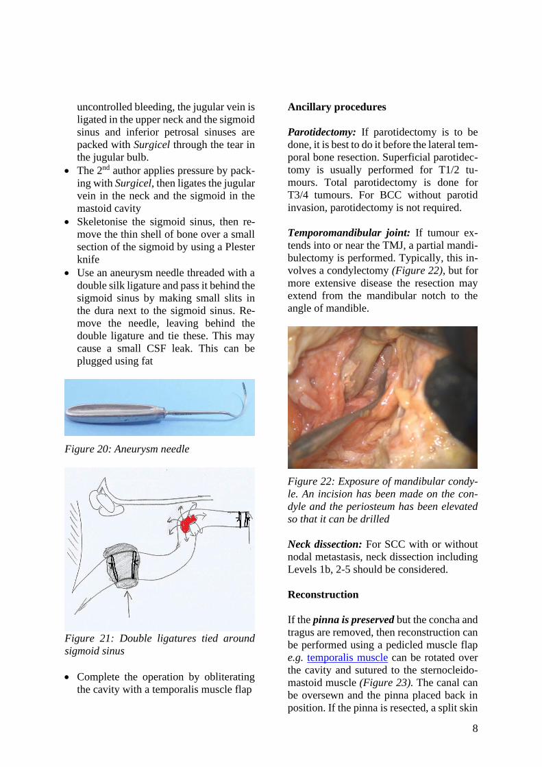
Figure 20: Aneurysm needle
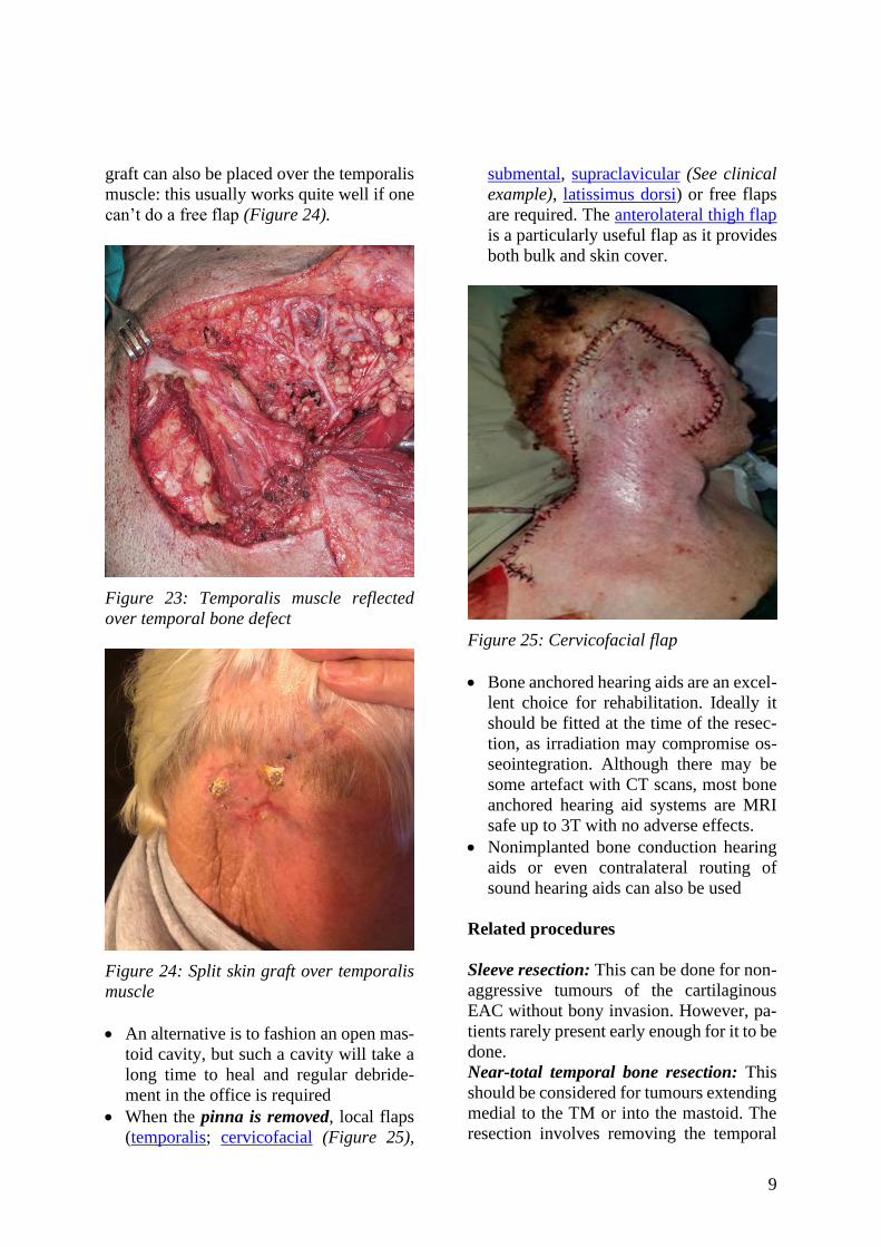
Figure 21: Double ligatures tied around
sigmoid sinus
• Complete the operation by obliterating
the cavity with a temporalis muscle flap
Ancillary procedures
Parotidectomy: If parotidectomy is to be
done, it is best to do it before the lateral tem-
poral bone resection. Superficial parotidec-
tomy is usually performed for T1/2 tu-
mours. Total parotidectomy is done for
T3/4 tumours. For BCC without parotid
invasion, parotidectomy is not required.
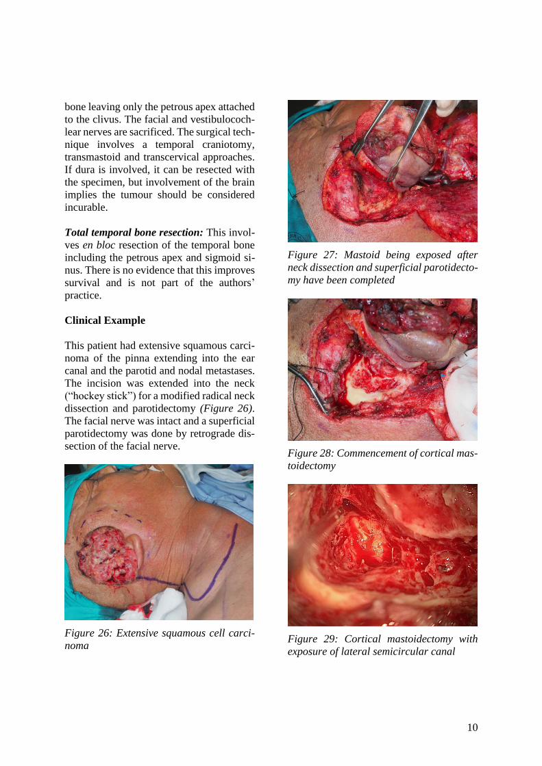
Temporomandibular joint: If tumour ex-
tends into or near the TMJ, a partial mandi-
bulectomy is performed. Typically, this in-
volves a condylectomy (Figure 22), but for
more extensive disease the resection may
extend from the mandibular notch to the
angle of mandible.
Figure 22: Exposure of mandibular condy-
le. An incision has been made on the con-
dyle and the periosteum has been elevated
so that it can be drilled
Neck dissection: For SCC with or without
nodal metastasis, neck dissection including
Levels 1b, 2-5 should be considered.
Reconstruction
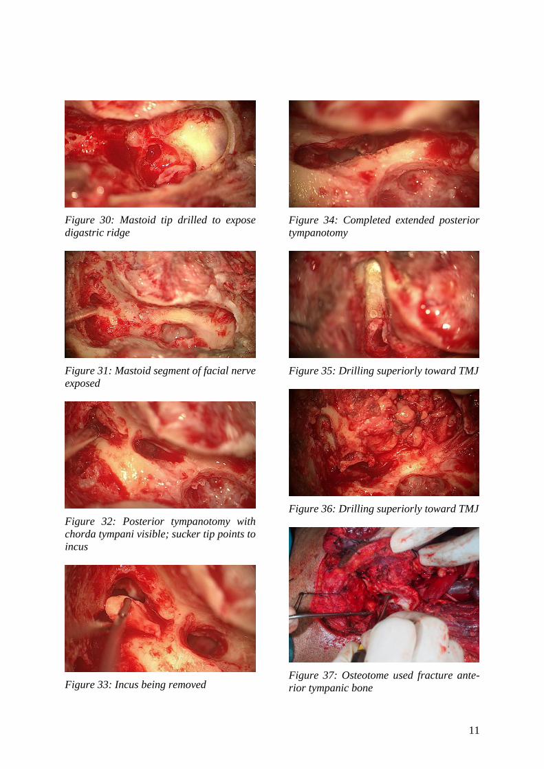
If the pinna is preserved but the concha and
tragus are removed, then reconstruction can
be performed using a pedicled muscle flap
e.g. temporalis muscle can be rotated over
the cavity and sutured to the sternocleido-
mastoid muscle (Figure 23). The canal can
be oversewn and the pinna placed back in
position. If the pinna is resected, a split skin
9
graft can also be placed over the temporalis
muscle: this usually works quite well if one
can’t do a free flap (Figure 24).
Figure 23: Temporalis muscle reflected
over temporal bone defect
Figure 24: Split skin graft over temporalis
muscle
• An alternative is to fashion an open mas-
toid cavity, but such a cavity will take a
long time to heal and regular debride-
ment in the office is required
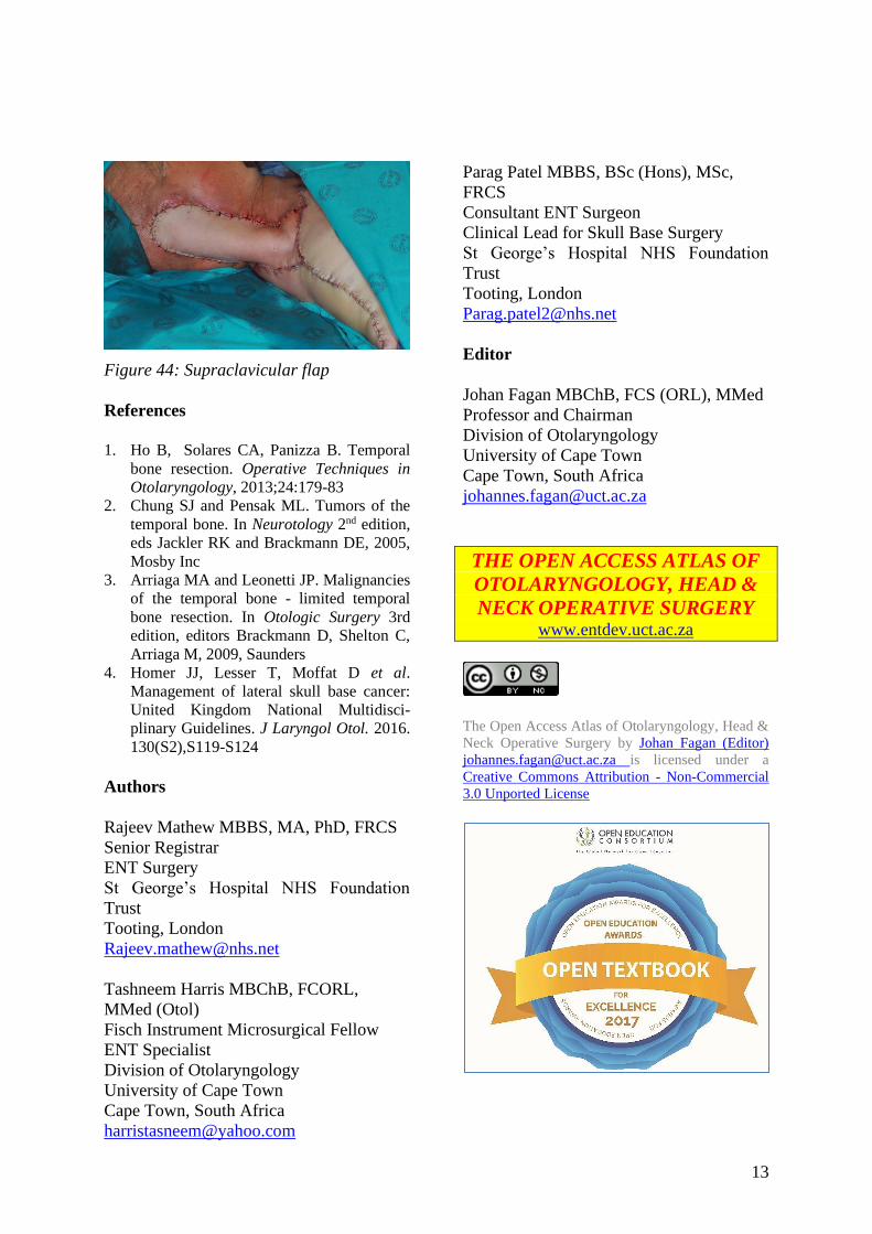
• When the pinna is removed, local flaps
(temporalis; cervicofacial (Figure 25),
submental, supraclavicular (See clinical
example), latissimus dorsi) or free flaps
are required. The anterolateral thigh flap
is a particularly useful flap as it provides
both bulk and skin cover.
Figure 25: Cervicofacial flap
• Bone anchored hearing aids are an excel-
lent choice for rehabilitation. Ideally it
should be fitted at the time of the resec-
tion, as irradiation may compromise os-
seointegration. Although there may be
some artefact with CT scans, most bone
anchored hearing aid systems are MRI
safe up to 3T with no adverse effects.
• Nonimplanted bone conduction hearing
aids or even contralateral routing of
sound hearing aids can also be used
Related procedures
Sleeve resection: This can be done for non-
aggressive tumours of the cartilaginous
EAC without bony invasion. However, pa-
tients rarely present early enough for it to be
done.
Near-total temporal bone resection: This
should be considered for tumours extending
medial to the TM or into the mastoid. The
resection involves removing the temporal
10
bone leaving only the petrous apex attached
to the clivus. The facial and vestibulococh-
lear nerves are sacrificed. The surgical tech-
nique involves a temporal craniotomy,
transmastoid and transcervical approaches.
If dura is involved, it can be resected with
the specimen, but involvement of the brain
implies the tumour should be considered
incurable.
Total temporal bone resection: This invol-
ves en bloc resection of the temporal bone
including the petrous apex and sigmoid si-
nus. There is no evidence that this improves
survival and is not part of the authors’
practice.
Clinical Example
This patient had extensive squamous carci-
noma of the pinna extending into the ear
canal and the parotid and nodal metastases.
The incision was extended into the neck
(“hockey stick”) for a modified radical neck
dissection and parotidectomy (Figure 26).
The facial nerve was intact and a superficial
parotidectomy was done by retrograde dis-
section of the facial nerve.
Figure 26: Extensive squamous cell carci-
noma
Figure 27: Mastoid being exposed after
neck dissection and superficial parotidecto-
my have been completed
Figure 28: Commencement of cortical mas-
toidectomy
Figure 29: Cortical mastoidectomy with
exposure of lateral semicircular canal
11
Figure 30: Mastoid tip drilled to expose
digastric ridge
Figure 31: Mastoid segment of facial nerve
exposed
Figure 32: Posterior tympanotomy with
chorda tympani visible; sucker tip points to
incus
Figure 33: Incus being removed
Figure 34: Completed extended posterior
tympanotomy
Figure 35: Drilling superiorly toward TMJ
Figure 36: Drilling superiorly toward TMJ
Figure 37: Osteotome used fracture ante-
rior tympanic bone
12
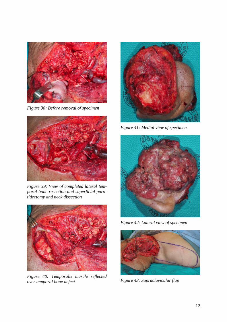
Figure 38: Before removal of specimen
Figure 39: View of completed lateral tem-
poral bone resection and superficial paro-
tidectomy and neck dissection
Figure 40: Temporalis muscle reflected
over temporal bone defect
Figure 41: Medial view of specimen
Figure 42: Lateral view of specimen
Figure 43: Supraclavicular flap
13
Figure 44: Supraclavicular flap
References
1. Ho B, Solares CA, Panizza B. Temporal
bone resection. Operative Techniques in
Otolaryngology, 2013;24:179-83
2. Chung SJ and Pensak ML. Tumors of the
temporal bone. In Neurotology 2nd edition,
eds Jackler RK and Brackmann DE, 2005,
Mosby Inc
3. Arriaga MA and Leonetti JP. Malignancies
of the temporal bone - limited temporal
bone resection. In Otologic Surgery 3rd
edition, editors Brackmann D, Shelton C,
Arriaga M, 2009, Saunders
4. Homer JJ, Lesser T, Moffat D et al.
Management of lateral skull base cancer:
United Kingdom National Multidisci-
plinary Guidelines. J Laryngol Otol. 2016.
130(S2),S119-S124
Authors
Rajeev Mathew MBBS, MA, PhD, FRCS
Senior Registrar
ENT Surgery
St George’s Hospital NHS Foundation
Trust
Tooting, London
Tashneem Harris MBChB, FCORL,
MMed (Otol)
Fisch Instrument Microsurgical Fellow
ENT Specialist
Division of Otolaryngology
University of Cape Town
Cape Town, South Africa
Parag Patel MBBS, BSc (Hons), MSc,
FRCS
Consultant ENT Surgeon
Clinical Lead for Skull Base Surgery
St George’s Hospital NHS Foundation
Trust
Tooting, London
Editor
Johan Fagan MBChB, FCS (ORL), MMed
Professor and Chairman
Division of Otolaryngology
University of Cape Town
Cape Town, South Africa
THE OPEN ACCESS ATLAS OF
OTOLARYNGOLOGY, HEAD &
NECK OPERATIVE SURGERY www.entdev.uct.ac.za
The Open Access Atlas of Otolaryngology, Head &
Neck Operative Surgery by Johan Fagan (Editor)
[email protected] is licensed under a
Creative Commons Attribution - Non-Commercial
3.0 Unported License