Embed Size (px)
Citation preview
©2015 MFMER | slide-1
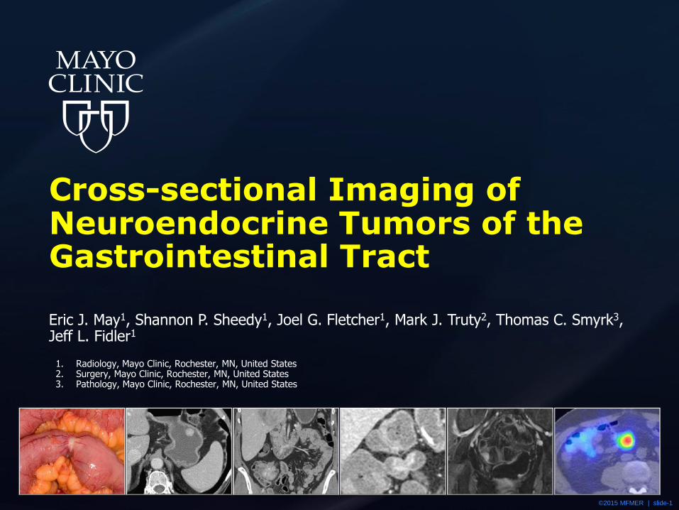
Cross-sectional Imaging of Neuroendocrine Tumors of the Gastrointestinal Tract
Eric J. May1, Shannon P. Sheedy1, Joel G. Fletcher1, Mark J. Truty2, Thomas C. Smyrk3, Jeff L. Fidler1
1. Radiology, Mayo Clinic, Rochester, MN, United States 2. Surgery, Mayo Clinic, Rochester, MN, United States 3. Pathology, Mayo Clinic, Rochester, MN, United States

©2015 MFMER | slide-2
Disclosures
• J. Fidler: Research support from Beekley Medical.

©2015 MFMER | slide-3
Learning Objectives
• Review the classification of neuroendocrine tumors
• Recognize the CT and MR appearances of GI tract neuroendocrine tumors, mimics, and pitfalls
• Understand appropriate imaging strategies for identifying neuroendocrine tumors
Target Audience:
• Radiologists and Gastroenterologists
©2015 MFMER | slide-4
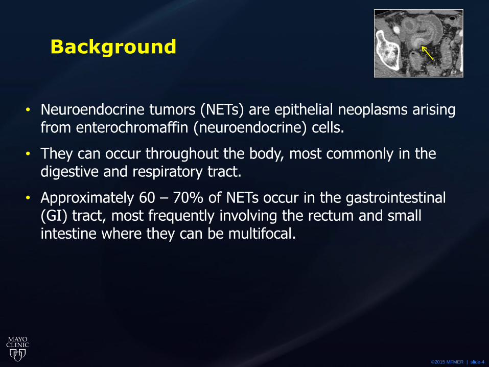
Background
• Neuroendocrine tumors (NETs) are epithelial neoplasms arising from enterochromaffin (neuroendocrine) cells.
• They can occur throughout the body, most commonly in the digestive and respiratory tract.
• Approximately 60 – 70% of NETs occur in the gastrointestinal (GI) tract, most frequently involving the rectum and small intestine where they can be multifocal.

©2015 MFMER | slide-5
Clinical Presentation
• Patients with NETs are often asymptomatic and incidentally detected, but they can present with a variety of symptoms such as:
• bowel obstruction • obscure GI bleeding • bowel ischemia • carcinoid syndrome
• NETs generally demonstrate a site-specific phenotype and behavior so clinical symptoms depend on the location of the primary lesion and the presence or absence of metastatic disease.
• NET cells may secrete specific peptide hormones such as serotonin,
insulin, gastrin, glucagon, somatostatin, etc., which can produce
clinically evident hormonal syndromes.

©2015 MFMER | slide-6
Classification
• GI NETs are typically separated into two major categories:
• Well-differentiated NETs
• Poorly-differentiated NETs
• The 2010 WHO classification system divides well-differentiated NETs into low (G1) and intermediate grades (G2)—both are referred to as carcinoid tumors.
• Poorly differentiated NETs are high-grade (G3) carcinomas, associated with a far worse prognosis.

©2015 MFMER | slide-7
Ki67 is a large nuclear protein expressed during cell proliferation. Its exact function is unknown, but it appears to be involved in cell cycle regulation and is
often used as a prognostic marker for NETs.
Boudreaux et al. Pancreas 2010
Histology and Grading Systems for NETs

©2015 MFMER | slide-8
Grade 1 – Well-differentiated NET cells are relatively uniform and have round to oval nuclei with abundant
granular cytoplasm. Cells are arranged in solid or trabecular clusters.
Grade 2 – Similar to grade 1, with increased mitotic rate. Immunostaining enables identification
of NET cells.
Grade 3 Large cell – Immunohistochemical expression of neuroendocrine markers is generally more limited in grade 3 NETs.
Grade 3 Small cell – Atypical mitoses, less cytoplasm, and a more sheet-like or diffuse architecture. Difficult to differentiate between small and large cell subtypes.

©2015 MFMER | slide-9
Grading & Outcomes
Oncologic outcomes and treatment recommendations are dependent on tumor grade, site, size, depth of invasion, growth characteristics, and extent of disease
(resectable or unresectable).
Yao et al. J Clin Oncol 2008 Boudreaux et al. Pancreas 2010

©2015 MFMER | slide-10
Incidence
• GI NETs are uncommon but not rare.
• The Surveillance Epidemiology and End Results (SEER, 1973–2004) registry showed an increasing incidence of small bowel NETs.
• 2.1/million in 1973 • 9.3/million in 2004
• The true incidence is likely much higher as hospitals are generally only required to report malignant cancers to state and national cancer registries, as a result many small localized NETs may be excluded from these registries.
In 2000, NETs surpassed adenocarcinomas as the most common small bowel tumor reported to the National Cancer Data Base (NCDB 1985-2005).
Boudreaux et al. Pancreas 2010 Bilimoria et al. Annals of Surgery 2009
Gross image with white
mesenteric metastasis
and mesenteric
retraction, typical for
NET.

©2015 MFMER | slide-11
Imaging
• The causes of the increasing incidence of GI NETs are unclear, but likely include improvements in and increased utilization of cross-sectional imaging and endoscopy. Increased physician awareness and possible environmental factors may also play a role.
• Specialized imaging techniques such as CT enterography and MR enterography offer improved sensitivity for detection of NETs.
• The variable imaging presentations of GI NETs will be reviewed, noting features that help to distinguish them from other GI neoplasms and mimics.

©2015 MFMER | slide-12
Esophageal
Invasive, hypermetabolic large cell NET in the distal esophagus. Esophageal NETs are exceedingly rare. They may be associated with adenocarcinoma or Barrett’s esophagus, but are difficult to prospectively differentiate from these diseases.
Invasive, poorly differentiated small cell NET in the distal esophagus, creating an abrupt high-grade stricture on esophagram and metabolically active on PET. Esophageal NETs most often occur in the distal esophagus.

©2015 MFMER | slide-13
Gastric
Type I somatostatin-avid gastric carcinoid lesions are often polypoid in nature.
Gastrinomas of stomach and pancreas with left parathyroid adenoma (type II)

©2015 MFMER | slide-14
Gastric
Ulcerated gastric NET with large perigastric nodal metastasis (type III)
Three types of gastric carcinoid tumors:
• Type 1 – Most common subtype. Presents with multiple small lesions. Associated with hypergastrinemia secondary to chronic atrophic gastritis. Chronic atrophic gastritis can be due either to autoimmune gastritis, which is often associated with pernicious anemia, or secondary to chronic H. pylori infection. Lesions < 1 cm require no treatment.
• Type 2 – Rare, also presents with multiple small lesions. Associated with Zollinger-Ellison syndrome (ZES) and multiple endocrine neoplasia type 1 (MEN-1).
• Type 3 – Sporadic subtype; no association with hypergastrinemia, chronic atrophic gastritis, autoimmune diseases, or MEN. Often presents with solitary gastric lesion and metastatic disease.
One of multiple gastric NETs (type I) in patient with chronic atrophic
gastritis and gastrinemia
Multiple polypoid gastric NETs of varying sizes (type II) in a patient with ZES and
islet cell tumor (not shown)
Binstock et al. AJR 2001

©2015 MFMER | slide-15
Duodenal
Sporadic somatostatin-avid duodenal gastrinoma with periduodenal metastases and gastric rugal thickening consistent with Zollinger-Ellison syndrome.
Duodenal gastrinomas in the setting of MEN-1. Lesions associated with MEN-1 are typically multiple and may present as intraluminal polyps or mural masses.
They are associated with pituitary and parathyroid adenomas (calipers).
©2015 MFMER | slide-16
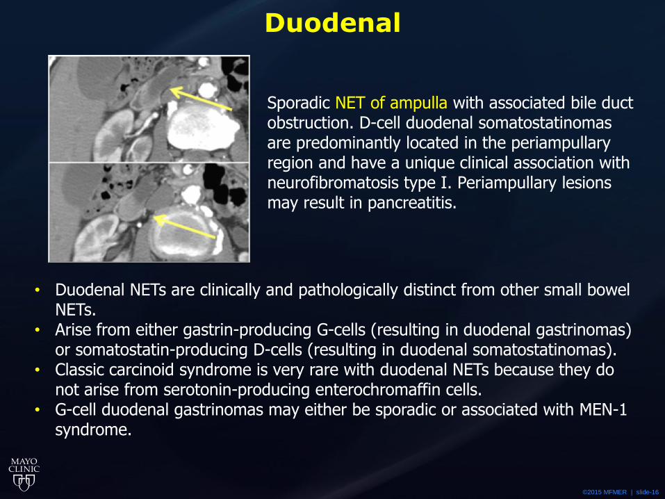
Duodenal
Sporadic NET of ampulla with associated bile duct obstruction. D-cell duodenal somatostatinomas are predominantly located in the periampullary region and have a unique clinical association with neurofibromatosis type I. Periampullary lesions may result in pancreatitis.
• Duodenal NETs are clinically and pathologically distinct from other small bowel NETs.
• Arise from either gastrin-producing G-cells (resulting in duodenal gastrinomas) or somatostatin-producing D-cells (resulting in duodenal somatostatinomas).
• Classic carcinoid syndrome is very rare with duodenal NETs because they do not arise from serotonin-producing enterochromaffin cells.
• G-cell duodenal gastrinomas may either be sporadic or associated with MEN-1 syndrome.

©2015 MFMER | slide-17
Small Bowel
Small bowel NETs originate intramurally and may appear as hyperenhancing nodular or asymmetric plaque-like lesions with associated puckering or retraction of the bowel wall. The gross image shows white tumor with transmural extension and puckering.
Small bowel NETs are frequently multifocal (~25% of cases) and are most frequently found in the distal ileum. Early nodal metastases (arrow head) are intimately associated with mesenteric vessels and spread toward the mesenteric root.
©2015 MFMER | slide-18
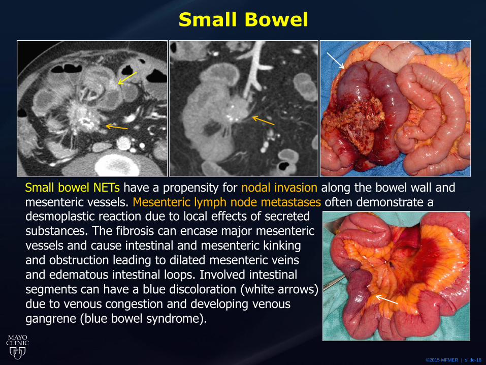
Small Bowel
Small bowel NETs have a propensity for nodal invasion along the bowel wall and mesenteric vessels. Mesenteric lymph node metastases often demonstrate a desmoplastic reaction due to local effects of secreted substances. The fibrosis can encase major mesenteric vessels and cause intestinal and mesenteric kinking and obstruction leading to dilated mesenteric veins and edematous intestinal loops. Involved intestinal segments can have a blue discoloration (white arrows) due to venous congestion and developing venous gangrene (blue bowel syndrome).

©2015 MFMER | slide-19
Small Bowel
Two patients with NETs in the ileum (example on left demonstrates associated serosal retraction (arrow head)) and active Crohn’s colitis involving the sigmoid colon. The association of Crohn’s disease and NETs is controversial, with several
cases observed at our institution.
NETs can occur within Meckel’s diverticula as demonstrated with this small hyperenhancing carcinoid tumor.
©2015 MFMER | slide-20
Appendiceal
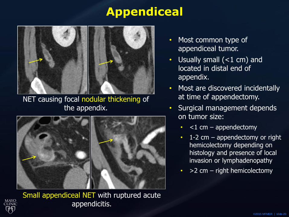
NET causing focal nodular thickening of the appendix.
Small appendiceal NET with ruptured acute appendicitis.
• Most common type of appendiceal tumor.
• Usually small (<1 cm) and located in distal end of appendix.
• Most are discovered incidentally at time of appendectomy.
• Surgical management depends on tumor size:
• <1 cm – appendectomy
• 1-2 cm – appendectomy or right hemicolectomy depending on histology and presence of local invasion or lymphadenopathy
• >2 cm – right hemicolectomy

©2015 MFMER | slide-21
Colon
Circumferential NET in the proximal ascending colon which directly invades into the adjacent pericolonic fat with local lymphadenopathy. Colonic/cecal
carcinoids are often exophytic and large (>5 cm) at presentation. The gross specimen shows an invasive cecal mass extending into the ileocecal valve.

©2015 MFMER | slide-22
Colon
• Colonic NETs are most frequently found in the right colon, often at or near, the ileocecal valve.
• Patients can remain asymptomatic for extended periods unless bleeding occurs, which may account for later stage presentations.
• Aggressive tumors with high proliferation rate often present with lymph node and liver metastases.
• Surgical treatment is similar to colonic adenocarcinoma but with overall worse outcomes.

©2015 MFMER | slide-23
Rectal
• Large rectal NETs may be indistinguishable from adenocarcinoma by CT and MR imaging.
• An asymmetric mural mass may suggest the diagnosis. • They are frequently asymptomatic and may be detected incidentally during
colonoscopy. • On CT or MR, rectal NETs may appear as small solitary submucosal nodules,
multiple nodules, or a large polypoid ulcerating mass. • Endoscopy and biopsy are required to confirm the diagnosis. • Small tumors (< 1 cm) tend to be localized and can be treated by
endoscopic resection.

©2015 MFMER | slide-24
Mimics – Enhancing GI masses
Tubulovillous adenoma
Hyperplastic polyp
Breast Ca Mets Lymphoma
Duodenal GIST
Sclerosing mesenteritis
Gastric GIST
©2015 MFMER | slide-25
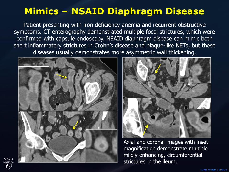
Mimics – NSAID Diaphragm Disease
Patient presenting with iron deficiency anemia and recurrent obstructive symptoms. CT enterography demonstrated multiple focal strictures, which were confirmed with capsule endoscopy. NSAID diaphragm disease can mimic both
short inflammatory strictures in Crohn’s disease and plaque-like NETs, but these diseases usually demonstrates more asymmetric wall thickening.
Axial and coronal images with inset magnification demonstrate multiple mildly enhancing, circumferential strictures in the ileum.

©2015 MFMER | slide-26
Pitfalls
Small bowel NET initially missed on routine CT performed with positive oral contrast. The same hyperenhancing, intraluminal mass clearly demonstrated on CT
enterography performed with neutral oral contrast.
Technical Issues:
• Positive oral contrast – May obscure small lesions.
• Thicker image slices – Limit visualization of small lesions.
• Suboptimal tumor contrast:
• Scan timing – NETs may be difficult to differentiate from bowel wall on venous phase.
• Low kV imaging – May improve image contrast and decrease dose.
• Injection rate – Higher injection rates (4-5 m/s) may improve tumor visualization.

©2015 MFMER | slide-27
Pitfalls
Enteric Delayed Arterial
Tiny small bowel NET – best demonstrated on the enteric phase, but still visualized on the early arterial and delayed phases
High-flow AVM – best seen on the arterial phase and not visualized on the delayed phase.
• Multiphase CT or MRI enterography may improved tumor visualization and help to distinguish NETs from other lesions based on enhancement characteristics.

©2015 MFMER | slide-28
Summary and Clinical Implications
• NETs can occur anywhere in the GI tract and represent the most common type of small bowel and appendiceal neoplasms.
• Understanding basic concepts regarding the pathologic classification of GI NETs and the ability to accurately characterize and stage NETs help radiologists and clinicians guide appropriate treatment and disease surveillance strategies.

©2015 MFMER | slide-29
Summary and Clinical Implications
• Frequently, small or early GI NETs may be subtle and overlooked, particularly on conventional cross-sectional imaging.
• Use of neutral oral contrast and multiphase CT and MR enterography aid detection of small GI NETs by optimizing luminal distention and tumoral enhancement.
• Although some of the CT and MR features of NETs are nonspecific, radiologists should be familiar with the common patterns and sites of disease involvement which can lead to the correct diagnosis.

©2015 MFMER | slide-30
References
• Bilimoria, K, et al. Small Bowel Cancer in the United States: Changes in Epidemiology, Treatment, and Survival Over the Last 20 Years. Annals of Surgery. 2009;249(1):63-71.
• Binstock AJ, et al. Carcinoid tumors of the stomach: a clinical and radiographic study. American Journal of Roentgenology. 2001;176(4):947–951.
• Bonekamp, D, et al. Role of computed tomography angiography in detection and staging of small bowel carcinoid tumors. World Journal of Radiology. 2015;7(9):220–235.
• Boudreaux JP, et al. The NANETS consensus guideline for the diagnosis and management of neuroendocrine tumors: well-differentiated neuroendocrine tumors of the jejunum, ileum, appendix, and cecum. Pancreas. 2010;39:753-766.
• Ganeshan, D, et al. Imaging Features of Carcinoid Tumors of the Gastrointestinal Tract. American Journal of Roentgenology. 2013;201:773-786.
• Levy AD, Sobin LH. From the archives of the AFIP: Gastrointestinal carcinoids: imaging features with clinicopathologic comparison. Radiographics. 2007;27:237–257.
• Yao JC, et al. One hundred years after "carcinoid": epidemiology of and prognostic factors for neuroendocrine tumors in 35,825 cases in the United States. Journal of Clinical Oncology. 2008;26:3063–3072.
Please direct correspondence to: [email protected]