Embed Size (px)
Citation preview
Core Curriculum V
Olecranon Fractures – A Case Based Approach to Understanding
Management
Jonathan M. Gross, MDDirector of Orthopedic Trauma
Staten Island University Hospital
Core Curriculum V
Introduction and Objectives• Design: Interactive Case Based• Objectives
– Review pertinent bone and soft tissue anatomy to understand fracture patterns and associated instability
– Review indications and strategies to stabilize fractures and restore stability
– Provide pearls to help minimize risk of surgical complications and illustrate key points of management
Core Curriculum V
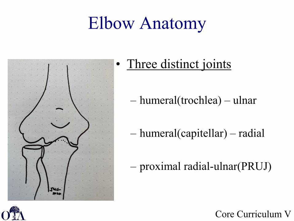
Elbow Anatomy
• Three distinct joints
– humeral(trochlea) – ulnar
– humeral(capitellar) – radial
– proximal radial-ulnar(PRUJ)
Core Curriculum VJMG 2020
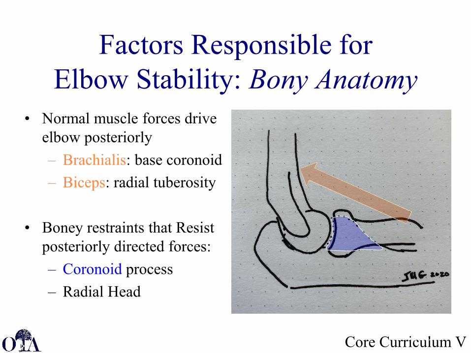
Factors Responsible for Elbow Stability: Bony Anatomy
• Normal muscle forces drive elbow posteriorly– Brachialis: base coronoid– Biceps: radial tuberosity
• Boney restraints that Resist posteriorly directed forces:– Coronoid process– Radial Head
Core Curriculum V
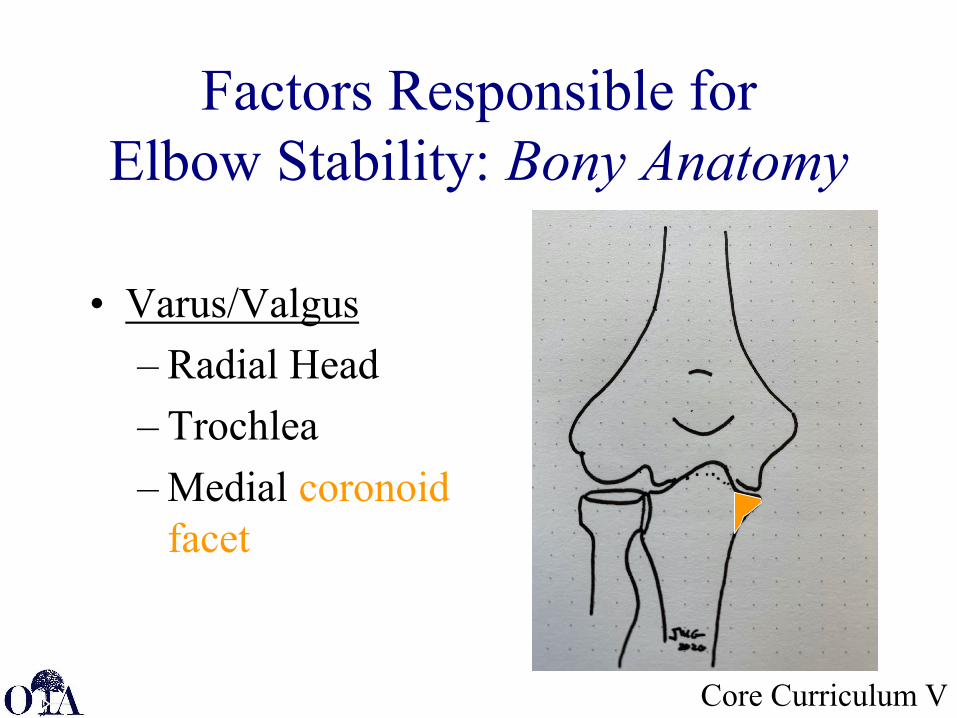
Factors Responsible for Elbow Stability: Bony Anatomy
• Varus/Valgus– Radial Head– Trochlea– Medial coronoid
facet
Core Curriculum V
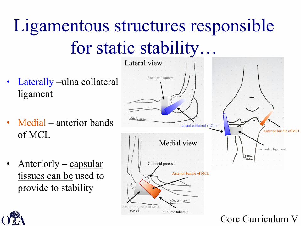
Ligamentous structures responsible for static stability…
JAAOS - Journal of the American Academy of Orthopaedic Surgeons16(9):519-529, September 2008.
• Laterally –ulna collateral ligament
• Medial – anterior bands of MCL
• Anteriorly – capsular tissues can be used to provide to stability
Coronoid process
Sublime tubercle
Anterior bundle of MCL
Posterior bundle of MCL
Anterior bundle of MCLLateral collateral (LCL)
Annular ligament
Annular ligament
Lateral view
Medial view
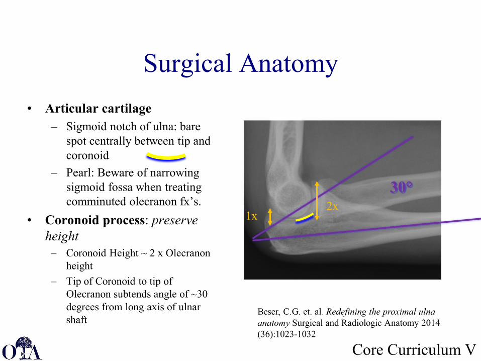
Surgical Anatomy
30°
Core Curriculum V
Beser, C.G. et. al. Redefining the proximal ulna anatomy Surgical and Radiologic Anatomy 2014 (36):1023-1032
1x2x
• Articular cartilage– Sigmoid notch of ulna: bare
spot centrally between tip and coronoid
– Pearl: Beware of narrowing sigmoid fossa when treating comminuted olecranon fx’s.
• Coronoid process: preserve height
– Coronoid Height ~ 2 x Olecranon height
– Tip of Coronoid to tip of Olecranon subtends angle of ~30 degrees from long axis of ulnar shaft
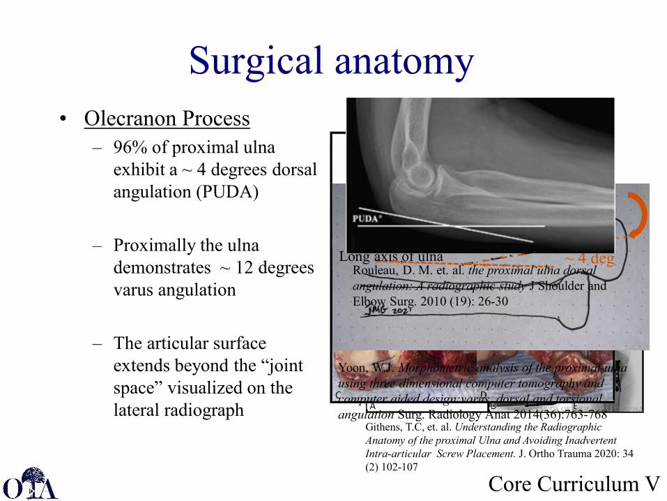
Surgical anatomy• Olecranon Process
– 96% of proximal ulna exhibit a ~ 4 degrees dorsal angulation (PUDA)
– Proximally the ulna demonstrates ~ 12 degrees varus angulation
– The articular surface extends beyond the “joint space” visualized on the lateral radiograph
Core Curriculum V
Githens, T.C, et. al. Understanding the Radiographic Anatomy of the proximal Ulna and Avoiding Inadvertent Intra-articular Screw Placement. J. Ortho Trauma 2020: 34 (2) 102-107
~ 4 degLong axis of ulna
Tip of Olecranon
Yoon, W.J. Morphometric analysis of the proximal ulna using three dimensional computer tomography and computer aided design:varus, dorsal and torsional angulation Surg. Radiology Anat 2014(36):763-768
Rouleau, D. M. et. al. the proximal ulna dorsal angulation: A radiographic study J Shoulder and Elbow Surg. 2010 (19): 26-30
Core Curriculum V
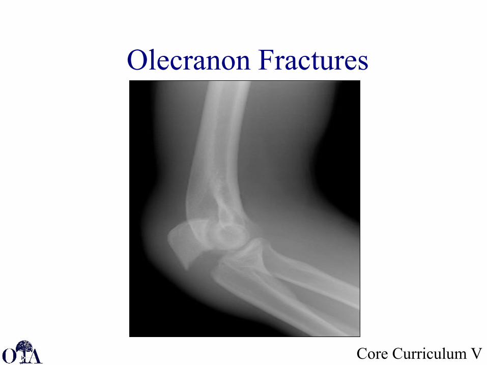
Olecranon Fractures
Core Curriculum V
Mechanism of Injury• Acute Tension overload: Tension applied by
the triceps with flexion of the elbow
• Direct Trauma
• Chronic overload: eg. stress fractures seen commonly with osteopaenic or pediatric patients
Core Curriculum V
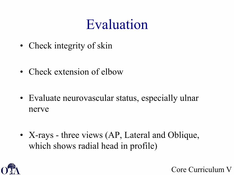
Evaluation• Check integrity of skin
• Check extension of elbow
• Evaluate neurovascular status, especially ulnar nerve
• X-rays - three views (AP, Lateral and Oblique, which shows radial head in profile)
Core Curriculum V
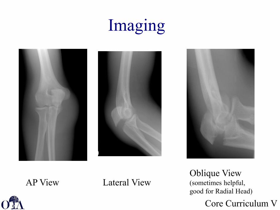
Imaging
AP View Lateral ViewOblique View (sometimes helpful, good for Radial Head)
Core Curriculum V
Classification
• Many Classifications:– Colton– Morrey– Schatzker– AO/ASIF– OTA
• Criteria– Displacement– Direction of fracture– Degree of comminution– Percent involvement– Associated injuries
Core Curriculum V
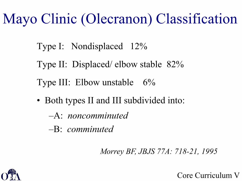
Type I: Nondisplaced 12%
Type II: Displaced/ elbow stable 82%
Type III: Elbow unstable 6%
• Both types II and III subdivided into:–A: noncomminuted–B: comminuted
Mayo Clinic (Olecranon) Classification
Morrey BF, JBJS 77A: 718-21, 1995Morrey BF, JBJS 77A: 718-21, 1995
Core Curriculum V
Treatment Objectives
• Restoration of elbow motion and prevention of stiffness– Goal is to begin early ROM
• Restoration and preservation of the elbow extensor mechanism.
• Restoration of the articular surface.• Prevention of complications.
Core Curriculum V
Treatment Methods• Nonoperative
– Indicated in low demand individual with stable elbow joint.
• (Duckworth, et. al. JBJS AM 2014:96,67-72 )• (Marot, V, et. al. Orthopedics & Traumatology:
Surgery & Research 2018:104, 79-82)
• Operative– Open reduction and internal fixation
• Tension band wire with pins or intramedullary screws• Plate
– Excision of olecranon and triceps repair• Comminuted, unreconstructable fractures• Typically, Elderly patients with loss of active elbow
extension
Core Curriculum V
Nonoperative Treatment• Classically Reserved for nondisplaced fractures
• Historically - Prolonged long arm cast was complicated by stiffness
• More Recent – short duration of “immobilization” provided reasonable results in low demand elderly...– Duckworth, et. al. (2017) 2 weeks collar and cuff, followed by
supervised ROM with physiotherapist– Immobilization prolonged for pain– Although loss of reduction, similar functional results to ORIF
group
Core Curriculum V
Indications for Surgery
• Disruption of extensor mechanism – Unable to actively extend elbow
• Articular incongruity– Any displaced fracture
Core Curriculum V
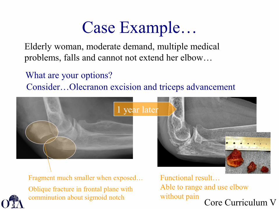
Case Example…
Fragment much smaller when exposed…
1 year later
Functional result…Able to range and use elbow without pain
Elderly woman, moderate demand, multiple medical problems, falls and cannot not extend her elbow…
Consider…Olecranon excision and triceps advancement
Oblique fracture in frontal plane with comminution about sigmoid notch
What are your options?
Core Curriculum V
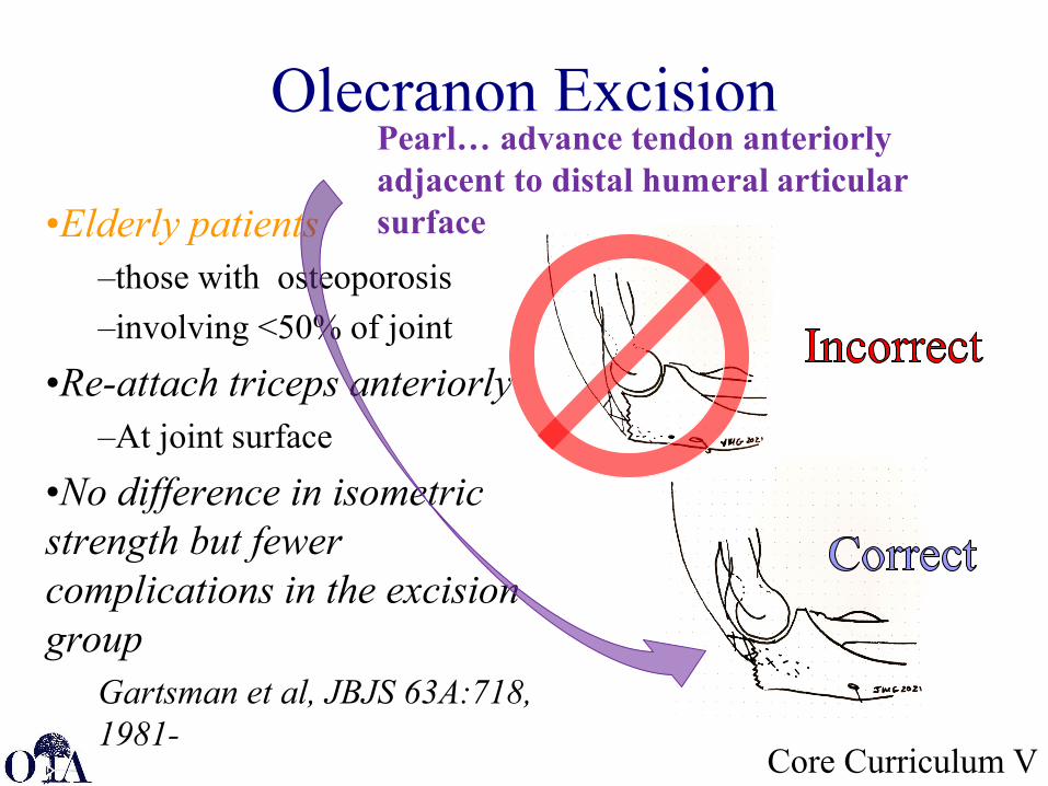
Olecranon Excision
•Elderly patients–those with osteoporosis –involving <50% of joint
•Re-attach triceps anteriorly–At joint surface
•No difference in isometric strength but fewer complications in the excision group
Gartsman et al, JBJS 63A:718, 1981-
Pearl… advance tendon anteriorly adjacent to distal humeral articular surface
Core Curriculum V
Positioning
• Posterior approach• Arm position
– Supine with arm across chest.– Lateral or prone also may be used.– Supine with arm on hand table
• Can Use Tourniquet (but may tighten extensor mechanism)
• Regional or general anesthesia
Core Curriculum V
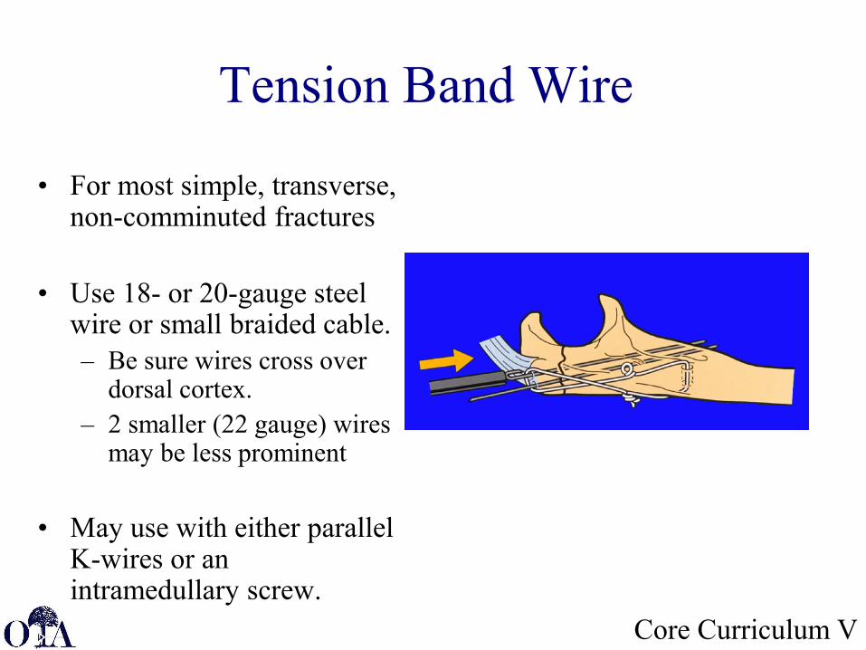
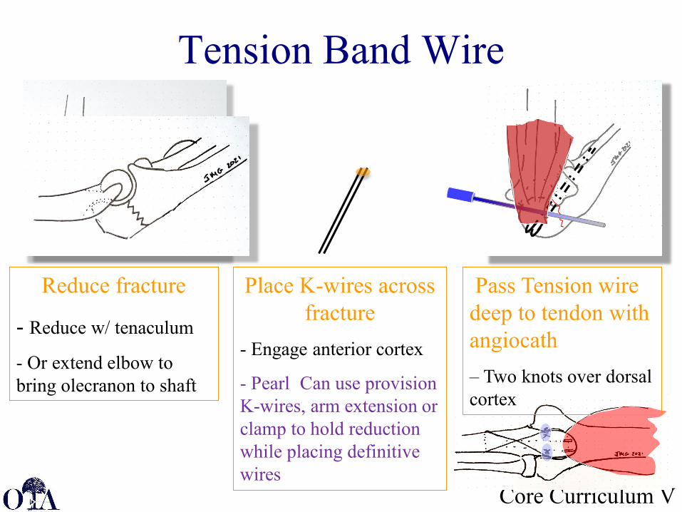
Tension Band Wire
• For most simple, transverse, non-comminuted fractures
• Use 18- or 20-gauge steel wire or small braided cable. – Be sure wires cross over
dorsal cortex.– 2 smaller (22 gauge) wires
may be less prominent
• May use with either parallel K-wires or an intramedullary screw.
Core Curriculum V
Reduce fracture
- Reduce w/ tenaculum
- Or extend elbow to bring olecranon to shaft
Place K-wires across fracture
- Engage anterior cortex
- Pearl: Can use provision K-wires, arm extension or clamp to hold reduction while placing definitive wires
Pass Tension wire deep to tendon with angiocath– Two knots over dorsal cortex
Tension Band Wire
Core Curriculum V
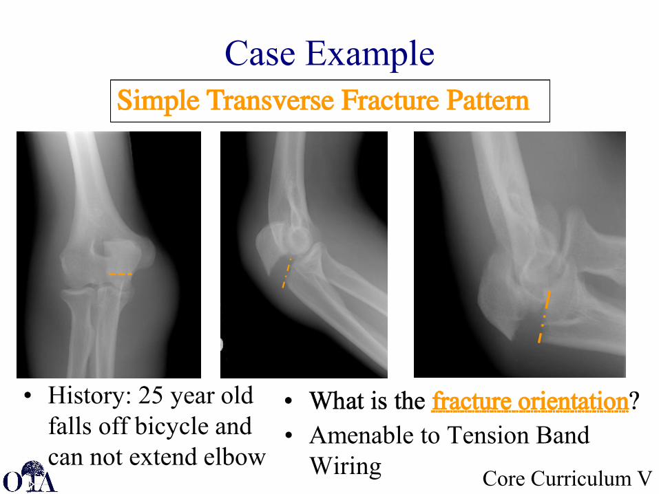
Case Example
• History: 25 year old falls off bicycle and can not extend elbow
• Amenable to Tension Band Wiring
Core Curriculum VJMG 2020
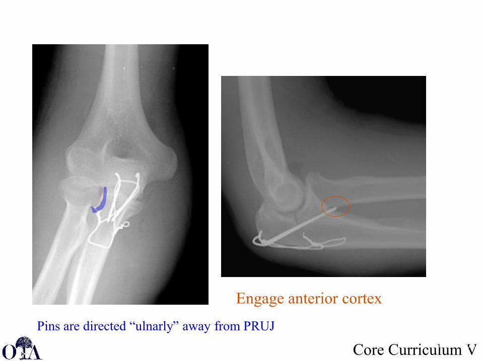
Engage anterior cortex
Pins are directed “ulnarly” away from PRUJ
Core Curriculum V
Potential Complications…
• K wires project though anterior cortex too far… irritate AIN– Solution, withdraw wire 5 mm prior to bending
wires over olecranon tip• K wires project to far radial… interfere with
proximal radio-ulnar joint – Solution, start wires more radial and aim more
ulnarly
JMG 2020
JMG 2020
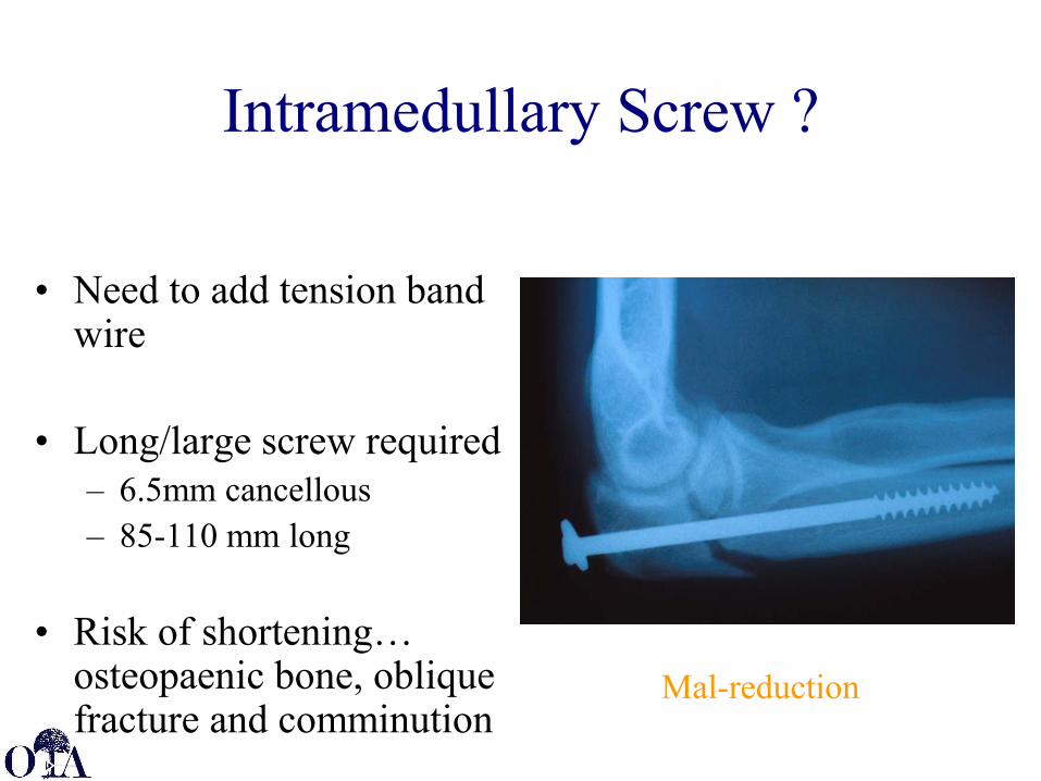
Intramedullary Screw ?
• Need to add tension band wire
• Long/large screw required– 6.5mm cancellous– 85-110 mm long
• Risk of shortening… osteopaenic bone, oblique fracture and comminution
Mal-reduction
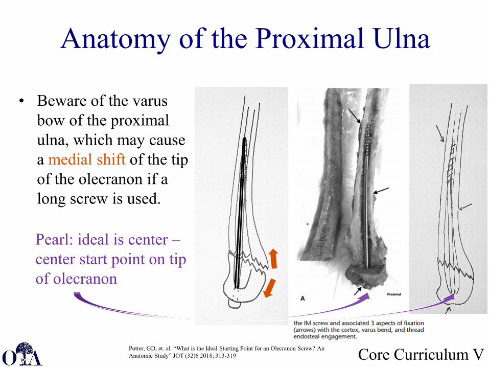
Anatomy of the Proximal Ulna
• Beware of the varus bow of the proximal ulna, which may cause a medial shift of the tip of the olecranon if a long screw is used.
Core Curriculum V
Pearl: ideal is center –center start point on tip of olecranon
Potter, GD, et. al. “What is the Ideal Starting Point for an Olecranon Screw? An Anatomic Study” JOT (32)6 2018; 313-319
Core Curriculum VJMG 2020
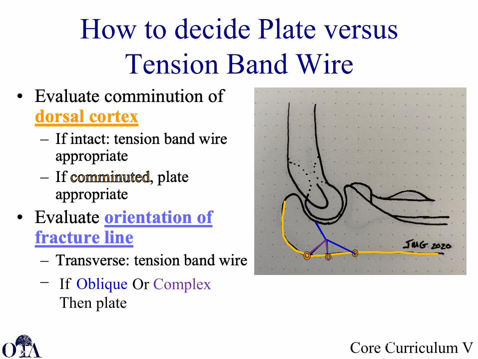
How to decide Plate versus Tension Band Wire
Oblique Or ComplexIfThen plate
JMG 2020
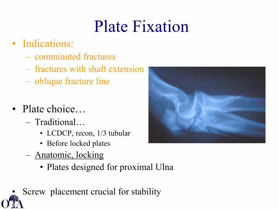
Plate Fixation• Indications:
– comminuted fractures– fractures with shaft extension– oblique fracture line
• Plate choice…– Traditional…
• LCDCP, recon, 1/3 tubular• Before locked plates
– Anatomic, locking• Plates designed for proximal Ulna
• Screw placement crucial for stability
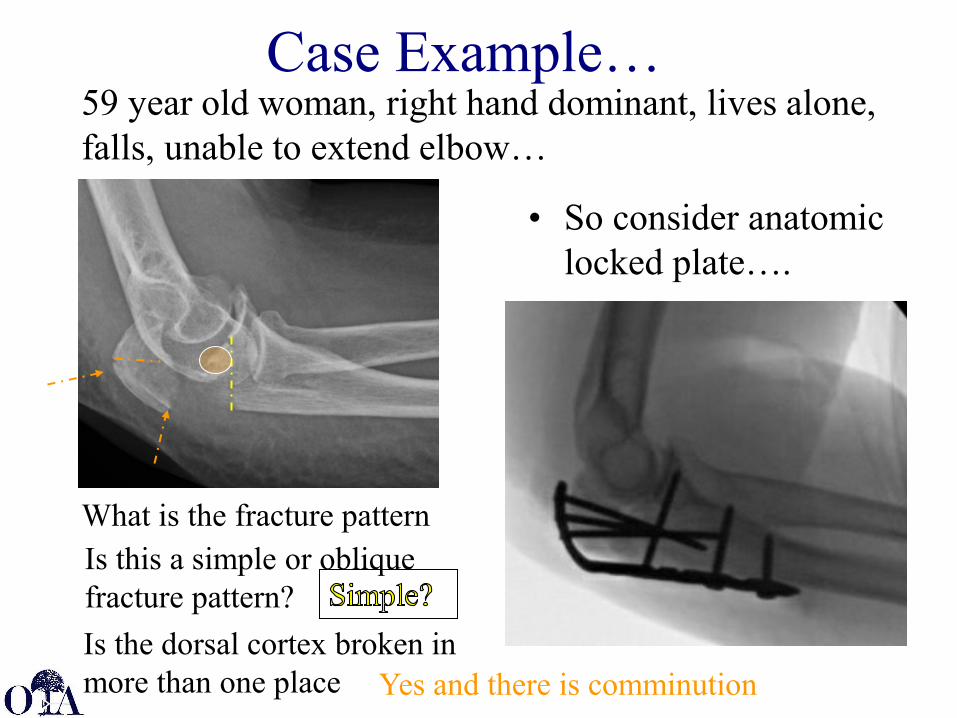
59 year old woman, right hand dominant, lives alone, falls, unable to extend elbow…
• So consider anatomic locked plate….
JMG 2020
Case Example…
What is the fracture pattern?
Is the dorsal cortex broken in more than one place?
Is this a simple or oblique fracture pattern?
Yes and there is comminution
Core Curriculum V
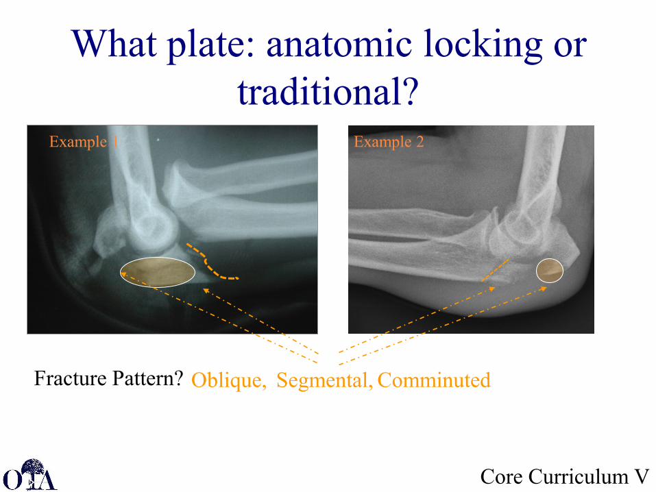
What plate: anatomic locking or traditional?
Example 1 Example 2
Fracture Pattern? Oblique, Segmental, Comminuted
Core Curriculum V
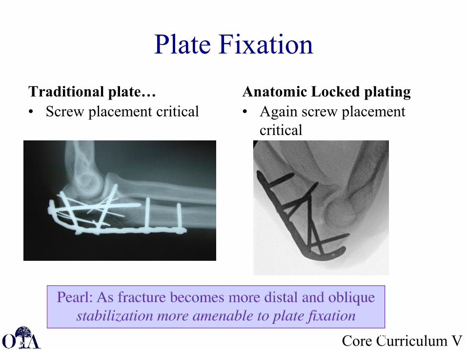
Plate FixationTraditional plate…• Screw placement critical
Anatomic Locked plating• Again screw placement
critical
JMG 2020
Courtesy: Fred Behrens, MD
Core Curriculum V
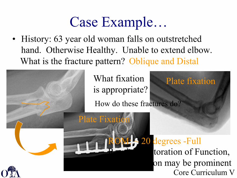
Case Example…• History: 63 year old woman falls on outstretched
hand. Otherwise Healthy. Unable to extend elbow.Oblique and DistalWhat is the fracture pattern?
Plate Fixation
What fixation is appropriate?
Plate fixation
Results: Restoration of Function, but all fixation may be prominent
How do these fractures do?
ROM: ~ 20 degrees -Full
Core Curriculum V
Locked plating• Relative indications
– Comminution– Osteoporotic bone
• Removal of hardware…– 18-53%; probably irrespective of locking or
non locking implant• Chen, M.J. et.al. Surgical and Nonoperative management of Olecranon
fractures in the Elderly: A Systematic Review and Meta-Analysis J Ortho Trauma 2021;35 (1) 10-16
• Bailey, C.S. et. al. “Outcome of Plate Fixation of Olecranon Fractures” J. Ortho Trauma 2001: 15 (8) 542-548
• Snoddy, MC, et. al. “Olecranon Fractures: factors influencing re-operation”Int Orthop. 2014:38(8) 1711-1716 JMG 2020
Core Curriculum V
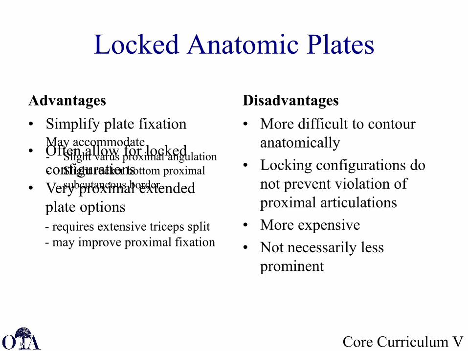
Locked Anatomic Plates
Advantages• Simplify plate fixation
Disadvantages• More difficult to contour
anatomically• Locking configurations do
not prevent violation of proximal articulations
• More expensive• Not necessarily less
prominent
May accommodate- Slight varus proximal angulation- Slight rocker bottom proximal
subcutaneous border
- requires extensive triceps split- may improve proximal fixation
• Often allow for locked configurations
• Very proximal extended plate options
Core Curriculum V
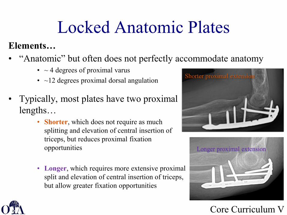
Locked Anatomic PlatesElements…• “Anatomic” but often does not perfectly accommodate anatomy
• ~ 4 degrees of proximal varus• ~12 degrees proximal dorsal angulation
• Typically, most plates have two proximal lengths…
• Shorter, which does not require as much splitting and elevation of central insertion of triceps, but reduces proximal fixation opportunities
• Longer, which requires more extensive proximal split and elevation of central insertion of triceps, but allow greater fixation opportunities
Shorter proximal extension
Longer proximal extension
Core Curriculum VJMG 2020
Proximal Ulna with Shaft Extension
Core Curriculum V
Plate Location
• No mechanical difference between posterior or lateral placement
(King et al, J Shoulder Elbow Surg 5:437, 1996)
• Less problems with plate prominence when placed laterally and one can get bicortical screw purchase
• Posterior Plate allows more advantageous screw placement– Coronoid screw– IM screw– Olecranon tip screw
Core Curriculum V
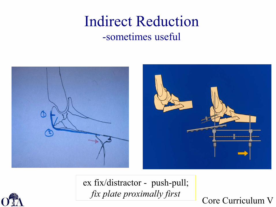
Indirect Reduction-sometimes useful
ex fix/distractor - push-pull; fix plate proximally first
Core Curriculum V
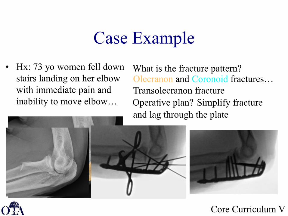
Case Example• Hx: 73 yo women fell down
stairs landing on her elbow with immediate pain and inability to move elbow…
What is the fracture pattern?
Coronoid fragment
comminutionOlecranon fragment
Olecranon and Coronoid fractures…Transolecranon fractureOperative plan? Simplify fracture and lag through the plate
Core Curriculum V
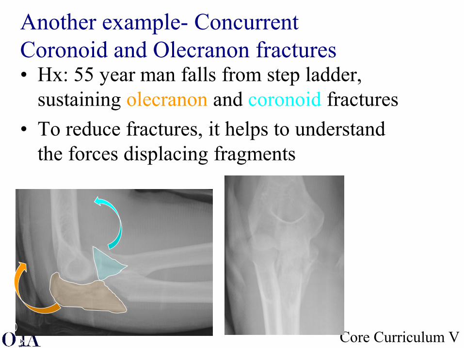
Another example- Concurrent Coronoid and Olecranon fractures• Hx: 55 year man falls from step ladder,
sustaining olecranon and coronoid fractures• To reduce fractures, it helps to understand
the forces displacing fragments
JMG 2020
Core Curriculum V
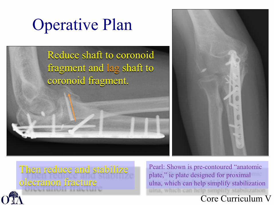
Operative Plan
Pearl: Shown is pre-contoured “anatomic plate,” ie plate designed for proximal ulna, which can help simplify stabilization
Reduce shaft to coronoid fragment and lag shaft to coronoid fragment.
Then reduce and stabilize olecranon fracture
JMG 2020
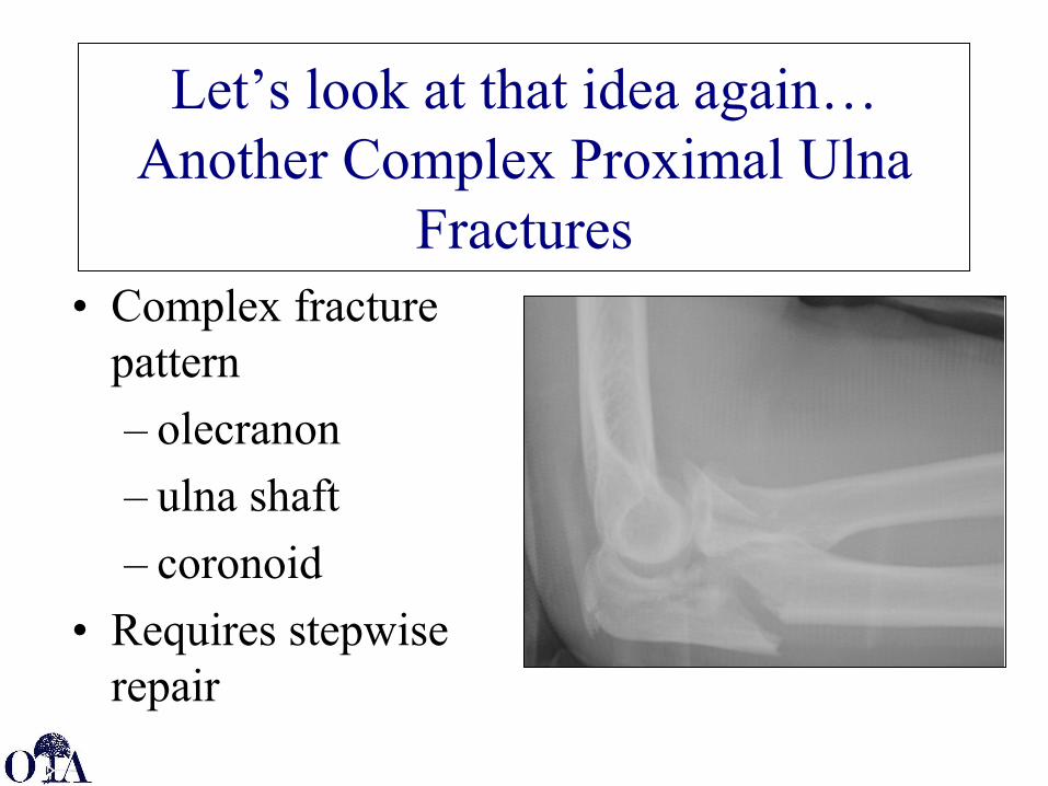
Let’s look at that idea again… Another Complex Proximal Ulna
Fractures• Complex fracture
pattern– olecranon– ulna shaft– coronoid
• Requires stepwise repair
Core Curriculum V
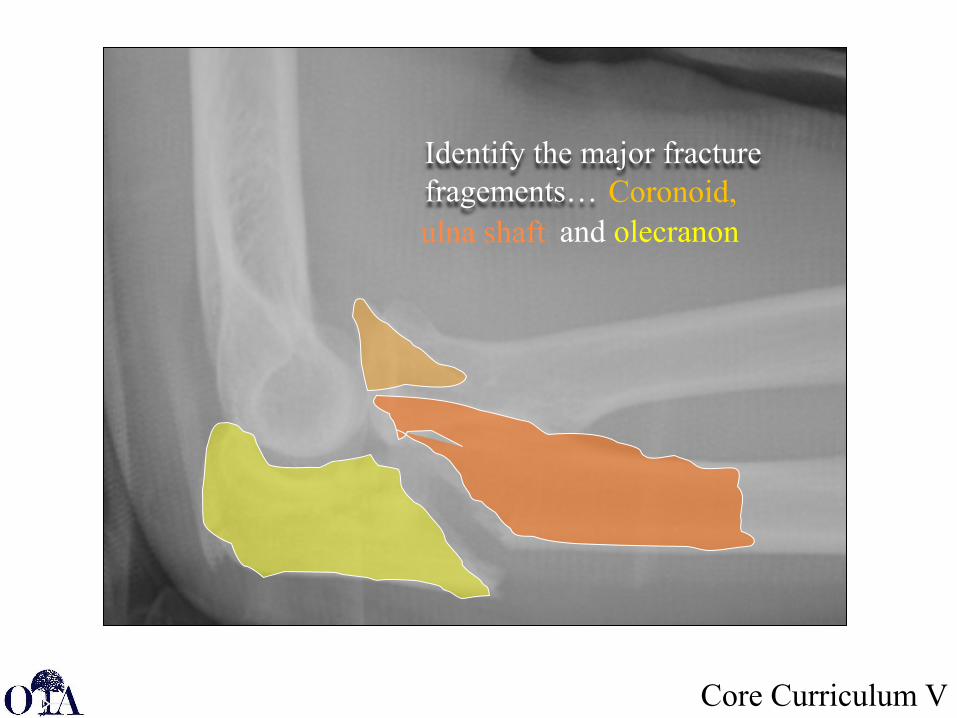
Identify the major fracture fragements… Coronoid,ulna shaft and olecranon
Core Curriculum V
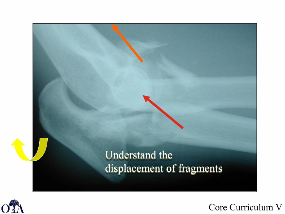
Understand the displacement of fragments
Core Curriculum V
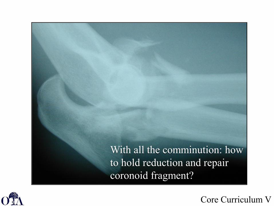
With all the comminution: how to hold reduction and repair coronoid fragment?
Core Curriculum V
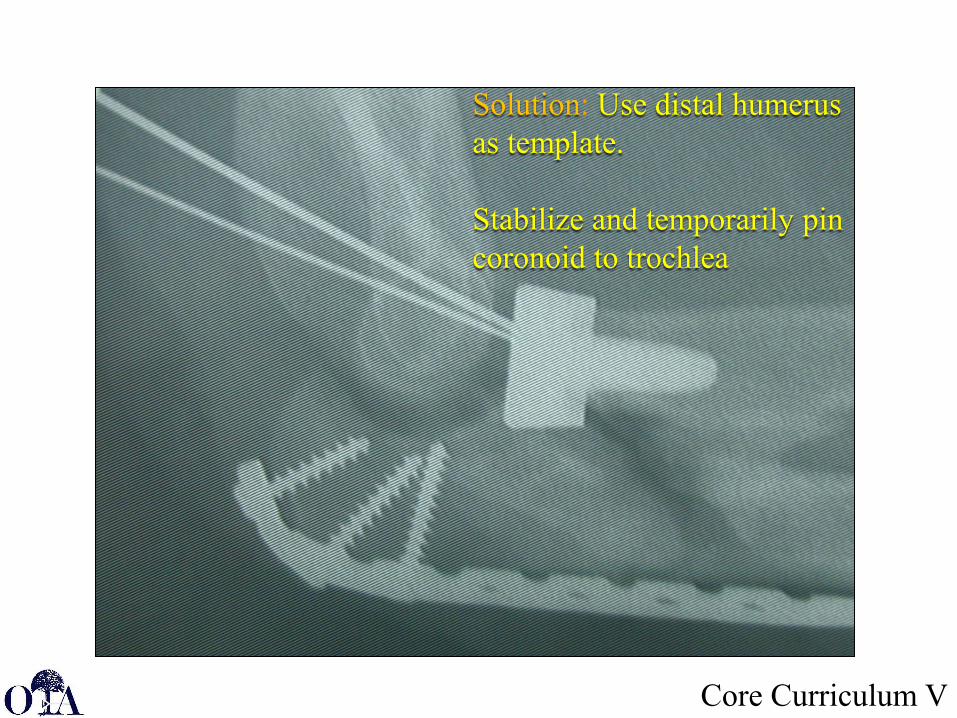
Solution: Use distal humerus as template.
Stabilize and temporarily pin coronoid to trochlea
Core Curriculum V
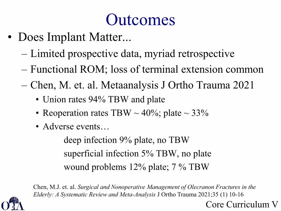
Outcomes• Does Implant Matter...
– Limited prospective data, myriad retrospective– Functional ROM; loss of terminal extension common– Chen, M. et. al. Metaanalysis J Ortho Trauma 2021
• Union rates 94% TBW and plate• Reoperation rates TBW ~ 40%; plate ~ 33%• Adverse events…
deep infection 9% plate, no TBWsuperficial infection 5% TBW, no platewound problems 12% plate; 7 % TBW
JMG 2020
Chen, M.J. et. al. Surgical and Nonoperative Management of Olecranon Fractures in the Elderly: A Systematic Review and Meta-Analysis J Ortho Trauma 2021;35 (1) 10-16

Core Curriculum V
Complications
Core Curriculum V
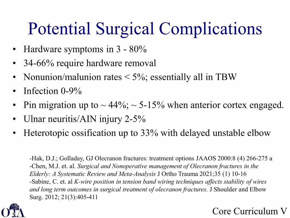
Potential Surgical Complications• Hardware symptoms in 3 - 80%• 34-66% require hardware removal• Nonunion/malunion rates < 5%; essentially all in TBW• Infection 0-9%• Pin migration up to ~ 44%; ~ 5-15% when anterior cortex engaged.• Ulnar neuritis/AIN injury 2-5%• Heterotopic ossification up to 33% with delayed unstable elbow
-Hak, D.J.; Golladay, GJ Olecranon fractures: treatment options JAAOS 2000:8 (4) 266-275 a-Chen, M.J. et. al. Surgical and Nonoperative management of Olecranon fractures in the Elderly: A Systematic Review and Meta-Analysis J Ortho Trauma 2021;35 (1) 10-16-Sabine, C. et. al K-wire position in tension band wiring techniques affects stability of wires and long term outcomes in surgical treatment of olecranon fractures. J Shoulder and Elbow Surg. 2012; 21(3):405-411
Core Curriculum V
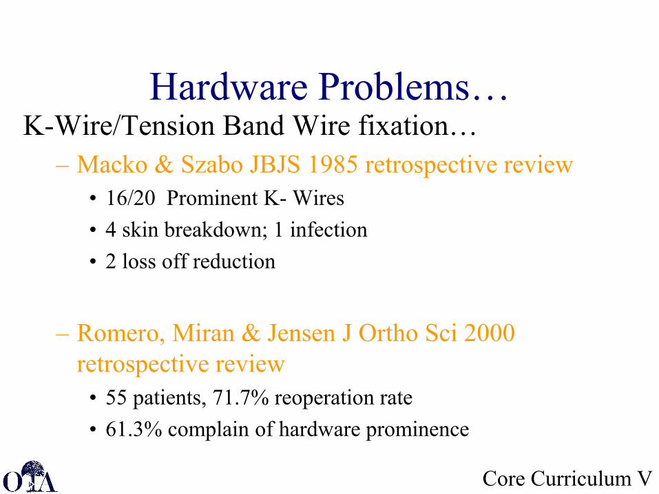
Hardware Problems…K-Wire/Tension Band Wire fixation…
– Macko & Szabo JBJS 1985 retrospective review• 16/20 Prominent K- Wires• 4 skin breakdown; 1 infection• 2 loss off reduction
– Romero, Miran & Jensen J Ortho Sci 2000 retrospective review
• 55 patients, 71.7% reoperation rate• 61.3% complain of hardware prominence
Core Curriculum V
Outcome
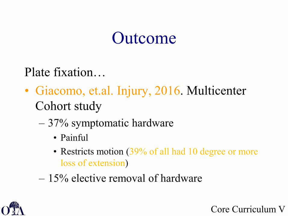
Plate fixation…• Giacomo, et.al. Injury, 2016. Multicenter
Cohort study– 37% symptomatic hardware
• Painful• Restricts motion (39% of all had 10 degree or more
loss of extension)– 15% elective removal of hardware
Core Curriculum V
Outcomes
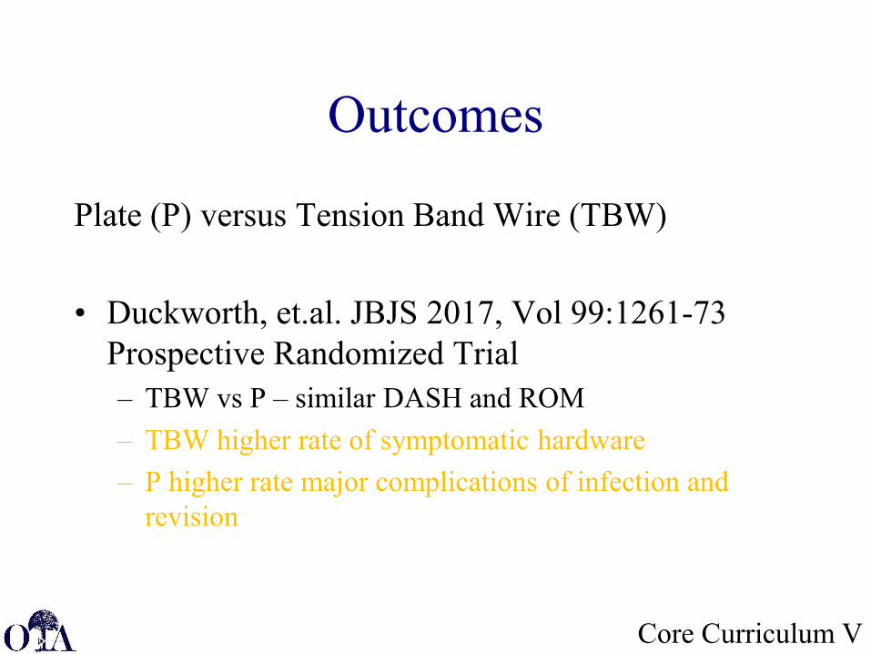
Plate (P) versus Tension Band Wire (TBW)
• Duckworth, et.al. JBJS 2017, Vol 99:1261-73 Prospective Randomized Trial– TBW vs P – similar DASH and ROM– TBW higher rate of symptomatic hardware– P higher rate major complications of infection and
revision
Core Curriculum V
Take Home Principle…• Treatment of olecranon fractures, requires an
understanding of the fracture pattern and the patient’s functional demands.
• So When addressing Olecranon fractures Understand:– Who is the patient/what are his or her demands?– What is the fracture pattern and is it associated with
other injuries?– If operative intervention indicated, run the check list to
develop the most effective and cost-conscious treatment for the the fracture
JMG 2020
JTC 2010
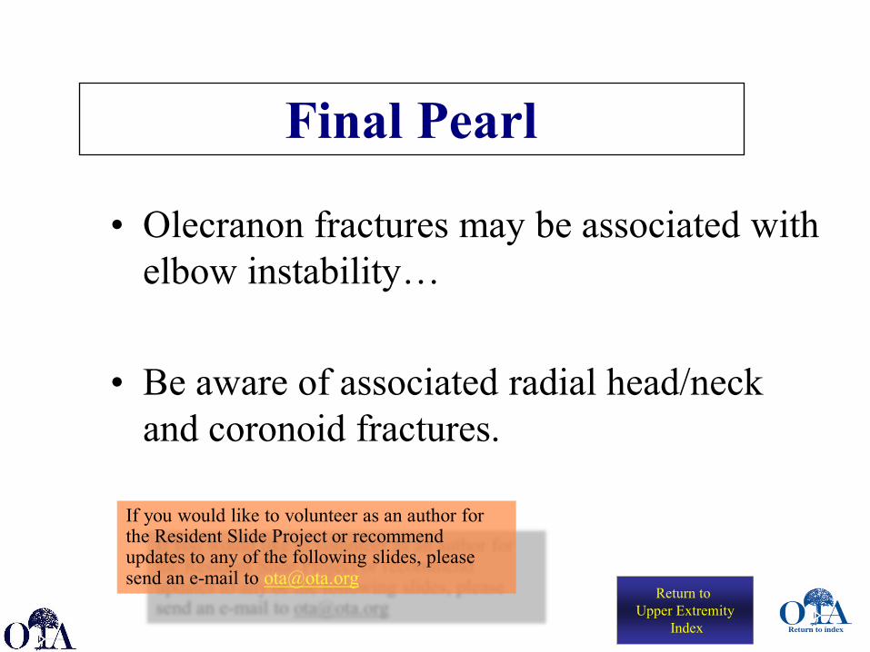
Final Pearl
• Olecranon fractures may be associated with elbow instability…
• Be aware of associated radial head/neck and coronoid fractures.
Return to Upper Extremity
Index
If you would like to volunteer as an author for the Resident Slide Project or recommend updates to any of the following slides, please send an e-mail to [email protected]
Core Curriculum V
Selective Bibliography• Oldie but Goodie Reviews…
– Hak, David J. MD; Golladay, Gregory J. MD Olecranon Fractures: Treatment Options; Journal of the American Academy of OrthopaedicSurgeons: July 2000 - Volume 8 - Issue 4 - p 266-275
– Rouleau, D.M.; Sandman, E.; Riet, RV Management of Fractures of Proximal Ulna JAAOS vol 21 (2013) issue 3:149-160
– Stein, S.P. Coronoid Process Fractures JAAOS vol 16 (2008) issue 9:519-529
JMG 2020
Core Curriculum V
Selective Bibliography• A few classics
– Gartsman GM, Sculco TP, Otis JC. Operative treatment of olecranon fractures. Excision or open reduction with internal fixation. The Journal of Bone and Joint surgery. American Volume. 1981 Jun;63(5):718-721.
– Doornberg J, Ring D, Jupiter JB. Effective treatment of fracture-dislocations of the olecranon requires a stable trochlear notch. Clin Orthop Relat Res. 2004 Dec;(429):292-300.
– Villanueva P, Osorio F, Commessatti M, Sanchez-Sotelo J. Tension-band wiring for olecranon fractures: analysis of risk factors for failure. J Shoulder Elbow Surg. 2006 May-Jun;15(3):351-6.
– Prayson MJ, Iossi MF, Buchalter D, Vogt M, Towers J (2008) Safe zone for anterior cortical perforation of the ulna during tension-band wire fixation: a magnetic resonance imaging analysis. J Shoulder Elbow Surg 17(1):121–125
JMG 2020
Core Curriculum V
Selective Bibliography• A few key recent references…
– Duckworth, AD, Clement, ND; Aitken, SA; Court-Brown, CM; The epidemiology of fractures of the proximal ulna. Injury 43(2012) 343-346
– Duckworth, AD, Clement, ND; Mc Eachen, JE; White, TO; Court-Brown, C; McQueen, M M Nonoperative Management of Displaced Olecranon fractures in Low Demand Elderly J Bone Joint Surg Am (2014) 96; 67-72
– De Giacomo, A. F. ; Tornetta, P; Sinicrope, B.J.; Cronin, P. K.; Althausen, P L.; Bray, T. J. ; Kain, M.S.; Marcantonio, A; Sagi, C; James, CR. Outcomes after plating of olecranon fractures: A multicenter evaluation, Injury, 47 (2016) Is7: 1466-1471,
– Duckworth, AD, Clement, ND; White, TO; Court-Brown, C; McQueen, M M Plate versus Tension Band Wire Fixation of Olecranon Fractures: A prospective randomized trial J Bone Joint Surg Am (2017) 99; 1261-12-73
JMG 2020
Core Curriculum V
Selective Bibliography
• A few key recent references (continued)…– Chen, M.J. et.al. Surgical and Nonoperative management of Olecranon fractures in
the Elderly: A Systematic Review and Meta-Analysis J Ortho Trauma 2021;35 (1) 10-16
– Githens, T.C, et. al. Understanding the Radiographic Anatomy of the proximal Ulna and Avoiding Inadvertent Intra-articular Screw Placement. J. Ortho Trauma 2020: 34 (2) 102-107