Embed Size (px)
Citation preview
Ocular Pharmacology
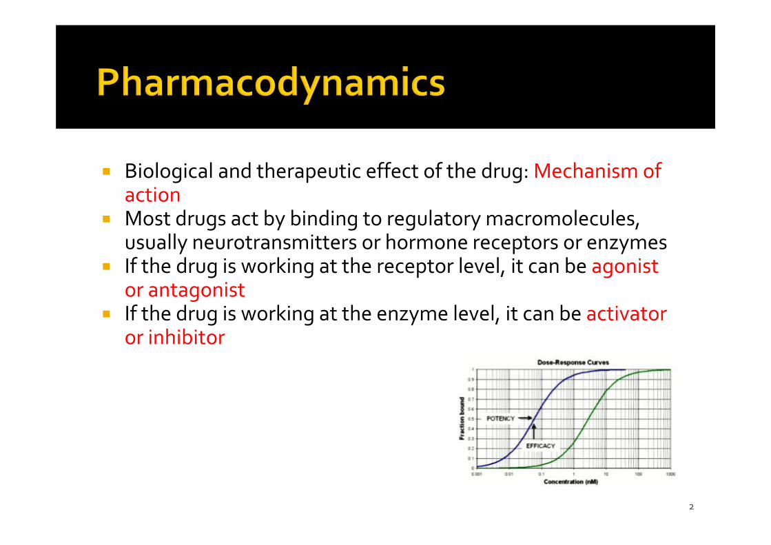
Biological and therapeutic effect of the drug: Mechanism of action
Most drugs act by binding to regulatory macromolecules, usually neurotransmitters or hormone receptors or enzymes
If the drug is working at the receptor level, it can be agonist or antagonist
If the drug is working at the enzyme level, it can be activator or inhibitor
2
3
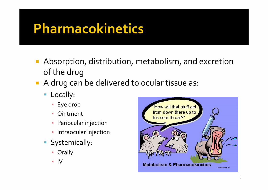
Absorption, distribution, metabolism, and excretion of the drug
A drug can be delivered to ocular tissue as: Locally:
▪ Eye drop▪ Ointment▪ Periocular injection▪ Intraocular injection
Systemically:▪ Orally▪ IV
4
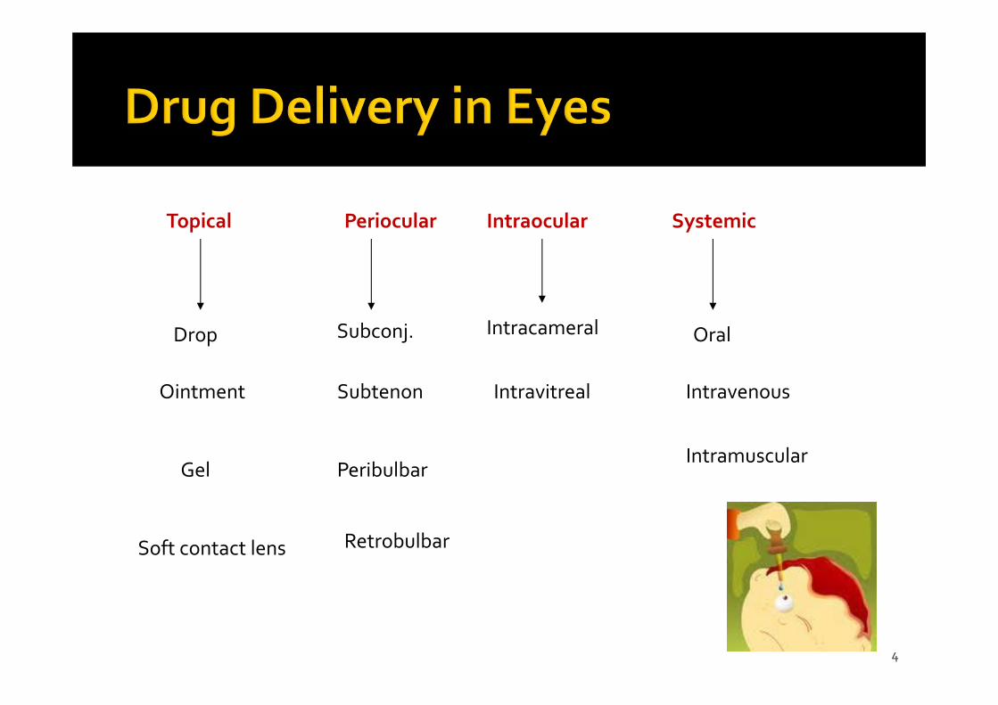
Topical Periocular Intraocular Systemic
Drop
Ointment
Gel
Soft contact lens
Subconj.
Subtenon
Peribulbar
Retrobulbar
Intracameral
Intravitreal
Oral
Intravenous
Intramuscular
5
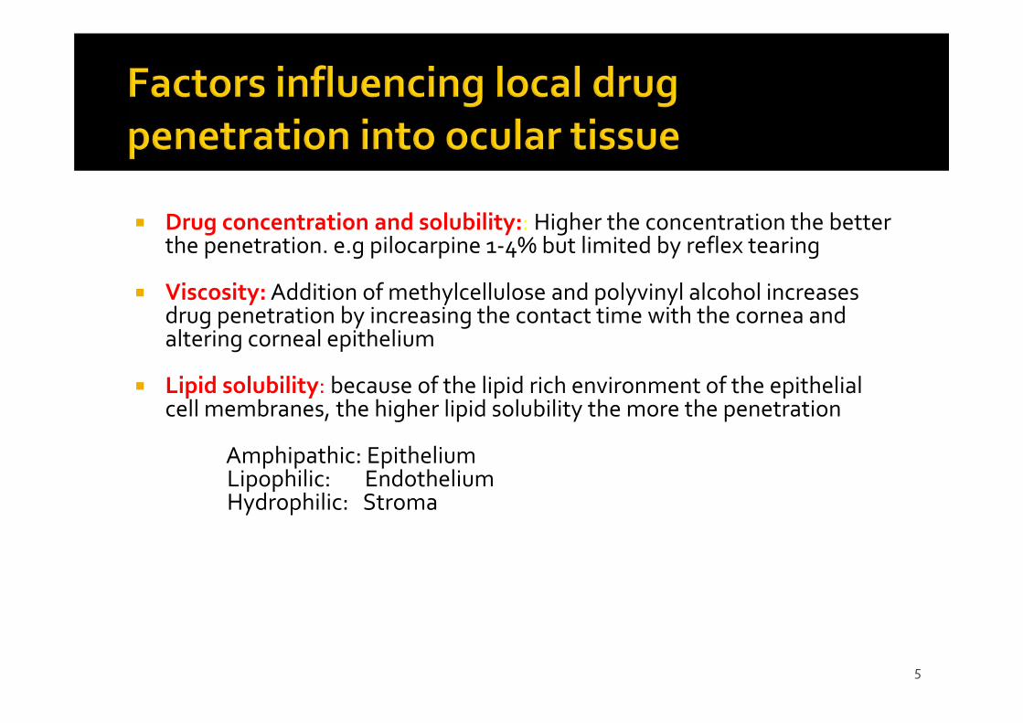
Drug concentration and solubility:: Higher the concentration the better the penetration. e.g pilocarpine 1‐4% but limited by reflex tearing
Viscosity:Addition of methylcellulose and polyvinyl alcohol increases drug penetration by increasing the contact time with the cornea and altering corneal epithelium
Lipid solubility: because of the lipid rich environment of the epithelial cell membranes, the higher lipid solubility the more the penetration
Amphipathic: EpitheliumLipophilic: EndotheliumHydrophilic: Stroma
6
Surfactants:: The preservatives used in ocular preparations alter cell membrane in the cornea and increase drug permeability e.g. benzylkonium and thiomersal
pH: the normal tear pH is 7.4 and if the drug pH is much different, this will cause reflex tearing
Drug tonicity:When an alkaloid drug is put in relatively alkaloid medium, the proportion of the uncharged form will increase, thus more penetration
Molecular weight and size
7
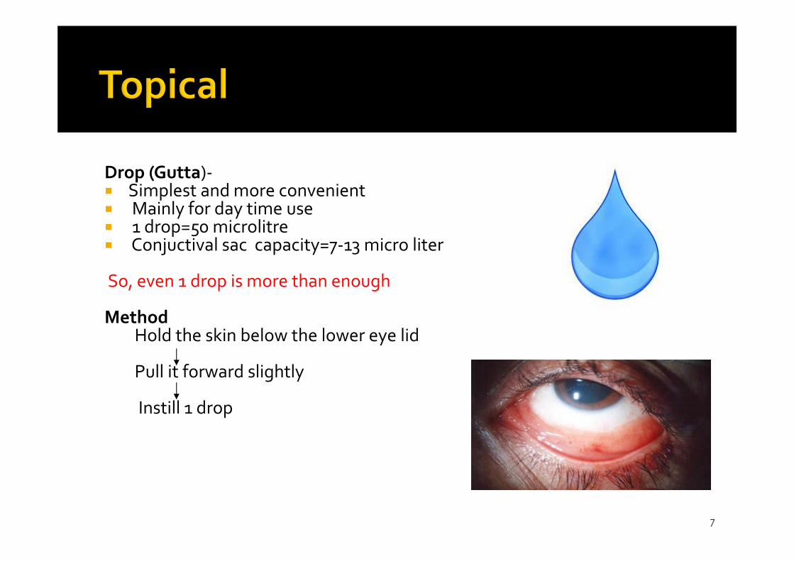
Drop (Gutta)‐ Simplest and more convenient Mainly for day time use 1 drop=50 microlitre Conjuctival sac capacity=7‐13 micro liter
So, even 1 drop is more than enough
MethodHold the skin below the lower eye lid
Pull it forward slightly
Instill 1 drop
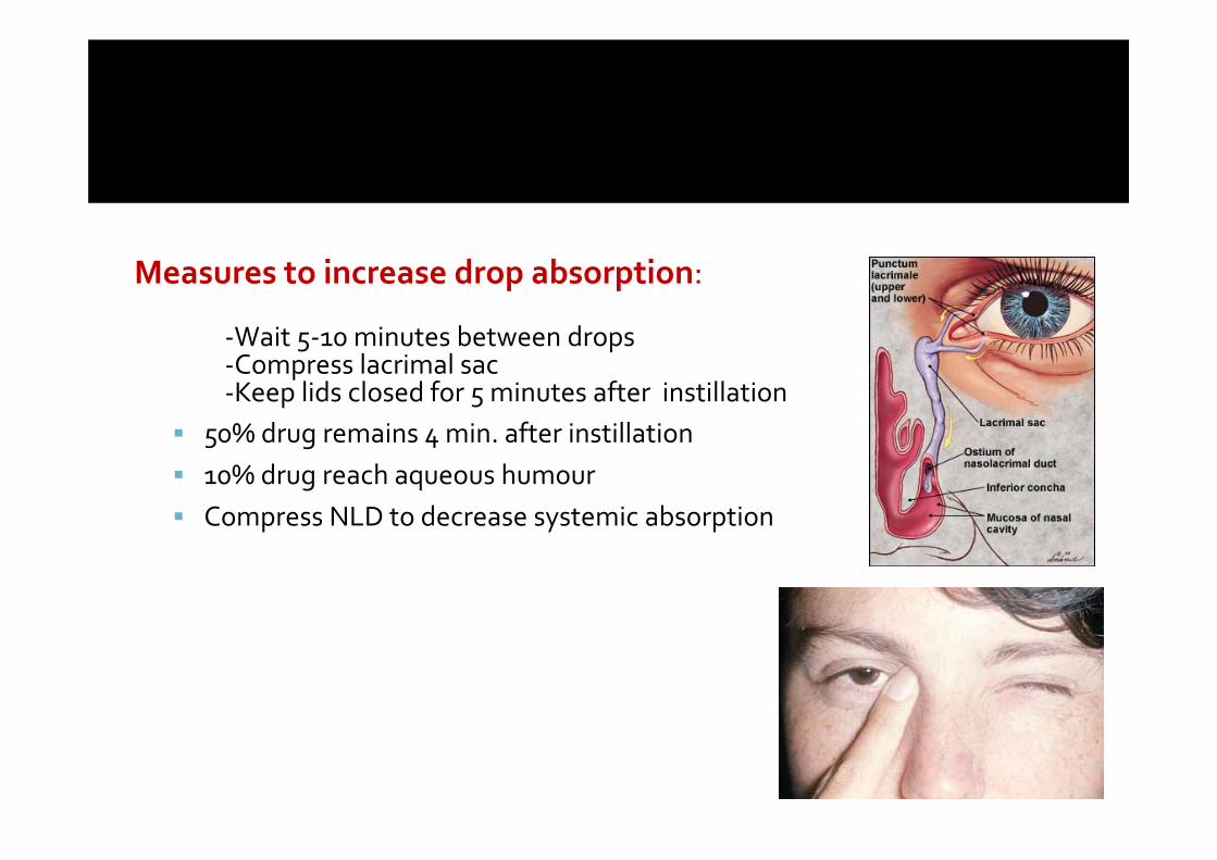
Measures to increase drop absorption:
‐Wait 5‐10 minutes between drops‐Compress lacrimal sac‐Keep lids closed for 5 minutes after instillation
50% drug remains 4 min. after instillation 10% drug reach aqueous humour Compress NLD to decrease systemic absorption
9
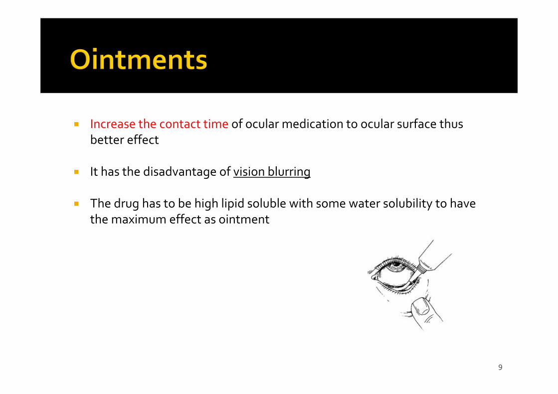
Increase the contact time of ocular medication to ocular surface thus better effect
It has the disadvantage of vision blurring
The drug has to be high lipid soluble with some water solubility to have the maximum effect as ointment
10
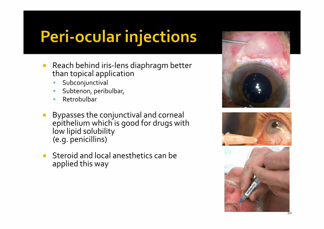
Reach behind iris‐lens diaphragm better than topical application Subconjunctival Subtenon, peribulbar, Retrobulbar
Bypasses the conjunctival and corneal epithelium which is good for drugs with low lipid solubility(e.g. penicillins)
Steroid and local anesthetics can be applied this way
11
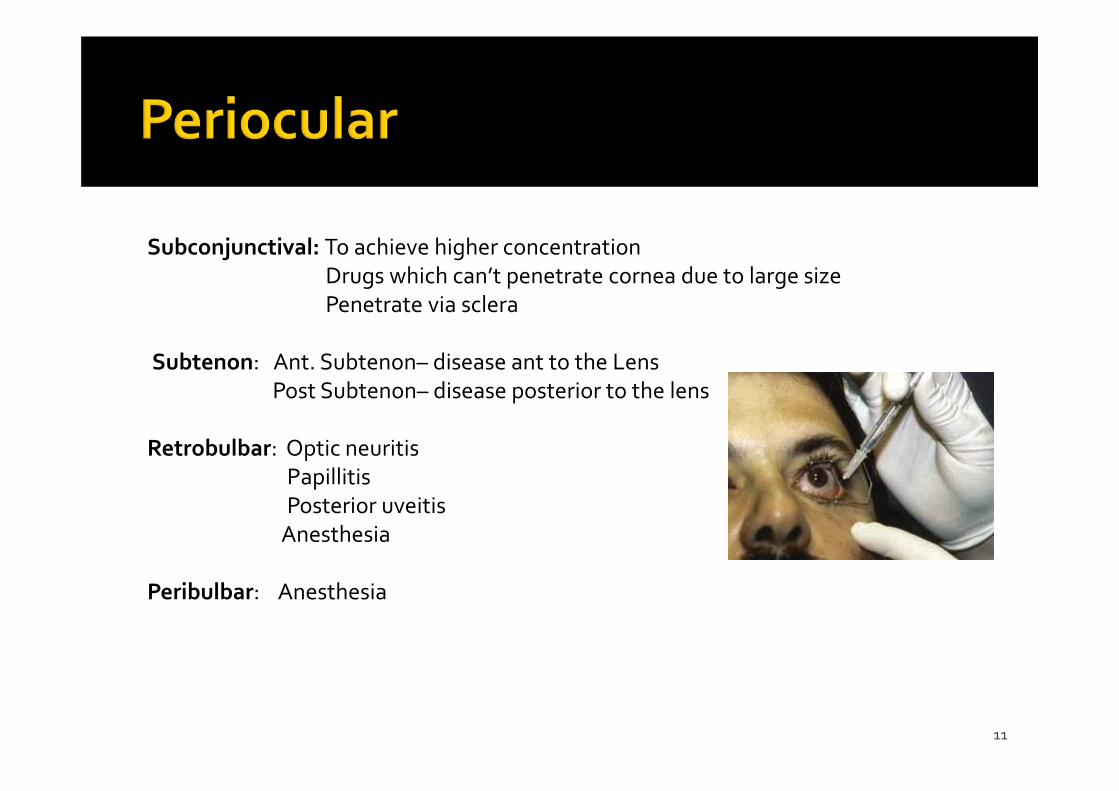
Subconjunctival: To achieve higher concentrationDrugs which can’t penetrate cornea due to large sizePenetrate via sclera
Subtenon: Ant. Subtenon– disease ant to the LensPost Subtenon– disease posterior to the lens
Retrobulbar: Optic neuritisPapillitisPosterior uveitisAnesthesia
Peribulbar: Anesthesia
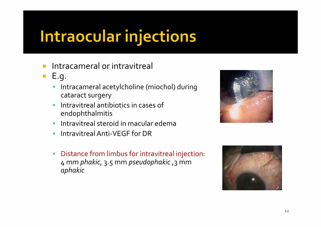
12
Intracameral or intravitreal E.g. Intracameral acetylcholine (miochol) during
cataract surgery Intravitreal antibiotics in cases of
endophthalmitis Intravitreal steroid in macular edema Intravitreal Anti‐VEGF for DR
Distance from limbus for intravitreal injection:4 mm phakic, 3.5 mm pseudophakic ,3 mm aphakic
13
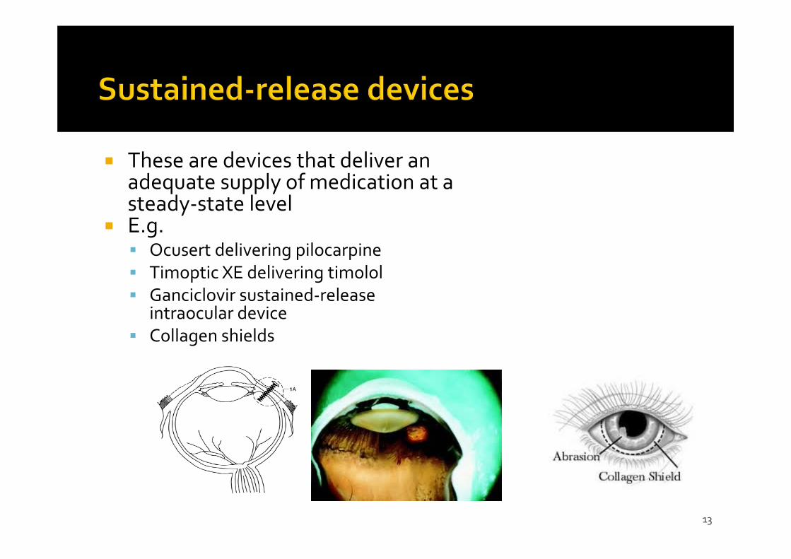
These are devices that deliver an adequate supply of medication at a steady‐state level
E.g. Ocusert delivering pilocarpine Timoptic XE delivering timolol Ganciclovir sustained‐release
intraocular device Collagen shields
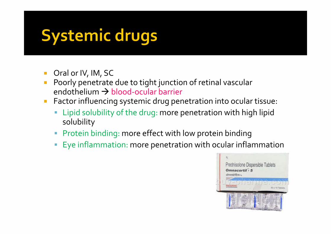
Oral or IV, IM, SC Poorly penetrate due to tight junction of retinal vascular
endothelium blood‐ocular barrier Factor influencing systemic drug penetration into ocular tissue: Lipid solubility of the drug: more penetration with high lipid
solubility Protein binding: more effect with low protein binding Eye inflammation: more penetration with ocular inflammation
15
Antibacterials (antibiotics) Antivirals Antifungal Mydriatics and cycloplegics Antiglaucoma Anti‐inflammatory agents Ocular Lubricants Antihistaminics Ocular diagnostic drugs Local anesthetics Ocular Toxicology
Corticosteroids
NSAIDs
16
Penicillins Cephalosporins Sulfonamides Tetracyclines Chloramphenicol Aminoglycosides Fluoroquinolones Vancomycin Macrolides
17
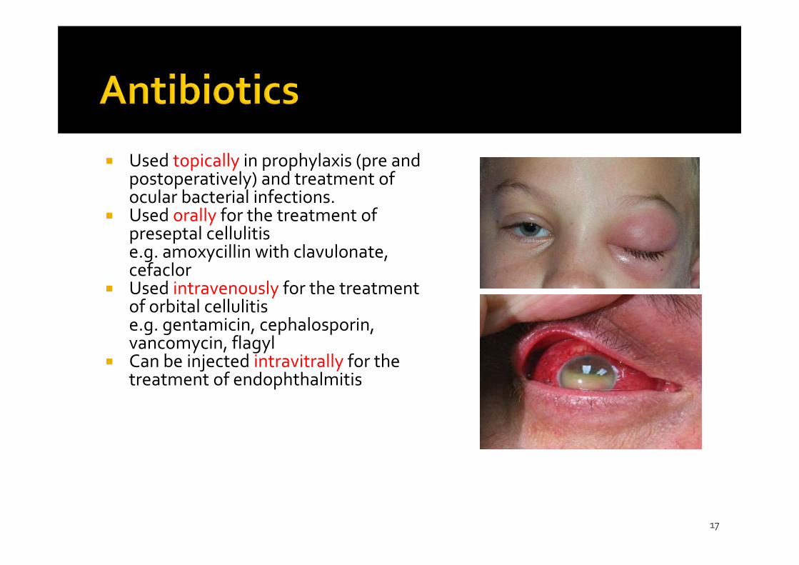
Used topically in prophylaxis (pre and postoperatively) and treatment of ocular bacterial infections.
Used orally for the treatment of preseptal cellulitise.g. amoxycillin with clavulonate, cefaclor
Used intravenously for the treatment of orbital cellulitise.g. gentamicin, cephalosporin, vancomycin, flagyl
Can be injected intravitrally for the treatment of endophthalmitis
18
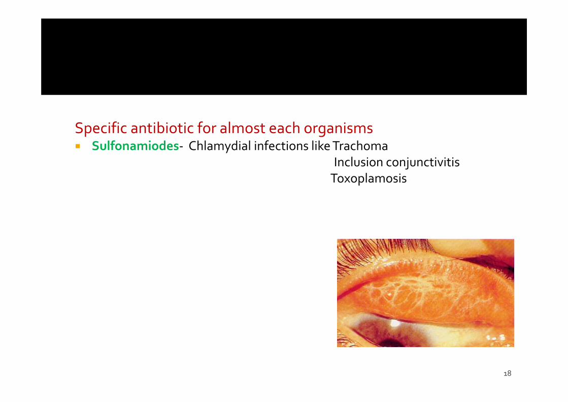
Specific antibiotic for almost each organisms Sulfonamiodes‐ Chlamydial infections like Trachoma
Inclusion conjunctivitisToxoplamosis
Cephalosporin 1 st generation
▪ Cephalothin, cefazolin, cephalexin▪ Active against G+ve and G‐ve▪ Not active against MRSA, Enterobacter, Proteus spp, P aeruginosa, Serratia,
enterococci 2 nd generation
▪ Cefamandole, cefoxitin, cefuroxime▪ Greater activity against G‐ve : H.influenzae, Enterobacter, Neisseria
3 rd generation▪ Cefotaxime, Ceftriaxone, Cefoperazone▪ Active against GNR > G+ve cocci : Serratia, Proteus, β‐lactamase H influenzae, anaerobe
▪ P.aeruginosa : ceftazidime, cefoperazone▪ Cefotaxime : good penetration blood‐ocular barrier
4 th generation Extended spectrum
Against gram‐positive organisms as 1 st generation Greater resistance to beta‐lactamases than 3 rd generation
Can cross blood brain barrier
Against nosocomial pathogens Cefepime, Cefluprenam, Cefozopran, Cefpirome, Cefquinome
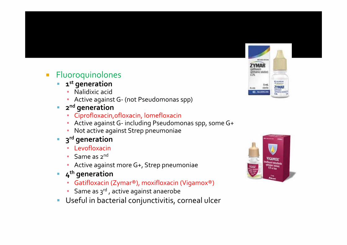
Fluoroquinolones 1st generation
▪ Nalidixic acid▪ Active against G‐ (not Pseudomonas spp)
2nd generation▪ Ciprofloxacin,ofloxacin, lomefloxacin▪ Active against G‐ including Pseudomonas spp, some G+▪ Not active against Strep pneumoniae
3rd generation▪ Levofloxacin▪ Same as 2nd
▪ Active against more G+, Strep pneumoniae 4th generation
▪ Gatifloxacin (Zymar®), moxifloxacin (Vigamox®)▪ Same as 3rd , active against anaerobe
Useful in bacterial conjunctivitis, corneal ulcer
22
Amino glycosides Mainly against Gm negative bacilli Bacterial protein synthesis inhibitors Gentamycin—0.3% eye drop Tobramycin‐ Pseudomonas 1% eye drop Neomycin—0.3‐0.5% eye drop
23
Tetracycline Inhibit protein synthesis Active against both gm+ and gm ‐, some fungi and Chlamydia
Chloromphenicol Broad spectrum ,bacteriostatic, gm+/gm‐, Chlamydia 0.5% Eye drop, ointment
Vancomycin Against MRSA or strep Useful in corneal ulcer, endophthalmitis
Polymyxin B + Neomycin Against Staph. aureus, Strep spp, GNR Useful in surface bacterial infection e.g. conjunctivitis, blepharitis
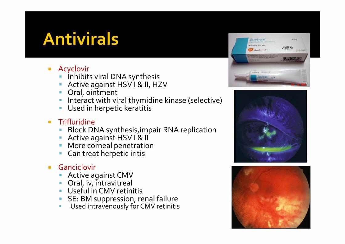
Acyclovir Inhibits viral DNA synthesis Active against HSV I & II, HZV Oral, ointment Interact with viral thymidine kinase (selective) Used in herpetic keratitis
Trifluridine Block DNA synthesis,impair RNA replication Active against HSV I & II More corneal penetration Can treat herpetic iritis
Ganciclovir Active against CMV Oral, iv, intravitreal Useful in CMV retinitis SE: BM suppression, renal failure Used intravenously for CMV retinitis
Basic fungal classificationa) Filamentous fungi Septate = Fusarium, Aspergillus Nonseptate = Mucor
b) Yeasts Candida, Cryptococcus
Most antifungal drugs act by attacking the membrane sterols of fungi (ergosterol), leaving mammalian sterols (cholesterol) unaffected
27
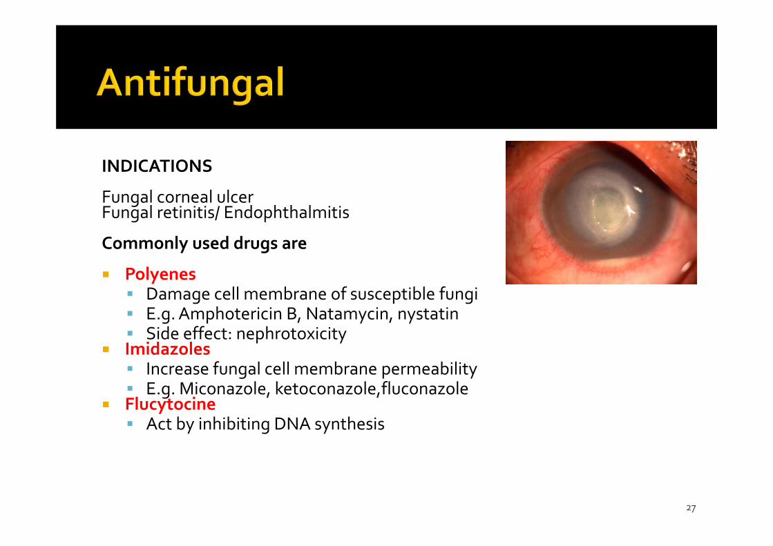
INDICATIONS
Fungal corneal ulcerFungal retinitis/ Endophthalmitis
Commonly used drugs are
Polyenes Damage cell membrane of susceptible fungi E.g. Amphotericin B, Natamycin, nystatin Side effect: nephrotoxicity
Imidazoles Increase fungal cell membrane permeability E.g. Miconazole, ketoconazole,fluconazole
Flucytocine Act by inhibiting DNA synthesis
28
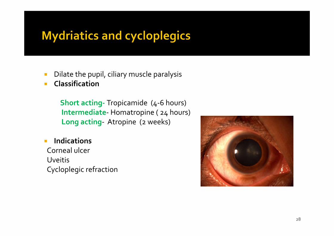
Dilate the pupil, ciliary muscle paralysis Classification
Short acting‐ Tropicamide (4‐6 hours)Intermediate‐ Homatropine ( 24 hours)Long acting‐ Atropine (2 weeks)
IndicationsCorneal ulcerUveitisCycloplegic refraction
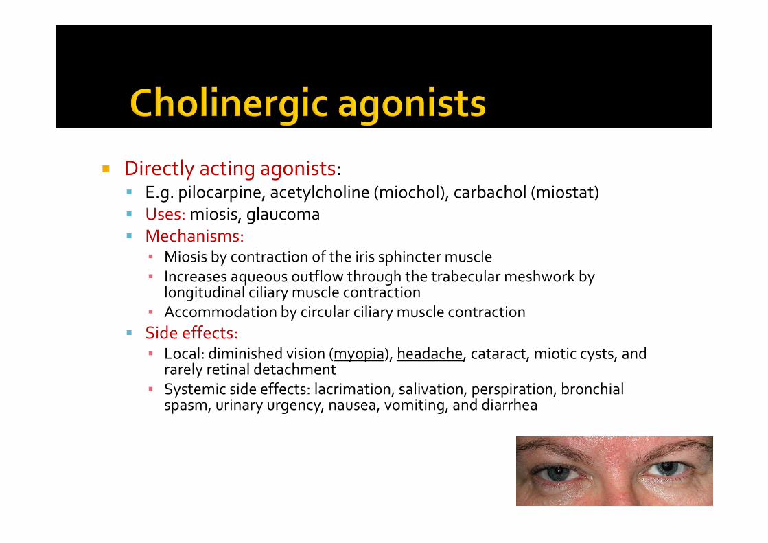
Directly acting agonists: E.g. pilocarpine, acetylcholine (miochol), carbachol (miostat) Uses: miosis, glaucoma Mechanisms:
▪ Miosis by contraction of the iris sphincter muscle ▪ Increases aqueous outflow through the trabecular meshwork by
longitudinal ciliary muscle contraction▪ Accommodation by circular ciliary muscle contraction
Side effects: ▪ Local: diminished vision (myopia), headache, cataract, miotic cysts, and
rarely retinal detachment▪ Systemic side effects: lacrimation, salivation, perspiration, bronchial
spasm, urinary urgency, nausea, vomiting, and diarrhea
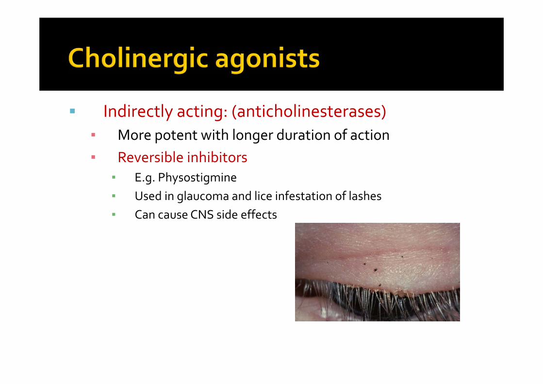
Indirectly acting: (anticholinesterases) ▪ More potent with longer duration of action▪ Reversible inhibitors
▪ E.g. Physostigmine▪ Used in glaucoma and lice infestation of lashes▪ Can cause CNS side effects

Irreversible:▪ e.g. Phospholine iodide▪ Uses: Accommodative esotropia▪ Side effects: Iris cyst and anterior subcapsular cataract
▪ C/I in angle closure glaucoma, asthma, Parkinsonism
▪ Causes apnea if used with succinylcholineor procaine
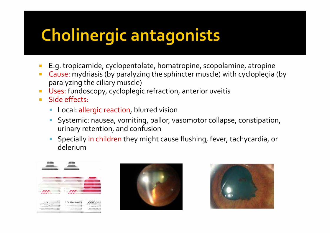
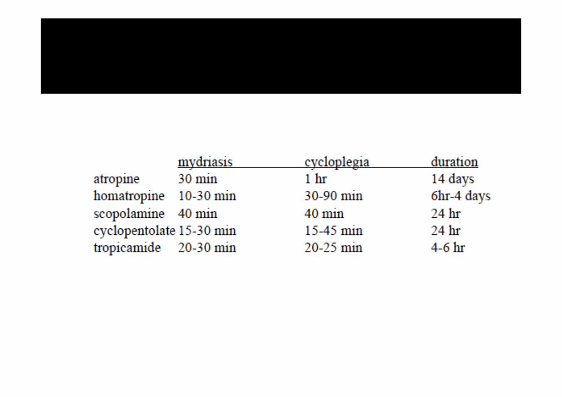
E.g. tropicamide, cyclopentolate, homatropine, scopolamine, atropine Cause: mydriasis (by paralyzing the sphincter muscle) with cycloplegia (by
paralyzing the ciliary muscle) Uses: fundoscopy, cycloplegic refraction, anterior uveitis Side effects: Local: allergic reaction, blurred vision Systemic: nausea, vomiting, pallor, vasomotor collapse, constipation,
urinary retention, and confusion Specially in children they might cause flushing, fever, tachycardia, or
delerium
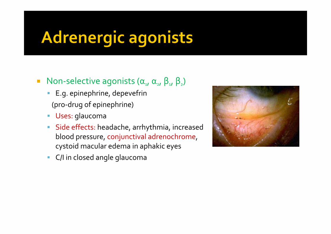
Non‐selective agonists (α1, α2, β1, β2) E.g. epinephrine, depevefrin(pro‐drug of epinephrine) Uses: glaucoma Side effects: headache, arrhythmia, increased
blood pressure, conjunctival adrenochrome, cystoid macular edema in aphakic eyes
C/I in closed angle glaucoma
Alpha‐1 agonists E.g. phenylephrine Uses: mydriasis (without cycloplegia), decongestant Adverse effect: Can cause significant increase in blood pressure specially in infant and
susceptible adults Rebound congestion Precipitation of acute angle‐closure glaucoma in patients with narrow
angles
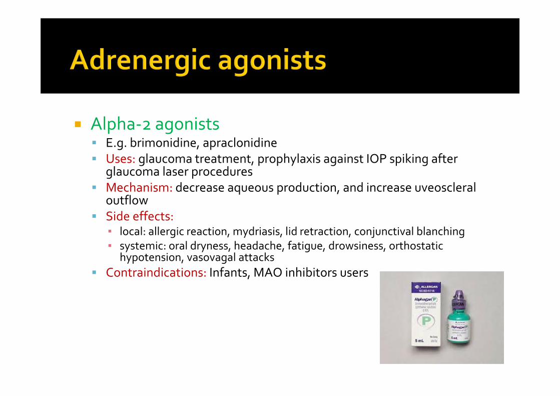
Alpha‐2 agonists E.g. brimonidine, apraclonidine Uses: glaucoma treatment, prophylaxis against IOP spiking after
glaucoma laser procedures Mechanism: decrease aqueous production, and increase uveoscleral
outflow Side effects:
▪ local: allergic reaction, mydriasis, lid retraction, conjunctival blanching▪ systemic: oral dryness, headache, fatigue, drowsiness, orthostatic
hypotension, vasovagal attacks Contraindications: Infants, MAO inhibitors users
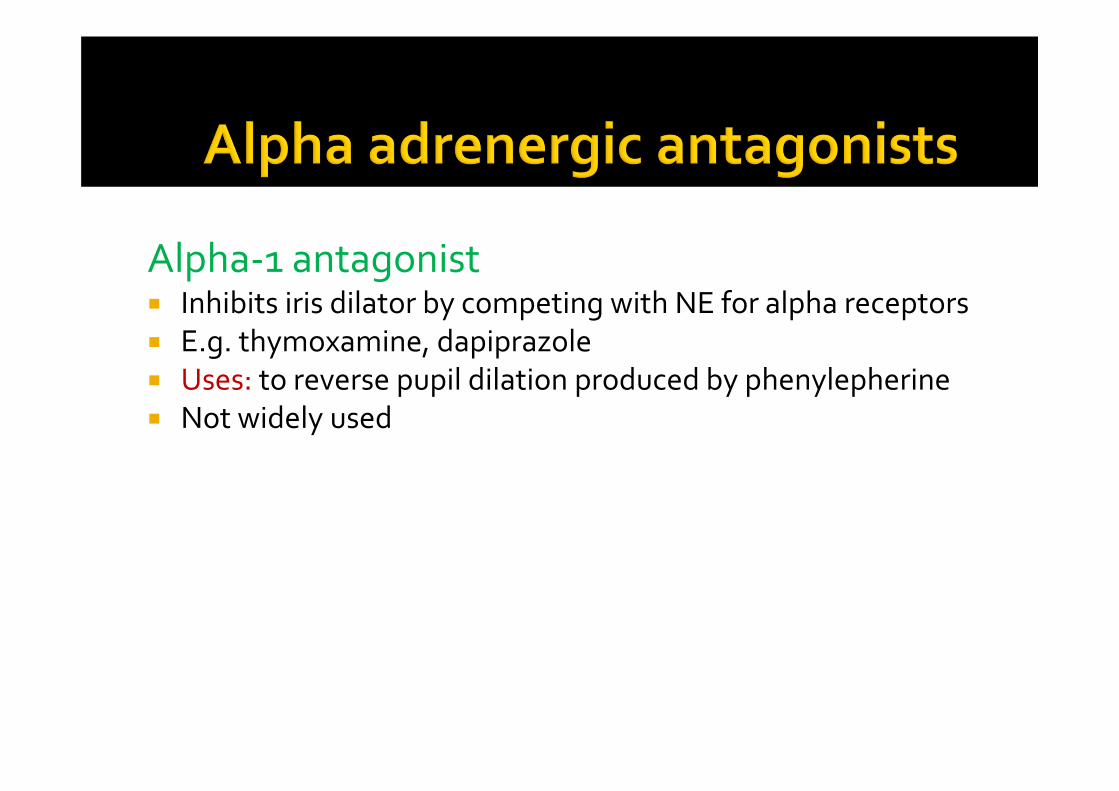
Alpha‐1 antagonist Inhibits iris dilator by competing with NE for alpha receptors E.g. thymoxamine, dapiprazole Uses: to reverse pupil dilation produced by phenylepherine Not widely used
38
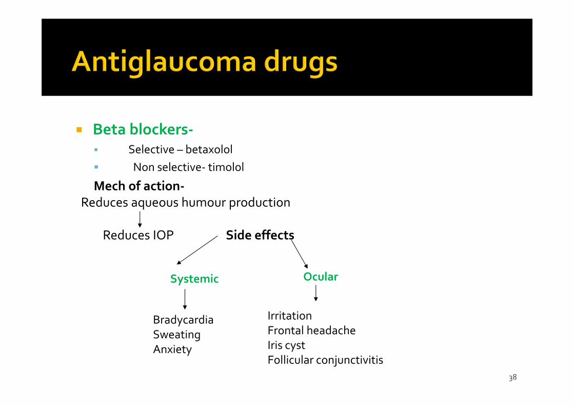
Beta blockers‐ Selective – betaxolol Non selective‐ timolol
Mech of action‐Reduces aqueous humour production
Reduces IOP Side effects
Systemic
BradycardiaSweatingAnxiety
Ocular
IrritationFrontal headacheIris cystFollicular conjunctivitis
39
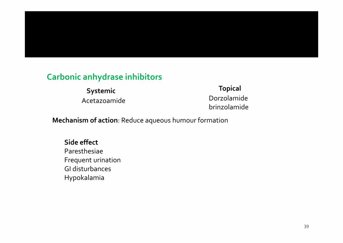
Carbonic anhydrase inhibitors
Systemic Topical
Acetazoamide Dorzolamidebrinzolamide
Mechanism of action: Reduce aqueous humour formation
Side effectParesthesiaeFrequent urinationGI disturbancesHypokalamia
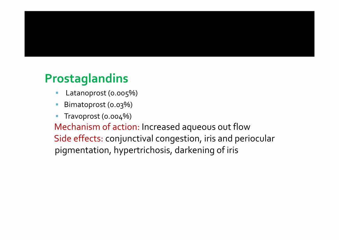
Prostaglandins Latanoprost (0.005%) Bimatoprost (0.03%) Travoprost (0.004%)Mechanism of action: Increased aqueous out flowSide effects: conjunctival congestion, iris and periocularpigmentation, hypertrichosis, darkening of iris
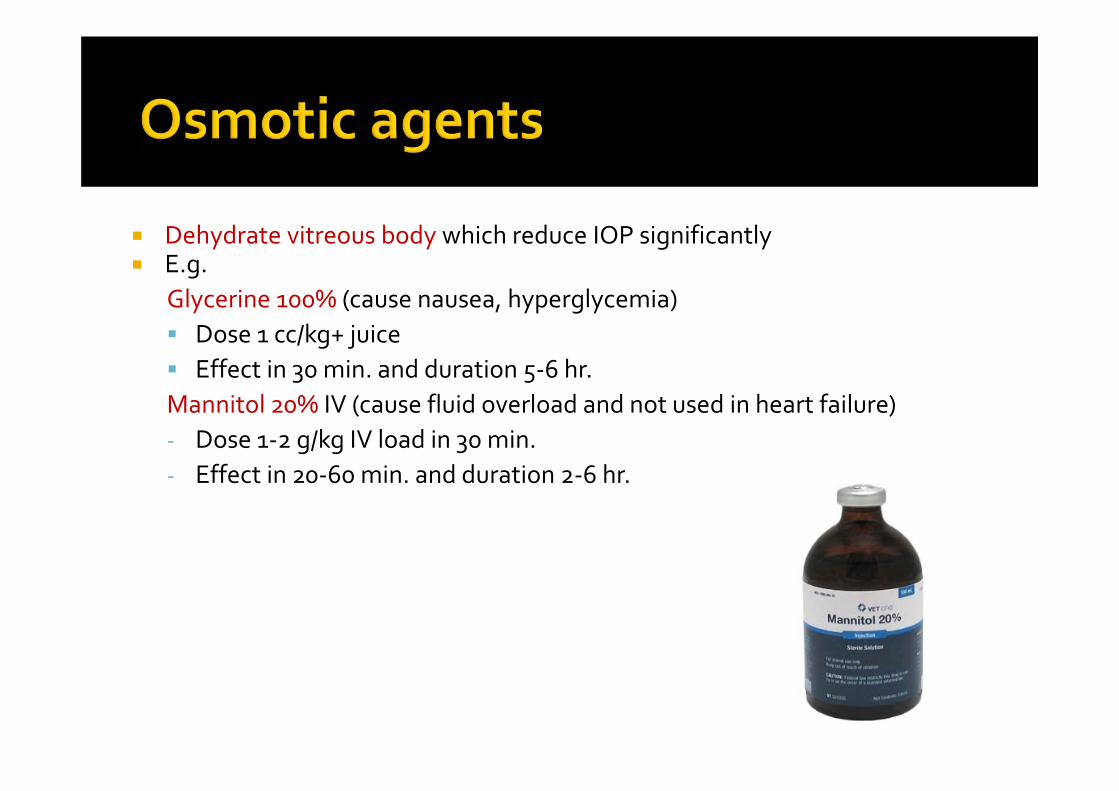
Dehydrate vitreous body which reduce IOP significantly E.g.
Glycerine 100% (cause nausea, hyperglycemia) Dose 1 cc/kg+ juice Effect in 30 min. and duration 5‐6 hr.Mannitol 20% IV (cause fluid overload and not used in heart failure)‐ Dose 1‐2 g/kg IV load in 30 min.‐ Effect in 20‐60 min. and duration 2‐6 hr.
42
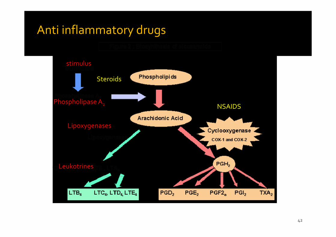
stimulus
Phospholipase A2
Lipoxygenases
Leukotrines
Steroids
NSAIDS
Anti inflammatory drugs
43
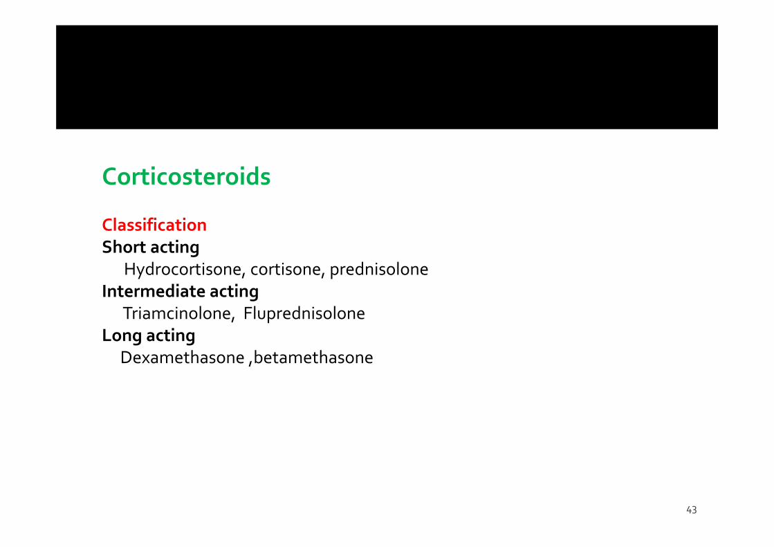
Corticosteroids
Classification Short acting
Hydrocortisone, cortisone, prednisoloneIntermediate acting
Triamcinolone, FluprednisoloneLong acting
Dexamethasone ,betamethasone
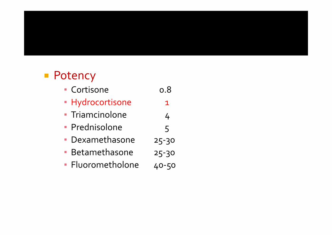
Potency▪ Cortisone 0.8▪ Hydrocortisone 1▪ Triamcinolone 4 ▪ Prednisolone 5▪ Dexamethasone 25‐30▪ Betamethasone 25‐30▪ Fluorometholone 40‐50
45
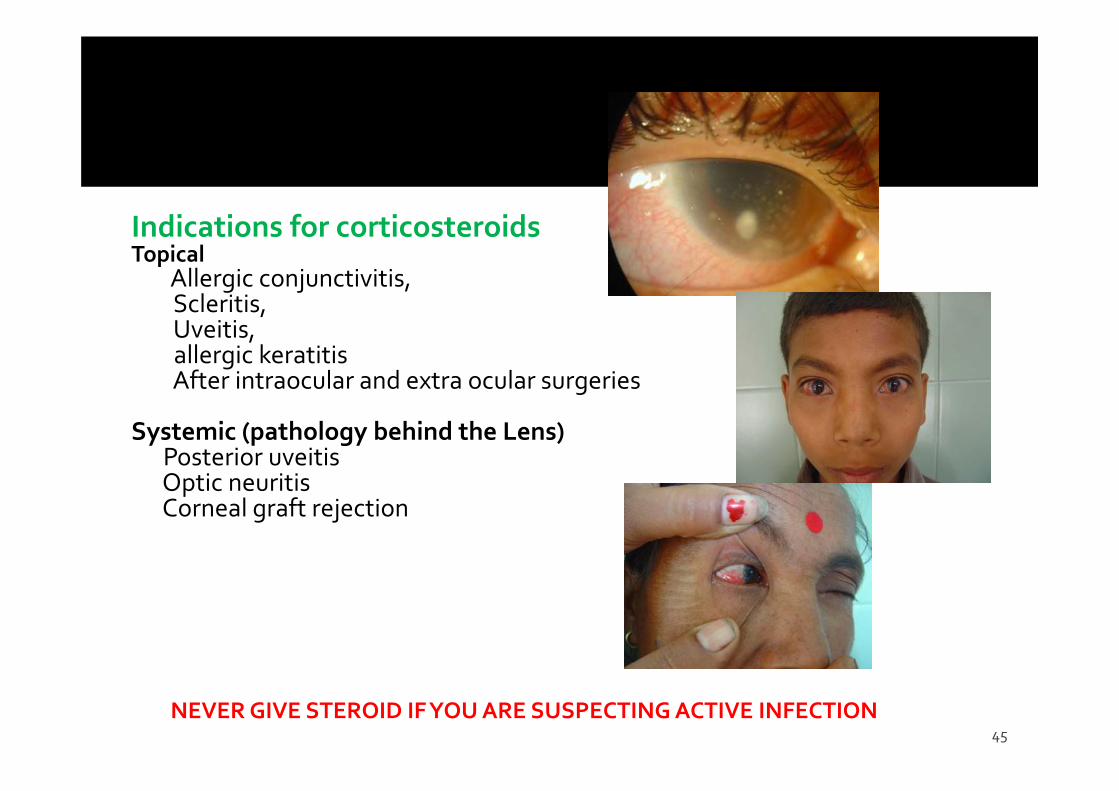
Indications for corticosteroidsTopical
Allergic conjunctivitis, Scleritis,Uveitis, allergic keratitisAfter intraocular and extra ocular surgeries
Systemic (pathology behind the Lens)Posterior uveitisOptic neuritisCorneal graft rejection
NEVER GIVE STEROID IF YOU ARE SUSPECTING ACTIVE INFECTION
46
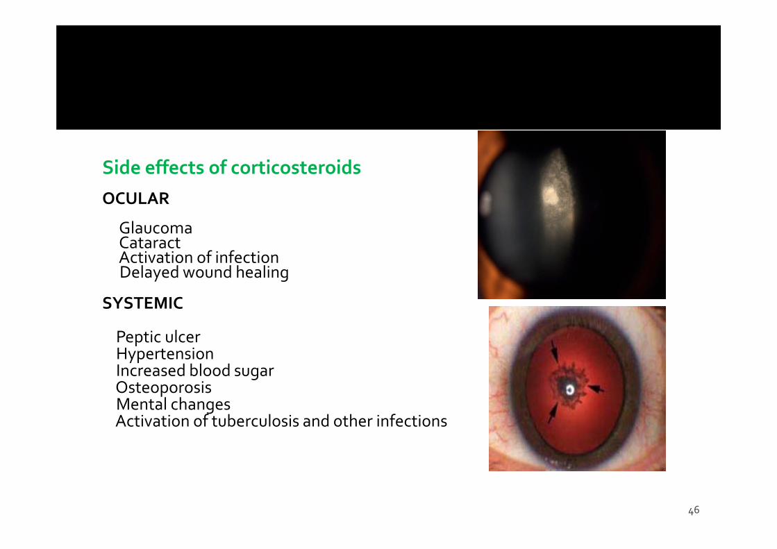
Side effects of corticosteroidsOCULAR
GlaucomaCataractActivation of infectionDelayed wound healing
SYSTEMIC
Peptic ulcerHypertensionIncreased blood sugarOsteoporosisMental changesActivation of tuberculosis and other infections
47
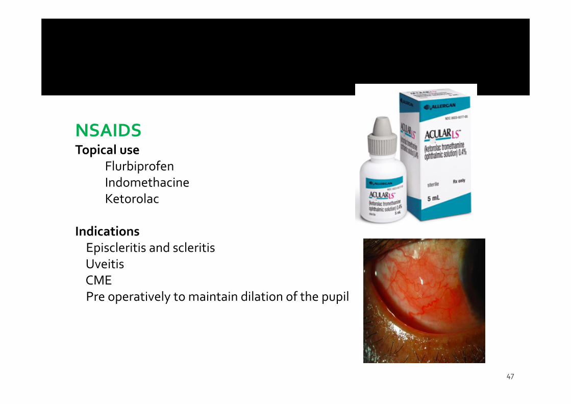
NSAIDSTopical use
FlurbiprofenIndomethacineKetorolac
IndicationsEpiscleritis and scleritisUveitisCMEPre operatively to maintain dilation of the pupil
48
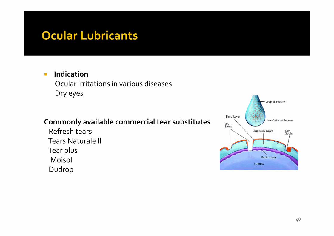
IndicationOcular irritations in various diseasesDry eyes
Commonly available commercial tear substitutesRefresh tearsTears Naturale IITear plusMoisolDudrop
Avoidance of allergens, cold compress, lubrications Antihistamines (e.g.pheniramine, levocabastine) Decongestants (e.g. naphazoline, phenylepherine, tetrahydrozaline) Mast cell stabilizers (e.g. cromolyn, lodoxamide, pemirolast,
nedocromil, olopatadine) NSAID (e.g. ketorolac) Steroids (e.g. fluorometholone, remixolone, prednisolone) Drug combinations
Pyrilamine maleate, pheniramine maleate, antazolinephosphate
H1 antihistamine Use in allergic conjunctivitis, irritation, pinguecula and
pterygium Can cause sedation, mydriasis and increase IOP
Phenylephrine‐ Alpha1 agonist‐ 0.12‐0.125%‐When expose to wind, heat :can cause oxidation‐Can cause mydriasis, blanching of conjunctival vessles, AACG, high BP
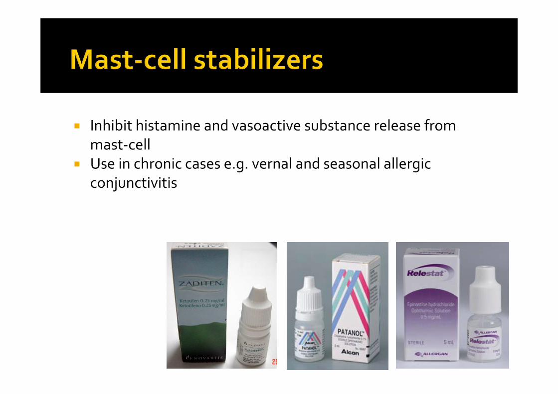
Inhibit histamine and vasoactive substance release from mast‐cell
Use in chronic cases e.g. vernal and seasonal allergic conjunctivitis
53
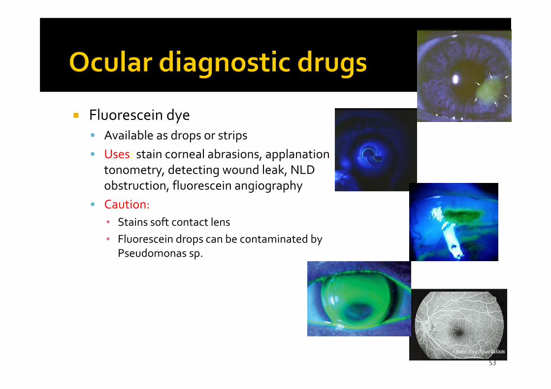
Fluorescein dye Available as drops or strips Uses: stain corneal abrasions, applanation
tonometry, detecting wound leak, NLD obstruction, fluorescein angiography
Caution:▪ Stains soft contact lens▪ Fluorescein drops can be contaminated by
Pseudomonas sp.
54
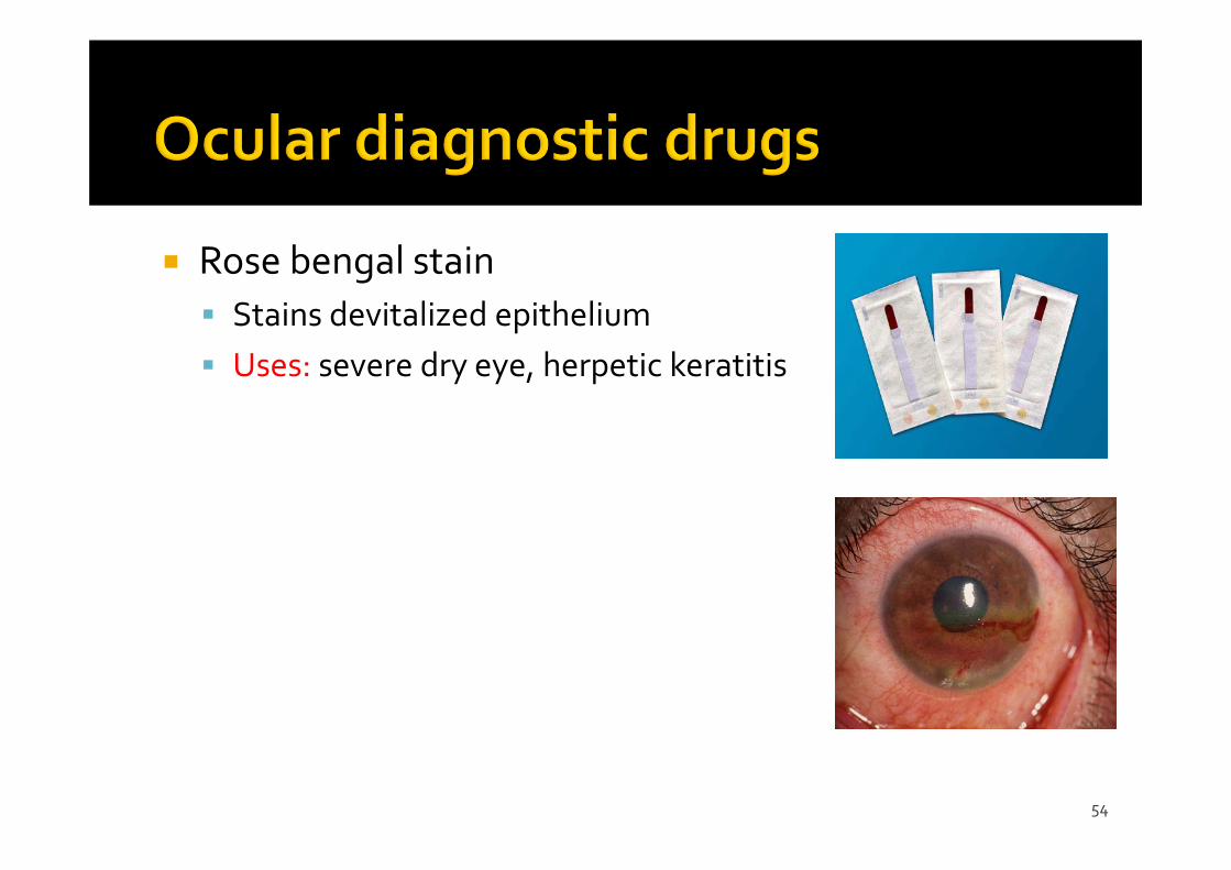
Rose bengal stain Stains devitalized epithelium Uses: severe dry eye, herpetic keratitis
55
Topical E.g. propacaine, tetracaine Uses: applanation tonometry, gonioscopy, removal of corneal foreign bodies, removal of sutures, examination of patients who cannot open eyes because of pain
Adverse effects: toxic to corneal epithelium, allergic reaction rarely
56
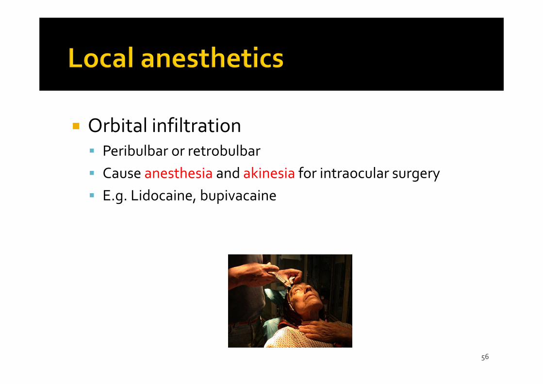
Orbital infiltration Peribulbar or retrobulbar Cause anesthesia and akinesia for intraocular surgery E.g. Lidocaine, bupivacaine
57
58
Mechanical injury from the bottle e.g. corneal abrasion Pigmentation: epinephrine‐adrenochrome Ocular damage:: e.g. topical anesthetics, benzylkonium Hypersensitivity:: e.g. atropine, neomycin, gentamicin Systemic effect:: Topical phenylephrine can increase BP
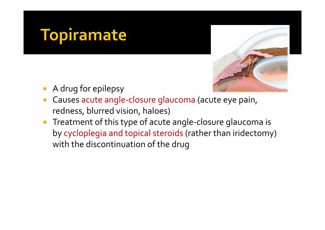
A drug for epilepsy Causes acute angle‐closure glaucoma (acute eye pain,
redness, blurred vision, haloes) Treatment of this type of acute angle‐closure glaucoma is
by cycloplegia and topical steroids (rather than iridectomy) with the discontinuation of the drug
60
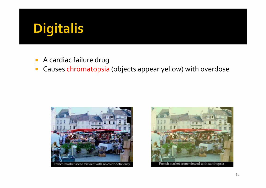
A cardiac failure drug Causes chromatopsia (objects appear yellow) with overdose
61
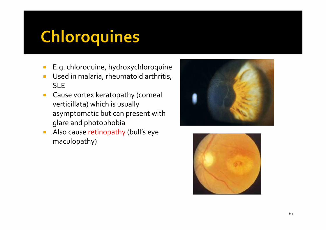
E.g. chloroquine, hydroxychloroquine Used in malaria, rheumatoid arthritis,
SLE Cause vortex keratopathy (corneal
verticillata) which is usually asymptomatic but can present with glare and photophobia
Also cause retinopathy (bull’s eye maculopathy)
62
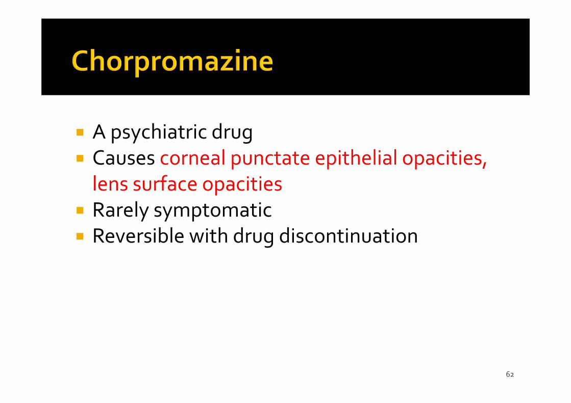
A psychiatric drug Causes corneal punctate epithelial opacities, lens surface opacities
Rarely symptomatic Reversible with drug discontinuation
63
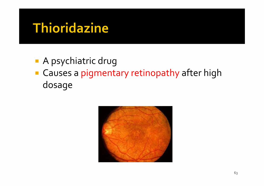
A psychiatric drug Causes a pigmentary retinopathy after high dosage
64
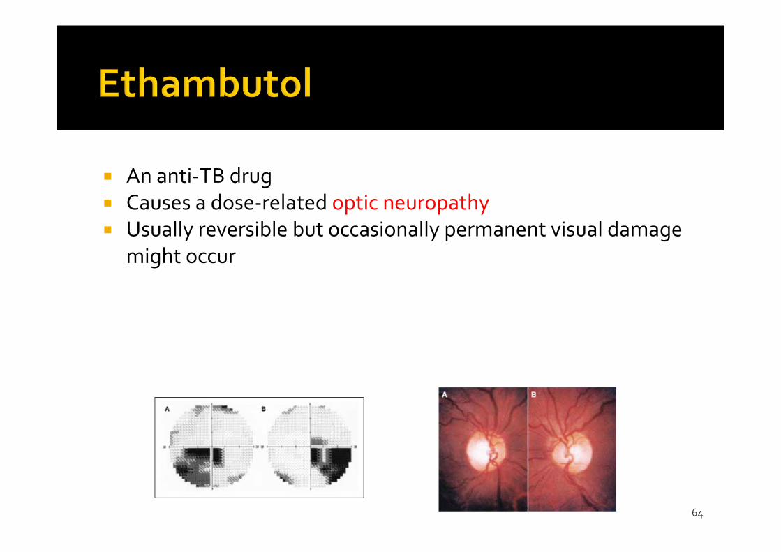
An anti‐TB drug Causes a dose‐related optic neuropathy Usually reversible but occasionally permanent visual damage
might occur
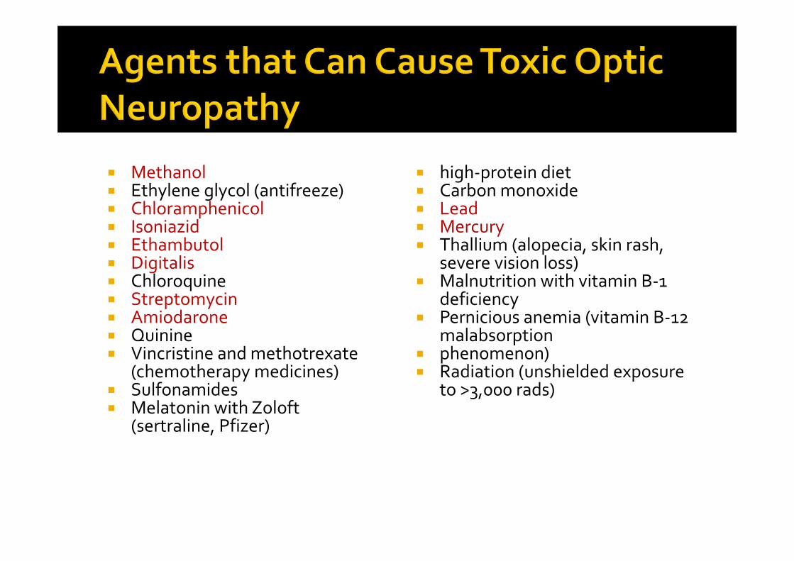
Methanol Ethylene glycol (antifreeze) Chloramphenicol Isoniazid Ethambutol Digitalis Chloroquine Streptomycin Amiodarone Quinine Vincristine and methotrexate
(chemotherapy medicines) Sulfonamides Melatonin with Zoloft
(sertraline, Pfizer)
high‐protein diet Carbon monoxide Lead Mercury Thallium (alopecia, skin rash,
severe vision loss) Malnutrition with vitamin B‐1
deficiency Pernicious anemia (vitamin B‐12
malabsorption phenomenon) Radiation (unshielded exposure
to >3,000 rads)
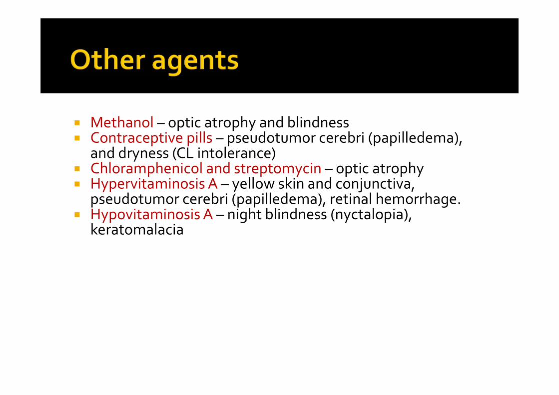
Methanol – optic atrophy and blindness Contraceptive pills – pseudotumor cerebri (papilledema),
and dryness (CL intolerance) Chloramphenicol and streptomycin – optic atrophy Hypervitaminosis A – yellow skin and conjunctiva,
pseudotumor cerebri (papilledema), retinal hemorrhage. Hypovitaminosis A – night blindness (nyctalopia),
keratomalacia


![WELCOME [gmch.gov.in]](https://img.pdfslide.us/doc/110x75/616a5d6311a7b741a351b2cf/welcome-gmchgovin.jpg)