Embed Size (px)
Citation preview
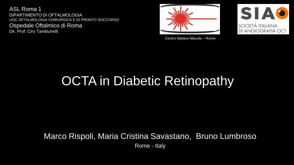
OCTA in Diabetic Retinopathy
Marco Rispoli, Maria Cristina Savastano, Bruno LumbrosoRome - Italy
Centro Italiano Macula – Rome
ASL Roma 1DIPARTIMENTO DI OFTALMOLOGIAUOC OFTALMOLOGIA CHIRURGICA E DI PRONTO SOCCORSO
Ospedale Oftalmico di RomaDir. Prof. Ciro Tamburrelli
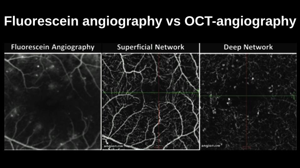
Fluorescein angiography vs OCT-angiography
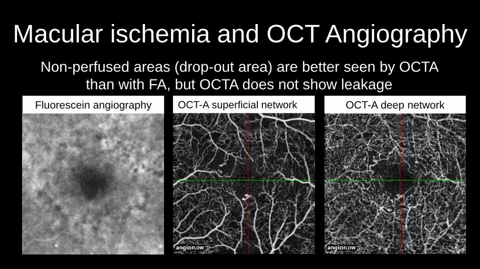
Macular ischemia and OCT AngiographyNon-perfused areas (drop-out area) are better seen by OCTA
than with FA, but OCTA does not show leakage
Fluorescein angiography OCT-A superficial network OCT-A deep network
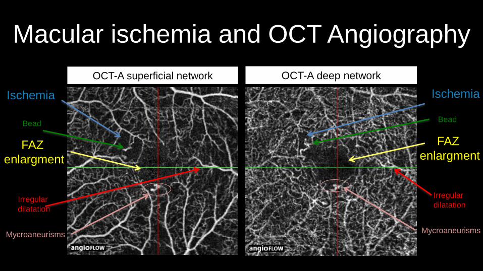
Macular ischemia and OCT Angiography
OCT-A superficial network OCT-A deep network
Ischemia
FAZ
enlargment
Ischemia
FAZ
enlargment
MycroaneurismsMycroaneurisms
BeadBead
Irregular
dilatation
Irregular
dilatation
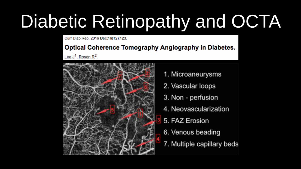
Diabetic Retinopathy and OCTA
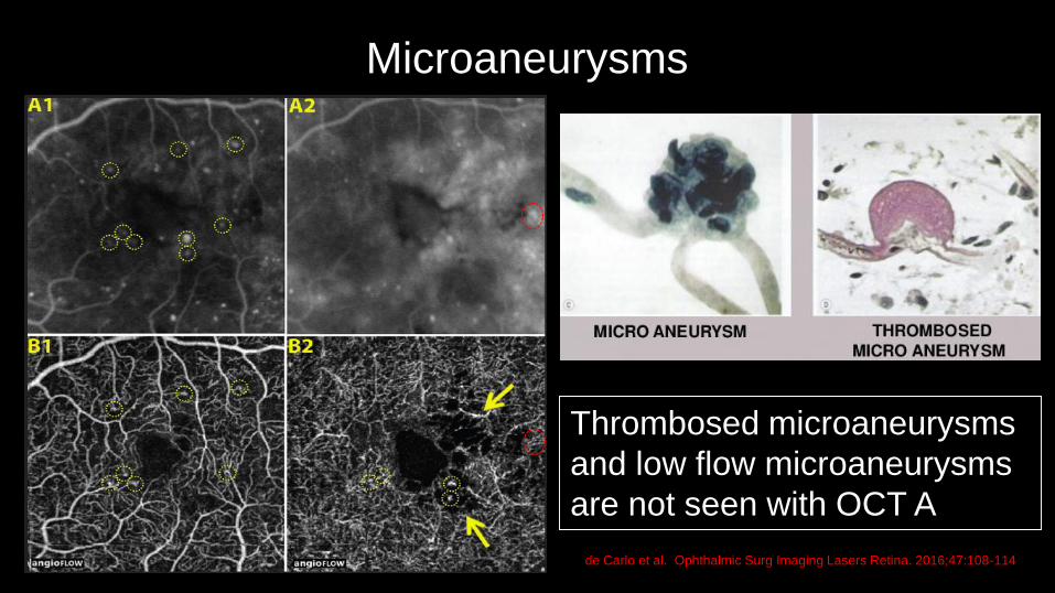
de Carlo et al. Ophthalmic Surg Imaging Lasers Retina. 2016;47:108-114
Thrombosed microaneurysms
and low flow microaneurysms
are not seen with OCT A
Microaneurysms
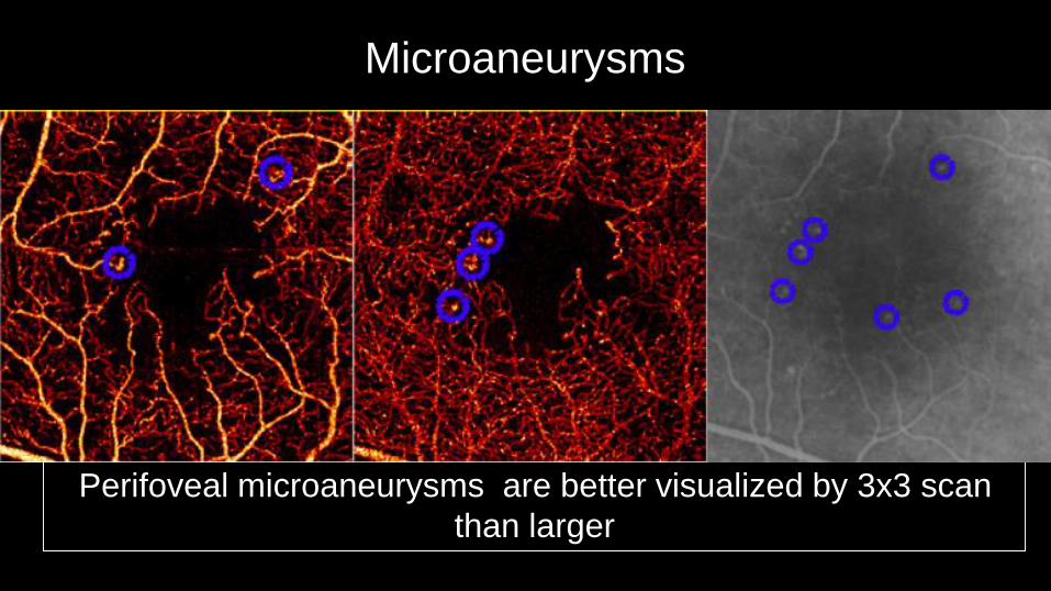
Perifoveal microaneurysms are better visualized by 3x3 scan
than larger
Microaneurysms
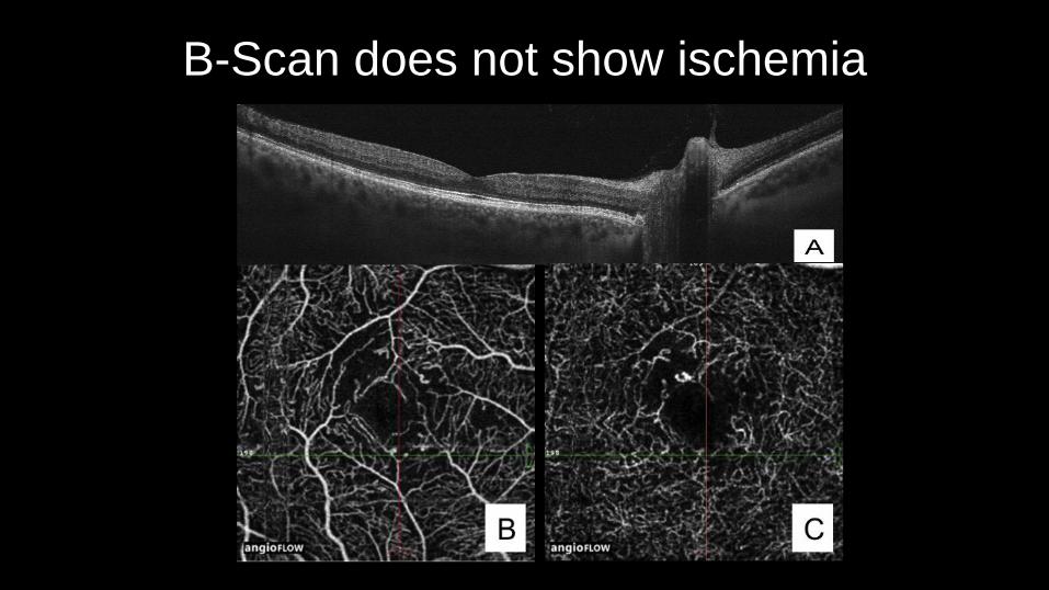
B-Scan does not show ischemia
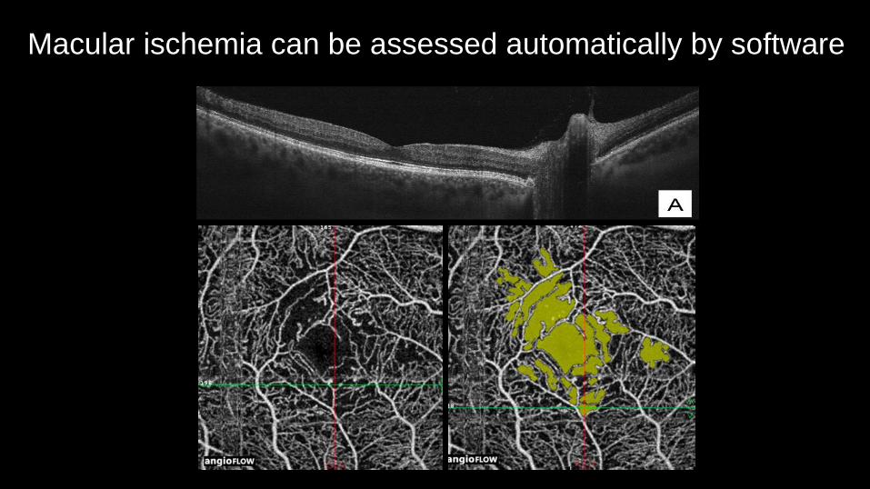
Macular ischemia can be assessed automatically by software
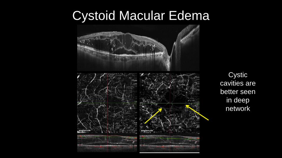
Cystoid Macular Edema
Cystic
cavities are
better seen
in deep
network
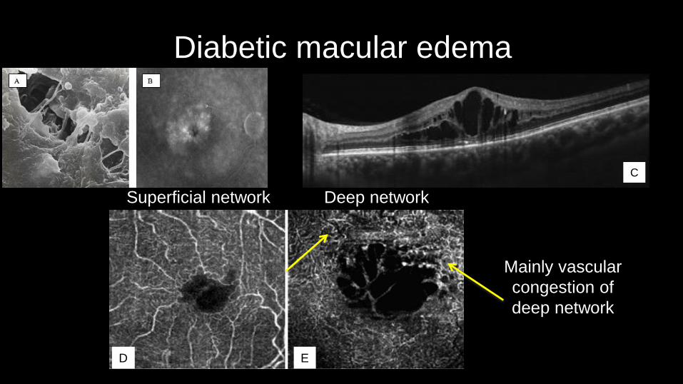
Diabetic macular edema
D E
C
Superficial network Deep network
Mainly vascular
congestion of
deep network
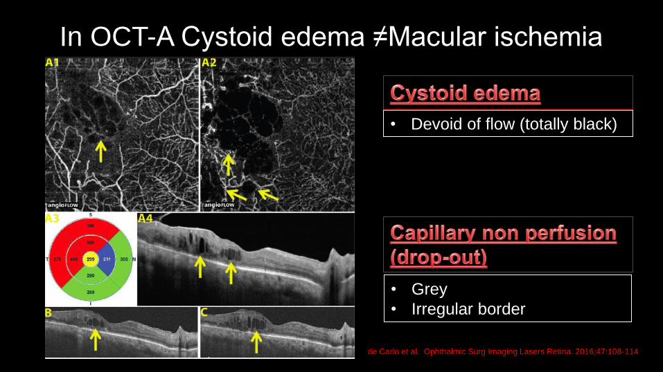
In OCT-A Cystoid edema ≠Macular ischemia
• Devoid of flow (totally black)
de Carlo et al. Ophthalmic Surg Imaging Lasers Retina. 2016;47:108-114
• Grey
• Irregular border

Superficial network
Macular ischemia: map densityDeep network
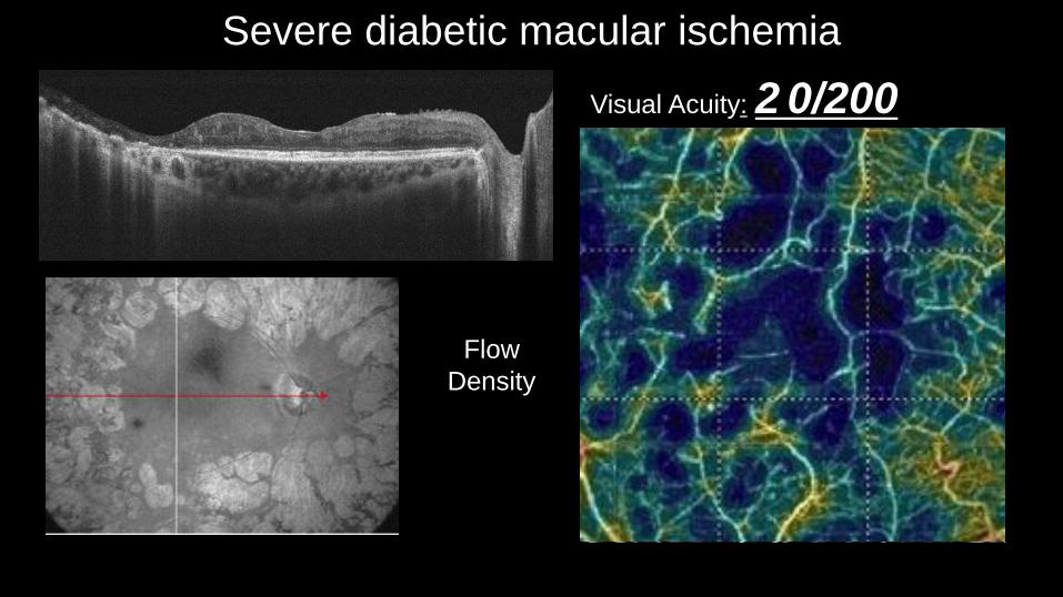
Severe diabetic macular ischemia
Visual Acuity: 2 0/200
NO flow
analysis
Flow
Density
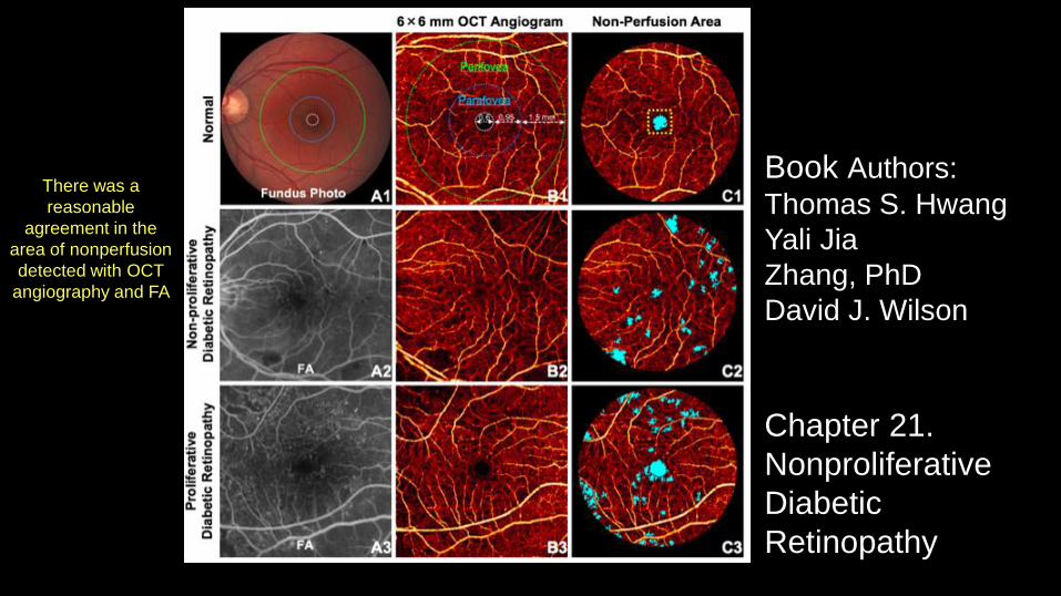
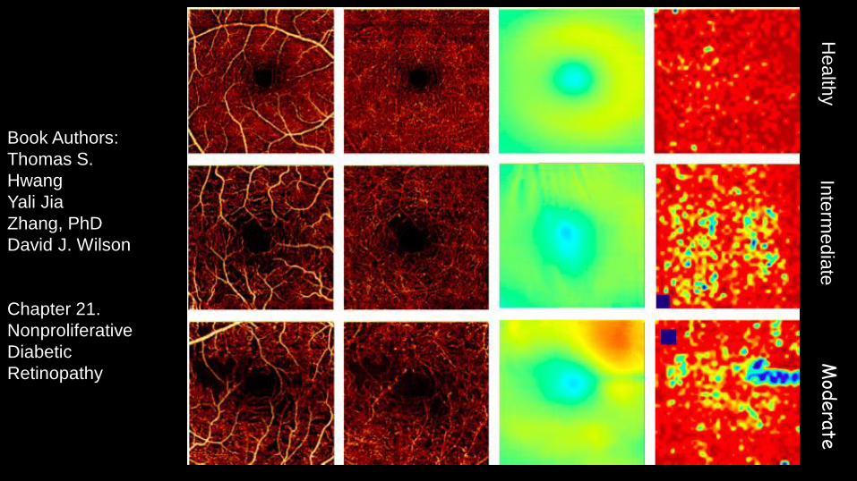
Book Authors:
Thomas S. Hwang
Yali Jia
Zhang, PhD
David J. Wilson
Chapter 21.
Nonproliferative
Diabetic
Retinopathy
There was a
reasonable
agreement in the
area of nonperfusion
detected with OCT
angiography and FA
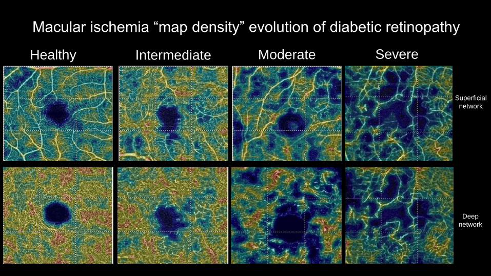
Macular ischemia “map density” evolution of diabetic retinopathy
Superficial
network
Deep
network
Healthy Intermediate Moderate Severe
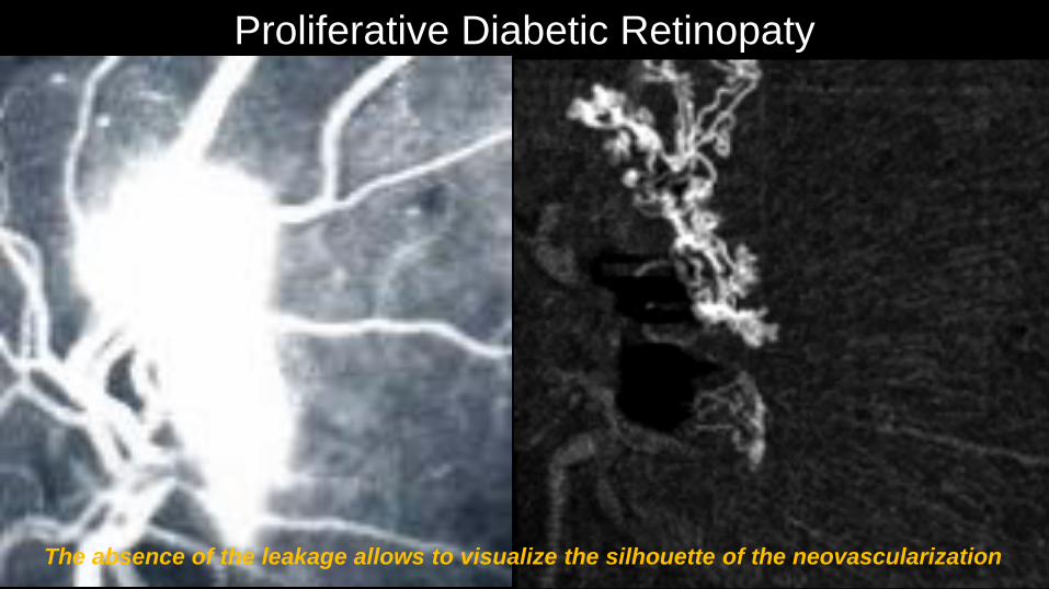
Proliferative Diabetic Retinopaty
The absence of the leakage allows to visualize the silhouette of the neovascularization

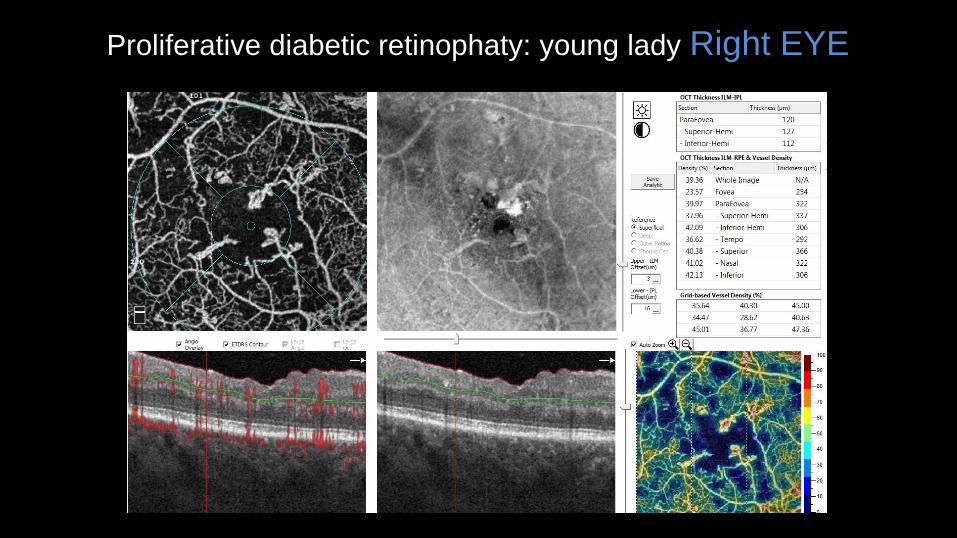
Proliferative diabetic retinophaty: young lady Right EYE
3
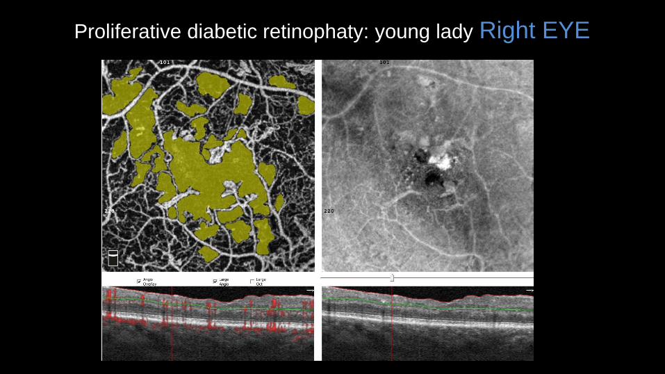
Proliferative diabetic retinophaty: young lady Right EYE
Proliferative diabetic retinophaty: young lady Right EYE
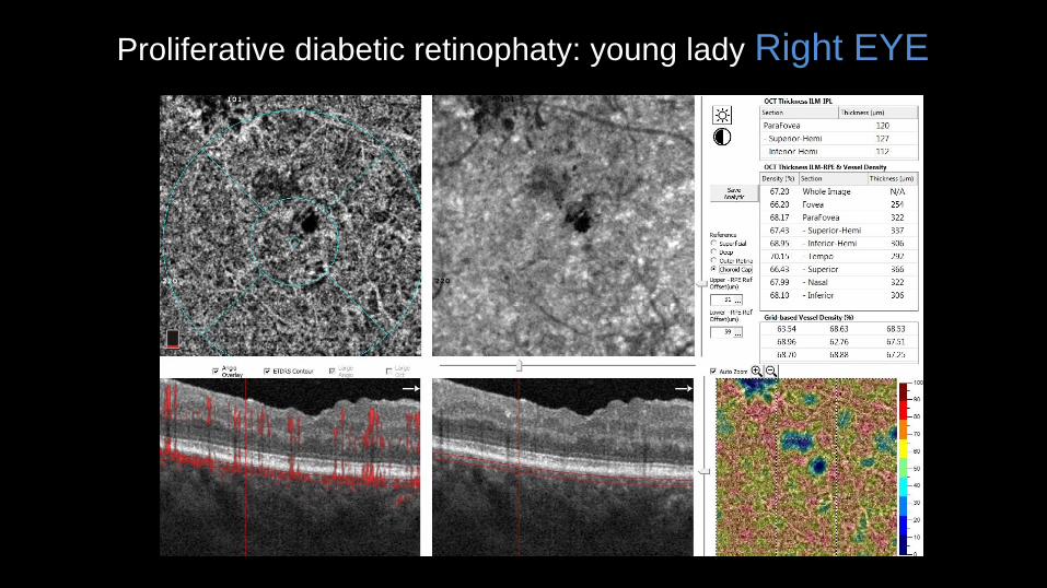
Proliferative diabetic retinophaty: young lady Right EYE
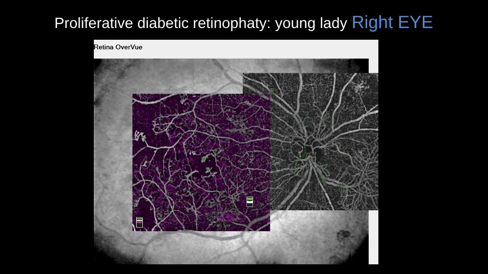
Proliferative diabetic retinophaty: young lady Right EYE
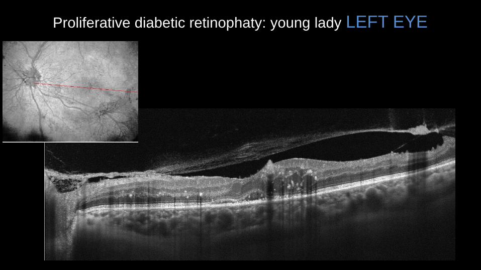
Proliferative diabetic retinophaty: young lady LEFT EYE
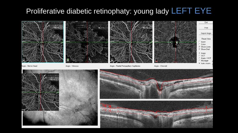
Proliferative diabetic retinophaty: young lady LEFT EYE

Proliferative diabetic retinophaty: young lady LEFT EYE
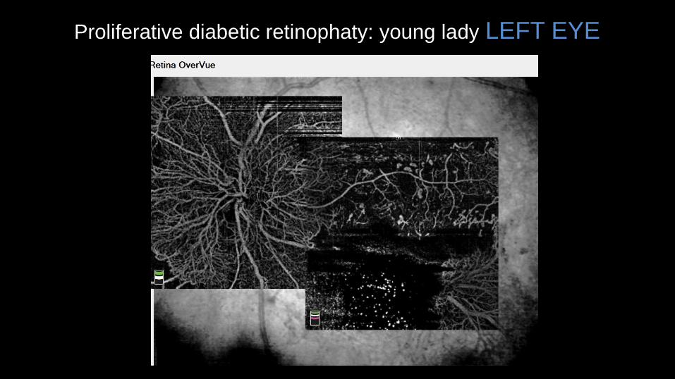
Proliferative diabetic retinophaty: young lady LEFT EYE
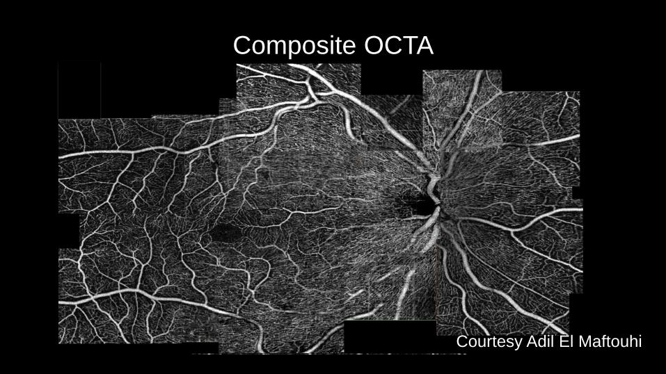
Composite OCTA
Courtesy Adil El Maftouhi

What about the choroid?
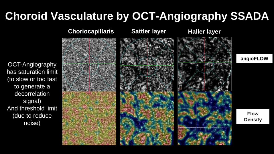
Choroid Vasculature by OCT-Angiography SSADA
OCT-Angiography
has saturation limit
(to slow or too fast
to generate a
decorrelation
signal)
And threshold limit
(due to reduce
noise)
Choriocapillaris Sattler layer Haller layer
angioFLOW
Flow
Density
Health
yIn
term
edia
te
Mod
erate
Book Authors:
Thomas S.
Hwang
Yali Jia
Zhang, PhD
David J. Wilson
Chapter 21.
Nonproliferative
Diabetic
Retinopathy
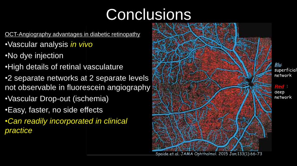
ConclusionsOCT-Angiography advantages in diabetic retinopathy
•Vascular analysis in vivo
•No dye injection
•High details of retinal vasculature
•2 separate networks at 2 separate levels
not observable in fluorescein angiography
•Vascular Drop-out (ischemia)
•Easy, faster, no side effects
•Can readily incorporated in clinical
practice
Red
Thank you
Marco Rispoli Bruno Lumbroso Maria Cristina Savastano

![Wide field and highly sensitive angiography based … angio.pdfneuroscience [11], dermatology [9], and gastroenterology [12]. To date, the spectral-domain OCT (SD-OCT) configuration](https://img.pdfslide.us/doc/110x75/5fcafac549a44e020304fbcc/wide-field-and-highly-sensitive-angiography-based-angiopdf-neuroscience-11-dermatology.jpg)