Embed Size (px)
Citation preview
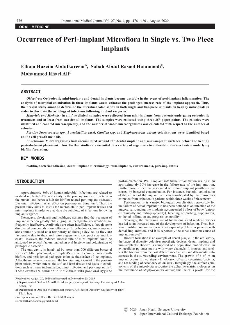
International Medical Journal Vol. 27, No. 4, pp. 476 - 480 , August 2020
ORAL MEDICINE
Occurrence of Peri-Implant Microflora in Single vs. Two Piece Implants
Elham Hazeim Abdulkareem1), Sabah Abdul Rasool Hammoodi1), Mohammed Rhael Ali2)
ABSTRACTObjectives: Orthodontic mini-implants and dental implants become unstable in the event of peri-implant inflammation. The
analysis of microbial colonisation in these implants would enhance the prolonged success rate of the implant approach. Thus, the present study aimed to determine the microbial colonisation in both single and two-piece implants on healthy individuals in order to elucidate the aetiology of infections following implant surgeries.
Materials and Methods: In all, five clinical samples were collected from mini-implants from patients undergoing orthodontic treatment and at least from two dental implants. The samples were collected using three 35# paper points. The colonies were identified and counted microscopically, and the number of viable microorganisms was calculated with respect to the number of colonies.
Results: Streptococcus spp., Lactobacillus casei, Candida spp. and Staphylococcus aureus colonisations were identified based on the cell growth methods.
Conclusion: Microorganisms had accumulated around the dental implant and mini-implant surfaces before the healing post-abutment placement. Thus, further studies are essential on a variety of organisms to understand the mechanism underlying biofilm formation.
KEY WORDSbiofilm, bacterial adhesion, dental implant microbiology, mini-implants, culture media, peri-implantitis
Received on August 20, 2019 and accepted on November 20, 20191) Department of Oral and Maxillofacial Surgery, College of Dentistry, University of Anbar Anbar, Iraq2) Department of Oral and Maxillofacial Surgery, College of Dentistry, University of Tikrit Tikrit, IraqCorrespondence to: Elham Hazeim Abdulkareem(e-mail:[email protected])
476
INTRODUCTION
Approximately 80% of human microbial infections are related to medical implants1). The oral cavity is the primary source of bacteria in the human, and hence a hub for biofilm-related peri-implant diseases1). Bacterial infection has an effect on peri-implant bone loss2). Thus, the present study aims to assess the microbiota in peri-implant tissues and mini-implants in order to elucidate the aetiology of infections following implant surgeries.
Nowadays, physicians and healthcare systems find the treatment of implant infection greatly challenging, as therapeutic interventions are frequently ineffective. Antibiotics are often insufficient, although some discovered compounds show efficiency. In orthodontics, mini-implants are commonly used as a temporary anchorage device, as they are favourable due to their arch wire engagement, compact size and low cost2). However, the reduced success rate of mini-implants could be attributed to several factors, including oral hygiene and colonisation of pathogenic bacteria2).
The oral cavity is inhabited by more than 700 different bacterial species3). After placement, an implant's surface becomes coated with biofilm, and periodontal pathogens colonise the surface of the implants. After the miniscrew placement, the bacteria might spread to the peri-im-plant sulcus, which infects the soft and hard tissues and leads to condi-tions such as tissue inflammation, minor infection and peri-implantitis4). These events are common in individuals with poor oral hygiene
post-implantation. Peri□ implant soft tissue inflammation results in an approximately 30% increase in the failure rate of the implantation. Furthermore, infections associated with bone implant prostheses are caused by bacterial contamination. For instance, bacterial colonisation of the surface of the implant had been corroborated by the miniscrews extracted from orthodontic patients within three weeks of placement4).
Peri-implantitis is a major biological complication responsible for the failure of dental implants5). It has been defined as an infection of the mucosa surrounding the implants accompanied by loss of bone (detect-ed clinically and radiographically), bleeding on probing, suppuration, epithelial infiltration and progressive mobility.
Strikingly, the increasing use of biomaterials and medical devices has led to an increased rate of the development of infection. Thus, bac-terial biofilm contamination is a widespread problem in patients with dental implantation, and it is reportedly the most common cause of implant removal6).
Biofilm formation is an example of dental plaque. In the oral cavity, the bacterial diversity colonises prosthetic devices, dental implants and mini-implants. Biofilm is composed of a population embedded in an extracellular polymer matrix with water channels. It protects and shel-ters the bacteria from the host defence mechanisms and detrimental sub-stances in the surrounding environment. The growth of biofilm on implant occurs in two steps: (1) adhesion of early colonising bacteria, and (2) binding of secondary colonisers7). Intriguingly, the surface com-ponents of the microbiota recognise the adhesive matrix molecules on the membrane of Staphylococcus aureus; this factor is pivotal for the
C 2020 Japan Health Sciences University & Japan International Cultural Exchange Foundation
Abdulkareem E. H. et al. 477
recognition of and adhesion to surfaces. Viruses, fungi, protozoa and bacteria also interact with the medical device and are involved in the biomaterial contamination. Consequently, biofilm can protect the bacte-ria. Furthermore, Streptococcus mutans is one of the most significant species that has been identified in biofilm on oral implants. It elevates the inf lammatory response and amplif ies any bone defects . Streptococcus gordonii is a pioneer colonising species which adheres to both tooth and implant surfaces to initiate biofilm formation8). Recent studies have demonstrated that opportunistic fungal cells, such as Candida albicans and Aspergillus, are associated with non-responding antibacterial treatments9). Moreover, S. aureus was the most frequent microorganism isolated in human infection and on the surface of metal-lic devices10).
Presently, clinicians are addressing the issue of inflammation of the supporting tissues due to the colonisation of bacteria. The formation of biofilm on metallic devices is already a major concern in the biomedical field11). Biofilm is a medical challenge because the antibiotics are often unable to diffuse inside the biofilm layer owing to the presence of the strains resistant to antibiotics11). Thus, the present study aims to examine the microbial profile in the head, around the inner surface of the titani-um implants before the placement of the prosthetic abutment and around the mini-implant.
MATERLALS AND METHODS
The ethics approval for the current study was granted by the Institutional Scientific Committee of the University of Anbar, Ramadi, Iraq under reference number 90 on 16/05/2018. Informed consent was
obtained from the participants. The patients were instructed to avoid food consumption and tooth brushing for one hour before the scheduled sampling session. Also, it was ensured that none of the individuals were suffering from any systemic diseases. All participants were examined clinically using a dental mirror and probe to detect the supragingival area around the dental implants and mini-implants. Subsequently, the supragingival plaque samples were collected using three #35 paper points and stored in 500 μl of sterile saline. A total of two samples were taken from the dental implants (Germany, NuclOSS) and five samples of mini-implants were obtained from patients undergoing orthodontic treatment (self-drilling titanium mini-implants, 1.4 mm diameter x 6-8 mm long, 3M, Abson, Korea) from males aged 20-51 years. Then, the samples were transported to the laboratory. The samples were inoculat-ed onto Petri plates containing blood and MacConkey's culture media, and the samples were then maintained at 37℃ for 24 h in aerobic condi-tions. The identification and numbers of colonies was done by micro-scope. Numbers of microorganisms were calculated from the numbers of colony forming units.
RESULTS
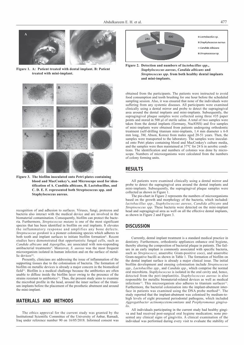
All patients were examined clinically using a dental mirror and probe to detect the supragingival area around the dental implants and mini-implants. Subsequently, the supragingival plaque samples were collected as shown in Figure 1.
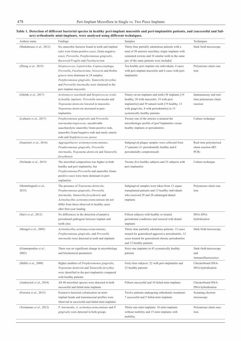
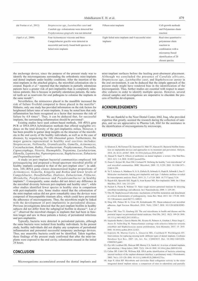
The pie-chart in Figure 2 represents the numbers of microorganisms based on the growth and morphology of the bacteria, which included: lactobacillus spp., Staphylococcus aureus, Candida albicans and Streptococcus spp. These bacteria were detected on the mini-implant head and supragingival area as well on all the effective dental implants, as shown in Figure 2 and Figure 3.
DISCUSSION
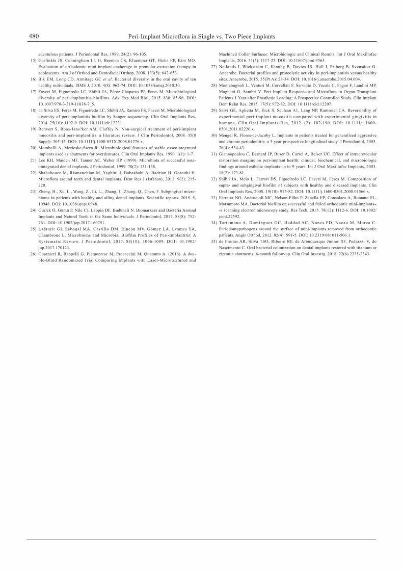
Currently, dental implant treatment is a standard medical practice in dentistry. Furthermore, orthodontic appliances enhance oral hygiene, thereby altering the composition of bacterial plaque in patients. The fail-ure in an early implant is commonly associated with specific bacteria, such as Streptococci, anaerobic Gram-positive cocci, and anaerobic Gram-negative bacilli as shown in Table 1. The formation of biofilm on the dental implant surface is already a major clinical issue. The initial biofilm development and ensuing colonisation include Streptococcus spp., Lactobacillus spp., and Candida spp., which comprise the normal oral microbiota. Staphylococcus is isolated in the oral cavity and, hence, detected from the peri-implantitis. Staphylococcus aureus is also responsible for metallic biomaterial-related devices as well as medical infections12). This microorganism also adheres to titanium surfaces12). Furthermore, the bacterial colonisation into the implant-abutment inter-face in patients was examined using the DNA probe method13). The study reported that the implant-abutment was colonised by medium-to-high levels of eight presumed periodontal pathogens, which included Aggregatibacter actinomycetemcomitans and Porphyromonas gingiva-lis.
All individuals participating in the current study had healthy gingi-va and had received post-surgical oral hygiene medication; none pre-sented any clinical signs of gingivitis. A clinical examination of the individual was performed during every visit to evaluate the stability of
Figure 1. A: Patient treated with dental implant. B: Patient treated with mini-implant.
Figure 2. Detection and numbers of lactobacillus spp., Staphylococcus aureus, Candida albicans and Streptococcus spp. from both healthy dental implants and mini-implants.
Figure 3. The biofilm inoculated onto Petri plates containing blood and MacConkey's, and Microscope used for iden-tification of A. Candida albicans, B. Lactobacillus, and C. D. E. F. represented both Streptococcus spp. and Staphylococcus aureus.
A
A
C
E
B
B
D
F
Peri-Implant Microflora in Single vs. Two Piece Implants478
Table 1. Detection of different bacterial species in healthy peri-implant mucositis and peri-implantitis patients, and (successful and fail-ure) orthodontic mini-implants, were analysed using different techniques.
Authors name Findings Samples Techniques
(Shahabouee et al., 2012) Six anaerobic bacteria found in teeth and implant Thirty-four partially edentulous patients with a Dark field microscope sulci were Gram-positive cocci, Gram-negative total of 50 anterior maxillary single implants with cocci, Prevotella, Porphyromonas gingivalis, cemented crowns and 34 similar teeth in the same Bacteroid Fragilis and Fusobacterium jaw of the same patients were included
(Zheng et al., 2015) Streptococcus, Leptotrichia, Capnocytophaga, Ten healthy peri-implant site individuals, 8 cases Polymerase chain reac Prevotella, Fusobacterium, Neisseria and Rothia with peri-implant mucositis and 6 cases with peri- tion genera were dominant in 24 samples. implantitis Porphyromonas gingivalis, Tannerella forsythia and Prevotella intermedia were clustered in the peri-implant mucositis
(Gürlek et al., 2017) Actinomyces naeslundi and Streptococcus oralis Ninety-seven implants and teeth (58 implants [19 Immunoassay and real- in healthy implants. Prevotella intermedia and healthy, 20 with mucositis, 19 with peri- time polymerase chain Treponema denticola lowered in mucositis. implantitis] and 39 natural teeth [19 healthy, 12 reaction Treponema denticola increased in peri- with gingivitis, 8 with periodontitis] in 15 implantitis systemically healthy patients.
(Lafaurie et al., 2017) Porphyromonas gingivalis and Prevotella Twenty-one of the articles evaluated the Culture technique intermedius/nigrescens, uncultivable microbiologic profile of peri?implantitis versus asaccharolytic anaerobic Gram-positive rods, healthy implants or periodontitis anaerobic Gram?negative rods and rarely enteric rods and Staphylococcus aureus
(Guarnieri et al., 2016) Aggregatibacter actinomycetemcomitans, Subgingival plaque samples were collected from Real-time polymerised Porphyromonas gingivalis, Prevotella 17 patients (11 periodontally healthy and 6 chain reaction (RT- intermedia, Treponema denticola and Tannerella periodontally compromised) PCR) forsythensis
(Neilands et al., 2015) The microbial composition was higher in both Twenty-five healthy subjects and 25 subjects with Culture technique healthy and peri-implantitis, but peri-implantitis Porphyromonas/Prevotella and anaerobic Gram- positive cocci were more dominant in peri- implantitis
(Montebugnoli et al., The presence of Treponema denticola, Subgingival samples were taken from 13 organ- Polymerase chain reac2015) Porphyromonas gingivalis, Prevotella transplanted patients and 13 healthy individuals tion intermedia, Tannerella forsythensis and who received 29 and 28 submerged dental Actinobacillus actinomycetemcomitans do not implants differ from those observed in healthy cases after first-year loading
(Salvi et al., 2012) No differences in the detection of putative Fifteen subjects with healthy or treated DNA-DNA periodontal pathogens between implant and periodontal conditions and restored with dental hybridisation tooth sites implants
(Mengel et al., 2005) Actinobacillus actinomycetemcomitans, Thirty-nine partially edentulous patients: 15 cases Dark-field microscopy Porphyromonas gingivalis, and Prevotella treated for generalised aggressive periodontitis, 12 intermedia were detected at teeth and implants cases treated for generalised chronic periodontitis and 12 healthy patients
(Giannopoulou et al., There was no significant change in microbiology Sixty-one implants in 45 systemically healthy Dark-field microscopy2003) and biochemical parameters patients and immunofluorescence
(Shibli et al., 2008) Higher numbers of Porphyromonas gingivalis, Forty-four subjects: 22 with peri-implantitis and Checkerboard DNA- Treponema denticola and Tannerella forsythia 22 healthy patients DNA hybridisation were identified in the peri-implantitis compared with healthy patients
(Andrucioli et al., 2018) All 40 microbial species were detected in both Fifteen successful and 10 failed mini-implants Checkerboard DNA- successful and failed mini-implants DNA hybridisation
(Ferreira et al., 2015) Extensive bacterial colonisation on mini- Twelve patients undergoing orthodontic treatment: Scanning electron implant heads and transmucosal profiles were 7 successful and 5 failed mini-implants microscopy observed in successful and failed mini-implants
(Tortamano et al., 2012) P. intermedia, A. actinomycetemcomitans and P. Thirty-one mini-implants: 16 mini-implants Polymerase chain reac- gingivalis were detected in both groups without mobility and 15 mini-implants with tion. mobility.
Abdulkareem E. H. et al. 479
the anchorage device, since the purpose of the present study was to identify the microorganisms surrounding the orthodontic mini-implants and dental implants under healthy conditions. After the insertion of the mini-implants in the attached gingiva, the microbial colonisation site is created. Rajesh et al.5) reported that the implants in partially edentulous patients have a greater risk of peri-implantitis than in completely eden-tulous patients; this is because in partially edentulous patients, the natu-ral teeth act as reservoirs for oral pathogens to colonise the implants in the same mouth14).
Nevertheless, the miniscrews placed in the mandible increased the risk of failure fivefold compared to those placed in the maxilla15). Hitherto, only one study has provided information on the risk factors for heightened failure rates of mini-implants. It may be noted here that only inflammation has been recognised as a factor that increases the risk of failure by 4.8 times15). Thus, it can be deduced that, for successful implants, the surrounding inflammation should be prevented1).
Existing studies have used culture-based methods, 16S rRNA gene PCR or DNA-DNA hybridisation methods that offered negligible evi-dence on the total diversity of the peri-implantitis milieu. However, it has been possible to garner deep insights on the structure of the microbi-ota in the oral cavity of the healthy individuals, as well as in the case of diseases, by sequencing the 16S ribosomal genes. Furthermore, the major genera represented in heal thy oral cavi t ies includes: Streptococcus, Veillonella, Granulicatella, Gamella, Actinomyces, Corynebacterium, Rothia, Fusobacterium, Porphyromonas, Prevotella, Capnocytophaga, Nisseria, Haemophilis, Treponema, Lactobacterium, Eikenella, Leptotrichia, Peptostreptococcus, Staphylococcus, Eubacteria and Propionibacterium16).
A study on peri-implant bacterial communities employed 16S pyrosequencing and proposed a broad-spectrum microbial profile of healthy implants compared to that of the peri-implant sites17). In addi-tion, 16S rRNA gene clones demonstrated higher proportions of Actinomyces, Gemella, Kingella and Rothia and lower levels of Campylobacter, Desulfobulbus, Dialister, Eubacterium, Filifactor, Mitsukella , Porphyromonas and Pseudoramibacter in healthy implants18). Consequently, some studies did not detect any difference in the microbial diversity between peri-implants and healthy sites19), while other studies identified fewer species in healthy sites in comparison with peri-implantitis sites. Some studies stated that the colonisation of the mini-implant sulcus did not grow remarkably since the devices were composed of biocompatible titanium alloy, which could have prevented the adherence of microorganisms. Thus, the microbiota might be linked with the development of peri-implantitis in periodontal disease. Previous investigations detected that the peri-implant biofilm in healthy subjects did not differ from the subgingival biofilm in disease20). Lee et al.21) observed the microbial changes in implants that had been in func-tion longer and saw in those patients a history of periodontal infections and peri-implantitis.
Typically, bacteria were detected in periodontal patients, although the same bacteria were isolated from healthy individuals. In the present study, healthy individuals did not display any symptoms of periodontal inflammation and presented successful temporary anchorage devices. Thus, any anaerobic bacteria could not be identified. Taken together, these findings of the present study confirmed that after the mini-im-plants were exposed to the oral cavity, colonisation ensued in the initial 24 hours.
CONCLUSION
Microorganisms accumulated around the dental implants and
mini-implant surfaces before the healing post-abutment placement. Al though we concluded the presence of Candida albicans , Streptococcus spp., Lactobacillus casei, and Staphylococcus aureus in the oral environment, it can be deduced that the simple approach of the present study might have rendered bias in the identification of the microorganism. Thus, further studies are essential with respect to anaer-obic cultures in order to identify multiple species. However, several clinical samples and investigations are imperative to elucidate the pro-cess of biofilm development.
ACKNOWLEDGEMENTS
We are thankful to the Noor Dental Center, Irbil, Iraq, who provided expertise that greatly, assisted the research during the collection of sam-ples, and we are appreciative to Pharma Lab, Irbil for the assistance in the identification of microorganisms by microscope.
REFERENCES
1) Khatoon Z, McTiernan CD, Suuronen EJ, Mah TF, Alarcon EI. Bacterial biofilm forma-tion on implantable devices and approaches to its treatment and prevention. Heliyon, 2018. 28; 4(12): e01067. DOI: 10.1016/j.heliyon.2018.e01067.
2) Ahmad N, Saad N. Effects of antibiotics on dental implants: a review. Clin Med Res, 2012. 4(1): 1-6. DOI: 10.4021/jocmr658w.
3) Zaura E, Keijser BJ, Huse SM, Crielaard W. Defining the healthy "core microbiome" of oral microbial communities. BMC Microbiol, 2009. 15; 9:259. DOI: 10.1186/1471-2180-9-259.
4) Yu T, Acharya A, Mattheos N, Li S, Ziebolz D, Schmalz G, Haak R, Schmidt J, Sun Y. Molecular mechanisms linking peri-implantitis and type 2 diabetes mellitus revealed by transcriptomic analysis. PeerJ, 2019. 21; 7: e7124. DOI: 10.7717/peerj.7124.
5) Rajesh KS, Spoorthi KH, Hegde S, Arun Kumar MS. Peri-implantitis 3600. Int J Dent Med Res, 2015. 1(6): 213-219.
6) Puckett S, Pareta R, Webster TJ. Nano rough micron patterned titanium for directing osteoblast morphology and adhesion. Int J Nanomedicine, 2008. 3: 229-241.
7) Otto M. Staphylococcal infections: mechanisms of biofilm maturation and detachment as critical determinants of pathogenicity. Annu Rev Med, 2013. 64: 175-88. DOI: 10.1146/annurev-med-042711-140023.
8) Ding AM, Palmer RJ Jr, Cisar JO, Kolenbrander PE. Shear-enhanced oral microbial adhesion. Appl Environ Microbiol, 2010. 76(4): 1294-7. DOI: 10.1128/AEM.02083-09.
9) Zarco MF, Vess TJ, Ginsburg GS. The oral microbiome in health and disease and the potential impact on personalized dental medicine. Oral Dis, 2012. 18(2): 109-20. DOI: 10.1111/j.1601-0825.2011.01851.x.
10) Izquierdo-Barba I, García-Martín JM, Álvarez R, Palmero A, Esteban J, Pérez-Jorge C, Arcos D, Vallet-Reg M. Nanocolumnar coatings with selective behavior towards osteoblast and Staphylococcus aureus proliferation; Acta Biomater, 2015. 15: 20-8. DOI: 10.1016/j.actbio.2014.12.023.
11) Esposito M, Murray-Curtis L, Grusovin MG, Coulthard P, Worthington HV. Interventions for replacing missing teeth: different types of dental implants. Cochrane Database Syst Rev, 2007. (4), Art . No.: CD003815. Doi. 10.1002/14651858.CD003815.pub3.
12) Pye AD, Lockhart DE, Dawson MP, Murray CA, Smith AJ. A review of dental implants and infection. J Hosp Infect, 2009. 72(2): 104-10. DOI: 10.1016/j.jhin.2009.02.010.
13) Callan DP, Cobb CM, Williams KB. DNA probe identification of bacteria colonizing internal-surfaces of the implant-abutment interface: a preliminary study. J Periodontol, 2005. 76(1): 115-120. DOI: 10.1111/j.1600-051X.2008.01274.x.
14) Aspe P, Allen RP. Microbiota and crevicular fluid collagenase activity in the osseo integrated dental implant sulcus: a comparison of sites in edentulous and partially
(de Freitas et al., 2012) Streptococcus spp., Lactobacillus casei and Fifteen mini-implants Cell-growth methods Candida spp. colonisations were detected but and Polymerase chain Porphyromonas gingivalis was not detected reaction
(Apel et al., 2009) Four Actinomyces viscosus and three Eight failed mini-implants and 4 successful mini- Real-time quantitative Campylobacter gracilis were detected in implants polymerase chain successful and rarely found both species in reaction in failed mini-implants combination with a microarray-based identification of 20 chosen species
Peri-Implant Microflora in Single vs. Two Piece Implants480
edentulous patients. J Periodontal Res, 1989. 24(2): 96-105. 15) Garfinkle JS, Cunningham LL Jr, Beeman CS, Kluemper GT, Hicks EP, Kim MO.
Evaluation of orthodontic mini-implant anchorage in premolar extraction therapy in adolescents. Am J of Orthod and Dentofacial Orthop, 2008. 133(5): 642-653.
16) Bik EM, Long CD, Armitage GC et al. Bacterial diversity in the oral cavity of ten healthy individuals. ISME J, 2010. 4(8): 962-74. DOI: 10.1038/ismej.2010.30.
17) Faveri M, Figueiredo LC, Shibli JA, Pérez-Chaparro PJ, Feres M. Microbiological diversity of peri-implantitis biofilms. Adv Exp Med Biol, 2015. 830: 85-96. DOI: 10.1007/978-3-319-11038-7_5.
18) da Silva ES, Feres M, Figueiredo LC, Shibli JA, Ramiro FS, Faveri M. Microbiological diversity of peri-implantitis biofilm by Sanger sequencing. Clin Oral Implants Res, 2014. 25(10): 1192-9. DOI: 10.1111/clr.12231.
19) Renvert S, Roos-Jans?ker AM, Claffey N. Non-surgical treatment of peri-implant mucositis and peri-implantitis: a literature review. J Clin Periodontol, 2008. 35(8 Suppl): 305-15. DOI: 10.1111/j.1600-051X.2008.01276.x.
20) Mombelli A, Mericske-Stern R. Microbiological features of stable osseointegrated implants used as abutments for overdentures. Clin Oral Implants Res, 1990. 1(1): 1-7.
21) Lee KH, Maiden MF, Tanner AC, Weber HP. (1999). Microbiota of successful osse-ointegrated dental implants. J Periodontol, 1999. 70(2): 131-138.
22) Shahabouee M, Rismanchian M, Yaghini J, Babashahi A, Badrian H, Goroohi H. Microflora around teeth and dental implants. Dent Res J (Isfahan), 2012. 9(2): 215-220.
23) Zheng, H., Xu, L., Wang, Z., Li, L., Zhang, J., Zhang, Q., Chen, F. Subgingival micro-biome in patients with healthy and ailing dental implants. Scientific reports, 2015. 5, 10948. DOI: 10.1038/srep10948.
24) Gürlek Ö, Gümü P, Nile CJ, Lappin DF, Buduneli N. Biomarkers and Bacteria Around Implants and Natural Teeth in the Same Individuals. J Periodontol, 2017. 88(8): 752-761. DOI: 10.1902/jop.2017.160751.
25) Lafaurie GI, Sabogal MA, Castillo DM, Rincón MV, Gómez LA, Lesmes YA, Chambrone L. Microbiome and Microbial Biofilm Profiles of Peri-Implantitis: A Systematic Review. J Periodontol , 2017. 88(10): 1066-1089. DOI: 10.1902/jop.2017.170123.
26) Guarnieri R, Rappelli G, Piemontese M, Procaccini M, Quaranta A. (2016). A dou-ble-Blind Randomized Trial Comparing Implants with Laser-Microtextured and
Machined Collar Surfaces: Microbiologic and Clinical Results. Int J Oral Maxillofac Implants, 2016. 31(5): 1117-25. DOI: 10.11607/jomi.4563.
27) Neilands J, Wickström C, Kinnby B, Davies JR, Hall J, Friberg B, Svensäter G. Anaerobe. Bacterial profiles and proteolytic activity in peri-implantitis versus healthy sites. Anaerobe, 2015. 35(Pt A): 28-34. DOI: 10.1016/j.anaerobe.2015.04.004.
28) Montebugnoli L, Venturi M, Cervellati F, Servidio D, Vocale C, Pagan F, Landini MP, Magnani G, Sambri V. Peri-Implant Response and Microflora in Organ Transplant Patients 1 Year after Prosthetic Loading: A Prospective Controlled Study. Clin Implant Dent Relat Res, 2015. 17(5): 972-82. DOI: 10.1111/cid.12207.
29) Salvi GE, Aglietta M, Eick S, Sculean A1, Lang NP, Ramseier CA. Reversibility of experimental peri-implant mucositis compared with experimental gingivitis in humans. Clin Oral Implants Res, 2012. (2) : 182-190. DOI: 10.1111/j .1600-0501.2011.02220.x.
30) Mengel R, Flores-de-Jacoby L. Implants in patients treated for generalized aggressive and chronic periodontitis: a 3-year prospective longitudinal study. J Periodontol, 2005. 76(4): 534-43.
31) Giannopoulou C, Bernard JP, Buser D, Carrel A, Belser UC. Effect of intracrevicular restoration margins on peri-implant health: clinical, biochemical, and microbiologic findings around esthetic implants up to 9 years. Int J Oral Maxillofac Implants, 2003. 18(2): 173-81.
32) Shibli JA, Melo L, Ferrari DS, Figueiredo LC, Faveri M, Feres M. Composition of supra- and subgingival biofilm of subjects with healthy and diseased implants. Clin Oral Implants Res, 2008. 19(10): 975-82. DOI: 10.1111/j.1600-0501.2008.01566.x.
33) Ferreira NO, Andrucioli MC, Nelson-Filho P, Zanella EP, Consolaro A, Romano FL, Matsumoto MA. Bacterial biofilm on successful and failed orthodontic mini-implants---a scanning electron microscopy study. Res Tech, 2015. 78(12): 1112-6. DOI: 10.1002/jemt.22592.
34) Tortamano A, Dominguez GC, Haddad AC, Nunes FD, Nacao M, Morea C. Periodontopathogens around the surface of mini-implants removed from orthodontic patients. Angle Orthod, 2012. 82(4): 591-5. DOI: 10.2319/081011-506.1.
35) de Freitas AR, Silva TSO, Ribeiro RF, de Albuquerque Junior RF, Pedrazzi V, do Nascimento C. Oral bacterial colonization on dental implants restored with titanium or zirconia abutments: 6-month follow-up. Clin Oral Investig, 2018. 22(6) 2335-2343.