Embed Size (px)
Citation preview
Journal of Environmental Science and Health, Part C, 28:147–171, 2010ISSN: 1059-0501 print / 1532-4095 onlineDOI: 10.1080/10590501.2010.504978
Occurrence, Efficacy,Metabolism, and Toxicityof Triclosan
Jia-Long Fang,1 Robin L. Stingley,1 Frederick A. Beland,1
Wafa Harrouk,2 Debbie L. Lumpkins,2 and Paul Howard1
1National Center for Toxicological Research, Food and Drug Administration,Jefferson, AR2Center for Drug Evaluation and Research, Food and Drug Administration,Silver Spring, MD
Triclosan has broad-spectrum anti-microbial activity against most gram-negative andgram-positive bacteria. It is widely used in personal care products, household items,medical devices, and clinical settings. Due to its extensive use, there is potential for hu-mans in all age groups to receive life-time exposures to triclosan, and, indeed, triclosanhas been detected in human tissues and the environment. Data gaps exist regardingthe chronic dermal toxicity and carcinogenicity of triclosan, which is needed for therisk assessment of triclosan. The US Food and Drug Administration (FDA) nominatedtriclosan to the National Toxicology Program (NTP) for toxicological evaluations. Cur-rently, the NTP is conducting several dermal toxicological studies to determine the car-cinogenic potential of triclosan, evaluate its endocrine and developmental-reproductiveeffects, and investigate the potential UV-induced dermal formation of chlorinated phe-nols and dioxins of triclosan. This paper reviews data on the human exposure, environ-mental fate, efficacy of anti-microbial activity, absorption, distribution, metabolism andelimination, endocrine disrupting effects, and toxicity of triclosan.
Keywords: triclosan; metabolism; endocrine disruption effect; toxicity
INTRODUCTION
Triclosan is a synthetic, lipid-soluble, broad-spectrum anti-microbial agentthat was first introduced in the health care industry in 1972 and in the tooth-paste in Europe in 1985 (1). In the United States, triclosan has been used for
This article is not subject to US copyright law.This article is not an official guidance or policy statements of US Food and Drug Admin-istration. No official support or endorsement by the US Food and Drug Administrationis intended or should be inferred.Address correspondence to Jia-Long Fang, National Center for Toxicological Re-search, Food and Drug Administration, Jefferson, Arkansas 72079, USA. E-mail:[email protected]
147
Downloaded By: [Fang, Jia-Long][FDA Biosciences Library] At: 15:11 20 September 2010
148 J.-L. Fang et al.
more than 40 years in personal care products, household items, medical de-vices, and hospitals to control the spread of bacteria (1). In 1997, the US Foodand Drug Administration (FDA) approved triclosan (0.3%) for use in ColgateTotal R© toothpaste to prevent gingivitis and cavities. This paper reviews dataon the human exposure, environmental fate, efficacy, absorption, distribution,metabolism and elimination, endocrine disrupting effects, and toxicity of tri-closan. We will not discuss whether the benefits of using triclosan-containingproducts in personal care and household outweigh the potential hazards as-sociated with this use, such as the emergence of antibiotic-resistant bacteria,because these issues are still the subject of ongoing scientific and public debate.
Triclosan is regulated by both the FDA and US Environmental ProtectionAgency (EPA). Within the FDA, triclosan is considered an over-the-counterdrug for use in hand soaps, toothpaste, deodorants, laundry detergent, fab-ric softeners, facial tissues, antiseptics for wound care, and medical devices.Triclosan is currently registered with the EPA under the Federal InsecticideFungicide and Rodenticide Act as an anti-microbial agent for the protection ofpolymers and plastics.
Chemical Identification
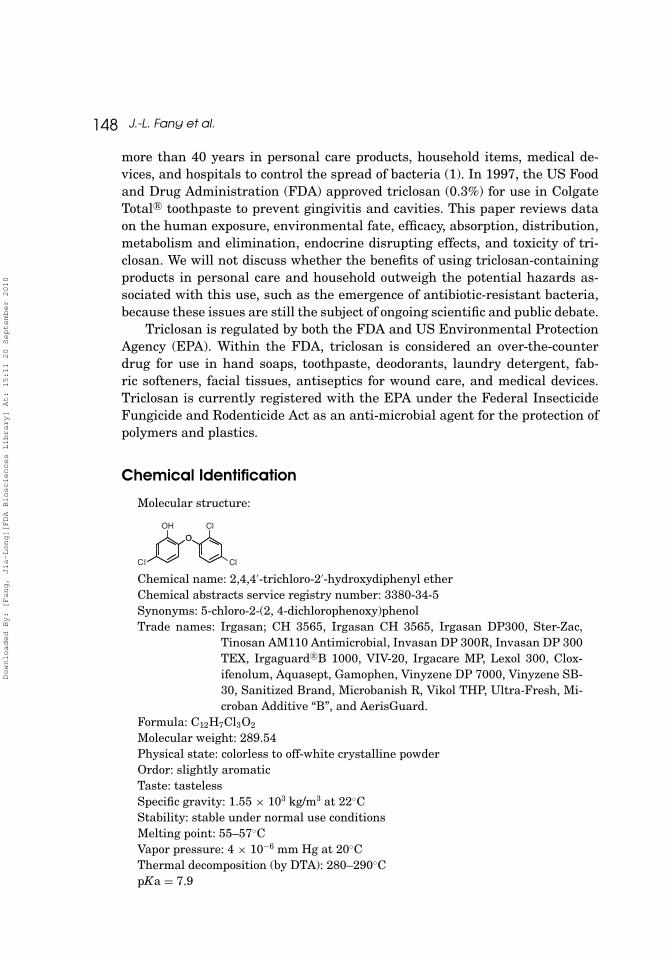
Molecular structure:
OO
Cl
OH Cl
Cl
Chemical name: 2,4,4′-trichloro-2′-hydroxydiphenyl etherChemical abstracts service registry number: 3380-34-5Synonyms: 5-chloro-2-(2, 4-dichlorophenoxy)phenolTrade names: Irgasan; CH 3565, Irgasan CH 3565, Irgasan DP300, Ster-Zac,
Tinosan AM110 Antimicrobial, Invasan DP 300R, Invasan DP 300TEX, Irgaguard R©B 1000, VIV-20, Irgacare MP, Lexol 300, Clox-ifenolum, Aquasept, Gamophen, Vinyzene DP 7000, Vinyzene SB-30, Sanitized Brand, Microbanish R, Vikol THP, Ultra-Fresh, Mi-croban Additive “B”, and AerisGuard.
Formula: C12H7Cl3O2
Molecular weight: 289.54Physical state: colorless to off-white crystalline powderOrdor: slightly aromaticTaste: tastelessSpecific gravity: 1.55 × 10³ kg/m3 at 22◦CStability: stable under normal use conditionsMelting point: 55–57◦CVapor pressure: 4 × 10⁻⁶ mm Hg at 20◦CThermal decomposition (by DTA): 280–290◦CpKa = 7.9
Downloaded By: [Fang, Jia-Long][FDA Biosciences Library] At: 15:11 20 September 2010
Occurrence, Efficacy, Metabolism, & Toxicity of Triclosan 149
Octanol-water partition constant (log Kow): 4.76Solubility: water, 0.01g/L; 0.1 N NaOH, 23.5 g/L; ethanol, acetone, propylene gly-
col, Tween 20, benzene, methylcellosolve, highly soluble (> 1,000 g/L)
HUMAN EXPOSURE TO TRICLOSAN
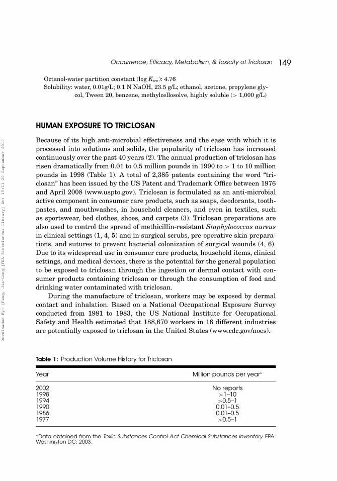
Because of its high anti-microbial effectiveness and the ease with which it isprocessed into solutions and solids, the popularity of triclosan has increasedcontinuously over the past 40 years (2). The annual production of triclosan hasrisen dramatically from 0.01 to 0.5 million pounds in 1990 to > 1 to 10 millionpounds in 1998 (Table 1). A total of 2,385 patents containing the word “tri-closan” has been issued by the US Patent and Trademark Office between 1976and April 2008 (www.uspto.gov). Triclosan is formulated as an anti-microbialactive component in consumer care products, such as soaps, deodorants, tooth-pastes, and mouthwashes, in household cleaners, and even in textiles, suchas sportswear, bed clothes, shoes, and carpets (3). Triclosan preparations arealso used to control the spread of methicillin-resistant Staphylococcus aureusin clinical settings (1, 4, 5) and in surgical scrubs, pre-operative skin prepara-tions, and sutures to prevent bacterial colonization of surgical wounds (4, 6).Due to its widespread use in consumer care products, household items, clinicalsettings, and medical devices, there is the potential for the general populationto be exposed to triclosan through the ingestion or dermal contact with con-sumer products containing triclosan or through the consumption of food anddrinking water contaminated with triclosan.
During the manufacture of triclosan, workers may be exposed by dermalcontact and inhalation. Based on a National Occupational Exposure Surveyconducted from 1981 to 1983, the US National Institute for OccupationalSafety and Health estimated that 188,670 workers in 16 different industriesare potentially exposed to triclosan in the United States (www.cdc.gov/noes).
Table 1: Production Volume History for Triclosan
Year Million pounds per year∗
2002 No reports1998 >1–101994 >0.5–11990 0.01–0.51986 0.01–0.51977 >0.5–1
∗Data obtained from the Toxic Substances Control Act Chemical Substances Inventory EPA:Washington DC; 2003.
Downloaded By: [Fang, Jia-Long][FDA Biosciences Library] At: 15:11 20 September 2010
150 J.-L. Fang et al.
The production and the widespread use of triclosan may result in it beingdisposed in sewage systems, which ultimately leads to environmental deposi-tion. As a result, triclosan is found in finished drinking water, surface water,wastewater, and environmental sediments, as well as in the bile of wild fish,indicating extensive contamination of aquatic ecosystems (7–10). Triclosan hasbeen detected in both raw and finished drinking water in Southern California,at levels of 56 and 49 ng per liter, respectively (7). Based on a US GeologicalSurvey conducted from 1999 through 2000, triclosan was detected in 85 of 139water samples from a network of streams across 30 US states (10). Triclosanwas also found in lakes and in a river in Switzerland at concentrations of up to74 ng per liter (9), and has been detected in bile samples of wild fish, caught atleast 1 km downstream of three different wastewater treatment plants in Swe-den, at concentrations ranging from 0.44 to 4.4 mg per kg fresh weight (11). Inaddition, methylated triclosan was present in fish and shellfish from the TamaRiver and Tokyo Bay in Japan (12) and in 45 samples corresponding to the in-fluents and effluents from eight domestic wastewater treatment plants locatedalong the Llobregat and Ebro rivers in Spain (13).
Triclosan has been identified in human breast milk. It was found in three offive randomly selected Swedish human milk samples, at concentrations rang-ing from <20 to 300 µg per kg lipid (11). The concentration of triclosan was 81and 345 µg per kg lipid in two of four milk samples collected in 2007 from fouranonymous lactating American women with no known occupational exposureto triclosan (14). Additionally, single samples of breast milk obtained from 62women, who had donated milk in 2001 to the Mothers Milk Bank in San Jose,CA, and Austin, TX, have been analyzed for triclosan. Triclosan levels rangedfrom undetectable (two samples) and barely detectable (nine samples) to read-ily detectable at the levels between 100 to 2,100 µg per kg lipid (51 samples)(15).
Triclosan has been detected in a pooled plasma sample from 10 randomlyselected male blood donors in Sweden (16). Sandborgh-Englund et al. (17)found triclosan in plasma at 0.1–8.1 ng per ml in 10 individuals, of which fivewere exposed and five were not exposed to triclosan via personal care products.Regardless of whether the nursing mothers used triclosan-containing soap, de-odorant, or toothpaste, triclosan was detected in their plasma and breast milk(18). The levels, however, were higher in the mothers who used products con-taining triclosan (0.4–38.0 ng per g in plasma and 0.022–0.95 ng per g in milk)than in those who did not (0.01–19 ng per g in plasma and <0.018–0.35 ng perg in milk).
The excretion of triclosan in urine has been reported at rates of 0.1–743 µgper day from 10 randomly selected Swedish men (17) and at concentrations of2.4–3,790 µg per liter of urine from a random selection of 2,517 participants 6years of age and older from the US general population (19).
Downloaded By: [Fang, Jia-Long][FDA Biosciences Library] At: 15:11 20 September 2010
Occurrence, Efficacy, Metabolism, & Toxicity of Triclosan 151
ENVIRONMENTAL FATE
Triclosan is readily chlorinated with sodium hypochlorite (20), and its chlo-rinated derivatives are also formed during the disinfection and deodorizationof water supplies and wastewater with sodium hypochlorite (21). Triclosan isconverted to di- and trichlorodibenzo-p-dioxin upon heating to temperaturesgreater than 400◦C to simulate incineration (22). Bleaching fabrics contain-ing triclosan with sodium hypochlorite, followed by combustion, leads to theformation of di-, tri-, and tetrachlorodibenzo-p-dioxins (22).
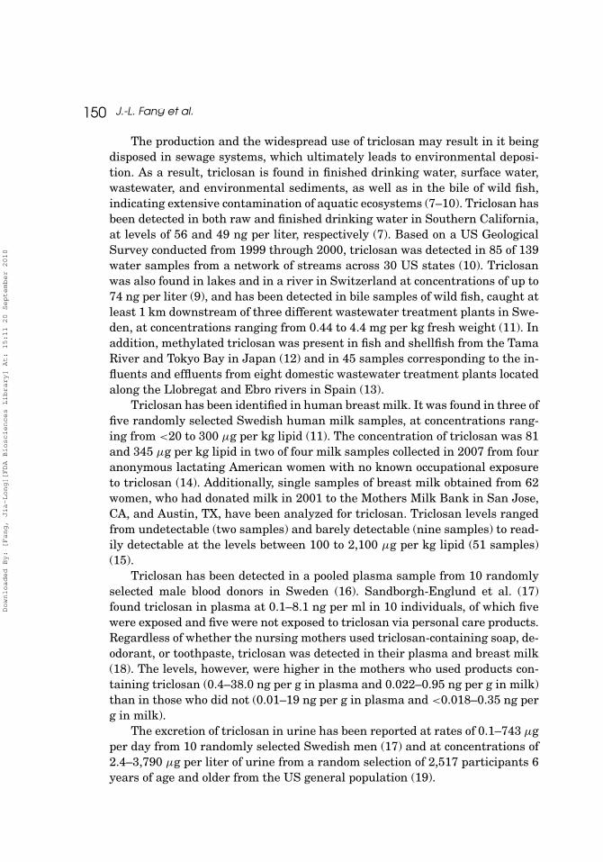
Photodegradation appears to be one of the major routes of elimination oftriclosan in aquatic environments (8, 23) and takes place at low light intensityunder UV (254, 313, or 365 nm) light, simulated solar light, or artificial whitelight. Triclosan photodegradation with UV (365 nm) and simulated solar lightirradiation exhibits first-order kinetic behavior, with both light sources givingsimilar kinetic parameters (24). The photodecomposition products of triclosanare illustrated in Figure 1. The photochemical formation of dichlorodibenzo-dioxin from triclosan in both solid phase and thin films of triclosan has beendemonstrated (20, 25). More recently, several highly toxic photoproducts, in-cluding 2,8-dichlorodibenzo-p-dioxin (23, 24, 26–32), 2,4-dichlorophenol (24,26, 28, 29), and possibly dichlorohydroxydibenzofuran (24, 29), were identi-fied in water samples. The photodegradation of triclosan and formation of 2,8-dichlorodibenzo-p-dioxin occur over a wide range of pH levels (3.0–9.0), withthe rate of formation being faster at basic pH (24, 29).
O
OH
Cl
Cl
Cl
Triclosan
2,8-Dichlorodibenzo-p-dioxin
Cl
Cl
HO
2,4-DichlorophenolDichlorohydroxydibenzofuran?
O
Cl O Cl
O
Cl O
Cl
2,7-Dichlorodibenzo-p-dioxin
OH
OClCl
or OH
Cl
Monochlorophenol
OHO
Cl2
Dichlorohydroxydiphenyl ether
O
OH
Cl
Cl
Cl
Triclosan
2,8-Dichlorodibenzo-p-dioxin
Cl
Cl
HO
2,4-DichlorophenolDichlorohydroxydibenzofuran?
O
Cl O Cl
O
Cl O
Cl
2,7-Dichlorodibenzo-p-dioxin
OH
OClCl
or OH
Cl
Monochlorophenol
OHO
Cl2
Dichlorohydroxydiphenyl ether
Figure 1: Simplified schematic of triclosan photodecomposition.
Downloaded By: [Fang, Jia-Long][FDA Biosciences Library] At: 15:11 20 September 2010
152 J.-L. Fang et al.
EFFICACY OF TRICLOSAN ON ANTI-MICROBIAL ACTIVITY
Mechanism of ActionTriclosan has been shown to intercalate into bacterial cell membranes and
disrupt membrane activities, without causing leakage of intracellular compo-nents (33, 34). In addition, triclosan is an inhibitor of the enoyl-reductase oftype II fatty acid synthase involved in the bacterial lipid biosynthesis (35–38).
At low doses, triclosan is bacteriostatic and, at higher doses, it becomesbactericidal (39, 40). Sub-lethal concentrations of triclosan favor a specific ac-tion against type II fatty acid synthase enoyl-reductase (FabI), while at bac-tericidal concentrations triclosan appears to act against multiple targets, in-cluding less specific targets such as the cell membrane (40). Triclosan also hasanti-viral, anti-fungal (1), and anti-malarial activity (41).
While triclosan has in vitro activity against a broad spectrum of bacteria,it is generally more effective against gram-positive than gram-negative bac-teria (1, 42). Triclosan is particularly effective against Staphylococcus aureus(1, 42, 43). However, some clinical isolates of Staphylococcus aureus are notas susceptible to triclosan due to the overexpression of FabI (44). In additionto its overexpression, the FabI from these isolates carries a single amino acidchange that prevents the formation of a stable triclosan-NAD+-FabI complex(44). In gram-negative bacteria, such as Pseudomonas aeruginosa, there areseveral multi-drug efflux pumps that remove a number of drugs, including tri-closan, from the cells (45, 46). In addition, some strains of a Pseudomonasaeruginosa contain a triclosan-resistant enoyl-acyl carrier protein reductaseFabV (47).
In SoapsThe efficacy of triclosan in soaps is equivocal. An evaluation of the avail-
able data by the American Medical Association in 2002 determined that, whenproperly used in clinical settings, triclosan-containing soaps were efficacious(48). A review of studies on the efficacy of triclosan in soap, however, revealedthat it does not reduce bacterial counts on hands to a greater extent than plainsoap unless it is used repeatedly and at relatively high concentrations (≥1%)compared to the 0.1–0.45% found in consumer anti-bacterial soaps (49).
There have been few appropriately designed studies to assess the impactof the use of triclosan-containing products on infection rates. One random-ized double-blind study evaluated the effect of the use of the anti-microbialproducts by consumers on the occurrence of infectious disease symptoms overa one-year period in 228 inner-city households. The study found no statisti-cal difference between use of “anti-microbial” household cleaners, detergents,and hand washes compared to the use of identically packaged products lacking
Downloaded By: [Fang, Jia-Long][FDA Biosciences Library] At: 15:11 20 September 2010
Occurrence, Efficacy, Metabolism, & Toxicity of Triclosan 153
anti-microbial ingredients (50). This study used several types of anti-microbialhousehold cleaners, such as a liquid hand washing soap containing triclosan,a liquid kitchen spray, an “all-purpose” hard-surface cleaner containing aquaternary-ammonium compound, and a laundry detergent containing oxy-genated bleach, and focused primarily on symptoms consistent with a viralillness as a measure of efficacy.
Studies using bacterial counts to determine the efficacy of triclosan-containing soaps have produced conflicting results. An epidemiological studyof household use over 11 months found that a hand washing soap containing0.2% triclosan was not significantly better at reducing bacterial levels onhands than a similar plain hand washing soap without triclosan (51). Whenoverall bacterial counts were used to determine the efficacy of soaps contain-ing triclosan, those with less than 1% triclosan were not significantly moreeffective than plain soap (49), except in 2 studies, one in which 0.3% triclosansoap was used 18 times daily for 5 days (52), and the other in which 0.75%triclosan was used in 2-min hand washes 3 times daily for 2 days (53). When1% triclosan soaps were compared to plain soap using bacterial counts as themeasure of efficacy, one study found no significant difference when hands werewashed using a standard surgical technique (54), but another study found that1% triclosan significantly reduced bacterial counts when hands were washedfor 30 sec or for 3 min (55).
In studies employing artificial contamination of hands with Serratiamarcescens, 10 hand washings for 10 sec each using 1.0% triclosan soap (56)or as little as one hand washing with 1.5% triclosan soap (57) reduced bac-terial counts significantly more than washing with plain soaps, as measuredby a log10 bacterial reduction. In a study using artificial contamination of fin-gertips with Escherichia coli, when hands were washed for 30 sec with 1.5% or2.0% triclosan soaps, reduction of bacterial counts was not significantly greaterthan when plain soap was used (58). A soap containing 2.0% triclosan exhib-ited residual anti-microbial activity on the forearm skin of 20 volunteers forup to at least 2 hr after three applications, as compared to plain soap (59).
In DeodorantsWhen used ad libitum for 6 months, deodorant sprays containing 0.15%
triclosan and anti-perspirant deodorant sprays containing 0.25% triclosan re-duced bacterial counts per cm2 of skin from 5.2 × 10⁵ to 1.4 × 10³ and 3.74 ×10², respectively (60). Bacterial levels returned to pre-test levels within 4 to7 days after stopping the use of triclosan-containing deodorants (60).
In DentifricesThere have been a number of reports of the anti-plaque and anti-gingivitis
efficacy of toothpaste containing triclosan (61–66). Volpe et al. (61) reviewed
Downloaded By: [Fang, Jia-Long][FDA Biosciences Library] At: 15:11 20 September 2010
154 J.-L. Fang et al.
the caries-preventive benefits of a triclosan/copolymer/fluoride dentifrice from13 independent, double-blind studies. Individuals using a dentifrice contain-ing triclosan/copolymer/fluoride had, on average, 27% less plaque (p < 0.01)than subjects using a fluoride dentifrice. The improved level of plaque controlwas accompanied by a 57% reduction in gingivitis severity index (p < 0.01)(61). Recently, a double-blind clinical study also demonstrated that a singleevening’s use of a dentifrice containing 0.3% triclosan/2.0% polyvinylmethylether/maleic acid copolymer/0.243% sodium fluoride in a 17% dual silica baseprovided a 28.4% reduction in oral malodor scores (p < 0.05) and 49.5% re-duction in microbial colony forming unit scores (p < 0.05) when compared toa commercially available dentifrice containing 0.243% sodium fluoride in a sil-ica base (63). When triclosan (0.3%) formulated with 2.0% Gantrez R© in denti-frices, there was effective anti-plaque and anti-gingivitis activity (65). Denti-frices with combinations of triclosan and soluble pyrophosphate or zinc citrate,however, were not effective against plaque and gingivitis (65). Following theuse of toothpaste containing triclosan, approximately 36% of the triclosan dosewas retained in the saliva and bacterial plaque (67, 68).
In SuturesSutures impregnated with triclosan have efficacy in vitro against gram-
positive and gram-negative bacteria, including isolates that are methicillinresistant (6). An in vivo challenge test was conducted to mimic a clinicallyrelevant environment. In this assay, control and test sutures were implantedsubcutaneously in the dorsal-lateral regions (control on the left side, test on theright side) of the same animal, and inoculated with a known number of bacte-ria. After a direct in vivo challenge, sutures with triclosan produced a nearly3-log reduction in the growth of Staphylococcus aureus in guinea pigs and a1-log reduction in the growth of E. coli in mice compared to control sutures(6, 69).
In PlasticsThere is no clear evidence available to demonstrate the efficacy of tri-
closan as an anti-microbial agent when combined with plastics. A low-densitypolyethylene film containing triclosan (1 g triclosan per kg polyethylene film)had a strong anti-microbial effect in in vitro simulated vacuum-packaged con-ditions against the psychrotrophic food pathogen L. monocytogenes, but did noteffectively reduce spoilage bacteria or the growth of L. monocytogenes on refrig-erated vacuum-packaged chicken breasts stored at 7◦C (70). A plastic wrap in-corporating 1,500 ppm of triclosan did not effectively reduce bacterial numberson refrigerated and vacuum packed meat surfaces (71). Triclosan-incorporatedplastics desorbed insufficient amounts of triclosan to inhibit bacterial growth
Downloaded By: [Fang, Jia-Long][FDA Biosciences Library] At: 15:11 20 September 2010

Occurrence, Efficacy, Metabolism, & Toxicity of Triclosan 155
when used in cutting boards (72). An anti-bacterial toothbrush containingtriclosan-coated tufts also failed to inhibit the bacterial growth when comparedto the regular toothbrush without the coated tufts (73).
ABSORPTION, DISTRIBUTION, METABOLISM, AND ELIMINATION
Triclosan reaches the systemic circulation via absorption through the mucousmembranes of the oral cavity (74) and gastrointestinal tract after oral expo-sure (17, 74, 75), through the skin after dermal exposure (76, 77), and throughmucosal tissues following intra-vaginal administration (78).
Several studies have reported the blood levels of total triclosan in humansfollowing use in either mouth rinses or dentifrices (17, 74, 75, 79). After 10healthy volunteers were exposed to a single dose of 4 mg triclosan by swal-lowing an oral mouthwash solution, the triclosan levels in plasma increasedrapidly from a median baseline of 0.4 ng per ml to a maximum concentra-tion of 218 ng per ml within 1 to 3 hrs (17). In another study, nine subjectswho ingested 20 ml of a 0.01% triclosan (2 mg) aqueous solution twice daily for21 days had blood triclosan levels between 150 and 174 ng per ml at 4 hrs afterthe morning dose. Nine other subjects who brushed twice daily with 1 g of den-tifrice containing 0.2% triclosan (2 mg) had blood levels between 15 and 21 ngper ml, which was approximately 9%–14% of that from subjects who ingestedan equivalent dose level of the aqueous solution (75). Measurable amounts oftriclosan appeared in the plasma of 21 healthy individuals at 15 min after asingle tooth brushing plus full ingestion of 1.25 g of dentifrice containing 0.3%triclosan. The peak plasma triclosan concentration occurred at 2–6 hrs afterthe dosing, and the mean peak plasma triclosan concentration was 243 ng perml. When this application was repeated three times daily for 12 days, the meanplasma triclosan concentrations ranged between 252 and 402 ng per ml, withan overall mean of 352 ng per ml (75). Other studies using a dentifrice con-taining triclosan at a concentration of 0.2%, 0.3%, or 0.6% daily for 2–12 weeksresulted in the blood levels of triclosan between 16 and 25 ng per ml (79). Dur-ing use of a mouth rinse containing 4.5 mg of triclosan for 30 sec twice dailyfor 21 days, mean plasma triclosan concentrations were 74.5–94.2 µg per ml,and it was estimated that about 2%–4% of the daily triclosan dose (9.0 mg)was absorbed and circulated in the blood (74). When absorbed through the oralmucous membrane from twice daily oral rinse with a mouth rinse containing0.03% triclosan for 21 days, triclosan levels in human blood plasma returnedto baseline approximately 8 days following the final exposure (74), and no ap-parent accumulation of triclosan in blood was observed (17, 74).
Dermal absorption of triclosan in humans and animals has been re-ported (79–82). Triclosan was detected in blood (0–189 ng per ml) and urine(0–5,600 ng per ml) following daily use of a dermal spray containing triclosanby 4 healthy males for 4 weeks or a soap bar containing 1% triclosan for
Downloaded By: [Fang, Jia-Long][FDA Biosciences Library] At: 15:11 20 September 2010
156 J.-L. Fang et al.
bathing and showering by 25 leukemic patients for 4 weeks and 6 healthymales for 48 weeks.
There is evidence that triclosan can be absorbed through the intact skin ofguinea pigs (80). In another study using ddY mice, Kanetoshi et al. (81) applied50 µl (32 mg triclosan per ml) [3H]triclosan (in ethanol: olive oil) to the mouseskin and quantified the absorption 6, 12, and 18 hrs later. Maximum levels of[3H]triclosan occurred between 12 and 18 hrs, with the greatest concentrationsin the gall bladder, liver, body fat, lungs, kidneys, blood, heart, testes, spleen,and brain. The levels in the tissues were approximately 14%–67% the levelsachieved in a comparable study where [3H]triclosan was given orally (82).
In in vitro studies, triclosan was shown to penetrate rat skin more rapidlyand extensively than human skin. Twenty-three percent of the dose penetratedcompletely through rat skin into receptor fluid by 24 hrs, whereas penetrationthrough human skin was only 6.3% of the applied dose (77) or about 0.7% of thedose administered in a transdermal adhesive formulation patch model (76).
A shampoo containing 0.05% (w/v) [3H]triclosan or an aerosol deodorantcontaining 0.1% (w/v) [3H]triclosan was applied to Wistar rat skin in a mannersimilar to consumer use, and the dermal penetration was calculated from theamount of radioactivity excreted by the rats. The penetration of triclosan fromshampoo was 197 ng per cm2 (3.3% of the applied amount) compared to 6.85 µgper cm2 from the aerosol deodorant (35.3% of the applied dose). These dataindicate that the composition and mode of use of different products containingtriclosan may be very important in determining the extent of penetration (83).
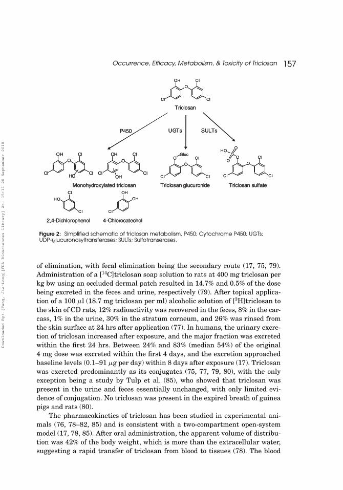
Triclosan is readily metabolized to glucuronide and sulfate conjugates(Figure 2). The conjugation of triclosan in the presence of human liver mi-crosomes or cytosol (84) and in skin (77) was demonstrated in vitro. Dermalmetabolism of triclosan in diffusion cells fitted with human skin following ap-plication of 7 µl of 64.5 mM [3H]triclosan showed that triclosan sulfate wasthe only metabolite in the skin at 4 hrs after application, whereas both thesulfate and glucuronide were present at 8 and 24 hrs after application (77).At all times, there was more unchanged triclosan than either of the conju-gates. Similar results were found in diffusion cells with rat skin (77). Tulpet al. (85) reported that both aromatic hydroxylation and cleavage of the etherbond of triclosan occurred in Wistar rats, as indicated by the presence of mono-hydroxylated triclosan, 2,4-dichlorophenol, and 4-chlorocatechol in the urineand feces, after oral administration a single dose of 500 mg triclosan per kgbody weight (bw) (Figure 2). Although triclosan is readily photodegraded intodichlorodibenzo-p-dioxin by heat and UV irradiation (20, 22, 23, 28, 31), therehave been no studies to investigate the metabolic formation of dichlorodibenzo-p-dioxin or chlorodibenzofurans on the skin in vivo.
Triclosan is excreted in the feces and urine. Rats and mice show predomi-nantly biliary excretion into the feces, whereas guinea pigs excrete the major-ity of the dose via the kidney. In humans, urinary excretion is the major route
Downloaded By: [Fang, Jia-Long][FDA Biosciences Library] At: 15:11 20 September 2010
Occurrence, Efficacy, Metabolism, & Toxicity of Triclosan 157
O
OH
Cl
Cl
Cl
Triclosan
P450 UGTs SULTs
OOH
Cl
Cl
ClHO
OOH
Cl
Cl
ClOH
Monohydroxylated triclosanCl
Cl
HO
2,4-Dichlorophenol 4-Chlorocatechol
OH
Cl
OH
Triclosan sulfateTriclosan glucuronide
O
O
Cl
Cl
Cl
S
OHO
OO
O
Cl
Cl
Cl
Gluc
O
OH
Cl
Cl
Cl
Triclosan
P450 UGTs SULTs
OOH
Cl
Cl
ClHO
OOH
Cl
Cl
ClOH
Monohydroxylated triclosanCl
Cl
HO
2,4-Dichlorophenol 4-Chlorocatechol
OH
Cl
OH
Triclosan sulfateTriclosan glucuronide
O
O
Cl
Cl
Cl
S
OHO
OO
O
Cl
Cl
Cl
Gluc
Figure 2: Simplified schematic of triclosan metabolism. P450; Cytochrome P450; UGTs;UDP-glucuronosyltransferases; SULTs; Sulfotranserases.
of elimination, with fecal elimination being the secondary route (17, 75, 79).Administration of a [14C]triclosan soap solution to rats at 400 mg triclosan perkg bw using an occluded dermal patch resulted in 14.7% and 0.5% of the dosebeing excreted in the feces and urine, respectively (79). After topical applica-tion of a 100 µl (18.7 mg triclosan per ml) alcoholic solution of [3H]triclosan tothe skin of CD rats, 12% radioactivity was recovered in the feces, 8% in the car-cass, 1% in the urine, 30% in the stratum corneum, and 26% was rinsed fromthe skin surface at 24 hrs after application (77). In humans, the urinary excre-tion of triclosan increased after exposure, and the major fraction was excretedwithin the first 24 hrs. Between 24% and 83% (median 54%) of the original4 mg dose was excreted within the first 4 days, and the excretion approachedbaseline levels (0.1–91 µg per day) within 8 days after exposure (17). Triclosanwas excreted predominantly as its conjugates (75, 77, 79, 80), with the onlyexception being a study by Tulp et al. (85), who showed that triclosan waspresent in the urine and feces essentially unchanged, with only limited evi-dence of conjugation. No triclosan was present in the expired breath of guineapigs and rats (80).
The pharmacokinetics of triclosan has been studied in experimental ani-mals (76, 78–82, 85) and is consistent with a two-compartment open-systemmodel (17, 78, 85). After oral administration, the apparent volume of distribu-tion was 42% of the body weight, which is more than the extracellular water,suggesting a rapid transfer of triclosan from blood to tissues (78). The blood
Downloaded By: [Fang, Jia-Long][FDA Biosciences Library] At: 15:11 20 September 2010
158 J.-L. Fang et al.
half-life (t1/2) of triclosan following intraperitoneal injection to guinea pigs andrats was 13 and 18 hrs, respectively (83), whereas the blood t1/2 of triclosan dur-ing the β-phase was 8.8 hrs after intravenous administration of 5 mg triclosanper kg bw to Wistar rats (78). Oral intubation of 5 mg triclosan per kg bw in a0.66% triclosan sodium lauryl sulfate solution or 0.2% triclosan toothpaste torats gave a t1/2 of 7–65 hrs for triclosan and its metabolites (glucuronide andsulfate conjugates) (79). After oral administration, the t1/2 of triclosan in ddYmouse liver was approximately 8 hrs (82). The t1/2 terminal blood/plasma con-centration phase for humans ranged between 6 and 63 hrs (17, 75). Triclosandoes not appear to sequester into a long-term compartment (e.g., fat) in thebody following single or repeated administration (74, 75, 79).
Due to its structural resemblance to polychlorinated biphenyls, triclosanhas been proposed to affect the hepatic mixed function oxidase system.Triclosan has been shown to increase the activities of aminopyrine N-demethylase, biphenyl 2-hydroxylase, biphenyl 4-hydroxylase, p-nitroanisoleO-demethylase, p-nitrophenetole O-deethylase, and 7-ethyoxycoumarin O-deethylase in male Wister rats (81, 86). The increase in the activities ofthese P450-dependent monooxygenases was associated with an elevation ofcytochrome P450 content (81) and the induction of cytochromes CYP2B1/2,3A2/1, and 4A1 (86). In male ddY mice, only aminopyrine N-demethylaseactivity was induced by triclosan (81). A subsequent study using rat hepa-tocytes cultured on Matrigel demonstrated that triclosan preferably inducedcytochromes CYP2B1/2, along with a slight increase in CYP3A (87). The studyalso showed that triclosan produced an accumulation of hydroxymethylbilane,and consequently uroporphyrin I, in rat hepatocytes by inhibiting uropor-phyrinogen III synthetase (87). Recently, triclosan was shown to be a selec-tive inhibitor of the glucuronidation and sulfonation of phenolic xenobiotics inhuman liver preparations in vitro (84).
Triclosan has been demonstrated to be a slow binding inhibitor of humanand goose type I fatty acid synthase and to inhibit partially enoyl-reductaseactivity type I fatty acid synthase with IC50 values between 10 and 50 µM(88). Triclosan, at similar concentrations, also inhibited cell growth of MCF-7and SK-BR-3 human breast cancer cells (88).
ENDOCRINE DISRUPTOR EFFECTS
Triclosan has been associated with endocrine disruption effects. In one study,triclosan was weakly androgenic, causing changes in the fin length and sex ra-tio of Japanese Medaka fish when exposure started at 2 days post-hatching for14 days (89), while in another study, triclosan was toxic and a weak estrogen,with the potential to induce vitellogenin in male Medaka (90). In male frogs,intraperitoneal injection of triclosan (4, 40, and 400 µg per g bw) resulted in nosignificant reduction in the levels of plasma vitellogenin or testosterone (91).
Downloaded By: [Fang, Jia-Long][FDA Biosciences Library] At: 15:11 20 September 2010
Occurrence, Efficacy, Metabolism, & Toxicity of Triclosan 159
Gee et al. (92) demonstrated that triclosan possesses intrinsic estrogenicand androgenic activity. In terms of estrogenic activity, triclosan could displaceestradiol from estrogen receptors of MCF-7 human breast cancer cells and fromrecombinant human ERα/ERβ. Triclosan, at 10 µM, completely inhibited theinduction of an androgen-responsive ERE-CAT reporter gene in MCF7 cells by10−4 µM 17β-estradiol and the stimulation of growth of MCF-7 human breastcancer cells by 10−4 µM 17β-estradiol. Triclosan, by itself (1.0 µM), increasedthe growth of MCF-7 cells over 21 days. With regard to androgenic activity,triclosan displaced testosterone from binding to the ligand-binding domain ofthe rat androgen receptor. Triclosan (0.1 µM) was able to inhibit the inductionof an androgen-responsive LTR-CAT reporter gene in S115+A mouse mam-mary tumor cells by 10−3 µM testosterone; 1.0 µM triclosan also inhibitedthe induction of the same reporter gene in T47D human breast cancer cellsby 10−2 µM testosterone. Triclosan, at 20 µM, antagonized the stimulation ofthe growth of S115+A mouse mammary tumor cells by 10−3 µM testosterone(92). Studies conducted in rats revealed that triclosan exposure did not alterandrogen-dependent tissue weights or the onset of preputial separation (93).
In North American bullfrogs, triclosan exposure during the pre-metamorphic stage altered the rate of triiodothyronine-induced metamorpho-sis and thyroid hormone receptor mRNA expression (94). In a 4-day oral studyusing weanling female rats, a dose-dependent decrease in serum thyroxine wasobserved (at 100 mg triclosan per kg bw and higher), with a no-observable-effect-level of 30 mg triclosan per kg bw per day (95). In a similar study usingweanling male rats exposed to triclosan by gavage from postnatal days 23 to53, triclosan significantly decreased total serum thyroxine in a dose-dependentmanner at 30 mg triclosan per kg bw and higher; triiodothyronine was de-ceased only at 200 mg triclosan per kg bw, and thyroid-stimulating hormonewas not affected at any dose of triclosan (93). The decrease in circulating thy-roxine was associated with an up-regulation of hepatic catabolism (96).
TOXICITY
Acute ToxicityTriclosan demonstrated a high threshold for severe toxicity in acute stud-
ies. The LD50 following oral administration is from 3,750 to >5,000 mg tri-closan per kg bw in rats, 4,350 mg triclosan per kg bw in mice, and >5,000 mgtriclosan per kg bw in dogs (42, 79, 97). The acute LD50 in neonatal ratsis 580 mg triclosan per kg bw, which is lower than that for adult rats (79).The route of administration has a significant influence on the toxicity of tri-closan. Intravenous administration displayed a greater degree of toxicity, withan LD50 of 19 mg triclosan per kg bw in mice and 29 mg triclosan per kg bw inrats (79, 97), while intraperitoneal injection led to LD50 values ranging from
Downloaded By: [Fang, Jia-Long][FDA Biosciences Library] At: 15:11 20 September 2010
160 J.-L. Fang et al.
184 to 1,090 mg triclosan per kg bw in mice (81, 98). The dermal LD50 of tri-closan applied to rabbit skin as a slurry in propylene glycol under an occludedpatch was ≥9,300 mg triclosan per kg bw (97). Subcutaneous administration oftriclosan at ethanol led to an LD50 of 14,700 mg triclosan per kg bw in rats (97).Body powders and soaps containing triclosan have been formulated as aqueousslurries and administered orally to rats (8 to > 250 mg triclosan per kg bw). Nodeaths were recorded (79). Likewise, there has been no mortality when bodylotions and shower gels containing triclosan have been administered orally torats (79).
Subchronic ToxicitySeveral human safety studies have been conducted in a total of 1,246 vol-
unteers using dental products (toothpaste, mouth rinses, or aqueous slurry)containing triclosan at concentrations ranging from 0.01% to 0.6%, for dura-tions of <1 week to >12 weeks. No adverse effects were noted at any time pe-riod, in any product, or at any triclosan concentration (79). In several studies,pre- and post-treatment blood chemistry tests for liver and kidney function aswell as hematological measurements were conducted. There was no differencebetween the control and treated populations (79).
The subchronic toxicity of triclosan has been investigated in rats, rabbits,dogs, and baboons, with time frames ranging from 3 days to 52 weeks, usingboth oral and dermal dosing (42, 79, 97). In general, toxicity was more evidentfollowing oral intubation or when administered by capsule than when mix-ing triclosan in the diet. The administration of triclosan in the diet to rabbits(125 mg triclosan per kg bw per day) and beagle dogs (25 mg triclosan per kgbw per day) for 13 weeks caused no symptoms or pathological changes. Whengiven in similar fashion to male rats at 150 and 300 mg triclosan per kg bwper day, hepatic and hematopoietic changes were observed (42, 79). Oral in-tubation of triclosan to rabbits for 13 weeks at 30 and 150 mg triclosan perkg bw per day induced mortality and hematologic changes. Baboons receivingup to 300 mg triclosan per kg bw daily by oral capsule for 52 weeks showedno pathological findings, although emesis and diarrhea were reported at 100and 300 mg triclosan per kg bw per day. Administration of triclosan in cap-sules to 5-month-old dogs for 13 weeks induced hepatic morphologic and func-tional changes at 25, 50, and 100 mg triclosan per kg bw per day and nephricand hematopoietic dysfunction at 100 and 200 mg triclosan per kg bw per day(42, 79).
DeSalva et al. (79) reviewed the potential toxic effects of the subchronicdermal administration of triclosan. There were four such dermal toxicitystudies in rats, three in rabbits, and one in 11-day-old beagle pups. Dermalapplication did not induce systemic toxicity, although skin irritation was ob-served in one of the rabbit studies at the doses of 15 and 30 mg triclosan per kg
Downloaded By: [Fang, Jia-Long][FDA Biosciences Library] At: 15:11 20 September 2010
Occurrence, Efficacy, Metabolism, & Toxicity of Triclosan 161
bw per day (42, 79). In addition, the manufacturer of triclosan (BASF; formerlyCiba) conducted two 14-day dermal toxicity studies in CD1 mice (99, 100) andone 14-day dermal toxicity study in Cr1:CD R©BR rats (101) where triclosanwas applied at doses of 0, 0.3, 0.6, 1.5, 3.0, or 6.0 mg triclosan per animal perday in propylene glycol (99) or acetone (100, 101). There were no dose-relatedclinical signs of toxicity in any of the studies; however, in both mice and rats,high doses of triclosan induced severe skin effects, including eschar and/orfissuring. Ulceration was also noted in mice. No-observable-adverse-effectlevels for 14 daily dermal doses of triclosan were 0.3 mg triclosan per day formice, 0.6 mg triclosan per day for female rats, and 1.5 mg triclosan per day formale rats (99–101).
In a 90-day dermal toxicity study conducted by Colgate-Palmolive in 1998,triclosan was applied at doses of 0, 10, 40, or 80 mg triclosan per kg bw per dayin propylene glycol for at least 6 hrs under gauze. There were no treatment re-lated systemic effects, although dermal erythema and/or edema were observedin all treatment groups, especially in the high-dose group. Histopathologicalexaminations indicated the presence of eschar and desquamation, hyperplasia/hyperkeratosis of epidermis, dermal inflammation, and focal necrosis at thetreated site among treated animals (102). The effects of chronic dermal expo-sure (i.e., 2 years) to triclosan are currently not known.
Skin SensitizationTriclosan is considered to have a low skin sensitizing potential. Skin sen-
sitization tests have been conducted in guinea pigs, using several modalities,such as intracutaneous injection of 0.1% triclosan in physiological saline andgum Arabica according to the method of Draize (103); intradermal injection of0.1 ml of 1% triclosan in 5% polyethylene glycol three times per week for a to-tal of 10 injections followed by a challenge injection 2 weeks later; and dermalapplication of 50 ppm triclosan in a water-isopropanol mixture for 6 days perweek for a period of 3 weeks followed by dermal challenge 3 weeks later. In allcases, there was no consistent triclosan-related sensitization (97). When thesplit adjuvant technique of Maguire (104) was used, 1 of 20 guinea pigs wassensitized by the triclosan treatment (105).
Triclosan presented no evidence for skin sensitization in male human sub-jects tested using the Draize test at concentrations up to 20% of triclosan (106).Skin sensitization was not observed in 20 human subjects who were assessedusing the maximization test of Kligman and Epstein (107). Consumer derma-tological products containing triclosan have been tested in human volunteers.Application of body powder (0.1% triclosan), body lotion (0.1% triclosan), orsoap (0.1%–0.25% triclosan), formulated as slurry or solution, to the skin of hu-man volunteers did not induce skin sensitization using a repeated insult patchtest or prophetic patch test (79). Several other studies conducted in human
Downloaded By: [Fang, Jia-Long][FDA Biosciences Library] At: 15:11 20 September 2010

162 J.-L. Fang et al.
volunteers also indicated that triclosan was not a sensitizer (97). In addition,triclosan was neither a phototoxicant nor a photosensitizing agent (97, 106).
There have been rare reports of contact dermatitis from triclosan-containing formulations. Two patients were reported to have allergic contactdermatitis, one caused by a deodorant foot-powder containing 0.2% triclosanand the other by a deodorant stick containing 0.12% triclosan (108). Both indi-viduals had positive patch tests to triclosan. Shortly afterward, a similar casewas documented (109). In addition, two patients were positive to triclosan afterroutine patch testing among 1,100 patients (110). In 1986, three cases of con-tact dermatitis were reported from exposure to 2.0% triclosan in petrolatumafter previous use of Logomel, a steroid/anti-microbial cream containing 3.0%triclosan (111). In a recent study of 103 patients patch and photopatch testedwith 2% triclosan in petrolatum, three had allergic contact reactions and nonehad photoallergic reactions (112).
Reproductive/Developmental ToxicityA two-generation reproduction study has been reported in rats and four
developmental studies have been conducted in mice, rabbits, and rats. All fivestudies were sponsored by triclosan manufacturers. The two-generational re-production study in rats was conducted at doses of 0, 300, 1,000, and 3,000 ppmtriclosan in the diet (equivalent to 0, 15, 50, and 150 mg triclosan per kg bwper day). There were no adverse effects on reproduction activity at any dosetested, although neonatal toxicity, which was indicated by reductions in sur-vival in the F1 and F2 litters and a slight increase in the incidence of dilatedkidneys, occurred in litters of dams administered 3,000 ppm triclosan (79).
Triclosan was found to have no teratologic effects in mice or rats (50 and100 mg triclosan per kg bw per day) or rabbits (10, 25, 50, and 100 mg triclosanper kg bw per day) following oral administration during organogenesis. Oraladministration of triclosan to pregnant mice (gestation days 1 to 16) resultedin maternal and fetal toxicity as represented by the death of dams, a reductionin the litter size, and a decrease in pup weights at 50 and 100 mg triclosan perkg bw per day (79).
Genotoxicity and MutagenicityThere have been 18 independent studies to assess the mutagenic poten-
tial of triclosan (79). These studies included in vitro test systems (bacterialreverse mutation assays, genetic mutation in yeast, gene mutation in mouselymphoma cells, chromosomal aberration test in Chinese hamster bone mar-row cells, and sex-linked recessive lethal test in Drosophila melanogaster) andin vivo assays (mouse-dominant lethal and spot tests, chromosomal aberration
Downloaded By: [Fang, Jia-Long][FDA Biosciences Library] At: 15:11 20 September 2010
Occurrence, Efficacy, Metabolism, & Toxicity of Triclosan 163
test in Chinese hamster, micronucleus test in Chinese hamster bone marrowcells, and chromosomal aberration test in mouse germinal epithelium). Sixteenof these studies showed no evidence of mutagenicity. Of the two positive tests,one was a weak positive, which could not be replicated. In a mouse in vivo so-matic mutation test (mammalian spot test), a positive response was seen at adose of 50 mg triclosan per kg bw, which could not be reproduced in a subse-quent study by other investigators (113). The Comet assay has indicated thattriclosan can lead to dose-dependent DNA damage in Closterium ehrenbergii;0.86 µM triclosan caused significant genotoxic effects and higher concentra-tions resulted in irreversibly altered the DNA strands (114). More recently,the genotoxicity of triclosan was evaluated using the somatic mutation and re-combination test in Drosophila melanogaster, using flies with normal bioacti-vation (a standard cross) and flies with increased cytochrome P450-dependentbiotransformation capacity (a high bioactivation cross). In this assay, triclosanproduced negative responses in both types of flies (115). Taken together, thepreponderance of data indicates triclosan is neither genotoxic nor mutagenic.
Chronic Toxicity/CarcinogenicityThe chronic oral toxicity/carcinogenicity of triclosan has been studied in
rats, mice, and hamsters. Recently, Rodricks et al. (116) summarized the re-sults and indicated that there was a significant increase in hepatocellular ade-nomas and carcinomas in mice, but not in rats or hamsters. These studies wereconducted by the triclosan industry to support the chronic oral use of marketedproducts under the new drug application path and the over-the-counter mono-graph path. While the conducted studies were deemed to be appropriate for theoral route of administration, the impact of chronic dermal route of exposure isnot yet known.
Lyman and Furia (97) summarized an 18-month dermal carcinogenicitystudy in Swiss white mice. The study consisted of five groups: an untreatedcontrol group, a vehicle control group using acetone, a positive control groupusing 0.01% 7,12-dimethylbenz[a]anthracene in acetone, and two test groupstreated with 0.5 or 1% triclosan in acetone. One hundred microliters of thetest solutions were applied to the shaved intrascapular region of mice 3 timesweekly. The positive control group exhibited an increased mortality and all themice had skin squamous cell carcinomas varying in the extent of differentia-tion as well as severe erythema, eschar formation, and edema at the site ofthe tumor development. In addition to these findings, there were changes inbody weight, food consumption, behavior, skin reactions, mortality, gross andmicroscopic pathology, and tumor formation in all groups, which compromisedinterpretation of the results (97). As such, a properly designed and conducteddermal carcinogenicity study is still needed.
Downloaded By: [Fang, Jia-Long][FDA Biosciences Library] At: 15:11 20 September 2010
164 J.-L. Fang et al.
REGULATORY POSITION
Under the monograph rulemaking, the FDA first issued a notice on the need fortoxicological data on triclosan in 1972 (117). Upon reviewing the available datain 1978, the FDA classified triclosan as a Category III product (insufficientinformation on the safety and effectiveness). As of today, triclosan is still aCategory III product due to insufficient data on the dermal carcinogenicitypotential of triclosan as a result of dermal application (118).
The FDA has recommended that a properly designed dermal carcinogenic-ity study be conducted with triclosan to provide reliable data on the effects oflong-term dermal triclosan exposure. The primary reasons for this recommen-dation include: (a) the high volume of dermal exposure to triclosan worldwide;(b) a significant level of exposure from various triclosan-containing productsin all age groups resulting in life-time durations of exposure; and (c) the lackof published data on the carcinogenic effects of long-term use of triclosan bythe dermal route. In addition, the FDA has recommended that studies be con-ducted to address the phototoxicity of triclosan because of (a) the photoactiva-tion to dioxin derivatives and (b) the use of triclosan on solar exposed skin.
In order to obtain dermal toxicity data on triclosan, the NTP is currentlyconducting several dermal toxicological studies to determine the carcinogenicpotential of triclosan, evaluate its endocrine and developmental-reproductiveeffects, and investigate the potential UV-induced formation of chlorinated phe-nols and dioxins of triclosan on skin.
ACKNOWLEDGMENT
This research was supported by Interagency Agreement 224–07-0007 betweenthe National Center for Toxicological Research, US Food and Drug Administra-tion and the National Institute for Environmental Health Sciences/NationalToxicology Program.
REFERENCES
1. Jones RD, Jampani HB, Newman JL, Lee AS. Triclosan: a review of effectivenessand safety in health care settings. Am J Infect Control. 2000;28:184–196.
2. Environmental Protection Agency. Toxic Substances Control Act Chemical Sub-stances Inventory. Washington, DC; 2003.
3. Glaser A. The ubiquitous triclosan: a common antibacterial agent exposed. Pesti-cides and You. 2004;24:12–17.
4. Brady LM, Thomson M, Palmer MA, Harkness JL. Successful control of endemicMRSA in a cardiothoracic surgical unit. Med J Aust. 1990;152:240–245.
5. Zafar AB, Butler RC, Reese DJ, Gaydos LA, Mennonna PA. Use of 0.3% triclosan(Bacti-Stat) to eradicate an outbreak of methicillin-resistant Staphylococcus aureus ina neonatal nursery. Am J Infect Control. 1995;23:200–208.
Downloaded By: [Fang, Jia-Long][FDA Biosciences Library] At: 15:11 20 September 2010
Occurrence, Efficacy, Metabolism, & Toxicity of Triclosan 165
6. Ming X, Rothenburger S, Nichols MM. In vivo and in vitro antibacterial efficacy ofPDS plus (polidioxanone with triclosan) suture. Surg Infect. 2008;9:451–457.
7. Loraine GA, Pettigrove ME. Seasonal variations in concentrations of pharmaceu-ticals and personal care products in drinking water and reclaimed wastewater in south-ern California. Environ Sci Technol. 2006;40:687–695.
8. Singer H, Muller S, Tixier C, Pillonel L. Triclosan: occurrence and fate of a widelyused biocide in the aquatic environment: field measurements in wastewater treatmentplants, surface waters, and lake sediments. Environ Sci Technol. 2002;36:4998–5004.
9. Lindstrom A, Buerge IJ, Poiger T, Bergqvist PA, Muller MD, Buser HR. Occur-rence and environmental behavior of the bactericide triclosan and its methyl derivativein surface waters and in wastewater. Environ Sci Technol. 2002;36:2322–2329.
10. Kolpin DW, Furlong ET, Meyer MT, Thurman EM, Zaugg SD, Barber LB,Buxton HT. Pharmaceuticals, hormones, and other organic wastewater contami-nants in U.S. streams, 1999–2000: a national reconnaissance. Environ Sci Technol.2002;36:1202–1211.
11. Adolfsson-Erici M, Pettersson M, Parkkonen J, Sturve J. Triclosan, a commonlyused bactericide found in human milk and in the aquatic environment in Sweden.Chemosphere. 2002;46:1485–1489.
12. Miyazaki T, Yamagishi T, Matsumoto M. Residues of 4-chloro-1-(2,4-dichlorophenoxy)-2-methoxybenzene(triclosan methyl) in aquatic biota. Bull EnvironContam Toxicol. 1984;32:227–232.
13. Farre M, Asperger D, Kantiani L, Gonzalez S, Petrovic M, Barcelo D. Assessmentof the acute toxicity of triclosan and methyl triclosan in wastewater based on the biolu-minescence inhibition of Vibrio fischeri. Anal Bioanal Chem. 2008;390:1999–2007.
14. Ye X, Bishop AM, Needham LL, Calafat AM. Automated on-line column-switchingHPLC-MS/MS method with peak focusing for measuring parabens, triclosan, and otherenvironmental phenols in human milk. Anal Chim Acta. 2008;622:150–156.
15. Dayan AD. Risk assessment of triclosan (Irgasan R©) in human breast milk. FoodChem Toxicol. 2007;45:125–129.
16. Hovander L, Malmberg T, Athanasiadou M, Athanassiadis I, Rahm S, Bergman,Wehler EK. Identification of hydroxylated PCB metabolites and other phenolichalogenated pollutants in human blood plasma. Arch Environ Contam Toxicol.2002;42:105–117.
17. Sandborgh-Englund G, Adolfsson-Erici M, Odham G, Ekstrand J. Pharmacoki-netics of triclosan following oral ingestion in humans. J Toxicol Environ Health.2006;69:1861–1873.
18. Allmyr M, Adolfsson-Erici M, McLachlan MS, Sandborgh-Englund G. Triclosan inplasma and milk from Swedish nursing mothers and their exposure via personal careproducts. Sci Total Environ. 2006;372:87–93.
19. Calafat AM, Ye X, Wong LY, Reidy JA, Needham LL. Urinary concentrations of tri-closan in the U.S. population: 2003–2004. Environ Health Perspect. 2008;116:303–307.
20. Kanetoshi A, Ogawa H, Katsura E, Kaneshima H. Chlorination of IrgasanDP300 and formation of dioxins from its chlorinated derivatives. J Chromatogr.1987;389:139–153.
21. Onodera S, Nishikawa T, Suzuki S. Chemical changes of organic compoundsin chlorinated water. XIV. Characterization and determination of halogenated or-ganics formed during chlorination of water from the Tama River. J Chromatogr.1987;409:259–270.
Downloaded By: [Fang, Jia-Long][FDA Biosciences Library] At: 15:11 20 September 2010
166 J.-L. Fang et al.
22. Kanetoshi A, Ogawa H, Katsura E, Kaneshima H, Miura T. Formation ofpolychlorinated dibenzo-p-dioxins upon combustion of commercial textile productscontaining 2,4,4′-trichloro-2′-hydroxydiphenyl ether (Irgasan DP300). J Chromatogr.1988;442:289–299.
23. Latch DE, Packer JL, Stender BL, van Overbeke J, Arnold WA, McNeill K. Aque-ous photochemistry of triclosan: formation of 2,4-dichlorophenol, 2,8-dichlorodibenzo-p-dioxin, and oligomerization products. Environ Toxicol Chem. 2005;24:517–525.
24. Sanchez-Prado L, Llompart M, Lores M, Garcıa-Jares C, Bayona JM, Cela R. Mon-itoring the photochemical degradation of triclosan in wastewater by UV light and sun-light using solid-phase microextraction. Chemosphere. 2006;65:1338–1347.
25. Kanetoshi A, Ogawa H, Katsura E, Kaneshima H, Miura T. Formation of poly-chlorinated dibenzo-p-dioxin from 2,4,4′-trichloro-2′-hydroxydiphenyl ether (Irgasan R©
DP300) and its chlorinated derivatives by exposure to sunlight. J Chromatogr.1988;454:145–155.
26. Son H-S, Ko G, Zoh K-D. Kinetics and mechanism of photolysis and TiO2 photo-catalysis of triclosan. J Hazard Mater. 2009;166:954–960.
27. Aranami K, Readman JW. Photolytic degradation of triclosan in freshwater andseawater. Chemosphere. 2007;66:1052–1056.
28. Yu JC, Kwong TY, Luo Q, Cai Z. Photocatalytic oxidation of triclosan. Chemo-sphere. 2006;65:390–399.
29. Sanchez-Prado L, Llompart M, Lores M, Fernandez-Alvarez M, Garcia-JaresC, Cela R. Further research on the photo-SPME of triclosan. Anal Bioanal Chem.2006;384:1548–1557.
30. Mezcua M, Gomez MJ, Ferrer I, Aguera A, Hernando MD, Fernandez-Alba AR.Evidence of 2,7/2,8-dibenzodichloro-p-dioxin as a photodegradation product of triclosanin water and wastewater samples. Anal Chim Acta. 2004;524:241–247.
31. Lores M, Llompart M, Sanchez-Prado L, Garcia-Jares C, Cela R. Confirmation ofthe formation of dichlorodibenzo-p-dioxin in the photodegradation of triclosan by photo-SPME. Anal Bioanal Chem. 2005;381:1294–1298.
32. Latch DE, Packer JL, Arnold WA, McNeill K. Photochemical conversion of tri-closan to 2,8-dichlorodibenzo-p-dioxin in aqueous solution. J Photochem Photobiol AChem. 2003;158:63–66.
33. Villalaın J, Mateo CR, Aranda FJ, Shapiro S, Micol V. Membranotropic effects ofthe antibacterial agent triclosan. Arch Biochem Biophy. 2001;390:128–136.
34. Guillen J, Bernabeu A, Shapiro S, Villalain J. Location and orientation of triclosanin phospholipid model membranes. Eur Biophys J. 2004;33:448–453.
35. Heath RJ, Li J, Roland GE, Rock CO. Inhibition of the Staphylococcus au-reus NADPH-dependent enoyl-acyl carrier protein reductase by triclosan and hex-achlorophene. J Biol Chem. 2000;275:4654–4659.
36. Levy CW, Roujeinikova A, Sedelnikova S, Baker PJ, Stuitje AR, Slabas AR, RiceDW, Rafferty JB. Molecular basis of triclosan activity. Nature. 1999;398:383–384.
37. Ward WHJ, Holdgate GA, Rowsell S, McLean EG, Pauptit RA, Clayton E, NicholsWW, Colls JG, Minshull CA, Jude DA, Mistry A, Timms D, Camble R, Hales NJ, BrittonCJ, Taylor IWF. Kinetic and structural characteristics of the inhibition of enoyl (acylcarrier protein) reductase by triclosan. Biochemistry. 1999;38:12514–12525.
38. Stewart MJ, Parikh S, Xiao G, Tonge PJ, Kisker C. Structural basis and mecha-nism of enoyl reductase inhibition by triclosan. J Mol Biol. 1999;290:859–865.
Downloaded By: [Fang, Jia-Long][FDA Biosciences Library] At: 15:11 20 September 2010
Occurrence, Efficacy, Metabolism, & Toxicity of Triclosan 167
39. Kampf G, Kramer A. Epidemiologic background of hand hygiene and evaluationof the most important agents for scrubs and rubs. Clin Microbiol Rev. 2004;17:863–893.
40. Yazdankhah SP, Scheie AA, Hoiby EA, Lunestad BT, Heir E, Fotland TO, Nater-stad K, Kruse H. Triclosan and antimicrobial resistance in bacteria: an overview. Mi-crob Drug Resist. 2006;12:83–90.
41. Rao SP, Surolia A, Surolia N. Triclosan: a shot in the arm for antimalarialchemotherapy. Mol Cell Biochem. 2003;253:55–63.
42. Bhargava HN, Leonard PA. Triclosan: applications and safety. Am J Infect Con-trol. 1996;24:209–218.
43. Bamber AI, Neal TJ. An assessment of triclosan susceptibility in methicillin-resistant and methicillin-sensitive Staphylococcus aureus. J Hosp Infect.1999;41:107–109.
44. Fan F, Yan K, Wallis NG, Reed S, Moore TD, Rittenhouse SF, DeWolf WE, Jr.,Huang J, McDevitt D, Miller WH, Seefeld MA, Newlander KA, Jakas DR, Head MS,Payne DJ. Defining and combating the mechanisms of triclosan resistance in clinicalisolates of Staphylococcus aureus. Antimicrob Agents Chemother. 2002;46:3343–3347.
45. Chuanchuen R, Beinlich K, Hoang TT, Becher A, Karkhoff-Schweizer RR,Schweizer HP. Cross-resistance between triclosan and antibiotics in Pseudomonasaeruginosa is mediated by multidrug efflux pumps: exposure of a susceptible mu-tant strain to triclosan selects nfxB mutants overexpressing MexCD-OprJ. AntimicrobAgents Chemother. 2001;45:428–432.
46. Chuanchuen R, Karkhoff-Schweizer RR, Schweizer HP. High-level triclosan re-sistance in Pseudomonas aeruginosa is solely a result of efflux. Am J Infect Control.2003;31:124–127.
47. Zhu L, Lin J, Ma J, Cronan JE, Wang H. Triclosan resistance of Pseudomonasaeruginosa PAO1 is due to FabV, a triclosan-resistant enoyl-acyl carrier protein reduc-tase. Antimicrob Agents Chemother. 2010;54:689–698.
48. Tan L, Nielsen NH, Young DC, Trizna Z. Use of antimicrobial agents in consumerproducts. Arch Dermatol. 2002;138:1082–1086.
49. Aiello AE, Larson EL, Levy SB. Consumer antibacterial soaps: effective or justrisky? Clin Infect Dis. 2007;45:S137–S147.
50. Larson EL, Lin SX, Gomez-Pichardo C, Della-Latta P. Effect of antibacterial homecleaning and handwashing products on infectious disease symptoms: a randomized,double-blind trial. Ann Intern Med. 2004;140:321–329.
51. Larson E, Aiello A, Lee LV, Della-Latta P, Gomez-Duarte C, Lin S. Short- andlong-term effects of handwashing with antimicrobial or plain soap in the community.J Community Health. 2003;28:139–150.
52. Larson E, Mayur K, Laughon BA. Influence of two handwashing frequencies onreduction in colonizing flora with three handwashing products used by health care per-sonnel. Am J Infect Control. 1989;17:83–88.
53. Lilly HA, Lowbury EJ. Disinfection of the skin with detergent preparations ofIrgasan DP 300 and other antiseptics. Br Med J. 1974;4:372–374.
54. Faoagali J, Fong J, George N, Mahoney P, O’Rourke V. Comparison of the immedi-ate, residual, and cumulative antibacterial effects of Novaderm R, Novascrub R, Beta-dine Surgical Scrub, Hibiclens, and liquid soap. Am J Infect Control. 1995;23:337–343.
55. Leyden JJ, McGinley KJ, Kaminer MS, Bakel J, Nishijima S, Grove MJ, Grove GL.Computerized image analysis of full-hand touch plates: a method for quantification of
Downloaded By: [Fang, Jia-Long][FDA Biosciences Library] At: 15:11 20 September 2010
168 J.-L. Fang et al.
surface bacteria on hands and the effect of antimicrobial agents. J Hosp Infect. 1991;18Suppl B:13–22.
56. Sickbert-Bennett EE, Weber DJ, Gergen-Teague MF, Sobsey MD, Samsa GP, Ru-tala WA. Comparative efficacy of hand hygiene agents in the reduction of bacteria andviruses. Am J Infect Control. 2005;33:67–77.
57. Bartzokas CA, Corkill JE, Makin T. Evaluation of the skin disinfecting activityand cumulative effect of chlorhexidine and triclosan handwash preparations on handsartificially contaminated with Serratia marcescens. Infect Control. 1987;8:163–167.
58. Ayliffe GA, Babb JR, Davies JG, Lilly HA. Hand disinfection: a comparison ofvarious agents in laboratory and ward studies. J Hosp Infect. 1988;11:226–243.
59. Bartzokas CA, Corkill JE, Makin T, Pinder DC. Assessment of the remanent an-tibacterial effect of a 2% triclosan-detergent preparation on the skin. J Hyg (Lond).1983;91:521–528.
60. Cox AR. Efficacy of the antimicrobial agent triclosan in topical deodorant products:recent developments in vivo. J Soc Cosmet Chem. 1987;38:223–231.
61. Volpe AR, Petrone ME, De Vizio W, Davies RM, Proskin HM. A review of plaque,gingivitis, calculus and caries clinical efficacy studies with a fluoride dentifrice contain-ing triclosan and PVM/MA copolymer. J Clin Dent. 1996;7 Suppl:S1–S14.
62. DeVizio W, Davies R. Rationale for the daily use of a dentifrice containing triclosanin the maintenance of oral health. Compend Contin Educ Dent. 2004;25:54–57.
63. Hu D, Zhang YP, DeVizio W, Proskin HM. A clinical investigation of the efficacyof two dentifrices for controlling oral malodor and plaque microflora overnight. J ClinDent. 2008;19:106–110.
64. Brading MG, Cromwell VJ, Green AK, DeBrabander S, Beasley T, Marsh PD.The role of triclosan in dentifrice formulations, with particular reference to a new 0.3%Triclosan calcium carbonate-based system. Int Dent J. 2004;54:291–298.
65. Gunsolley JC. A meta-analysis of six-month studies of antiplaque and antigingivi-tis agents. J Am Dent Assoc. 2006;137:1649–1657.
66. Ciancio SG. Improving our patients’ oral health: the role of a triclosan/copolymer/fluoride dentifrice. Compend Contin Educ Dent. 2007;28:178–180, 82–83.
67. Gilbert RJ, Fraser SB, Van Der Ouderaa FJ. Oral disposition of triclosan(2,4,4′-trichloro-2′-hydroxydiphenyl ether) delivered from a dentifrice. Caries Res.1987;21:29–36.
68. Gilbert RJ, Williams PE. The oral retention and antiplaque efficacy of triclosan inhuman volunteers. Br J Clin Pharmacol. 1987;23:579–583.
69. Storch ML, Rothenburger SJ, Jacinto G. Experimental efficacy study of coatedVICRYL plus antibacterial suture in guinea pigs challenged with Staphylococcus au-reus. Surg Infect (Larchmt). 2004;5:281–288.
70. Vermeiren L, Devlieghere F, Debevere J. Effectiveness of some recent antimicro-bial packaging concepts. Food Addit Contam. 2002;19 Suppl:163–171.
71. Cutter CN. The effectiveness of triclosan-incorporated plastic against bacteria onbeef surfaces. J Food Prot. 1999;62:474–479.
72. Junker LM, Hay AG. Effects of triclosan incorporation into ABS plastic on biofilmcommunities. J Antimicrob Chemother. 2004;53:989–996.
73. Efstratiou M, Papaioannou W, Nakou M, Ktenas E, Vrotsos IA, Panis V. Contam-ination of a toothbrush with antibacterial properties by oral microorganisms. J Dent.2007;35:331–337.
Downloaded By: [Fang, Jia-Long][FDA Biosciences Library] At: 15:11 20 September 2010

Occurrence, Efficacy, Metabolism, & Toxicity of Triclosan 169
74. Lin YJ. Buccal absorption of triclosan following topical mouthrinse application.Am J Dent. 2000;13:215–217.
75. Bagley DM, Lin YJ. Clinical evidence for the lack of triclosan accumulation fromdaily use in dentifrices. Am J Dent. 2000;13:148–152.
76. Chedgzoy P, Winckle G, Heard CM. Triclosan: release from transdermal adhe-sive formulations and in vitro permeation across human epidermal membranes. Int JPharm. 2002;235:229–236.
77. Moss T, Howes D, Williams FM. Percutaneous penetration and dermalmetabolism of triclosan (2,4, 4′-trichloro-2′-hydroxydiphenyl ether). Food Chem Toxicol.2000;38:361–370.
78. Siddiqui WH, Buttar HS. Pharmacokinetics of triclosan in rat after intravenousand intravaginal administration. J Environ Pathol Toxicol. 1979; 2:861–871.
79. DeSalva SJ, Kong BM, Lin YJ. Triclosan: a safety profile. Am J Dent.1989;2:185–196.
80. Black JG, Howes D, Rutherford T. Percutaneous absorption and metabolism ofIrgasan DP300. Toxicology. 1975;3:33–47.
81. Kanetoshi A, Katsura E, Ogawa H, Ohyama T, Kaneshima H, Miura T. Acute tox-icity, percutaneous absorption and effects on hepatic mixed function oxidase activitiesof 2,4,4′-trichloro-2′-hydroxydiphenyl ether (Irgasan DP300) and its chlorinated deriva-tives. Arch Environ Contam Toxicol. 1992;23:91–98.
82. Kanetoshi A, Ogawa H, Katsura E, Okui T, Kaneshima H. Disposition and excre-tion of Irgasan DP300 and its chlorinated derivatives in mice. Arch Environ ContamToxicol. 1988;17:637–644.
83. Black JG, Howes D. Percutaneous absorption of triclosan from toilet preparations.J Soc Cosmet Chem. 1975;26:205–215.
84. Wang L-Q, Falany CN, James MO. Triclosan as a substrateand inhibitor of 3′-phosphoadenosine 5′-phosphosulfotransferase and UDP-glucuronosyl transferase inhuman liver fractions. Drug Metab Dispos. 2004;32:1162–1169.
85. Tulp MT, Sundstrom G, Martron LB, Hutzinger O. Metabolism of chlorodiphenylethers and Irgasan DP 300. Xenobiotica. 1979;9:65–77.
86. Hanioka N, Jinno H, Nishimura T, Ando M. Effect of 2,4,4′-trichloro-2′-hydroxydiphenyl ether on cytochrome P450 enzymes in the rat liver. Chemosphere.1997;34:719–730.
87. Jinno H, Hanioka N, Onodera S, Nishimura T, Ando M. Irgasan DP 300 (5-chloro-2-(2,4-dichlorophenoxy)-phenol) induces cytochrome P450s and inhibits haem biosyn-thesis in rat hepatocytes cultured on Matrigel. Xenobiotica. 1997;27:681–692.
88. Liu B, Wang Y, Fillgrove KL, Anderson VE. Triclosan inhibits enoyl-reductase oftype I fatty acid synthase in vitro and is cytotoxic to MCF-7 and SKBr-3 breast cancercells. Cancer Chemother Pharmacol. 2002;49:187–193.
89. Foran CM, Bennett ER, Benson WH. Developmental evaluation of a potential non-steroidal estrogen: triclosan. Mar Environ Res. 2000;50:153–156.
90. Ishibashi H, Matsumura N, Hirano M, Matsuoka M, Shiratsuchi H, IshibashiY, Takao Y, Arizono K. Effects of triclosan on the early life stages and reproduc-tion of medaka Oryzias latipes and induction of hepatic vitellogenin. Aquat Toxicol.2004;67:167–179.
91. Matsumura N, Ishibashi H, Hirano M, Nagao Y, Watanabe N, Shiratsuchi H,Kai T, Nishimura T, Kashiwagi A, Arizono K. Effects of nonylphenol and triclosan on
Downloaded By: [Fang, Jia-Long][FDA Biosciences Library] At: 15:11 20 September 2010
170 J.-L. Fang et al.
production of plasma vitellogenin and testosterone in male South African clawed frogs(Xenopus laevis). Biol Pharm Bull. 2005;28:1748–1751.
92. Gee RH, Charles A, Taylor N, Darbre PD. Oestrogenic and androgenic activity oftriclosan in breast cancer cells. J Appl Toxicol. 2008;28:78–91.
93. Zorrilla LM, Gibson EK, Jeffay SC, Crofton KM, Setzer WR, Cooper RL, Stoker TE.The effects of triclosan on puberty and thyroid hormones in male Wistar rats. ToxicolSci. 2009;107:56–64.
94. Veldhoen N, Skirrow RC, Osachoff H, Wigmore H, Clapson DJ, GundersonMP, Van Aggelen G, Helbing CC. The bactericidal agent triclosan modulates thyroidhormone-associated gene expression and disrupts postembryonic anuran development.Aquat Toxicol. 2006;80:217–227.
95. Crofton KM, Paul KB, DeVito MJ, Hedge JM. Short-term in vivo exposure tothe water contaminant triclosan: evidence for disruption of thyroxine. Environ ToxicolPharmacol. 2007;24:194–197.
96. Paul KB, Hedge JM, DeVito MJ, Crofton KM. Short-term exposure to triclosandecreases thyroxine in vivo via upregulation of hepatic catabolism in young Long-Evansrats. Toxicol Sci. 2010;113:367–379.
97. Lyman FL, Furia T. Toxicology of 2, 4, 4′-trichloro-2′-hydroxy-diphenyl ether. IMSInd Med Surg. 1969;38:64–71.
98. Miller TL, Lorusso DJ, Deinzer ML. The acute toxicity of nonachloropredioxinand 3- and 4-hydroxynonachlorodiphenyl ether in mice. J Toxicol Environ Health.1982;10:699–707.
99. Burns JM, Moore MR, Dehler D, Arrington Jr. JF, Ridgway R, Thakur AK,Smyth M, Centanni NM, Palmer MF, Hassan A. 14-Day repeated dose dermal studyof triclosan in mice (CHV6718–101). FDA Docket 1975N-0183H, OTC Volume Number119;2001.
100. Burns JM, Moore MR, Dehler D, Arrington Jr. JF, Ridgway R, Thakur AK, SmythM, Centanni NM, Palmer MF, Hassan A. 14-Day repeated dose dermal study of tri-closan in CD-1 mice (CHV2763-100). FDA Docket 1975N-0183H, OTC Volume Number120;2001.
101. Burns JM, Moore MR, Dehler D, Arrington Jr. JF, Ridder R, Thakur AK, SmythM, Centanni NM, Palmer MF, Hassan A. 14-Day repeated dose dermal study oftriclosan in rats (CHV6718-102). FDA Docket 1975N-0183H, OTC Volume Number118;2001.
102. Trimmer GW, Hostetler KA, Phillips RD, Forgash RC, Frank ER, Elliott MA,Lonardo EE, Letinski DJ, Stillman JE, Jackson JR, McGrath JL, Harris RL, MorrisCF, Clinton JM. 90-Day subchronic dermal toxicity study in the rat with satellite groupwith Irgasan DP300 (MRD-92-399). FDA Docket 1975N-0183H, OTC Volume Number116;1994.
103. Draize JH. Dermal Toxicity. In: The Appraisal of the Safety of Chemicals in Food,Drugs and Cosmetics. Austin, Texas: Association of Food and Drug Officials of theUnited States; 1959:46–59.
104. Maguire HCJ. The bioassay of contact allergens in the guinea pig. J Soc CosmetChem. 1973;24:151–162.
105. Lachapelle JM, Tennstedt D. Low allergenicity of triclosan. Predictive testing inguinea pigs and in humans. Dermatologica. 1979;158:379–783.
106. Marzulli FN, Maibach HT. Antimicrobials: experimental contact sensitization inman. J Soc Cosmet Chem. 1973;24:399–421.
Downloaded By: [Fang, Jia-Long][FDA Biosciences Library] At: 15:11 20 September 2010
Occurrence, Efficacy, Metabolism, & Toxicity of Triclosan 171
107. Kligman AM, Epstein W. Updating the maximization test for identifying contactallergens. Contact Dermatitis. 1975;1:231–239.
108. Roed-Petersen J, Auken G, Hjorth N. Contact sensitivity to Irgasan DP 300. Con-tact Dermatitis. 1975;1:293–294.
109. Hindson TC. Irgasan DP300 in a deodorant. Contact Dermatitis. 1975;1:328.
110. Wahlberg JE. Routine patch testing with Irgasan DP 300. Contact Dermatitis.1976;2:292.
111. Veronesi S, de Padova MP, Vanni D, Melino M. Contact dermatitis to triclosan.Contact Dermatitis. 1986;15:257–258.
112. Steinkjer B, Braathen LR. Contact dermatitis from triclosan (Irgasan DP 300).Contact Dermatitis. 1988;18:243–244.
113. Russell LB, Montgomery CS. Use of the mouse spot test to investigate the muta-genic potential of triclosan (Irgasan DP300). Mutat Res. 1980;79:7–12.
114. Ciniglia C, Cascone C, Giudice RL, Pinto G, Pollio A. Application of methodsfor assessing the geno- and cytotoxicity of triclosan to C. ehrenbergii. J Hazard Mater.2005;122:227–232.
115. Rodrigues F, Lehmann M, do Amaral VS, Reguly ML, de Andrade HH. Geno-toxicity of three mouthwash products, Cepacol, Periogard, and Plax, in the Drosophilawing-spot test. Environ Mol Mutagen. 2007;48:644–649.
116. Rodricks JV, Swenberg JA, Borzelleca JF, Maronpot RR, Shipp AM. Triclosan:a critical review of the experimental data and development of margins of safety forconsumer products. Crit Rev Toxicol. 2010;40:422–484.
117. FR. Federal Register Notice 37 FR 6775. 1972.
118. FR. Federal Register Notice 59 FR 31402. 1994.
Downloaded By: [Fang, Jia-Long][FDA Biosciences Library] At: 15:11 20 September 2010


![Tesis Gingivitis[1]](https://img.pdfslide.us/doc/110x75/577d1f8a1a28ab4e1e90cde0/tesis-gingivitis1.jpg)