Embed Size (px)
Citation preview

COGNITIVE NEUROSCIENCE
Object size modulates fronto-parietal activity duringreaching movements
Vincenza Tarantino, Teresa De Sanctis, Elisa Straulino, Chiara Begliomini and Umberto CastielloDipartimento di Psicologia Generale, Universit�a di Padova, Via Venezia 8, 35131 Padova, Italy
Keywords: event-related potentials, human, kinematics, object size, reaching, visuo-motor integration
Abstract
In both monkeys and humans, reaching-related sensorimotor transformations involve the activation of a wide fronto-parietal network.Recent neurophysiological evidence suggests that some components of this network host not only neurons encoding the direction ofarm reaching movements, but also neurons whose involvement is modulated by the intrinsic features of an object (e.g. size andshape). To date, it has yet to be investigated whether a similar modulation is evident in the human reaching-related areas. To fill thisgap, we asked participants to reach towards either a small or a large object while kinematic and electroencephalographic signals wererecorded. Behavioral results showed that the precision requirements were taken into account and the kinematics of reaching wasmodulated depending on the object size. Similarly, reaching-related neural activity at the level of the posterior parietal and premotorcortices was modulated by the level of accuracy determined by object size. We therefore conclude that object size is engaged in theneural computations for reach planning and execution, consistent with the results from physiological studies in non-human primates.
Introduction
In order to perform a successful reaching movement towards anobject, signals about the limb starting position, eye position and tar-get location have to be combined and integrated into common, dis-tributed spatial representations (Buneo et al., 2002; Battaglia-Mayeret al., 2003; Mascaro et al., 2003; Shadmehr & Wise, 2005). In bothhumans and monkeys, a central role for such integration is played bya neural circuit involving the frontal and parietal cortex, the so-called‘dorsal visual stream’ (for review see Culham et al., 2006).By means of single-unit recording techniques, a number of studies
have demonstrated the presence of visuo-motor-related neuronswithin the parieto-occipital (Galletti et al., 1996, 1997; Battaglia-Mayer et al., 2000; Fattori et al., 2001, 2005) and intra-parietal(Grefkes & Fink, 2005) sulci, and premotor dorsal and premotorventral cortices (Hoshi & Tanji, 2004a,b). Furthermore, a parietalreach region lying in the medial bank of the intra-parietal sulcus, aregion probably corresponding to the medial intra-parietal area, hasbeen defined (Andersen & Buneo, 2002; Buneo et al., 2002; Con-nolly et al., 2003; Gail & Andersen, 2006).Results from human neuroimaging studies appear to fit nicely
with the neurophysiological results reported above. Reaching-relatedactivation has been revealed within motor and premotor areas (Dec-ety et al., 1992; Grafton et al., 1996; Kawashima et al., 1996;Kertzman et al., 1997), and within specific sectors of the parietalcortex, namely the medial intra-parietal sulcus (Prado et al., 2005;Cavina-Pratesi et al., 2010; Konen et al., 2013) and precuneus
(Connolly et al., 2003; Astafiev et al., 2004; Grefkes et al., 2004;Grefkes & Fink, 2005; Filimon et al., 2009).Complementary to these approaches, evoked-related potentials
(ERPs) measured by electroencephalography (EEG) have shownP300-like components related to reaching in premotor, motor andparietal areas (Berndt et al., 2002; McDowell et al., 2002; Naranjoet al., 2007; Bozzacchi et al., 2012).A recent particularly noticeable finding has been that neural
recording in the monkey shows that one of the areas of the dorso-medial pathway, the medial posterior parietal area V6A, hosts neu-rons that, in addition to being sensitive for the direction of armreaching movements (Fattori et al., 2001, 2005), are also sensitiveto intrinsic features of target objects such as shape (Fattori et al.,2012). A result that is in line with the evidence that, in humans, thekinematic organisation of reaching is affected by the precisionrequirements related to intrinsic features of objects, such as size,despite a change in the distal program (i.e. hand shaping) is notimplied (MacKenzie et al., 1987; Gentilucci et al., 1991).To date, it has yet to be investigated whether, in humans, the
fronto-parietal network alerted during the planning and execution ofreaching movements is modulated by the intrinsic features ofobjects. To fill this gap, our study investigated kinematic and EEGsignals while participants performed a reaching action towards anobject that could be of either small or large size.
Materials and methods
Participants
Twenty-two students, recruited from the Faculty of Psychology atthe University of Padua, took part in the study. They had a mean
Correspondence: Umberto Castiello, as above.E-mail: [email protected]
Received 29 October 2013, revised 9 January 2014, accepted 13 January 2014
© 2014 Federation of European Neuroscience Societies and John Wiley & Sons Ltd
European Journal of Neuroscience, Vol. 39, pp. 1528–1537, 2014 doi:10.1111/ejn.12512

age of 23.68 years (SD 2.49; range 19–28 years; 11 females) andwere all right-handed, as measured by the Edinburgh HandednessInventory (Oldfield, 1971), with normal or corrected-to-normalvision, and without neurological or psychiatric pathologies. Theexperimental procedures were approved by the ethical committee ofthe University of Padua and were carried out in accordance with theprinciples of the revised Helsinki Declaration (World Medical Asso-ciations General Assembly, 2008). Written consent was obtained foreach participant.
Apparatus and procedures
The participant was seated on a height-adjustable chair so that thethorax pressed gently against the front edge of the table and thefeet were supported. The position of the head was controlled bymeans of a head–chin rest. A pressure-sensitive starting switchwas positioned 15 cm anterior to the midline of the participant’sthorax. With the hypothenar eminence of the right hand placedupon this switch, the starting position was slight shoulder flexionand 70–80° of internal rotation, 90° of elbow flexion, semipro-nation of the forearm, and 5–10° wrist extension. The experimentalstimuli were either a small (3 cm diameter) or large (7 cm diame-ter) wooden spherical object (Fig. 1A). The object was placedupon the working surface 30 cm directly in front of a pressure-sensitive starting switch (Fig. 1A). The visual availability of theobject was controlled via Plato liquid crystal shutter glasses(Translucent Technologies, Toronto, ON, Canada) worn by the par-ticipant throughout the test (Fig. 1A). Under computer control, theshutters changed from opaque to transparent within 10 ms andreturned to opaque in 2 ms. Participants were requested to performa reaching task in which they were asked to touch the object whilemaintaining the hand in a closed fist (the fist posture was the samefor both small and large objects). The fist posture was chosen soas to minimise distal involvement (see Kinematic recording anddata processing section below). Once the participants were com-fortable with the task, they performed a total of 80 trials, 40 trialstowards the large object and 40 trials towards the small object.The sequence of events was as follows. At the start, the shutterglasses were in a closed (opaque) state. At the time that the shut-ter glasses opened (i.e. became transparent), the object become vis-ible. The participant was instructed to start the reaching movementat the opening of the shutter glasses. The shutter glasses remainedopen for the entire duration of the movement until the handreturned to the starting position. Trials were administered in twoblocks presented in a pseudorandom order. The ERPs and kine-matic recordings started at the time that the shutter glasses becametransparent (Fig. 1B). Because the size of the sphere to be reachedwas unpredictable, participants could plan the specific movementonly after the object was visually available. Therefore, a planningphase was involved in the task, taking place in the time windowoccurring between the opening of the shutter glasses and the startof the movement.
Kinematic recording and data processing
A reflective passive marker (0.25 cm diameter) was attached to thewrist (radial aspect of the distal styloid process of the radius)(Fig. 1A). Furthermore, a marker was attached to the thumb (ulnarside of the nail) (Fig. 1A) so as to be sure that the thumb was notbehaving differently when reaching for the large, rather than thesmall object. The fist posture did not allow for differential move-ments of the remaining fingers (knuckles). We anticipated that
preliminary analyses for the thumb did not show significantdifferences depending on object size for all of the considered depen-dent measures (P-values > 0.05). Movements were recorded withthe SMART system (BTS, Milan, Italy). This consisted of six infra-red cameras (sampling rate 200 Hz) inclined at an angle of 45° tothe vertical, and placed around the table (Fig. 1A). The calibratedworking space was a parallelepiped (length 50 cm, breadth 50 cm,height 50 cm) from which the spatial error measured from stationaryand moving objects was 0.4 mm. The coordinates of the markerwere reconstructed with an accuracy of 1/3000 over the field ofview and sent to a host computer. The SD of the reconstructionerror was 1/3000 for the vertical (Y) axis and 1.4/3000 for the twohorizontal (X and Z) axes. The SMART analyzer software packagewas used to assess the data. This gave a three-dimensional recon-struction of the marker positions. The data were then filtered using afinite impulse response linear filter-transition band of 1 Hz (sharpen-ing variable, 2; cutoff frequency, 10 Hz). Reaching was assessed byanalysing the trajectory and velocity profiles of the wrist marker.The reaction time was defined as the time interval between theopening of the liquid crystal lenses and the release of the start but-ton upon which the hand was resting. The movement duration wascalculated as the time between movement onset (defined as the timeat which the button press was released) and the end of the action(defined as the time when the reaching hand touched the target).The dependent variables were: (i) reaction time, (ii) movement dura-tion, (iii) time and amplitude of peak velocity of the wrist marker,(iv) time from peak velocity to the end of the movement (decelera-tion time), (v) time and amplitude of the maximum height of thewrist trajectory, and (vi) trajectory length.
Electrophysiological recording and data processing
The EEG was acquired by a portable amplifier system (SD-MRI;Micromed, Mogliano Veneto, Italy) from an array of 30 tin elec-trodes embedded in an elastic cap (ElectroCap International, Inc.)according to the 10–20 International System (AEEGS, 1991). Themontage included the following scalp positions: Fp1, Fpz, Fp2, F7,F3, Fz, F4, F8, FT7, FC3, FCz, FC4, FT8, T3, C3, Cz, C4, T4,TP7, CP3, CPz, CP4, TP8, T5, P3, Pz, P4, T6, O1 and O2. Allelectrodes were referenced to linked mastoids. The ground electrodewas placed in AFz. The impedance of all electrodes was kept below5 kΩ. The signal were digitised at a sampling rate of 512 Hz (16bit AD converter), and high-pass filtered at 0.15 Hz. Data process-ing was performed by Brain Vision Analyzer 2 software (BrainProducts GmbH, Gilching, Germany). Continuous EEG was offlinelow-pass filtered at 30 Hz. Epochs were extracted time-locked to thetime that the shutter glasses opening and lasted 2000 ms. The timewindow considered encompassed the time at which the shutterglasses opened and the time at which the object was contacted (seeFig. 1B). Artifacts were corrected by means of independent compo-nent analysis applied on all epochs together, regardless of objectsize. The independent component analysis correction was performedby using a toolbox in the EEGLAB software (Delorme & Makeig,2004). This analysis allows for the identification of the independentcomponents in the segmented EEG signal by simultaneously takinginto account frequency, timing and location on the scalp. This pro-cedure helps in isolating artifactual components, such as blinks andhead muscle contraction (Jung et al., 2000). In addition, epochs con-taining amplitude deflection greater than �75 lV were rejected forall of the recorded channels prior to further analysis. The signal wasthen baseline-corrected against the mean voltage during the 200 msprior to object appearance. Epochs containing erroneous movements
© 2014 Federation of European Neuroscience Societies and John Wiley & Sons LtdEuropean Journal of Neuroscience, 39, 1528–1537
The effects of object size in reaching movements 1529

were discarded. A mean of 38.88 epochs (SD = 1.27) were includedwithin the statistical analyses. Based on visual inspection of grandaverage waveforms and amplitude scalp maps, the following ERPcomponents were statistically analysed – amplitude and latency ofP300, i.e. the positive peak evoked at 200–400 ms following objectappearance at parietal sites (P3, Pz and P4); amplitude and latencyof N400, i.e. the negative peak occurring at 300–500 ms after objectappearance at frontal (F3, Fz and F4), fronto-central (FC4, FCz andFC3), and central (C3, Cz and C4) sites; and mean amplitude of thesustained negativity observed in the 400–800 ms time window atfrontal (F3, Fz and F4), fronto-central (FC4, FCz and FC3), central(C3, Cz and C4), and parietal (P3, Pz and P4) sites.
Data analysis
The mean values for reaction time, movement duration and eachkinematic measure were entered within ANOVAS with object size
(small, large) as a within-subjects factor. The ERP componentswere analysed by means of separate repeated-measure ANOVAS (seeResults). The alpha level of significance was fixed at 0.05. Beforerunning the analyses, we checked for all of the main assumptionsbehind this statistical parametric model (i.e. normality and spheric-ity). The Kolmogorov–Smirnov test revealed that the normalityassumption was satisfied. In all ANOVAS, the Mauchly test showedthat the sphericity assumption was not violated. The effect size ofANOVA results was quantified by means of partial eta-square values(g2
p). Post hoc comparisons of ANOVA were corrected by the Bon-ferroni method. Correlation analyses by means of Pearson’s r coef-ficient were performed between kinematic and ERP measures.Namely, the mean peak and latency values (the maximum ERPamplitude value measured in a specific time window and its corre-sponding point in time) of P300 and N400 components, and meanamplitude within the 400–800 ms time window, at relevant sites,were considered.
A
B
Fig. 1. (A) The experimental set-up. (B) The timeline of events, within which ERP and kinematic data were recorded.
© 2014 Federation of European Neuroscience Societies and John Wiley & Sons LtdEuropean Journal of Neuroscience, 39, 1528–1537
1530 V. Tarantino et al.
Results
Reaction time
The reaction time results suggested that participants reacted faster toexecute a reaching movement towards a large (479 � 86 ms) than asmall (492 � 97.25 ms) object. However, this difference onlyapproached significance (P = 0.053).
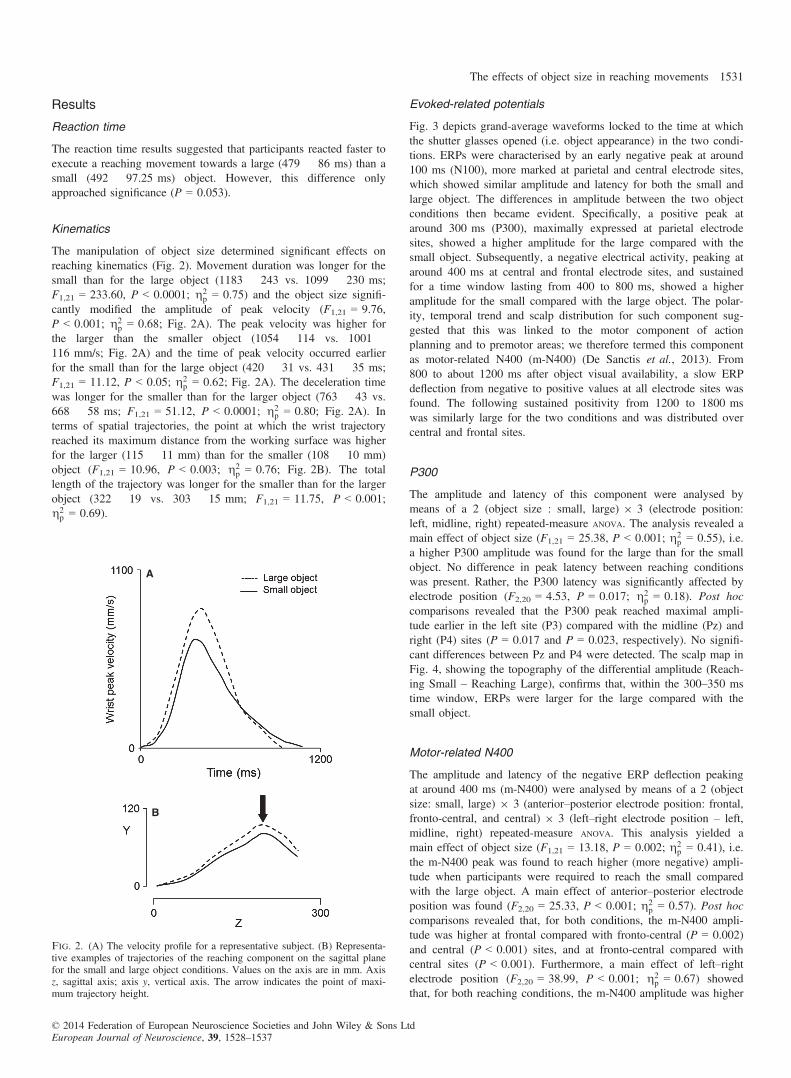
Kinematics
The manipulation of object size determined significant effects onreaching kinematics (Fig. 2). Movement duration was longer for thesmall than for the large object (1183 � 243 vs. 1099 � 230 ms;F1,21 = 233.60, P < 0.0001; g2
p = 0.75) and the object size signifi-cantly modified the amplitude of peak velocity (F1,21 = 9.76,P < 0.001; g2
p = 0.68; Fig. 2A). The peak velocity was higher forthe larger than the smaller object (1054 � 114 vs. 1001 �116 mm/s; Fig. 2A) and the time of peak velocity occurred earlierfor the small than for the large object (420 � 31 vs. 431 � 35 ms;F1,21 = 11.12, P < 0.05; g2
p = 0.62; Fig. 2A). The deceleration timewas longer for the smaller than for the larger object (763 � 43 vs.668 � 58 ms; F1,21 = 51.12, P < 0.0001; g2
p = 0.80; Fig. 2A). Interms of spatial trajectories, the point at which the wrist trajectoryreached its maximum distance from the working surface was higherfor the larger (115 � 11 mm) than for the smaller (108 � 10 mm)object (F1,21 = 10.96, P < 0.003; g2
p = 0.76; Fig. 2B). The totallength of the trajectory was longer for the smaller than for the largerobject (322 � 19 vs. 303 � 15 mm; F1,21 = 11.75, P < 0.001;g2p = 0.69).
Evoked-related potentials
Fig. 3 depicts grand-average waveforms locked to the time at whichthe shutter glasses opened (i.e. object appearance) in the two condi-tions. ERPs were characterised by an early negative peak at around100 ms (N100), more marked at parietal and central electrode sites,which showed similar amplitude and latency for both the small andlarge object. The differences in amplitude between the two objectconditions then became evident. Specifically, a positive peak ataround 300 ms (P300), maximally expressed at parietal electrodesites, showed a higher amplitude for the large compared with thesmall object. Subsequently, a negative electrical activity, peaking ataround 400 ms at central and frontal electrode sites, and sustainedfor a time window lasting from 400 to 800 ms, showed a higheramplitude for the small compared with the large object. The polar-ity, temporal trend and scalp distribution for such component sug-gested that this was linked to the motor component of actionplanning and to premotor areas; we therefore termed this componentas motor-related N400 (m-N400) (De Sanctis et al., 2013). From800 to about 1200 ms after object visual availability, a slow ERPdeflection from negative to positive values at all electrode sites wasfound. The following sustained positivity from 1200 to 1800 mswas similarly large for the two conditions and was distributed overcentral and frontal sites.
P300
The amplitude and latency of this component were analysed bymeans of a 2 (object size : small, large) 9 3 (electrode position:left, midline, right) repeated-measure ANOVA. The analysis revealed amain effect of object size (F1,21 = 25.38, P < 0.001; g2
p = 0.55), i.e.a higher P300 amplitude was found for the large than for the smallobject. No difference in peak latency between reaching conditionswas present. Rather, the P300 latency was significantly affected byelectrode position (F2,20 = 4.53, P = 0.017; g2
p = 0.18). Post hoccomparisons revealed that the P300 peak reached maximal ampli-tude earlier in the left site (P3) compared with the midline (Pz) andright (P4) sites (P = 0.017 and P = 0.023, respectively). No signifi-cant differences between Pz and P4 were detected. The scalp map inFig. 4, showing the topography of the differential amplitude (Reach-ing Small – Reaching Large), confirms that, within the 300–350 mstime window, ERPs were larger for the large compared with thesmall object.
Motor-related N400
The amplitude and latency of the negative ERP deflection peakingat around 400 ms (m-N400) were analysed by means of a 2 (objectsize: small, large) 9 3 (anterior–posterior electrode position: frontal,fronto-central, and central) 9 3 (left–right electrode position – left,midline, right) repeated-measure ANOVA. This analysis yielded amain effect of object size (F1,21 = 13.18, P = 0.002; g2
p = 0.41), i.e.the m-N400 peak was found to reach higher (more negative) ampli-tude when participants were required to reach the small comparedwith the large object. A main effect of anterior–posterior electrodeposition was found (F2,20 = 25.33, P < 0.001; g2
p = 0.57). Post hoccomparisons revealed that, for both conditions, the m-N400 ampli-tude was higher at frontal compared with fronto-central (P = 0.002)and central (P < 0.001) sites, and at fronto-central compared withcentral sites (P < 0.001). Furthermore, a main effect of left–rightelectrode position (F2,20 = 38.99, P < 0.001; g2
p = 0.67) showedthat, for both reaching conditions, the m-N400 amplitude was higher
A
B
Fig. 2. (A) The velocity profile for a representative subject. (B) Representa-tive examples of trajectories of the reaching component on the sagittal planefor the small and large object conditions. Values on the axis are in mm. Axisz, sagittal axis; axis y, vertical axis. The arrow indicates the point of maxi-mum trajectory height.
© 2014 Federation of European Neuroscience Societies and John Wiley & Sons LtdEuropean Journal of Neuroscience, 39, 1528–1537
The effects of object size in reaching movements 1531

at midline compared with left (P < 0.001) and right (P < 0.001)sites. The post hoc analysis of the anterior–posterior 9 left–rightelectrode position interaction (F4,18 = 27.67, P < 0.001; g2
p = 0.59)revealed that, for both conditions, at left and right sites the m-N400
amplitude increased progressively from central to frontal areas(P < 0.003), whereas at midline sites it was more widely distributedalong the anterior–posterior direction (only at FCz was the ampli-tude higher compared with Cz, P = 0.004).
Fig. 3. The plot depicts grand-average ERP waveforms locked to the time at which the shutter glasses opened (i.e. object appearance) for Reaching Small andReaching Large conditions.
Reaching Small - Reaching Large
800 – 1000 ms
–2.50 μV 0.00 μV 2.50 μV
200 – 400 400 – 600 600 – 800
1000 – 1200 1200 – 1400 1400 – 1600 1600 – 1800 1800 – 2000 ms
Fig. 4. The scalp maps show the topography of the differential ERP amplitude between Reaching Small and Reaching Large conditions, from 200 to 2000 ms.
© 2014 Federation of European Neuroscience Societies and John Wiley & Sons LtdEuropean Journal of Neuroscience, 39, 1528–1537
1532 V. Tarantino et al.

Neither the object size nor electrode position effect was found onpeak latency. In summary, the m-N400 showed a higher amplitudefor the small than for the large object at all electrode sites considered.Specifically, the maximum peak value was reached at FCz (smallobject : Mean Amplitude = �13.51 � 4.09 lV, Mean Latency =410.15 � 54.85 ms; large object : Mean Amplitude = �11.29� 3.73 lV, Mean Latency = 409.96 � 53.83 ms). The differentialscalp distribution for the m-N400 component, depicted in Fig. 4clearly shows that in this time window the ERPs were higherand more negative for the small object condition at frontal andcentral areas.
400–800 ms
As shown in Fig. 4, a sustained potential was observed from 400 to800 ms at frontal, fronto-central, central, and parietal electrode sites(F3, Fz, F4, FC3, FCz, FC4, C3, Cz, C4, P3, Pz and P4). The meanERP amplitude in this time window was analysed. Similarly tom-N400 results, the 2 (object size) 9 4 (anterior–posterior electrodeposition) 9 3 (left–right electrode position) ANOVA revealed that thepotential reached an overall higher (more negative) mean ERPamplitude for the small compared with the large object in all frontal,fronto-central and central electrode sites (main effect of object size– F1,21 = 10.63, P = 0.004; g2
p = 0.35). A significant main effect ofanterior–posterior electrode position (F2,20 = 42.68, P < 0.001;g2p = 0.68) revealed that, for both reaching conditions, the mean
amplitudes were progressively larger from parietal to frontal areas(all P-values < 0.009), whereas they did not differ between frontaland fronto-central sites. As for the m-N400, a significant maineffect of left–right electrode position (F2,20 = 37.03, P < 0.001;g2p = 0.65) showed that the mean ERP amplitude within the 400–
800 ms time window was maximal at midline compared with both
left and right sites (P-values < 0.001). The post hoc analysis of theanterior–posterior 9 left–right electrode position interaction(F4,18 = 27.67, P < 0.001; g2
p = 0.59) revealed that, for both reach-ing conditions such sustained negativity at right sites was progres-sively larger from parietal to frontal sites, whereas at left sites nodifferences were found between frontal and fronto-central sites, andat midline electrodes no differences were found between frontal,fronto-central and central sites. This result reflects an equal distribu-tion of such a component at Fz, FCz and Cz electrodes. The maxi-mum mean values of this sustained activity were found at FCz(small object : Mean Amplitude = �7.58 � 2.78 lV; large object :Mean Amplitude = �5.85 � 3.57 lV).
Correlations between kinematic and evoked-related potentialmeasures
The mean amplitude and latency of P300 were averaged across theP3, Pz and P4 electrodes; the mean amplitude and latency of m-N400was considered where such a component was maximally expressed(i.e. at FCz). These values were correlated with the reaction time,movement time, and time of peak velocity (Fig. 5A). A positive cor-relation was found between movement time and m-N400 latency forboth the small and the large objects (small object – r = 0.59,P = 0.005; large object – r = 0.45, P = 0.039). Figure 5B illustratesthe individual mean individual latency values of the m-N400 compo-nent and individual movement times for the two conditions.
Discussion
The aim of the present study was to investigate the kinematics andERP activity during reaching movements performed towards either alarge or a small object. Differently from previous studies, we did
A
B
Fig. 5. (A) The timeline of events (Movement Start, Peak Velocity, and Movement End) together with the ERP grand-average waveforms at representativesites (FCz and Pz). (B) The correlation between individual movement time and individual m-N400 latency in Reaching Small and Reaching Large conditions.
© 2014 Federation of European Neuroscience Societies and John Wiley & Sons LtdEuropean Journal of Neuroscience, 39, 1528–1537
The effects of object size in reaching movements 1533

not investigate ERPs evoked by a cue anticipating a specific object’sintrinsic features, but by the target object itself. Such an approachmay allow the examination of how information about an object’sgeometric properties is transformed into specific motor programsmore directly. Overall, the results indicate that object size deter-mines a modulation in the timing and amplitude of specific kine-matic landmarks and ERP components during reaching movements.In particular, the novelty of the present study resides in the fact that(i) the modulation of parietal activity to object size precedes thebeginning of the movement and (ii) fronto-parietal areas aremodulated by object size although this property does not need to beintegrated in the motor act.Before we discuss how our results fit with previous studies, it is
worth clarifying that previous experiments in humans haveemployed a variety of tasks to investigate the behavioral and neuralcorrelates of reaching. These tasks include reach-to-touch (Pellijeffet al., 2006; Levy et al., 2007; Cavina-Pratesi et al., 2010), pointing(Connolly et al., 2000, 2003; DeSouza et al., 2000; Astafiev et al.,2003; Fernandez-Ruiz et al., 2007; Hagler et al., 2007), and joystickmanipulation (Grefkes et al., 2004). These tasks differ widely in theextent of arm movement, purpose and cortical recruitment (Culham& Valyear, 2006; Culham et al., 2006; Filimon et al., 2009). Fur-thermore, these tasks also differ in terms of initial hand posture, afactor that has the ability to influence the unfolding of reachingmovements (Kritikos et al., 1998). Therefore, we cannot excludethat adopting a different task might have brought differentoutcomes.Consistent with previous reports, arm trajectories changed their
shape when targets of different size were used, and this effect waschiefly due to modifications in the deceleration phase (MacKenzieet al., 1987; Gentilucci et al., 1991; Castiello, 2001). Fitt’s law(1954) was found to apply, given that movement time increased asa function of task difficulty. The movement time was longer andmaximum velocity was lower for smaller objects requiring a greaterlevel of accuracy. Altogether these findings indicate that the size ofthe object had the ability to selectively influence the execution of areaching movement. This is an important aspect of the present studybecause, in order to ascertain the effects that such differential pro-cessing might have on ERPs, it is necessary to demonstrate that theparticipants’ movement showed differential kinematic signaturesdepending on reach conditions.For an efficient reaching movement the brain must integrate infor-
mation about the selected arm with information about the selectedtarget. The general consensus is that this integrative action is accom-plished through interactions between posterior parietal and premotorareas of the brain in both monkeys (Kalaska et al., 1997; Wiseet al., 1997; Caminiti et al., 1998) and humans (Grafton et al.,1996; Thoenissen et al., 2002; Astafiev et al., 2003; Connolly et al.,2003; Medendorp et al., 2003, 2005; Culham et al., 2006; Beurzeet al., 2007; Gallivan et al., 2011; Bozzacchi et al., 2012; Konenet al., 2013). Our EEG recordings corroborate these findings, reveal-ing that the planning and execution of reaching movements evolvesacross several cortical areas within the fronto-parietal networkfollowing a specific timing (Weinrich et al., 1984; Kalaska &Crammond, 1992; Glover et al., 2012).Differences in amplitude between the small and the large object
conditions become evident over parietal sites at around 300 ms(P300), during the planning phase of the movement. This activityreflects the involvement of parietal areas in the planning of reachingmovements (Culham et al., 2006; Beurze et al., 2007, 2009; Galli-van et al., 2011; Konen et al., 2013). These areas include part ofeither the classic parietal reach region identified in the macaque
(Andersen & Buneo, 2002; Bhattacharyya et al., 2009) and areaV6A (e.g. Fattori et al., 2005; Bosco et al., 2010) or their putativehuman homologue, the superior parieto-occipital cortex region (Con-nolly et al., 2003; Gallivan et al., 2009; Cavina-Pratesi et al.,2010).The P300 peak amplitude was higher for the large than for the
small object condition. This finding might indicate the greateramount of visuo-spatial information to be extracted from largerobjects, reflecting parietal activity that, in both humans and maca-ques, appears to serve a variety of visuo-motor and attention-relatedfunctions. For instance, it might be concerned with the encoding ofthree-dimensional visual features of objects for action (Gallivanet al., 2011; Fattori et al., 2012) and the integration of both targetand effector-specific information for movements (Beurze et al.,2009). In this respect, attention research indicates that the focus ofattention can be modulated depending on the size of the area overwhich focal attention is allocated (Castiello & Umilt�a, 1990, 1992).Furthermore, this finding is also in line with recent functional mag-netic resonance imaging research showing that parietal areas, suchas the anterior intra-parietal sulcus, are involved in integrating infor-mation about real three-dimensional objects, such as the object sizeand the grasp-relevant dimension (Monaco et al., 2013). In addition,it agrees with neurophysiological findings showing that neurons inarea V6A are influenced by spatial attention. The general suggestionis that this area, primarily involved in visuo-motor transformationfor reaching, may form a neural basis for coupling attention to thepreparation of reaching movements (Galletti et al., 2010). Overall,this particular finding might provide additional evidence for the inte-gration of visuo-motor and attention-related processes during move-ment planning (Baldauf & Deubel, 2010; Gallivan et al., 2011;Konen et al., 2013).Overall, the results concerned with parietal activity fit with neuro-
physiological findings suggesting that areas of the dorso-medialpathway are sensitive to intrinsic features of target objects such asshape (Fattori et al., 2010, 2012).In terms of frontal regions, we found a negative electrical activity,
peaking at around 400 ms following object appearance (m-N400),that was evident over central and frontal electrode sites. The spatio-temporal characteristics of the m-N400 might be assimilated to anindex of motor planning and it is strongly influenced by motor vari-ables. The polarity, timing and scalp distribution suggest that such acomponent reflects motor planning and that it is linked to premotoractivity (Shibasaki & Hallett, 2006).In the frontal cortex of monkeys, premotor dorsal and premotor
ventral neurons are shown to be involved in different aspects ofreaching movements (Hoshi & Tanji, 2000, 2002, 2004a,b,c, 2006;Calton et al., 2002; Hoshi et al., 2005). Similarly, in humans, elec-trophysiological (Naranjo et al., 2007), neuroimaging (Beurzeet al., 2007; Grol et al., 2007; Glover et al., 2012) and neuropsy-chological (Heilman & Gonzalez Rothi, 1993) evidence indicatesthat premotor cortices are central to the process of reach planning.Our findings are in agreement with these views, by showing thatpremotor cortices are activated during reaching preparation. Impor-tantly, the m-N400 peak had a later onset and a wider fronto-cen-tral distribution for the small than for the large object. This resultdemonstrates that premotor activity during reach planning is con-cerned not only with reach direction or the integration of targetlocation with information about the selected effector (Kertzmanet al., 1997; Hoshi & Tanji, 2000, 2002, 2004a,b,c, 2006; Batista& Andersen, 2001; Buneo et al., 2002; Calton et al., 2002;Medendorp et al., 2005), but also with the intrinsic features ofobjects.
© 2014 Federation of European Neuroscience Societies and John Wiley & Sons LtdEuropean Journal of Neuroscience, 39, 1528–1537
1534 V. Tarantino et al.

A point worth noting is that the difference in amplitude betweenthe small and the large objects remained significant up to 800 ms.This suggests that the size-dependent modulation of premotor activ-ity noticed during reach planning spreads into the execution phaseof the action, implying that, before the action can begin, the motorprogramme has to be fully formulated and that kinematic planningmight be fully fledged during the online control phase of the move-ment. In this respect, our behavioral results might support this view.Whereas there was a (non-significant) tendency for reaction time tobe longer for the small than the large object, the time to peak veloc-ity occurred significantly earlier for the small than for the largeobject. This indicates that planning continues to be influential and isoptimised early in the movement. Such a gradual crossover betweenplanning and control systems has the benefit of allowing for smoothrather than jerky corrections (Wolpert & Ghahramani, 2000; Glover,2004). The differences between the small and the large object mayreflect the need for additional sensorimotor control mechanisms forthe more accurate condition (i.e. small object). In this respect, thepresent and previous psychophysical studies demonstrated that, asobject size decreased, subjects had longer movement times, slowerspeeds, and more asymmetrical hand-speed profiles (Gentilucciet al., 1991; Berthier et al., 1996).Altogether, the above-mentioned findings suggest that both prepa-
ratory and execution activity along the fronto-parietal circuit under-lying reaching are modulated by object size. This result can beexplained in terms of the intimate relationship between reaching andgrasping components during prehension movements (Jeannerod,1984). It is known that grasping in humans and macaques activatesparietal and premotor areas that overlap with reach-related activa-tions (Tann�e-Gari�epy et al., 2002; Culham et al., 2003; Raos et al.,2004; Castiello, 2005; Davare et al., 2006; Fattori et al., 2009,2010, 2012). Therefore, it might be conceivable that the neural net-work that controls proximal movements in reaching-to-grasp hasinformation about object size, given that the two components shouldact in concert in order to determine the timing of hand preshapingduring reaching. But why should the proximal neural channel besensitive to object size during reaching alone, given that the distalprogram remains unmodified for small and large objects? In ouropinion, it would be difficult to conceive how the reaching channelcould act without extracting information regarding object size. Fromthe mere fact that it occupies space, an object must have a size, andto locate it necessarily entails information about its dimension. Inthis respect, our findings might provide a novel demonstration thatthe reaching and grasping phases are represented by overlappingparieto-frontal circuits, suggesting a lack of strict functional segrega-tion between parieto-frontal circuits for grasping and reaching inmonkeys (e.g. Fattori et al., 2010) and humans (Grol et al., 2007;Filimon et al., 2009; Filimon et al. 2010).As a final issue, we found that, for both the large and the small
objects, the individual mean latency for the premotor m-N400 com-ponent significantly correlated with the individual mean for move-ment time. According to behavioral evidence, reaching movementsare characterised by a ballistic and a feedback-based phase. The bal-listic phase is a product of a feedforward system that defines the ini-tial state of the limb and the goal. The feedback phase is used at theend of this movement to achieve an accurate contact with the object.An alternative possibility is that the second phase is controlled, as isthe first, by a feedforward system that takes into account the objectsize and accordingly sets its duration. This might indicate that anestimate of movement time, possibly performed at the level of pre-motor areas, might serve to plan the amount of online controlrequired during the final part of the movement.
In summary, the present study demonstrates that the use of con-verging techniques with different characteristics might allow betterunderstanding of how the human brain controls the reaching func-tion. In particular, it presents the timing of activation of the corticalregions engaged for the planning and execution of a human reach,starting from the early coding of the intrinsic features of the objectto the motor plan that leads to the actualisation of the movements.Although these findings confirm previous evidence concerned withreach planning and execution in general, they add to previous litera-ture demonstrating that, in humans, the neural network underlyingreaching movements is modulated by object size.
Abbreviations
EEG, electroencephalography; ERP, evoked-related potential; m-N400,motor-related N400
References
AEEGS (1991) American Electroencephalographic Society guidelines for stan-dard electrode position nomenclature. J. Clin. Neurophysiol., 8, 200–202.
Andersen, R.A. & Buneo, C.A. (2002) Intentional maps in posterior parietalcortex. Annu. Rev. Neurosci., 25, 189–220.
Astafiev, S.V., Shulman, G.L., Stanley, C.M., Snyder, A.Z., Van Essen, D.C. &Corbetta, M. (2003) Functional organization of human intraparietal and frontalcortex for attending, looking, and pointing. J. Neurosci., 23, 4689–4699.
Astafiev, S.V., Stanley, C.M., Shulman, G.L. & Corbetta, M. (2004) Extras-triate body area in human occipital cortex responds to the performance ofmotor actions. Nat. Neurosci., 7, 542–548.
Baldauf, D. & Deubel, H. (2010) Attentional landscapes in reaching andgrasping. Vision Res., 50, 999–1013.
Batista, A.P. & Andersen, R.A. (2001) The parietal reach region codes thenext planned movement in a sequential reach task. J. Neurophysiol., 85,539–544.
Battaglia-Mayer, A., Ferraina, S., Mitsuda, T., Marconi, B., Genovesio, A.,Onorati, P., Lacquaniti, F. & Caminiti, R. (2000) Early coding of reachingin the parieto-occipital cortex. J. Neurophysiol., 83, 2374–2391.
Battaglia-Mayer, A., Caminiti, R., Lacquaniti, F. & Zago, M. (2003) Multiplelevels of representation of reaching in the parieto-frontal network. Cereb.Cortex, 13, 1009–1022.
Berndt, I., Franz, V.H., Bulthoff, H.H. & Wascher, E. (2002) Effects ofpointing direction and direction predictability on event-related lateraliza-tions of the EEG. Hum. Movement Sci., 21, 387–410.
Berthier, N.E., Clifton, R.K., Gullapalli, V., McCall, D.D. & Robin, D.J.(1996) Visual information and object size in the control of reaching.J. Motor Behav., 28, 187–197.
Beurze, S.M., de Lange, F.P., Toni, I. & Medendorp, W.P. (2007) Integrationof target and effector information in the human brain during reach plan-ning. J. Neurophysiol., 97, 88–199.
Beurze, S.M., de Lange, F.P., Toni, I. & Medendorp, W.P. (2009) Spatialand effector processing in the human parietofrontal network for reachesand saccades. J. Neurophysiol., 101, 3053–3062.
Bhattacharyya, R., Musallam, S. & Andersen, R.A. (2009) Parietal reachregion encodes reach depth using retinal disparity and vergence angle sig-nals. J. Neurophysiol., 102, 805–816.
Bosco, A., Breveglieri, R., Chinellato, E., Galletti, C. & Fattori, P. (2010)Reaching activity in the medial posterior parietal cortex of monkeys ismodulated by visual feedback. J. Neurosci., 30, 14773–14785.
Bozzacchi, C., Giusti, M.A., Pitzalis, S., Spinelli, D. & Di Russo, F. (2012)Awareness affects motor planning for goal-oriented actions. Biol. Psychol.,89, 503–514.
Buneo, C.A., Jarvis, M.R., Batista, A.P. & Andersen, R.A. (2002) Directvisuomotor transformations for reaching. Nature, 416, 632–636.
Calton, J.L., Dickinson, A.R. & Snyder, L.H. (2002) Non-spatial, motor-spe-cific activation in posterior parietal cortex. Nat. Neurosci., 5, 580–588.
Caminiti, R., Ferraina, S. & Mayer, A.B. (1998) Visuomotor transformations:early cortical mechanisms of reaching. Curr. Opin. Neurobiol., 8, 753–761.
Castiello, U. (2001) The effects of abrupt onset of 2-D and 3-D distractorson prehension movements. Percept. Psychophys., 63, 1014–1025.
Castiello, U. (2005) The neuroscience of grasping. Nat. Rev. Neurosci., 6,726–736.
© 2014 Federation of European Neuroscience Societies and John Wiley & Sons LtdEuropean Journal of Neuroscience, 39, 1528–1537
The effects of object size in reaching movements 1535

Castiello, U. & Umilt�a, C. (1990) Size of the attentional focus and efficiencyof processing. Acta Psychol. (Amst.), 73, 195–209.
Castiello, U. & Umilt�a, C. (1992) Splitting focal attention. J. Exp. Psychol.Human., 18, 837–848.
Cavina-Pratesi, C., Monaco, S., Fattori, P., Galletti, C., McAdam, T.D.,Quinlan, D.J., Goodale, M.A. & Culham, J.C. (2010) fMRI reveals theneural substrates of arm transport and grip formation in reach-to-graspactions in humans. J. Neurosci., 30, 10306–10323.
Connolly, J.D., Goodale, M.A., DeSouza, J.F., Menon, R.S. & Vilis, T.(2000) A comparison of frontoparietal fMRI activation during anti-sac-cades and anti-pointing. J. Neurophysiol., 84, 1645–1655.
Connolly, J.D., Andersen, R.A. & Goodale, M.A. (2003) FMRI evidencefor a parietal reach region in the human brain. Exp. Brain Res., 153,140–145.
Culham, J.C. & Valyear, K.F. (2006) Human parietal cortex in action. Curr.Opin. Neurol., 16, 205–212.
Culham, J.C., Danckert, S.L., De Souza, J.F., Gati, J.S., Menon, R.S. &Goodale, M.A. (2003) Visually guided grasping produces fMRI activationin dorsal but not ventral stream brain areas. Exp. Brain Res., 153, 180–189.
Culham, J.C., Cavina-Pratesi, C. & Singhal, A. (2006) The role of parietalcortex in visuomotor control: what have we learned from neuroimaging?Neuropsychologia, 44, 2668–2684.
Davare, M., Andres, M., Cosnard, G., Thonnard, J.L. & Olivier, E. (2006)Dissociating the role of ventral and dorsal premotor cortex in precisiongrasping. J. Neurosci., 26, 2260–2268.
De Sanctis, T., Tarantino, V., Straulino, E., Begliomini, C. & Castiello, U.(2013) Co-registering kinematics and evoked related potentials duringvisually guided reach-to-grasp movements. PLoS ONE, 8, e65508.
Decety, J., Kawashima, R., Guly�as, B. & Roland, P.E. (1992) Preparation forreaching: a PET study of the participating structures in the human brain.NeuroReport, 3, 761–764.
Delorme, A. & Makeig, S. (2004) EEGLAB: an open source toolbox foranalysis of single-trial EEG dynamics. J. Neurosci. Meth., 134, 9–21.
DeSouza, J.F., Dukelow, S.P., Gati, J.S., Menon, R.S., Andersen, R.A. &Vilis, T. (2000) Eye position signal modulates a human parietal point-ing region during memory guided movements. J. Neurosci., 20, 5835–5840.
Fattori, P., Gamberini, M., Kutz, D.F. & Galletti, C. (2001) “Arm-reaching”neurons in the parietal area V6A of the macaque monkey. Eur. J. Neuro-sci., 13, 2309–2313.
Fattori, P., Kutz, D.F., Breveglieri, R., Marzocchi, N. & Galletti, C. (2005)Spatial tuning of reaching activity in the medial parieto-occipital cortex(area V6A) of macaque monkey. Eur. J. Neurosci., 22, 956–972.
Fattori, P., Breveglieri, R., Marzocchi, N., Filippini, D., Bosco, A. & Galletti,C. (2009) Hand orientation during reach-to-grasp movements modulatesneuronal activity in the medial posterior parietal area V6A. J. Neurosci.,29, 1928–1936.
Fattori, P., Raos, V., Breveglieri, R., Bosco, A., Marzocchi, N. & Galletti, C.(2010) The dorsomedial pathway is not just for reaching: grasping neuronsin the medial parieto-occipital cortex of the macaque monkey. J. Neuro-sci., 30, 342–349.
Fattori, P., Breveglieri, R., Raos, V., Bosco, A. & Galletti, C. (2012) Visionfor action in the macaque medial posterior parietal cortex. J. Neurosci.,32, 3221–3234.
Fernandez-Ruiz, J., Goltz, H.C., DeSouza, J.F., Vilis, T. & Crawford, J.D.(2007) Human parietal “reach region” primarily encodes intrinsic visualdirection, not extrinsic movement direction, in a visual motor dissociationtask. Cereb. Cortex, 17, 2283–2292.
Filimon, F., Nelson, J.D., Huang, R.S. & Sereno, M.I. (2009) Multipleparietal reach regions in humans: cortical representations for visual andproprioceptive feedback during on-line reaching. J. Neurosci., 29, 2961–2971.
Filimon, F. (2009) Human cortical control of hand movements: Parietofrontalnetworks for reaching, grasping, and pointing. Neuroscientist, 16, 388–340.
Fitt, P.M. (1954) The information capacity of the human motor system incontrolling the amplitude of movement. J. Exp. Psychol. Human., 47,381–391.
Gail, A. & Andersen, R.A. (2006) Neural dynamics in monkey parietal reachregion reflect context-specific sensorimotor transformations. J. Neurosci.,26, 9376–9384.
Galletti, C., Fattori, P., Battaglini, P.P., Shipp, S. & Zeki, S. (1996) Func-tional demarcation of a border between areas V6 and V6A in the superiorparietal gyrus of the macaque monkey. Eur. J. Neurosci., 8, 30–52.
Galletti, C., Fattori, P., Kutz, D.F. & Battaglini, P.P. (1997) Arm movement-related neurons in the visual area V6A of the macaque superior parietallobule. Eur. J. Neurosci., 9, 410–413.
Galletti, C., Breveglieri, R., Lappe, M., Bosco, A., Ciavarro, M. & Fattori, P.(2010) Covert shift of attention modulates the ongoing neural activity in areaching area of the macaque dorsomedial visual stream. PLoS ONE, 5,e15078.
Gallivan, J.P., Cavina-Pratesi, C. & Culham, J. (2009) Is that within reach?fMRI reveals that the human superior parieto-occipital cortex encodesobjects reachable by the hand. J. Neurosci., 29, 4381–4391.
Gallivan, J.P., McLean, D.A., Valyear, K.F., Pettypiece, C.E. & Culham,J.C. (2011) Decoding action intentions from preparatory brain activity inhuman parieto-frontal networks. J. Neurosci., 31, 9599–9610.
Gentilucci, M., Castiello, U., Corradini, M.L., Scarpa, M. & Umilt�a, C.(1991) Influence of different types of grasping on the transport componentof prehension movements. Neuropsychologia, 29, 361–378.
Glover, S. (2004) Separate visual representations in the planning and controlof action. Behav. Brain Sci., 27, 3–24.
Glover, S., Wall, M.B. & Smith, A.T. (2012) Distinct cortical networks sup-port the planning and online control of reaching-to-grasp in humans. Eur.J. Neurosci., 35, 909–915.
Grafton, S.T., Arbib, M.A., Fadiga, L. & Rizzolatti, G. (1996) Localizationof grasp representations in humans by positron emission tomography. Exp.Brain Res., 112, 103–111.
Grefkes, C. & Fink, G.R. (2005) The functional organization of the intrapari-etal sulcus in humans and monkeys. J. Anat., 207, 3–17.
Grefkes, C., Ritzl, A., Zilles, K. & Fink, G.R. (2004) Human medial intrapa-rietal cortex subserves visuomotor coordinate transformation. NeuroImage,23, 1494–1506.
Grol, M.J., Majdandzic, J., Stephan, K.E., Verhagen, L., Dijkerman, H.C.,Bekkering, H., Verstraten, F.A.J. & Toni, I. (2007) Parieto-frontal connec-tivity during visually guided grasping. J. Neurosci., 27, 11877–11887.
Hagler, D.J. Jr., Riecke, L. & Sereno, M.I. (2007) Parietal and superior fron-tal visuospatial maps activated by pointing and saccades. NeuroImage, 35,1562–1577.
Heilman, K.M. & Gonzalez Rothi, L.J. (1993) Apraxia. In Heilman, K.M. &Valenstein, E. (Eds), Clinical Neuropsychology. Oxford University Press,New York, NY, pp. 141–150.
Hoshi, E. & Tanji, J. (2000) Integration of target and body-part informationin the premotor cortex when planning action. Nature, 408, 466–470.
Hoshi, E. & Tanji, J. (2002) Contrasting neuronal activity in the dorsal andventral premotor areas during preparation to reach. J. Neurophysiol., 87,1123–1128.
Hoshi, E. & Tanji, J. (2004a) Functional specialization in dorsal and ventralpremotor areas. Prog. Brain Res., 143, 507–511.
Hoshi, E. & Tanji, J. (2004b) Differential roles of neuronal activity inthe supplementary and presupplementary motor areas: from informationretrieval to motor planning and execution. J. Neurophysiol., 92,3482–3499.
Hoshi, E. & Tanji, J. (2004c) Area-selective neuronal activity in the dorsolat-eral prefrontal cortex for information retrieval and action planning. J. Neuro-physiol., 91, 2707–2722.
Hoshi, E. & Tanji, J. (2006) Differential involvement of neurons in the dor-sal and ventral premotor cortex during processing of visual signals foraction planning. J. Neurophysiol., 95, 3596–3616.
Hoshi, E., Sawamura, H. & Tanji, J. (2005) Neurons in the rostral cingulatemotor area monitor multiple phases of visuomotor behavior with modestparametric selectivity. J. Neurophysiol., 94, 640–656.
Jeannerod, M. (1984) The timing of natural prehension movements. J. MotorBehav., 16, 235–254.
Jung, T.P., Humphries, C., Lee, T.W., McKeown, M.J., Iragui, V. & Sejnow-ski, T.J. (2000) Removing electroencephalographic artifacts by blindsource separation. Psychophysiology, 37, 163–178.
Kalaska, J.F. & Crammond, D.J. (1992) Cerebral cortical mechanisms ofreaching movements. Science, 255, 1517–1523.
Kalaska, J.F., Scott, S.H., Cisek, P. & Sergio, L.E. (1997) Cortical control ofreaching movements. Curr. Opin. Neurobiol., 7, 849–859.
Kawashima, R., Satoh, K., Itoh, H., Ono, S., Furumoto, S., Gotoh, R.,Koyama, M., Yoshioka, S., Takahashi, T., Takahashi, K., Yanagisawa, T.& Fukuda, H. (1996) Functional anatomy of GO/NO-GO discrimina-tion and response selection – a PET study in man. Brain Res., 728,79–89.
Kertzman, C., Schwarz, U., Zeffiro, T.A. & Hallett, M. (1997) The role ofposterior parietal cortex in visually guided reaching movements in humans.Exp. Brain Res., 114, 170–183.
© 2014 Federation of European Neuroscience Societies and John Wiley & Sons LtdEuropean Journal of Neuroscience, 39, 1528–1537
1536 V. Tarantino et al.

Konen, C.S., Mruczek, R.E.B., Montoya, J.L. & Kastner, S. (2013)Functional organization of human posterior parietal cortex: grasping- andreaching-related activations relative to topographically organized cortex.J. Neurophysiol., 109, 2897–2908.
Kritikos, A., Jackson, G.M. & Jackson, S.R. (1998) The influence of initialhand posture on the expression of prehension parameters. Exp. Brain Res.,119, 9–16.
Levy, I., Schluppeck, D., Heeger, D.J. & Glimcher, P.W. (2007) Specificityof human cortical areas for reaches and saccades. J. Neurosci., 27, 4687–4696.
MacKenzie, C.L., Marteniuk, R.G., Dugas, C., Liske, D. & Eickmeier, B.(1987) 3-Dimensional movement trajectories in fitts task – implications forcontrol. Q. J. Exp. Psychol. A, 39, 629–647.
Mascaro, M., Battaglia-Mayer, A., Nasi, L., Amit, D.J. & Caminiti, R. (2003)The eye and the hand: neural mechanisms and network models for oculo-manual coordination in parietal cortex. Cereb. Cortex, 13, 1276–1286.
McDowell, K., Jeka, J.J., Sch€oner, G. & Hatfield, B.D. (2002)Behavioral and electrocortical evidence of an interaction between probabil-ity and task metrics in movement preparation. Exp. Brain Res., 144,303–313.
Medendorp, W.P., Goltz, H.C., Vilis, T. & Crawford, J.D. (2003) Gaze-cen-tered updating of visual space in human parietal cortex. J. Neurosci., 23,6209–6214.
Medendorp, W.P., Goltz, H.C., Crawford, J.D. & Vilis, T. (2005) Integrationof target and effector information in human posterior parietal cortex forthe planning of action. J. Neurophysiol., 93, 954–962.
Monaco, S., Cheng, Y., Medendorp, P.W., Crawford, J.D., Fiehler, K. &Henriques, Y.P.D. (2013) Functional magnetic resonance imagingadaptation reveals the cortical networks for processing grasp-relevantobject properties. Cereb. Cortex, doi:10.1093/cercor/bht006. [Epub aheadof print].
Naranjo, J.R., Brovelli, A., Longo, R., Budai, R., Kristeva, R. & Battaglini,P.P. (2007) EEG dynamics of the frontoparietal network during reachingpreparation in humans. NeuroImage, 34, 1673–1682.
Oldfield, R.C. (1971) The assessment and analysis of handedness: the Edin-burgh inventory. Neuropsychologia, 9, 97–113.
Pellijeff, A., Bonilha, L., Morgan, P.S., McKenzie, K. & Jackson, S.R.(2006) Parietal updating of limb posture: an event-related fMRI study.Neuropsychologia, 44, 2685–2690.
Prado, J., Clavagnier, S., Otzenberger, H., Scheiber, C., Kennedy, H. & Pere-nin, M.T. (2005) Two cortical systems for reaching in central and periph-eral vision. Neuron, 48, 849–858.
Raos, V., Umilt�a, M.A., Gallese, V. & Fogassi, L. (2004) Functional proper-ties of grasping-related neurons in the dorsal premotor area F2 of themacaque monkey. J. Neurophysiol., 92, 1990–2002.
Shadmehr, R. & Wise, S.P. (2005) Computational Neurobiology of Reachingand Pointing: A Foundation for Motor Learning. MIT Press, Cambridge,MA.
Shibasaki, H. & Hallett, M. (2006) What is the Bereitschaftspotential? Clin.Neurophysiol., 17, 2341–2356.
Tann�e-Gari�epy, J., Rouiller, E.M. & Boussaoud, D. (2002) Parietal inputs todorsal versus ventral premotor areas in the macaque monkey: evidence forlargely segregated visuomotor pathways. Exp. Brain Res., 145, 91–103.
Thoenissen, D., Zilles, K. & Toni, I. (2002) Differential involvement of pari-etal and precentral regions in movement preparation and motor intention.J. Neurosci., 22, 9024–9034.
Weinrich, M., Wise, S.P. & Mauritz, K.H. (1984) A neurophysiologicalstudy of the premotor cortex in the rhesus monkey. Brain, 107, 385–414.
Wise, S.P., Boussaoud, D., Johnson, P.B. & Caminiti, R. (1997) Premotorand parietal cortex: corticocortical connectivity and combinatorial compu-tations. Annu. Rev. Neurosci., 20, 25–42.
Wolpert, D.M. & Ghahramani, Z. (2000) Computational principles of move-ment neuroscience. Nat. Neurosci., 3, 1212–1217.
World Medical Associations General Assembly (2008) World Medical Asso-ciation Declaration of Helsinki. Ethical principles for medical researchinvolving human subjects 2000. Adopted by the 18th WMA GeneralAssembly, Helsinki (Finland) June 1964 and 59th WMA General Assem-bly, Seoul (Republic of Korea), October 2008.
© 2014 Federation of European Neuroscience Societies and John Wiley & Sons LtdEuropean Journal of Neuroscience, 39, 1528–1537
The effects of object size in reaching movements 1537