Embed Size (px)
Citation preview
Rom J Morphol Embryol 2014, 55(2 Suppl):675–681
ISSN (print) 1220–0522 ISSN (on-line) 2066–8279
CCAASSEE RREEPPOORRTT
Fronto-parietal pial extension of a basal cell carcinoma of the scalp – case report
IONICA PIRICI1), OTILIA MĂRGĂRITESCU2), DANIELA CERNEA3), LOREDANA ELENA STOICA4), CĂLIN-GABRIEL ŞARLĂ5), DANIEL PIRICI6)
1)Research Center for Microscopic Morphology and Immunology, Department of Morphology, University of Medicine and Pharmacy of Craiova, Romania
2)Department of Neurosurgery, University of Medicine and Pharmacy of Craiova, Romania 3)Department of Anesthesiology, University of Medicine and Pharmacy of Craiova, Romania 4)Department of Dermatology, University of Medicine and Pharmacy of Craiova, Romania 5)Department of Physical Education and Sports, “Vasile Goldiş” Western University, Arad, Romania 6)Department of Research Methodology, University of Medicine and Pharmacy of Craiova, Romania
Abstract Basal cell carcinoma is the most common malignant tumor of the skin, and it develops most frequently on the head and neck regions. Although most of these tumors are slow growing and with a limited evolution, the existence of some aggressive variants accompanied by a complete neglect from the patient may occasionally lead to invasion of the face and organs of the head and neck. Even though, intracranial invasion of basal cell carcinoma of the scalp is a rare presentation. We describe here the case of a woman who developed an aggressive and neglected morpheiform basal cell carcinoma (ulcus terebrans), which showed a complete invasion through the skull, but with an apparent self-limitation to the pia mater.
Keywords: basal cell carcinoma, ulcus terebrans, intracranial extension, osteolytic tumor, immunohistochemical pattern.
Introduction
Basal Cell Carcinoma (BCC) is the most common malignant tumor of the skin, and also the most common type of cancer [1]. It is derived from the basal cell layer of the epidermis or basaloid cells of the skin appendages, and is almost always found in the areas of sun exposed hairy skin. Typically, these are slow growing tumors that metastasize exceptionally, but on occasion they can become destructive and spread locally by peripheral extension [2]. Some tumor subtypes, as the morpheaform variant, may increase in size or behave like “ulcus terebrans” destroying the skin and the deeper adjacent tissues. Most of these aggressive forms reside on the head and neck regions, implying both a drastic limitation of treatment options, and esthetic and functional disabilities [3, 4]. Periocular BCC for example may become rapidly inoperable or may require eyeball exenteration. Cases with intracranial invasion towards the brain from BCC of the scalp are rare in the literature, most of them being the result of neglect, and large infiltrative tumors eventually lead to the death of these patients [5–10].
We report herein the case of a woman who presented with a neglected large ulcerated basal cell carcinoma of the scalp, with intracranial extension and intensive osteolysis. The patient died after a septic shock, and the tumor was characterized histopathologically.
Case presentation
The 55 years-old female patient was admitted in the Intensive Care Unit of the Emergency County Hospital
of Craiova, Romania, with a suppurated left fronto-parietal wound and motor deficit in the right sight of the body. The patient was conscious but uncooperative at the moment of presentation, and her condition deteriorated rapidly in the same day towards a comatose state (grade 7 on the Glasgow scale). From the affirmations of her relatives, she had been diagnosed three years ago with a skin tumor of the forehead and she had refused any treatment during this time, with the ulcerated tumor increasing in size especially in the last month.
Clinical data, imaging and evolution
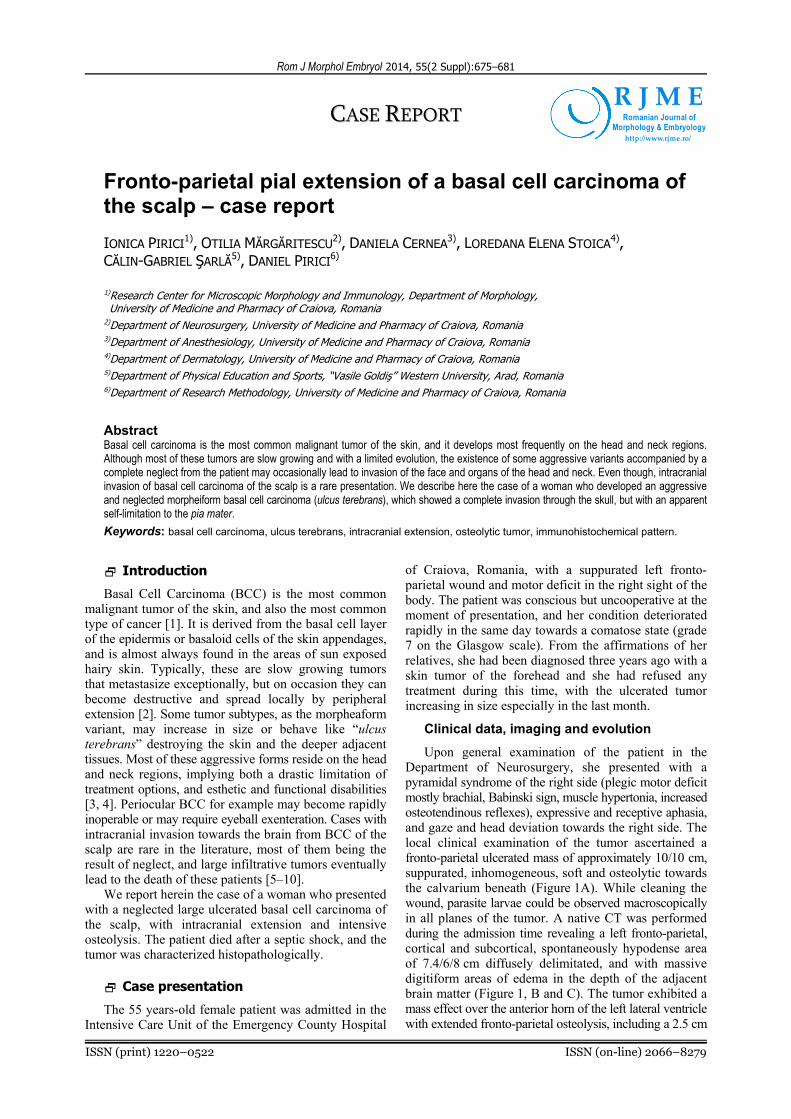
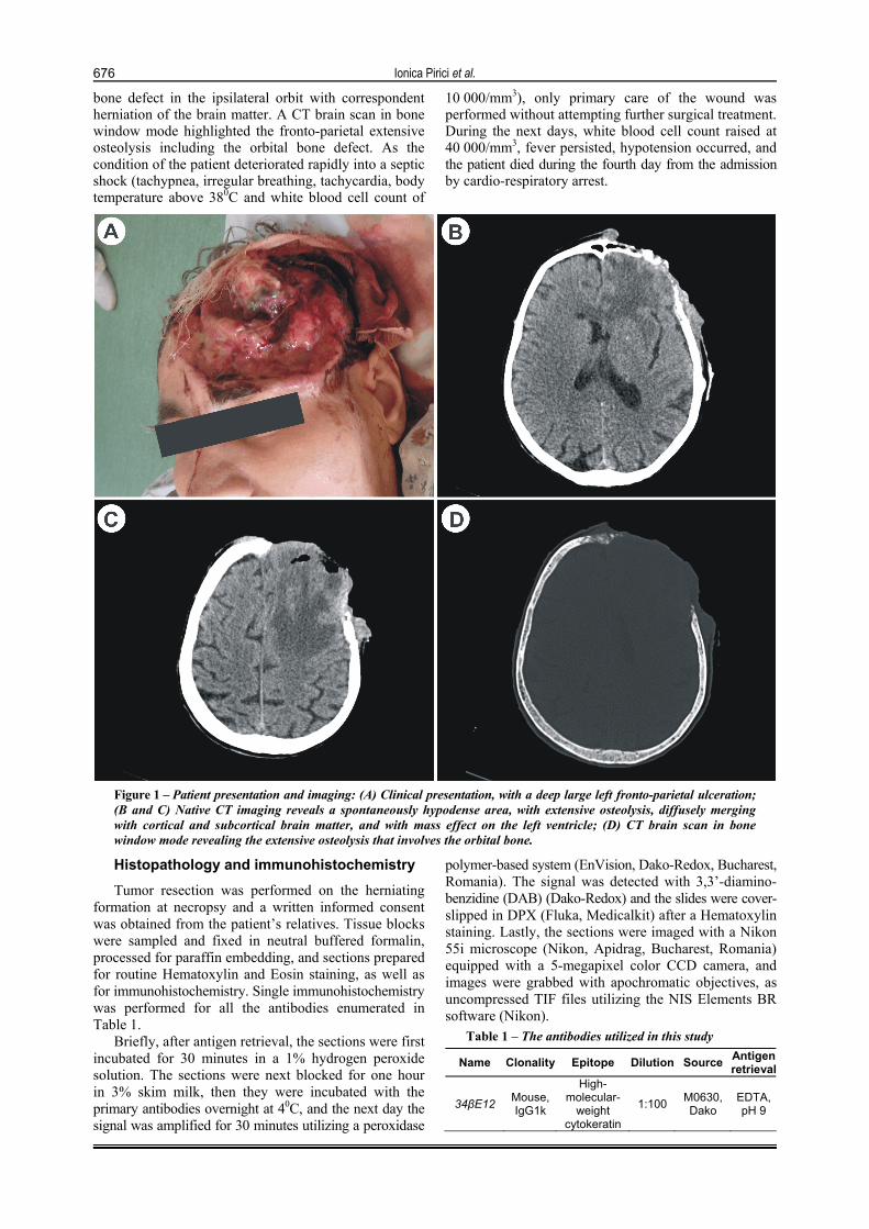
Upon general examination of the patient in the Department of Neurosurgery, she presented with a pyramidal syndrome of the right side (plegic motor deficit mostly brachial, Babinski sign, muscle hypertonia, increased osteotendinous reflexes), expressive and receptive aphasia, and gaze and head deviation towards the right side. The local clinical examination of the tumor ascertained a fronto-parietal ulcerated mass of approximately 10/10 cm, suppurated, inhomogeneous, soft and osteolytic towards the calvarium beneath (Figure 1A). While cleaning the wound, parasite larvae could be observed macroscopically in all planes of the tumor. A native CT was performed during the admission time revealing a left fronto-parietal, cortical and subcortical, spontaneously hypodense area of 7.4/6/8 cm diffusely delimitated, and with massive digitiform areas of edema in the depth of the adjacent brain matter (Figure 1, B and C). The tumor exhibited a mass effect over the anterior horn of the left lateral ventricle with extended fronto-parietal osteolysis, including a 2.5 cm
R J M ERomanian Journal of
Morphology & Embryologyhttp://www.rjme.ro/
Ionica Pirici et al.
676
bone defect in the ipsilateral orbit with correspondent herniation of the brain matter. A CT brain scan in bone window mode highlighted the fronto-parietal extensive osteolysis including the orbital bone defect. As the condition of the patient deteriorated rapidly into a septic shock (tachypnea, irregular breathing, tachycardia, body temperature above 380C and white blood cell count of
10 000/mm3), only primary care of the wound was performed without attempting further surgical treatment. During the next days, white blood cell count raised at 40 000/mm3, fever persisted, hypotension occurred, and the patient died during the fourth day from the admission by cardio-respiratory arrest.
Figure 1 – Patient presentation and imaging: (A) Clinical presentation, with a deep large left fronto-parietal ulceration; (B and C) Native CT imaging reveals a spontaneously hypodense area, with extensive osteolysis, diffusely merging with cortical and subcortical brain matter, and with mass effect on the left ventricle; (D) CT brain scan in bone window mode revealing the extensive osteolysis that involves the orbital bone.
Histopathology and immunohistochemistry
Tumor resection was performed on the herniating formation at necropsy and a written informed consent was obtained from the patient’s relatives. Tissue blocks were sampled and fixed in neutral buffered formalin, processed for paraffin embedding, and sections prepared for routine Hematoxylin and Eosin staining, as well as for immunohistochemistry. Single immunohistochemistry was performed for all the antibodies enumerated in Table 1.
Briefly, after antigen retrieval, the sections were first incubated for 30 minutes in a 1% hydrogen peroxide solution. The sections were next blocked for one hour in 3% skim milk, then they were incubated with the primary antibodies overnight at 40C, and the next day the signal was amplified for 30 minutes utilizing a peroxidase
polymer-based system (EnVision, Dako-Redox, Bucharest, Romania). The signal was detected with 3,3’-diamino-benzidine (DAB) (Dako-Redox) and the slides were cover-slipped in DPX (Fluka, Medicalkit) after a Hematoxylin staining. Lastly, the sections were imaged with a Nikon 55i microscope (Nikon, Apidrag, Bucharest, Romania) equipped with a 5-megapixel color CCD camera, and images were grabbed with apochromatic objectives, as uncompressed TIF files utilizing the NIS Elements BR software (Nikon).
Table 1 – The antibodies utilized in this study
Name Clonality Epitope Dilution Source Antigen retrieval
34βE12 Mouse, IgG1k
High-molecular-
weight cytokeratin
1:100 M0630, Dako
EDTA, pH 9
Fronto-parietal pial extension of a basal cell carcinoma of the scalp – case report
677
Name Clonality Epitope Dilution Source Antigen retrieval
Bcl-2 Mouse, IgG1k
BCL2 oncoprotein,
blocker of apoptosis
1:100 M0887, Dako
EDTA, pH 9
Ber-EP4 Mouse, IgG1k
Epithelial specific antigen
1:300 M0804, Dako
EDTA, pH 9
CD10 Mouse,
IgG1
common acute
lymphocytic leukemia antigen
1:100 M7308, Dako
0.1 M Citrate pH 6
Cytokeratin 8
Rabbit, polyclonal
Low-molecular-
weight cytokeratin
1:100
ab52949, Abcam,
Medicalkit, Craiova, Romania
0.1 M Citrate pH 6
Cytokeratin 20
Mouse, IgG2a
Low-molecular-
weight cytokeratin
1:50 M7019, Dako
0.1 M Citrate pH 6
GFAP Rabbit,
polyclonal
Intermediate filaments of astrocytes
1:30 000 Z0334, Dako
0.1 M Citrate pH 6
NSE Mouse, IgG1k
Cells of neuronal
origin 1:200
M0873, Dako
0.1 M Citrate pH 6
Pan-cytokeratin AE1/AE3
Mouse, IgG
High and low molecular
mass cytokeratins
1:100
Dako, Redox,
Bucharest,Romania
0.1 M Citrate pH 6
Vimentin Mouse, IgG1k
Intermediate filaments of
mesenchymal cells
1:100 M0725, Dako
0.1 M Citrate pH 6
α-SMA Mouse, IgG2a
Smooth muscle actin
1:200 M0851, Dako
EDTA, pH 9
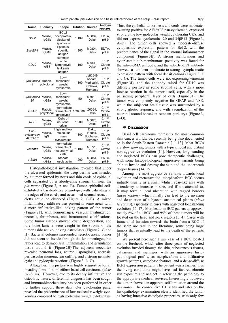
Histopathological investigation revealed that under the ulcerated epidermis, the deep dermis was invaded by a tumor formed by nests and thin cords of epithelial cells separated by a fibrohyaline stroma, till near the pia mater (Figure 2, A and B). Tumor epithelial cells exhibited a basaloid-like phenotype, with palisading at the edges of the cords, and occasional stromal retraction clefts could be observed (Figure 2, C–E). A mixed inflammatory infiltrate was present in some areas with a more infiltrative-like looking pattern of the tumor (Figure 2F), with hemorrhages, vascular hyalinization, necrosis, thromboses, and intratumoral calcifications. Some tumor islands showed cystic degeneration, and rare bone lamella were caught in the stroma of the tumor aside active-looking osteoclasts (Figure 2, G and H). Bacterial colonies surrounded necrotic areas. Tumor did not seem to invade through the leptomeninges, but rather lead to desmoplasia, inflammation and granulation tissue around it (Figure 2B).The adjacent neocortex revealed neuronal loss, neuropil spongiosis, necrosis, perivascular mononuclear cuffing, and a strong gemisto-cytic and pylocytic reactions (Figure 3, L–O).
Altogether, this picture was highly suggestive for an invading form of morpheiform basal cell carcinoma (ulcus terebrans). However, due to its deeply infiltrative and osteolytic nature, differential diagnosis has been sought and immunohistochemistry has been performed in order to further support these data. Our cytokeratin panel revealed the predominance of low molecular weight cyto-keratins compared to high molecular weight cytokeratins.
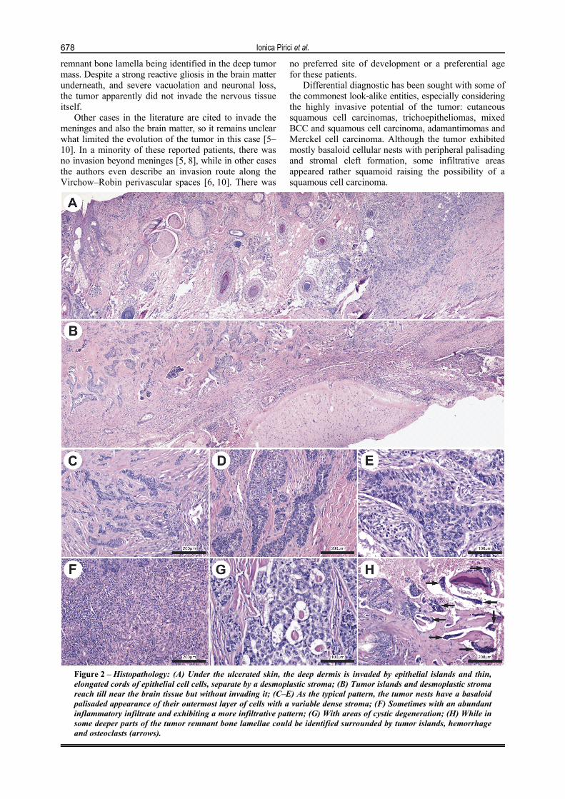
Thus, the epithelial tumor nests and cords were moderate-to-strong positive for AE1/AE3 pan-cytokeratin, expressed strongly the low molecular weight cytokeratin CK8, and did not express cytokeratins 20 and 34βE13 (Figure 3, A–D). The tumor cells showed a moderate-diffuse cytoplasmic expression pattern for Bcl-2, with the predominance of the signal in the stromal inflammatory component (Figure 3E). A strong membranous and cytoplasmic sub-membranous positivity was found for the anti-α-SMA antibody, and the anti-Ber-EP4 antibody showed a uniform moderate-to-strong cytoplasmatic expression pattern with focal densifications (Figure 3, F and G). The tumor cells were not expressing vimentin (Figure 3I), and the antibody raised for CD10 was diffusely positive in some stromal cells, with a more intense reaction in the tumor itself, especially in the palisading peripheral layer of cells (Figure 3J). The tumor was completely negative for GFAP and NSE, while the subjacent brain tissue was surrounded by a strong gliotic response, and with vacuolization of the neuropil around shrunken remnant perikarya (Figure 3, L–O).
Discussion
Basal cell carcinoma represents the most common skin cancer worldwide, recently being also documented so in the South-Eastern Romania [11–13]. Most BCCs are slow growing tumors with a typical local and distant non-aggressive evolution [14]. However, long-standing and neglected BCCs can pose therapeutic challenges, with some histopathological aggressive variants being able to invade and destroy the skin and the underneath deeper tissues [14, 15].
Among the most aggressive variants towards local evolution and metastazation, morpheaform BCC occurs initially usually as a small whitish scar-like area with a tendency to increase in size, and if not attended to, it may form a local ulceration with ragged borders (ulcus rodens), which finally can lead to the invasion and destruction of subjacent anatomical planes (ulcus terebrans), especially in cases with neglected longstanding evolution [15–17]. Morpheaform BCC gathers up approxi-mately 6% of all BCC, and 95% of these tumors will be located on the head and neck regions [3, 4]. Cases with intracranial invasion towards the brain from a BCC of the scalp are rare in the literature, some being large tumors that eventually lead to the death of the patients [5–10].
We present here such a rare case of a BCC located on the forehead, which after three years of neglected evolution invaded through the skin, subcutaneous tissues, calvarium and meninges, with an aggressive histo-pathological profile, as morpheaform and infiltrative growth patterns, osteolytic features, and a dense-diffuse Bcl-2 expression pattern. The patient was a farmer, thus the living conditions might have had favored chronic sun exposure and neglect in referring the pathology to the appropriate medical services. Interestingly however, the tumor showed an apparent self-limitation around the pia mater. The consecutive CT scans and later on the histopathology examination clearly identified the tumor as having intensive osteolytic properties, with only few
Ionica Pirici et al.
678
remnant bone lamella being identified in the deep tumor mass. Despite a strong reactive gliosis in the brain matter underneath, and severe vacuolation and neuronal loss, the tumor apparently did not invade the nervous tissue itself.
Other cases in the literature are cited to invade the meninges and also the brain matter, so it remains unclear what limited the evolution of the tumor in this case [5–10]. In a minority of these reported patients, there was no invasion beyond meninges [5, 8], while in other cases the authors even describe an invasion route along the Virchow–Robin perivascular spaces [6, 10]. There was
no preferred site of development or a preferential age for these patients.
Differential diagnostic has been sought with some of the commonest look-alike entities, especially considering the highly invasive potential of the tumor: cutaneous squamous cell carcinomas, trichoepitheliomas, mixed BCC and squamous cell carcinoma, adamantimomas and Merckel cell carcinoma. Although the tumor exhibited mostly basaloid cellular nests with peripheral palisading and stromal cleft formation, some infiltrative areas appeared rather squamoid raising the possibility of a squamous cell carcinoma.
Figure 2 – Histopathology: (A) Under the ulcerated skin, the deep dermis is invaded by epithelial islands and thin, elongated cords of epithelial cell cells, separate by a desmoplastic stroma; (B) Tumor islands and desmoplastic stroma reach till near the brain tissue but without invading it; (C–E) As the typical pattern, the tumor nests have a basaloid palisaded appearance of their outermost layer of cells with a variable dense stroma; (F) Sometimes with an abundant inflammatory infiltrate and exhibiting a more infiltrative pattern; (G) With areas of cystic degeneration; (H) While in some deeper parts of the tumor remnant bone lamellae could be identified surrounded by tumor islands, hemorrhage and osteoclasts (arrows).
Fronto-parietal pial extension of a basal cell carcinoma of the scalp – case report
679
Figure 3 – Immunohistochemistry: the tumor nests were positive for (A) pan-cytokeratin AE1/AE3, (B) cytokeratin 8, and negative for (C) cytokeratin 20 and (D) 34βE12; (E) Diffusely positive for Bcl-2; (F) Showed a strong sub-membranous staining for α-SMA; (G) A moderate staining for Ber-EP4; (H and I) Was negative for vimentin; (J and K) Diffusely positive for CD10 especially in the peripheral layer of cells of the respective nests; (L) And although the tumor itself was not positive for GFAP or (N) NSE; The remnant nervous tissue and leptomeninges were populated by intense astrogliosis (M); With (O) vacuolization of the neuropil and neuronal loss.
Immunohistochemistry for Bcl-2 and Ber-EP4 showed diffuse positivity in the tumor cells, while the classical pattern of a squamous cell carcinoma would have been a negative for both these markers [18]. The association of basaloid tumor nests with more infiltrative areas raised the possibility of a mixed BCC and squamous cell
carcinoma (SCC). Although no typical squamous foci could be identified in our sections, a mixed BCC and SCC should be deemed negative for Ber-EP4, Bcl-2 and positive for the squamous epithelial marker 34βE12 [19]. Regarding the role of Ber-EP4 to differentiate between BCC from a mixed BCC and SCC, in a study, all the

Ionica Pirici et al.
680
considered BCC cases were clearly positive, while mixed carcinomas showed only some areas of positivity [20]. On this line, in the present case, the diffuse cytoplasmatic staining for Ber-EP4 and Bcl-2 throughout the tumor pointed out to the necessary differences in order to support the diagnostic of BCC. Moreover, our case was completely negative for 34βE12. Trichoepithelioma has a frequent location on the scalp, and it can mimic the appearance of a BCC microscopically by consisting in multiple nodules of basaloid cells that tend to form primitive hair follicle-germ structures with admixed fibromyxoid stroma in between. The stroma is usually denser and more cellular than in a typical BCC, but the occasional infiltrative pattern of this case needed further distinctions. Smooth muscle actin has been showed to be present in a high number of BCCs of the skin, while cutaneous squamous cell carcinomas and trichoepithe-liomas are negative [21]. Anti-smooth muscle actin showed here a strong sub-membranous expression pattern in the epithelial nests, concurring for the diagnostic of BCC [22]. CD10 expression in stromal cells around tumor epithelial nests is also useful for differentiating tricho-epitheliomas from BCC, where nests of basaloid cells are picking up the stain with negative surrounding stromal cells [23]. In our case, the tumor nests were positive for CD10, especially in the peripheral layer of cells, differentiating it thus from a trichoepithelioma. Although a reduced α-SMA and Bcl-2 expression in BCC has been associated with aggressive phenotypes, interestingly, this was not the profile of the present case despite its extremely aggressive clinical behavior, although the Bcl-2 expression pattern was more dense-diffuse rather than dense [24, 25]. The nests of basaloid cells with palisading at their periphery admixed with more irregular tumor islands might create the impression of inner masses of elongated nuclei and stellate cytoplasm connecting as fine cords around empty spaces, as seen in adamantimomas. Vimentin was not expressed by our tumor, and besides clearly showing its non-mesenchymal origin, also helped in differentiating it form adamanti-momas [26]. The nests of compact cells with finely granular and dusty chromatin could also represent the pattern of a focal-trabecular type of Merkel cell carcinoma [27]. Anti-NSE immunostaining revealed a massive neuronal loss/degeneration in the peritumoral CNS tissue, but the tumor itself was not positive for NSE, and together with the negativity for anti-CK20 antibody, these ensured the differential diagnostic from a Merkel cell carcinoma [28]. As described in the literature for other series of BCCs, the tumor cells were strongly positive for a specific anti-CK8 antibody [29].
Due to the aggressive infiltrative pattern and osteolytic features, it is thus important first of all to increase the awareness of the population upon the possible evolution of these classically low malignant tumors, but also in cases of more aggressive presentation to consider the surgical approach based on the possibility that a neglected tumor may develop in time to such an aggressive extent. Classical treatment options for BCC include surgical excision, cryosurgery, as well as different cytostatics or superficial radiotherapy. On the other hand, a rapid diagnostic of locally aggressive BCC may also refer
these cases to new emerging therapeutic options, like the relatively recent FDA-approved Erivedge (Vismodegib), which acts by interfering with abnormal signaling in the Hedgehog pathway. This novel small molecule binds to smoothened (SMO) and leads to inhibition of an abnormal activation of the Hedgehog pathway, and has been showed to reduce the tumor sizes in 43% of locally advanced BCC cases, and in 30% of BCC metastases [30].
Conclusions
The conclusion of this report is twofold. First of all, even for difficult localizations and aggressive immuno-phenotypes, the development of large destructive BCCs appear less due to the aggressive characters of the tumors, but rather to treatment refusal and patients’ attitude. Second, it is important to increase the awareness of the public that untreated aggressive BCCs may be life threatening by direct invasion of the face and head organs, as well as through the consecutive sepsis.
Acknowledgments This paper was published under the frame of European
Social Fund, Human Resources Development Operational Programme 2007–2013, Project No. POSDRU/159/1.5/ S/136893.
Author contribution All authors have contributed equally to the present
work.
References [1] Gupta AK, Daigle D, Martin G, Basal cell carcinoma, Skinmed,
2014, 12(1):33–38; quiz 38. [2] Keen RR, Elzay RP, Basal cell carcinoma from mucosal
surface of lower lip: report of case, J Oral Surg Anesth Hosp Dent Serv, 1964, 22:453–455.
[3] Bastiaens MT, Hoefnagel JJ, Bruijn JA, Westendorp RG, Vermeer BJ, Bouwes Bavinck JN, Differences in age, site distribution, and sex between nodular and superficial basal cell carcinoma indicate different types of tumors, J Invest Dermatol, 1998, 110(6):880–884.
[4] Scrivener Y, Grosshans E, Cribier B, Variations of basal cell carcinomas according to gender, age, location and histopathological subtype, Br J Dermatol, 2002, 147(1):41–47.
[5] Ko CB, Walton S, Keczkes K, Extensive and fatal basal cell carcinoma: a report of three cases, Br J Dermatol, 1992, 127(2):164–167.
[6] Parizel PM1, Dirix L, Van den Weyngaert D, Lambert JR, Scalliet P, Van Oosterom AT, De Schepper AM, Deep cerebral invasion by basal cell carcinoma of the scalp, Neuroradiology, 1996, 38(6):575–577.
[7] Long SD, Kuhn MJ, Wynstra JH, Intracranial extension of basal cell carcinoma of the scalp, Comput Med Imaging Graph, 1993, 17(6):469–471.
[8] Kovarik CL, Stewart D, Barnard JJ, Lethal basal cell carcinoma secondary to cerebral invasion, J Am Acad Dermatol, 2005, 52(1):149–151.
[9] Mathieu D, Fortin D, Intracranial invasion of a basal cell carcinoma of the scalp, Can J Neurol Sci, 2005, 32(4):546–548.
[10] Schroeder M, Kestlmeier R, Schlegel J, Trappe AE, Extensive cerebral invasion of a basal cell carcinoma of the scalp, Eur J Surg Oncol, 2001, 27(5):510–511.
[11] Dreier J, Felderer L, Barysch M, Rozati S, Dummer R, Basal cell carcinoma: a paradigm for targeted therapies, Expert Opin Pharmacother, 2013, 14(10):1307–1318.
[12] Epstein EH Jr, Skin cancer: basal cell carcinoma – pay your money, take your choice, Nat Rev Clin Oncol, 2013, 10(9): 489–490.
Fronto-parietal pial extension of a basal cell carcinoma of the scalp – case report
681
[13] Pirici A, Study of the interaction between stroma and epithelium in basal cell carcinoma. A histological and clinico-statistical approach, PhD Thesis, University of Medicine and Pharmacy of Craiova, Romania, 2011.
[14] Rubin AI, Chen EH, Ratner D, Basal-cell carcinoma, N Engl J Med, 2005, 353(21):2262–2269.
[15] Crowson AN, Basal cell carcinoma: biology, morphology and clinical implications, Mod Pathol, 2006, 19(Suppl 2):S127–S147.
[16] Sonntag M, Reifenberger J, Megahed M, Schulte KW, Ulcus terebrans. Therapy options and their limits, Hautarzt, 2004, 55(10):983–985.
[17] Birbilis T, Ulcus terebrans: an unusual cause of paraparesis, J Neurol Neurosurg Psychiatry, 2003, 74(12):1643.
[18] Swanson PE, Fitzpatrick MM, Ritter JH, Glusac EJ, Wick MR, Immunohistologic differential diagnosis of basal cell carcinoma, squamous cell carcinoma, and trichoepithelioma in small cutaneous biopsy specimens, J Cutan Pathol, 1998, 25(3): 153–159.
[19] Vasudev P, Boutross-Tadross O, Radhi J, Basaloid squamous cell carcinoma: two case reports, Cases J, 2009, 2:29351.
[20] Beer TW, Shepherd P, Theaker JM, Ber EP4 and epithelial membrane antigen aid distinction of basal cell, squamous cell and basosquamous carcinomas of the skin, Histopathology, 2000, 37(3):218–223.
[21] Miller RT, Immunohistochemistry in the differential diagnosis of cutaneous basal cell carcinoma and squamous cell carcinoma, ProPath, THE FOCUS – Immunohistochemistry, 2004, December 2014.
[22] Winters R, Naud S, Evans MF, Trotman W, Kasznica P, Elhosseiny A, Ber-EP4, CK1, CK7 and CK14 are useful markers for basaloid squamous carcinoma: a study of 45 cases, Head Neck Pathol, 2008, 2(4):265–271.
[23] Heidarpour M, Rajabi P, Sajadi F, CD10 expression helps to differentiate basal cell carcinoma from trichoepithelioma, J Res Med Sci, 2011, 16(7):938–944.
[24] Uzquiano MC, Prieto VG, Nash JW, Ivan DS, Gong Y, Lazar AJ, Diwan AH, Metastatic basal cell carcinoma exhibits reduced actin expression, Mod Pathol, 2008, 21(5):540–543.
[25] Saladi RN, Singh F, Wei H, Lebwohl MG, Phelps RG, Use of Ber-EP4 protein in recurrent metastatic basal cell carcinoma: a case report and review of the literature, Int J Dermatol, 2004, 43(8):600–603.
[26] Benassi MS, Campanacci L, Gamberi G, Ferrari C, Picci P, Sangiorgi L, Campanacci M, Cytokeratin expression and distribution in adamantinoma of the long bones and osteo-fibrous dysplasia of tibia and fibula. An immunohistochemical study correlated to histogenesis, Histopathology, 1994, 25(1): 71–76.
[27] Smith PD, Patterson JW, Merkel cell carcinoma (neuro-endocrine carcinoma of the skin), Am J Clin Pathol, 2001, 115(Suppl):S68–S78.
[28] Sirikanjanapong S, Melamed J, Patel RR, Intraepidermal and dermal Merkel cell carcinoma with squamous cell carcinoma in situ: a case report with review of literature, J Cutan Pathol, 2010, 37(8):881-885.
[29] Kooy AJ, Tank B, Vuzevski VD, Van Joost T, Expression of cytokeratin 8 and low molecular weight cytokeratins in human basal cell carcinoma, Anticancer Res, 1995, 15(2):241–247.
[30] Meiss F, Zeiser R, Vismodegib, Recent Results Cancer Res, 2014, 201:405–417.
Corresponding author Daniel Pirici, Lecturer, MD, PhD, Department of Research Methodology, University of Medicine and Pharmacy of Craiova, 2 Petru Rareş Street, 200349 Craiova, Romania; Phone +40742–758 934, e-mail: [email protected] Received: February 3, 2014
Accepted: July 2, 2014