Embed Size (px)
Citation preview

Charles University in Prague Faculty of Science
Department of Zoology
ORAL MORPHOGENESIS IN THE MEXICAN AXOLOTL:
DEVELOPMENTAL ORIGIN OF TOOTH GERMS IN EVOLUTIONARY CONTEXTS
ORÁLNÍ MORFOGENEZE AXOLOTLA: PŮVOD ZUBŮ V EVOLUČNÍM KONTEXTU
Master thesis
VLADIMÍR SOUKUP
Supervisor: Mgr. Robert Černý, Ph.D.
Prague 2008


Herewith I declare that I carried out this Master thesis only by myself, with the use of cited
literature.
Prague September 5, 2008

ABSTRACT Mouth in the majority of vertebrates develops throughout an ectodermal stomodeum
which posteriorly contacts the foregut endoderm, together forming an oropharyngeal
membrane. This ecto-/ endodermal membrane gradually thins and become eventually
perforated, which causes opening of the stomodeal cavity into the pharynx. Teeth are then
understood as organs arising within the stomodeal part of the mouth, where the ectodermal
epithelium produces tooth enamel and neural crest mesenchymal cells form dentine and tooth
pulp. This project was meant to study the dynamics of the ectoderm and endoderm during the
formation of mouth and teeth in the Mexican axolotl. By utilizing transplantations of the oral
ectoderm from GFP-transgenic embryos and injections of fluorescent tracer DiI into the
endoderm, it was possible to follow the fate of both germ-layers during the course of
embryonic development into details. By using this approach it was demonstrated that the
mouth in the axolotl develops in a different way, i.e. via the stomodeal collar. Teeth were
found to arise within the stomodeal collar ectoderm as well as in the more posteriorly situated
endodermal areas. Moreover, some tooth germs were generated also directly at the ecto-/
endodermal boundary. Thus, the formation of teeth does not seem to primarily depend on the
distribution of different germ-layer epithelia, but, more likely, on strictly defined places
within the oropharynx. The evolutionary origin of teeth should not, therefore, be derived from
denticles, which hypothetically settled the oral area in a mechanistic kind of shift, either from
the external ectoderm or from the pharyngeal endoderm. Teeth should rather be assumed as
neural-crest-derived elements, which arose primarily in the oropharyngeal area only after an
odontogenic potential for their production was achieved here and which were probably never
dependent on the germ-layer derivation of the epithelium.

ABSTRAKT U většiny obratlovců se ústa vyvíjejí pomocí ektodermálního stomodea, které
posteriorně kontaktuje faryngeální entoderm a tvoří s ním orofaryngeální membránu. Tato
ekto-/ entodermální membrána se postupně ztenčuje, až dojde k jejímu protržení, což způsobí
otevření stomodeální dutiny do faryngu. Zuby jsou chápány jakožto orgány vznikající ve
stomodeální části úst, kde ektodermální epitel produkuje sklovinu zubu a mezenchym
neurální lišty tvoří dentin a zubní pulpu. V této práci byla studována dynamika ektodermu a
entodermu během vývoje úst a zubů u axolotla mexického. Transplantací orálního ektodermu
z GFP-transgenních embryí a injikací fluorescenční značky DiI do entodermu příjemce bylo
možné sledovat osud obou zárodečných vrstev během embryonálního vývoje. Tímto
přístupem bylo zjištěno, že se ústa u axolotla vyvíjejí odlišným způsobem, a to pomocí
ektodermálního stomodeálního límce. Zuby pak vznikají jak v ektodermální oblasti
stomodeálního límce, tak i v posteriornějších entodermálních místech. Navíc, některé zubní
zárodky jsou generovány přímo na ekto-/ entodermální hranici. Tvorba zubů tedy nezávisí na
rozložení epitelů různého zárodečného původu, ale spíše na konkrétních definovaných
místech v rámci orofaryngu. Evoluční původ zubů by tedy neměl být odvozován z dentikul,
které hypoteticky osídlily ústa mechanistickým přesunem, ať už z povrchového ektodermu
nebo z faryngeálního entodermu. Zuby by tedy spíše mohly být chápány jakožto elementy,
které se objevily primárně v orofaryngeální oblasti poté, co zde vznikl odontogenní potenciál
pro jejich tvorbu a které pravděpodobně nikdy nezávisely na zárodečném původu konkrétního
epitelu.

CONTENTS
ACKNOWLEDGEMENTS.........................................................................................................................................8
1. INTRODUCTION ....................................................................................................................................................9
2. CURRENT KNOWLEDGE ON THE SUBJECT.............................................................................................11
2.1. DEVELOPMENT OF A TOOTH..............................................................................................................................11 2.2. EVOLUTIONARY ORIGIN OF TEETH...................................................................................................................13
2.2.1. External “Ectodermal” Origin of Teeth (Outside-in Scenario) ............................................................13 2.2.2. Internal “Endodermal” Origin of Teeth (Inside-out Scenario) ............................................................15
2.3. ORAL VS. PHARYNGEAL TEETH IN FISHES .......................................................................................................18 2.4. DEVELOPMENT OF MOUTH IN VERTEBRATES ..................................................................................................20 2.5. DEVELOPMENT OF MOUTH IN URODELES ........................................................................................................22
2.5.1. Development of Teeth in Urodeles ..........................................................................................................24
3. MATERIALS AND METHODS ..........................................................................................................................27
3.1. HANDLING OF THE EMBRYOS............................................................................................................................27 3.2. GFP ECTODERM TRANSPLANTATIONS.............................................................................................................27 3.3. DII INJECTIONS..................................................................................................................................................27 3.4. SECTIONING .......................................................................................................................................................28 3.5. IMMUNOSTAINING .............................................................................................................................................28 3.6. RESIN HISTOLOGY .............................................................................................................................................29 3.7. SOFTWARE, MICROSCOPES ...............................................................................................................................29
4. RESULTS.................................................................................................................................................................30
4.1. HISTOLOGICAL OBSERVATIONS........................................................................................................................30 4.2. TRANSPLANTATIONS OF GFP ECTODERM........................................................................................................30 4.3. MOUTH DEVELOPMENT IN AXOLOTL ...............................................................................................................32 4.4. MOUTH OPENING...............................................................................................................................................32 4.5. ADENOHYPOPHYSIS...........................................................................................................................................33 4.6. NON-ECTODERMAL TEETH ...............................................................................................................................33 4.7. DII INJECTIONS AND GFP TRANSPLANTATIONS..............................................................................................34 4.8. ENDODERMAL DERIVATION OF TEETH.............................................................................................................34 4.9. DUAL ORIGIN OF TOOTH ENAMEL EPITHELIA .................................................................................................35 4.10. GERM-LAYER DERIVATION AND DISTRIBUTION OF TEETH ..........................................................................35
5. DISCUSSION ..........................................................................................................................................................37
5.1. DEVELOPMENT OF MOUTH IN URODELES AND IN OTHER VERTEBRATES ......................................................37 5.2. ORAL DEVELOPMENT VIA THE STOMODEAL COLLAR .....................................................................................38 5.3. ON THE PRESENCE AND HOMOLOGY OF THE OROPHARYNGEAL MEMBRANE................................................40 5.4. RELATION OF THE OROPHARYNGEAL MEMBRANE TO TEETH.........................................................................41

5.5. TOOTH ORIGIN IN EVOLUTIONARY CONTEXTS ................................................................................................42 5.6. ECTO-/ ENDODERMAL BOUNDARY AS THE SITE FOR TOOTH INITIATION .......................................................44 5.7. TOOTH INDUCTION: EPITHELIUM OR MESENCHYME? .....................................................................................45 5.8. ARE AXOLOTL TEETH HOMOLOGOUS DESPITE DIFFERENT EMBRYONIC DEVELOPMENT?...........................46
6. CONCLUSIONS .....................................................................................................................................................49
7. LIST OF LITERATURE.......................................................................................................................................51
SUPPLEMENTS ........................................................................................................................................................... I
FIGURES....................................................................................................................................................................... I List of Figures........................................................................................................................................................ I Abbreviations used in Figures............................................................................................................................. II
HISTOLOGICAL TECHNIQUES..................................................................................................................................XII JB-4 Embedding .................................................................................................................................................XII Histological Staining........................................................................................................................................ XIII
PRESENTATIONS OF THE PROJECT ..........................................................................................................................XV Scientific Talks ...................................................................................................................................................XV Posters............................................................................................................................................................... XVI Research Article ............................................................................................................................................... XVI

8
ACKNOWLEDGEMENTS This project was initiated on the basis of ideas and discussions between Prof. Ivan
Horáček and Dr. Robert Černý and has been supervised by Dr. Robert Černý at the
Department of Zoology, Charles University in Prague already since my Bachelor studies. The
experimental approaches, which constitute the key-stone of this work, were, however, carried
out in the laboratory of Dr. Hans-Henning Epperlein at the Department of Anatomy,
Technical University in Dresden, however all subsequent analyses were carried out in our
laboratories in Prague. These three scientists are acknowledged for their kind help and
suggestions during the course of development of this project. However, I would like to
express my thanks especially to Dr. Robert Černý for his fruitful discussions on the subject,
for his enthusiasm and simply for the way he is, both in the scientific and personal life.
I am also greatly indebted to other people, whose kind help made this scientific work
possible, most notably to: Elly Tanaka and Heino Andreas (Dresden) for supplies of axolotl
eggs, Levan Mchedlishvili (Dresden) for the instructions of microinjector handling and loan
of DiI, Jan Černý (Prague) for discussions on and loans of antibodies, Ondřej Šebesta
(Prague) for technical assistance in microscopy and Karel Kleisner (Prague) for the discussion
on tooth homology.
My personal thanks are addressed to my girlfriend, to all friends of mine, to my
siblings and to my parents. I could not imagine my life without love, support, help and
understanding of all these people.
During this project, I was supported by COST B23 (Oral Facial Development and
Regeneration), SMWK (Dresden) and the Ministry of Education, Youth and Sports of the
Czech Republic (Grant Nr. 113100004).

9
1. INTRODUCTION Teeth represent one of the most important evolutionary novelties within the vertebrate
clade. The appearance of these cone-shaped hard structures within gnathostomes can be
understood as a milestone that, together with jaws, enabled diversification of vertebrates as
active predators and, thus, completely changed relations within the Paleozoic seas. Although
it is generally accepted that teeth evolved from denticles1, the question about the place of
origin of these denticles is still a hot topic of for recent scientific discussions. Although some
researchers suggest that teeth evolved from external ectodermal denticles that reached the oral
area in an “outside-in” manner (sensu Reif 1982), others propose that rather pharyngeal
endodermal denticles were those to give rise to teeth according to an “inside-out” scenario
(Smith & Coates 1998).
Tooth development is most extensively studied in the mouse, where teeth are assumed
to develop from stomodeal ectodermal epithelium that secretes enamel and neural crest
mesenchyme that produces dentine and tooth pulp (Thesleff & Sharpe 1997; Jernvall &
Thesleff 2000). This model organism, however, possesses highly derived dentition not only
among mammals, but also among other vertebrates. Nevertheless, the aspects of mouse tooth
development are apodictically applied also to other species. Therefore, teeth are understood as
structures developing from the ectoderm and neural crest whatever their position is in
whatever species. However, such contribution of the germ-layers to teeth sometimes seems to
be very improbable. Teeth developing deep within the pharynx of some extant fishes would
suggest their derivation from rather endodermal (than ectodermal) epithelium. Moreover, the
precise germ-layer origin of oral teeth cannot be easily identified due to difficulties to
satisfactorily distinguish ectoderm from endoderm at critical stages of tooth germ initiation.
Hence, after perforation of the oropharyngeal membrane, which constitutes the natural
boundary between ectoderm and endoderm, the distribution of these germ-layers is unknown
even in such model animals like mouse, chick or zebrafish.
Based on this theoretical background, the purpose of this project was to contribute to
our knowledge on the development and evolution of vertebrate teeth by testing the direct
contribution of ectoderm and endoderm to tooth germs. The most suitable model organism for
such analysis seemed to be the Mexican axolotl Ambystoma mexicanum (Shaw, 1789), from
several reasons.
1 denticles = dentine including elements

10
1) The axolotl as other urodele amphibians represent interesting model organisms,
because the development of mouth does not advance through the stages of ectodermal
stomodeum as in other vertebrates (Johnston 1910; Adams 1924). Moreover, past
studies have questioned the dogmatic view on teeth as derivatives of ectoderm and
neural crest cells (Adams 1924; de Beer 1947).
2) Urodeles are maybe the most suitable vertebrates to study formation of mouth and
teeth and development in general. Their development is generally slow and its rate can
be easily controlled by temperature, they have large mesolecital eggs that are easy to
manipulate and enable experimental-embryological approaches.
3) A new strain of fluorescent transgenic axolotls (Sobkow et al. 2006) is recently
available. Transplantations of certain tissue from such fluorescent embryo into the
non-fluorescent host enables following the fate of this tissue during the course of
embryonic and larval development into great details.
4) I have worked with axolotl embryos and studied the development of mouth and
teeth in urodeles already during my Bachelor degree (Soukup 2006).
During this project, I have utilized GFP (Green Fluorescent Protein) transgenic
embryos of the Mexican axolotl (Sobkow et al. 2006) together with experimental
embryological approaches and performed a fate-mapping of ectoderm and endoderm during
the course of mouth and tooth development. Taking an advantage of transplantations of
labelled tissues and injections of cell tracer, this project was meant to study:
1) the dynamics of ectoderm and endoderm during the formation of mouth in urodeles,
and
2) the contribution of ectoderm and endoderm to developing teeth.

11
2. CURRENT KNOWLEDGE ON THE SUBJECT 2.1. Development of a tooth
Teeth, together with scales, hair, feathers, claws and multiple glands among others, are
considered to be derivatives of integument (Kardong 1995). However, despite the diversity in
form and function, all these structures develop at the interface of epithelium and underlying
mesenchyme and seem to be regulated by common developmental mechanisms, i.e. sequential
reciprocal interactions between these tissues (Pispa & Thesleff 2003).
The first visible sign of tooth development is a local thickening of the epithelial layer,
which forms a placode. Mesenchymal cells directly under this thickening later condensate into
a papilla. Still later, the thickened epithelial layer starts to invaginate and proliferate into the
underlying mesenchyme to form a tooth bud and subsequently a tooth cup. Continued folding
of the epithelial-mesenchymal junction finally outlines the future shape of the tooth. In
mammals, folding of the epithelial layer is ruled by a transitional structure called enamel knot.
Enamel knot represents a cluster of non-dividing cells within the epithelium (Jernvall et al.
1994) that secrete multiple growth factors both to surrounding epithelia and mesenchyme and
directs either activation or inhibition of proliferation in these tissues (Jernvall et al. 1998;
Jernvall & Thesleff 2000; for a computational-developmental model see Salazar-Ciudad &
Jernvall 2002). On the account of reciprocal interactions, mesenchymal cells form a dental
pulp and become odontoblasts that generate dentine. Epithelial cells, on the other hand,
differentiate into an enamel organ, where ameloblasts produce enamel. Secretion of protein
matrix and anorganic material takes place at the epithelial-mesenchymal junction.
The epithelial-mesenchymal contact is, as mentioned above, a place of reiterative
reciprocal signalling from the initiation of tooth germ to hard tissue deposition (Thesleff &
Sharpe 1997; Jernvall & Thesleff 2000; Tucker & Sharpe 2004). Signalling involves multiple
molecules belonging to fibroblast growth factor (FGF), transforming growth factor β (TGFβ),
hedgehog (Hh), Wnt and tumor necrosis factor (TNF) families. It is beyond the scope of this
study to review all the genetic mechanisms that govern the development of the tooth. For the
purpose of this study it is more interesting to have a look at tooth induction and its tissue
origin. Mouse tissue recombination studies identified that the first instructive signal for
odontogenesis comes from the oral epithelium to the adjacent mesenchyme (Mina & Kollar
1987; Lumsden 1988). This signal goes hand in hand with the first regionalization of the
mandibular arch by antagonistic signalling of Bmp4 and Fgf8 (Tucker et al. 1999) and leads
to a specification of places of tooth development (Neubüser et al. 1997; Tucker et al. 1998).

12
The odontogenic potential, however, subsequently shifts from the epithelium into the
mesenchyme (Peters & Balling 1999). Mesenchymal expression of multiple homeobox-
containing transcription factors then defines the type of developing tooth (Sharpe 1995;
Thomas & Sharpe 1998).
It is generally accepted that tooth enamel epithelium (later ameloblasts) is a derivative
of ectoderm, whereas dental mesenchyme (later odontoblasts) is produced by neural crest
cells2. Such scheme is however apodictically applied to all vertebrates, although germ-layer
derivation of teeth has been shown in a relatively low number of species. By means of precise
fate-mapping3 methods, the dental mesenchyme has been shown to be a neural crest
derivative only in rodents (Imai et al. 1996; Chai et al. 2000) and urodeles (Chibon 1967a;
1967b), but its neural crest origin is assumed also for other vertebrates (Smith & Hall 1990;
1993). Similarly, ectodermal origin of enamel epithelium was demonstrated by precise fate-
mapping only in urodeles (Chibon 1970), but ectodermal origin is proposed also for mouse
(e.g. Jernvall & Thesleff 2000), bony fishes (oral dentition; Sire & Huysseune 1996; Sire et
al. 1998; Sire 2001) and other vertebrates as well (Reif 1982; Kardong 1995; Pispa & Thesleff
2003). However, teeth of many actinopterygians are found deep within pharynx, beyond the
possible influence of ectoderm, where the epithelium is proposed to be rather of endodermal
origin (Huysseune et al. 2002). No fate-mapping study has, however, been performed to
confirm this suggestion.
The epithelial germ-layer derivation of such pharyngeal teeth4 is not well known,
however, even the germ-layer origin of teeth in the oral area is questionable. In the oral area,
ectoderm can be clearly distinguished from endoderm during initial stages of development,
because these two germ-layers are in contact at an oropharyngeal membrane. After rupture of
this membrane, which causes opening of the mouth, the natural limit between both germ-
layers is lost. Later distribution of ectoderm and endoderm in the mouth cannot be determined
by means of classical histology, because cell compartments, characteristic for ectoderm and
2 Neural crest is a transient population of cells that originates at the limit of neural and non-neural ectoderm during the closure of the neural tube (Le Douarin & Kalcheim 1999). These cells undergo an extensive latero-ventral migration through the whole body (Cerny et al. 2004) to reach multiple positions within the embryo. At these positions, neural crest is the source of multiple cells and tissues, such as pigment cells, cartilage, bone, nerves etc. (Le Douarin & Kalcheim 1999). 3 Fate-mapping is a term for mapping the fate of desired tissue. At the initial stage, this tissue is labelled by a stable marker, which can be easily followed during the course of development and which is finally found within all the derivatives of this tissue (Stern & Fraser 2001). 4 Pharyngeal teeth are teeth found within the pharyngeal cavity of various fishes. They can be situated either loosely in the pharyngeal mucosa or associated with branchial arches.

13
endoderm at initial stages of development, are gradually using up. Cells of both germ-layers,
thus, become indistinguishable already before any signs of development of teeth.
The germ-layer origin of vertebrate teeth is, however, key to the understanding of
tooth evolution. Possible endodermal contribution to tooth formation revealed by fate-
mapping studies could have direct influence on our understanding of tooth evolutionary
scenarios. A series of these studies performed throughout various vertebrate taxa could
indicate the role of germ-layers during the evolution of vertebrates, whether or not there is the
endodermal contribution to teeth, or whether there were shifts in either contribution of ecto-/
endoderm in relation to developing teeth.
2.2. Evolutionary Origin of Teeth Recent opinions on the evolutionary origin of teeth in extant vertebrates are
predominated by two opposing theories. It used to be generally accepted that teeth evolved
from external dermal denticles and that is why they should develop from ectodermal
epithelium (“outside-in” theory, sensu Reif 1982). More recently, however, it was proposed
that teeth could evolve from denticles situated in the pharyngeal region, which would support
their endodermal derivation (“inside-out” theory, Smith & Coates 1998; 2000; 2001). No
experimental evidence has, however, been put forth to support endodermal origin of teeth
(until nowadays) and, therefore, the “classical” ectodermal evolutionary origin is rather
accepted, although the influence of the latter is increasing. These theories are discussed in
details bellow.
2.2.1. External “Ectodermal” Origin of Teeth (Outside-in Scenario) It was proposed already a long time ago, however, mainly based on tissue
composition, that teeth are structures homologous to external dermal denticles, but it had
taken quite a long time, until a suitable theoretical model explaining the mechanism of
evolutionary denticle-to-tooth-transition was brought in. The nowadays widely accepted
“Odontode Regulation Theory” (Reif 1982) offered such a model and replaced the previous
“Lepidomorial Theory” (see Janvier 1996 and Donoghue 2002 for aspects of Lepidomorial
Theory).
The Odontode Regulation Theory is based on studies of dentition and squamation in
sharks and comprises an odontode as its most fundamental unit. Odontode is the primary
element of the dermal skeleton of all vertebrates (Reif 1982). It is a composite hard structure,

14
which consists of dentine surrounding central pulp, acellular or cellular bone at the base
anchoring it into dermis and a hypermineralized cap composed of enamel or enameloid
(which, however, need not be necessarily present). It develops at the interface of outer
ectodermal epithelium and underlying neural crest mesenchyme. Although the odontode was
first defined as a “hard tissue unit corresponding very closely to teeth and difficult to
distinguish from them by any rational criteria” (Ørvig 1967), it was later extended to include
teeth as well (Reif 1982). Teeth were understood to have not evolved independently de novo,
but rather by means of a modification of the morphogenetic program for denticles. The term
odontode thus comprises two structures: dermal denticles (scale-like elements of external
skeleton found mostly on the body surface) and teeth. The main difference between these
manifestations of the odontode is that dermal denticles are always generated superficially (e.g.
Reif 1980), whereas teeth develop from a deep epithelial invagination called dental lamina
(Reif 1982). For more information about odontodes and their evolutionary changes and
derivatives see Ørvig (1977) and Huysseune & Sire (1998).
According to the Odontode Regulation Theory, the evolution of teeth from dermal
denticles starts from a situation of undifferentiated odontodes arranged uniformly on the body
surface. These odontodes form inhibitory fields around themselves preventing formation of
other odontodes in their vicinity (Osborn 1978). New odontodes can be added only apart from
the inhibitory field, i.e. during the growth of the animal or after shedding of the preceding
odontode. This mechanism is, however, very undesirable especially along the jaw margins,
where odontodes could have functioned as grasping or crushing elements. Loss of odontode
naturally enables formation of a new one, but, at this place, the developing germ would be
very vulnerable. Moreover, the development of the new odontode at the jaw margin just after
the loss of its predecessor is not efficient, because it would take quite a long time for the
successive germ to become functional. The epithelial invagination along the whole length of
the jaws (called dental lamina), however, solve the problem of superficially developing
odontodes. Odontode germs are, thus, generated deep within the dental lamina, far away from
inhibitory fields of other superficially forming odontodes, and in the area, where they cannot
be easily damaged. These are generated in advance of need, quite quickly compensate their
predecessors and gradually increase in size according to the body growth. Dental lamina thus
offers a series of replacement germs (now teeth), a “tooth family”, while functioning tooth is
still at the place.
Originally, the evolution of teeth as derivatives of the dental lamina was proposed to
take place only after the evolution of jaws (Reif 1982; Mallatt 1996). Jaws are seen to have

15
evolved from anterior pharyngeal arch(es), as they enabled closure of the mouth during
forceful expiration through pharyngeal slits (Mallatt 1996). It was only later, when jaws
enlarged and were augmented by continuously replacing teeth to become an apparatus for
grasping the prey. Hence, teeth as derivatives of dental lamina are proposed to be a
synapomorphy of Chondrichthyes and Teleostomi, since Placodermi, the basal jawed
vertebrates possessed only non-tooth bearing gnathalia (Reif 1982, but see Smith & Johanson
2003).
The term “odontode” has, however, gradually become more or less theoretical, in spite
of being concretely defined, and is nowadays mainly used as a model of predecessor or
archetype of both dermal denticles and teeth. This can be seen for example in the studies of
dermal skeleton in fishes (Sire & Huysseune 1996; Sire et al. 1998; Sire 2001), where teeth
are suggested rather to precede than to evolve from dermal denticles. By comparison of
developmental stages of individual dermal elements, Sire & Huysseune (2003) propose that
teeth evolved from isolated external odontodes (represented by placoid scales of sharks) 450
My ago, while dermal denticles, having more similar ontogeny and structure with teeth, arose
much later, ca 100 My ago. Thus, the following evolutionary sequence was suggested: (1)
isolated odontodes, (2) teeth, (3) dermal denticles. Teeth would in this case represent only
intermediate products later giving rise to dermal denticles. Such suggestion is based also on
the presence of teeth found on the surface of the head of some teleosts (Sire 2001). Dermal
denticles are then proposed to have evolved from teeth that escaped from their original
positions in the mouth and were spread along the body surface. Since the extra-oral teeth are
found in several non-related species (Sire 2001), it was suggested that this change in position
and subsequent hypothetical tooth-to-denticle shift could happen even many times during the
evolution of vertebrates.
Whether this evolutionary tooth-to-denticle shift happened or not, remains to be
elucidated. Nevertheless, according to this generally accepted “outside-in” scenario, the
external skin odontodes were captured in the oral cavity and were incorporated into the jaw
apparatus. Hence, odontodes, as derivatives of exoskeleton, are assumed to have always been
developing in the ectodermal area. Teeth are, thus, understood as derivatives of ectodermal
epithelium and underlying mesenchyme from their very evolutionary beginning.
2.2.2. Internal “Endodermal” Origin of Teeth (Inside-out Scenario) The Odontode Regulation Theory is an influential concept in that it offers logical
scenario on the evolutionary change of denticles into composite patterned teeth and clearly

16
demarks teeth from other dermal skeletal units by features of dental lamina (Reif 1982). On
the other hand, important points against this theory were addressed, namely that no transition
from denticles into teeth has ever been found even in sharks (see even Reif 1982) and that it
includes an assumption that teeth evolved only after the appearance of jaws (Smith & Coates
1998; 2000).
Another hypothesis on the evolutionary origin of teeth was, therefore, put forth (Smith &
Coates 1998). According to the “inside-out” scenario (Smith & Coates 1998; 2000; 2001),
teeth have evolved from denticles found within the pharyngeal cavity, i.e. areas lined by
endodermal (rather than ectodermal) epithelium.
The “inside-out” theory is based on the presence of denticles associated with
pharyngeal arches of Loganellia scotica (Van der Brugghen & Janvier 1993; Janvier 1996)
(an agnathan vertebrate from the group Thelodonti). Pharyngeal denticles of Loganellia have
been redescribed by Smith & Coates (1998) as a series of hard exoskeletal units arranged in a
whorl-like manner representing a family of subsequently developing denticles. The denticle
family likely grew in one direction by a precise apposition of successive elements at the end
of this series. This pattern organization might propose exact spatial and temporal regulation of
initiation, development, polarity, shape and size of successive denticles (Smith & Coates
1998). Such characteristics are, however, claimed to be features of dental lamina, meaning
that these denticles should be regarded, according to the definition by Reif (1982), as true
teeth. Whether the dental lamina really existed in Loganellia, cannot, however, be
demonstrated.
Similar families (tooth-whorls) have been found also in basal gnathostomes such as
stem Chondrichthyes, Acanthodii, basal Osteichthyes and also recent Chondrichthyes (Janvier
1996, pp. 125, 137-138, 145, 196, 203; Smith & Coates 1998 and citations therein). These
tooth-whorls are associated either with branchial arches or with the jaw margins (along their
whole length, or in symphyseal or parasymphyseal positions), as clusters of either unicuspid
or multicuspid teeth with fused bases. They can vary from being a bow-shaped series of four
teeth, or complete spirals consisting of many teeth gradually increasing in size (Reif 1982).
Although teeth are generally regarded as derivatives of ectoderm and neural crest, the
inside-out theory proposes evolution of teeth from endodermal pharyngeal areas (Smith &
Coates 1998; 2000; 2001). Teeth are seen to originate from patterned pharyngeal denticles
similar to those found in Loganellia, where the patterning mechanisms were intrinsic to the
pharyngeal endodermal epithelium. The patterning mechanisms were then co-opted from the
endodermal pharyngeal epithelium into ectodermal oral epithelium. The advantage of this co-

17
option would be in generating the denticle/ tooth-whorls within the ectodermal jaw epithelium
and functioning as grasping apparatus. Teeth of extant vertebrates would, thus, according to
the inside-out theory, be derivatives of ancient odontogenic developmental mechanisms co-
opted from endodermal regions.
Tooth-whorls are assumed to be an ancient character acquired already before the
appearance of jawed vertebrates, which led Smith & Coates (2001) to the proposal that they
could represent a synapomorphy of loganellid thelodonts and primitive gnathostomes.
Although no tooth-whorls have been found within several groups of this proposed clade (e.g.
jawless Osteostraci or basal jawed Placodermi), denticles organized into rows were reported
at the rear of the gill chamber of some placoderms (Johanson & Smith 2003; 2005). Based on
the different spatial arrangement of these denticles when compared to the pattern of outer skin
denticles, the pharyngeal denticles are regarded as derivatives of different patterning
mechanisms. These denticles are, therefore, proposed to have developed from endodermal
epithelium and are claimed to share the same developmental mechanisms with tooth-whorls
of Loganellia (Johanson & Smith 2003; 2005).
Although the inside-out theory assumes the shift from pharyngeal denticles into oral
tooth-whorls, there appears an inconsistency in proposals of later evolution of these elements
(in spite of being addressed by the very same author). Formerly, it was proposed that ancient
mechanisms for tooth development underwent shift from endodermal into ectodermal
epithelium and that it was primarily the endodermal epithelium, where teeth have evolved
(Smith & Coates 1998; 2000). However, just the very same authors proposed later that it is
the boundary of ecto- and endoderm, which is central to tooth development (Smith 2003;
Johanson & Smith 2003). Hence, the ecto-/ endodermal border was proposed to function as
the place of initiation, development and subsequent replacement of teeth within first
vertebrate dentitions, whether situated on the pharyngeal arches or in the oral area.
Development of the whole dentition was, therefore, proposed to start from a single
symphyseal tooth germ, which acts as an inductor of other teeth. A waving ecto-/ endodermal
border along the whole length of the jaw functions as the place of initiation of these tooth
germs. These founders subsequently induce development of other members, which together
form tooth families. This theoretical model is based on the assumption of ecto-/ endodermal
border as the place of origin of teeth; however, such proposal is faded by the inability to map
the distribution of ectoderm and endoderm in the mouth of fossil vertebrates. Moreover, even
in extant vertebrates, the origin of oral epithelia is also not known due to a considerable lack
of fate-mapping studies.
18
Summarizing this “inside-out” theory out, dermal and pharyngeal denticles are
proposed to share common regulation of morpho- and histogenesis, and are therefore assumed
to be homologous at this level. However, the latter differ significantly from the former in a
whorl-like pattern which suggests different mechanisms of development. These whorls were
initially formed in the pharyngeal portion of oropharynx prior to the evolution of jaws and
were later co-opted for oral areas. Teeth have probably always been associated with the ecto-/
endodermal boundary, no matter whether this border is situated at pharyngeal arches or
associated with jaws. Teeth in extant vertebrates would therefore be generated by mechanisms
that evolved primarily in the pharyngeal areas.
Despite being based on paleontological data, the origin of teeth from pharyngeal
denticles of agnathan vertebrates was questioned by many paleontologists. Purnell (2001)
found oral denticles comparable to those of Loganellia in some members of the Heterostraci
group (another agnathan clade). These denticles were organized into a pattern as well;
however, they gradually integrated with external headshield denticles that possessed different
pattern. This led to the suggestion that external skin denticles in Heterostraci must have given
rise to oral denticles rather according to “outside-in” than “inside-out” scenario (Purnell
2001). Next, Donoghue & Smith (2001) showed that Loganellia is a derived thelodont and
pointed out that no comparable organized pharyngeal denticles have been found within other
members of this clade! Pharyngeal denticles of Loganellia and tooth-whorls of basal jawed
vertebrates would thus be convergently achieved and could not in this way be considered as a
synapomorphy of these lineages. According to these points, the proposal that teeth evolved
according to “inside-out” theory clearly needs more developmental as well as paleontological
evidence.
2.3. Oral vs. Pharyngeal Teeth in Fishes After appearance of reports that vertebrate teeth could have evolved from pharyngeal,
i.e. endodermal areas, much effort has been put into studies of pharyngeal teeth of extant bony
fishes (Laurenti et al. 2004; Jackman et al. 2004; Fraser et al. 2004; 2006a; 2006b; Borday-
Birraux et al. 2006; Debiais-Thibaud et al. 2007). Pharyngeal teeth are found either
distributed loosely in the pharyngeal mucosa or associated with branchial arches. Small
toothlets can be found even in a large pharyngeal sac behind the last branchial arch in
stromateoid fishes (Haedrich 1967). The most specialized teeth within fishes seem to be those
of cypriniform teleosts (e.g. zebrafish) being situated exclusively on the fifth ceratobranchials

19
(Huysseune et al. 1998; Van der heyden et al. 2000). Although fate-mapping of endoderm in
zebrafish revealed its presence in the pharyngeal region (Warga & Nüsslein-Volhard 1999), it
is of great interest that no labelled tissue was found in developing tooth germs5. Presence of
endoderm in the pharynx does not, however, necessarily mean that teeth do develop from this
germ-layer. Edwards (1929) documented ingrowth of ectodermal tissue at the contact areas of
ecto- and endoderm between pharyngeal arches and its migration on the surface of endoderm
in the carp. Pharyngeal teeth in extant bony fishes are, nevertheless, generally considered as
endodermal derivatives (Huysseune et al. 2002; Stock 2001).
Modern studies comparing oral and pharyngeal teeth in fishes could reveal whether
there are differences in expression patterns of genes and could thus find out whether these
differences are correlated with ectodermal or endodermal derivation of teeth. Such studies
performed until now have, however, found mostly similar expression of studied genes. For
example, expressions of Dlx2 and Dlx4 during mouse tooth development correlate with
expression patterns of their zebrafish orthologs dlx2a, dlx2b and dlx4a, dlx4b (Borday-
Birraux et al. 2006). Spatial and temporal expression patterns of trout Shh, Pitx-2 and Bmp-4
in tooth germs do not differ from their mouse orthologs (Fraser et al. 2004). Expression sites
of Fgfs and their downstream genes are also similar within mouse and zebrafish (Jackman et
al. 2004). Shared expression patterns of all these genes suggest that they have similar roles
within tooth primordia in vertebrates and that these genes are required for the proper
development of teeth in all vertebrates (Jackman et al. 2004; Fraser et al. 2004; Borday-
Birraux et al. 2006).
Some small differences were, however, found between the gene expression patterns
during the development of oral and pharyngeal teeth. Fraser et al. (2004) found a
downregulation of pitx-2 after initiation of a tooth germ in the pharyngeal region, whereas its
expression was not altered in marginal, palatal or lingual teeth of a trout. Next, whereas
mouse Dlx1 and Dlx6 are expressed in dental mesenchyme during dental papilla stage (Zhao
et al. 2000), their zebrafish orthologs (dlx1a and dlx6a) have not even been found in
developing tooth germs (Borday-Birraux et al. 2006). The absence of expression of dlx1a and
dlx6a could be, however, related to the relative absence of dental papilla during tooth
development in the zebrafish. Zebrafish tooth anlagen, most significantly, lack expression of
fgf8 and its downstream gene pax9 (Jackman et al. 2004). Fgf8 is however a crucial gene for
the development of mouse teeth in that it is necessary for advancement beyond the tooth bud
5 This is, however, not due to any technical bias, but because in this paper, the endodermal contribution to pharyngeal teeth was not primarily sought.

20
stage (Peters et al. 1998). Jackman et al. (2004) concluded that, very probably, other Fgf
paralogs are expressed in zebrafish tooth primordia to compensate for fgf8. The synchronous
absence of pax9 and fgf8 from zebrafish dental mesenchyme could indicate a fundamental
difference between the development of oral and pharyngeal teeth. Alternative explanation is
that differences in pax9 and fgf8 expression are due to accumulated changes in the genetic
control of tooth development because of a long separate history of Actinopterygii and
Sarcopterygii. Whether such differences can be found between oral (supposedly ectodermal)
and pharyngeal (supposedly endodermal) teeth in fish, remains unknown (zebrafish, the
model organism in genetic and developmental studies, possesses pharyngeal teeth only).
Laurenti et al. (2004) found expression of eve1 gene in the tooth epithelium of the first
developing tooth in zebrafish. Paralogs of this gene (evx1 and evx2), however, are not
expressed during the development of teeth in mouse. This led to suggestions that either
different regulatory pathways could contribute to tooth development in mouse and zebrafish,
or that oral and pharyngeal teeth evolved as convergent elements being under different
molecular control. Similar study on the expression of eve1 gene in medaka (Debiais-Thibaud
et al. 2007), a teleost possessing both pharyngeal and oral teeth, however revealed its presence
during the development of oral as well as pharyngeal teeth. eve1 thus represents a gene that
was probably incorporated into developmental mechanism producing teeth within the
actinopterygian lineage, and is not an example of a difference between oral and pharyngeal
dentitions.
It can, therefore, be concluded, based on the conserved expressions of multiple genes
within oral as well as pharyngeal teeth in fishes (Fraser et al. 2004; 2006a; 2006b; Debiais-
Thibaud et al. 2007) when compared to similar expressions in the mouse (Jackman et al.
2004; Fraser et al. 2004; 2006a; Borday-Birraux et al. 2006) that teeth are probably
homologous structures (at least within Osteichthyes) and that oral and pharyngeal teeth do not
differ significantly (but see Tucker & Sharpe 2004). Minor differences in expression patterns
between oral (ectodermal) and pharyngeal (endodermal) teeth are probably either due to
separate histories of these teeth, or due to positional cues, e.g. influence of Hox-code in the
pharyngeal, but not oral, areas (Hunt et al. 1991; Krumlauf 1993).
2.4. Development of Mouth in Vertebrates During early vertebrate development, from gastrulation onwards, the embryo consists
of three germ-layers: ectoderm outwards, endoderm inwards and mesoderm between them.

21
The only place, where the mesoderm is never present, is the area of prospective mouth
(Adelman 1932). Anterior endoderm ends blindly at this place and contacts prospective oral
ectoderm. It is only at later stages of development when this oral ectoderm produces a blind
pocket-like structure called stomodeum. The stomodeum represents a primitive oral cavity.
The stomodeal cavity is, at first, separated from the pharyngeal endoderm by an
oropharyngeal membrane, but the membrane is later perforated causing a continuation of oral
and pharyngeal cavities.
Oropharyngeal membrane thus represents a transient structure during development of
the mouth. It consists of two layers: stomodeal (oral) ectoderm situated anteriorly and foregut
(pharyngeal) endoderm situated posteriorly. The membrane gradually thins from a double-
layered to a single-layered unit (by intercalation of anterior ectodermal and posterior
endodermal cells) until it is finally perforated (Dickinson & Sive 2006). The natural boundary
between ectoderm and endoderm is, thus, lost and its later position within the oral area is not
precisely known due to the inability to distinguish between cells of these germ-layers.
Interestingly, the mechanism of perforation of the oropharyngeal membrane can be
driven by different processes in various vertebrates. For example, in frogs and mouse, the
perforation of the membrane is caused by apoptotic cells (Watanabe et al. 1984; Poelmann et
al. 1985; Dickinson & Sive 2006). Alternatively, in chick and hamster, the surrounding
epithelia seem to proliferate intensively, which causes tension and subsequent rupture of the
membrane (Waterman 1977; Waterman & Balian 1980; Waterman & Schoenwolf 1980;
Miller & Olcott 1989). Thus, either apoptotic membrane cells, or increased proliferation in
surrounding epithelia can lead to formation of minute holes in the oropharyngeal membrane
and results in the opening of the stomodeum into the foregut (Waterman & Schoenwolf 1980;
Poelmann et al. 1985).
Just in front of the oropharyngeal membrane, there appears an invagination of cells at
the median dorsal surface of ectodermal stomodeum. This is a direct predecessor of an
anterior lobe of hypophysis (adenohypophysis). It appears either as a pocket-like structure,
Rathke’s pouch (sharks, Baumgartner 1915; caecilians, Laubman 1925; Teipel 1932; reptiles,
Baumgartner 1916; marsupials Parker 1917), or as a solid ingrowth of cells (Amia, Reighard
& Mast 1908; Smith 1914; frogs, Atwell 1918). Prospective adenohypophysis migrates on the
surface of prosencephalon, loses its connection with ectoderm and finally contacts evaginated
part of diencephalon, the infundibulum; together forming an anterior pituitary. According to
classical morphological observations, adenohypophysis was proposed to be a derivative of
external ectodermal (epidermal) epithelium. However, it has been demonstrated by fate-

22
mapping that its predecessor tissue can be found within the inner layer of transverse neural
fold (chick, Couly & Le Douarin 1985; Xenopus, Eagleson et al. 1986; mouse, Kawamura &
Kikuyama 1992; Kouki et al. 2001).
2.5. Development of Mouth in Urodeles Although notes on the mouth development in urodeles go as far as to Goette (1869),
Orr (1889), Honssay (1893) and Röse (1895) and their aspects have been studied quite
extensively (Johnston 1910; Landacre 1921; Adams 1924; 1931; Marcus 1930; 1932;
Gerhardt 1932; Reisinger 1933; Ströer 1933; Balinsky 1947; de Beer 1947; Chibon 1970;
Takahama et al. 1988), the primary inability to satisfactorily distinguish between ectoderm
and endoderm made it often quite difficult to precisely determine the exact distribution of
these tissues during development of the mouth. Since majority of these studies was non-
experimental and made by means of classical histology, their results did necessarily lead to
very different interpretations on the morphodynamics and final arrangement of ectoderm and
endoderm. Nevertheless, the general process of mouth development was described quite
satisfactorily and in thorough details and was supported by a number of studies.
Mouth in urodeles develops in a completely different manner than in other vertebrates.
The whole prospective oral area is filled with a mass or cluster of cells of “oral endoderm”.
Moreover, the ectoderm does not create a pocket-like stomodeum, but instead it forms the so
called “ectodermal” or “stomodeal collar” (Landacre 1921; de Beer 1947; see also schematics
on Fig. 17). The collar is formed by single layers of ectodermal cells, each of which is
situated directly dorsal or ventral to the endodermal foregut and ensheath it in a ring-like
manner.
The development of mouth in urodeles begins by an invagination of the inner
ectodermal layer between the foregut endoderm and surrounding tissues, while the outer
ectodermal layer stays in place and covers the whole oral area. The invagination occurs along
the borders of contact of ectoderm and endoderm and continues on the surface of the oral
endoderm. It is not known, whether the cells directly between the oral endoderm and the outer
ectodermal layer at the place of prospective oral opening undergo apoptosis or whether they
are included into the collar, but after the invagination occurs, there is no inner ectodermal
layer between these two tissues. Migrating inner ectodermal cells gradually cover larger and
larger part of oral endoderm as a “sleeve” (Adams 1924; for a 3D model of stomodeal collar
see Reisinger 1933, Fig. 39). The mouth cavity has not yet been formed. It appears only later

23
as a horizontal cleft in the middle of the oral endodermal cluster of cells. Although, in one
report, it was observed that the cleft is proceeding anteriorly from the lumen of the foregut to
finally reach the surface of the embryo (Johnston 1910), Takahama et al. (1988) found several
gaps among endodermal cells that become connected to each other, to the outer surface or to
the lumen of foregut to finally cause opening of the mouth. Endodermal cells detach, during
this process, from each other and join the walls of future mouth. Thus, “the oral cavity is lined
by endoderm, but possessing a collar of ectoderm dorsal to its roof and ventral to its floor”
(Landacre 1921; p. 26). Degenerating cells were observed infrequently in the oral ectoderm,
whereas there were none of these in the endoderm (Takahama et al. 1988). No basal lamina at
the contact between ectoderm and endoderm was found in any stage of development
(Takahama et al. 1988).
The development of adenohypophysis is rather ingrowth-like than pocket-like and
resembles that of fishes and anurans (Reighard 1908; Smith 1914; Atwell 1918). Prospective
adenohypophysis is situated in the inner layer of the anterior median part of the ectodermal
stomodeal collar, but its possible origin can be seen in the basal layer of transverse neural fold
(see 2.4. Development of Mouth in Vertebrates). Cells of prospective adenohypophysis
represent in fact the leading edge of the collar and are, thus, first cells which invaginate
(Kingsley & Thyng 1904). Adenohypophysial primordium gradually extends posterodorsally
along the surface of the oral endoderm until its posterior part gets into contact with an
infundibulum. Finally, the contact with the rest of the stomodeal collar is broken and the
adenohypophysis continues its development in connection with the infundibulum (Atwell
1921; Copeland 1943).
From the problematica mentioned in the first paragraph of this chapter, there appear
differences in observations of the distribution of the germ-layers during the course of
development. Although Adams (1924) states that the outer ectodermal layer stays intact and
always covers the oral area, Johnston (1910) observes loss of this layer, which would bring
endodermal cells exposed to the outer surface of the embryo and parallels this to the
beginning of the rupture of oropharyngeal membrane of other vertebrates. The distribution
and extent of the ectodermal collar is also insufficiently described in some studies. Adams
(1924) reports ectoderm reaching approximately behind the level of inner choanae on the
palate and to the middle of the lower jaw. On the other hand, other authors propose much
more massive migration of cells of stomodeal collar, reaching up to oesophagus (Marcus
1930; 1932; Gerhardt 1931; 1932). This would lead to ectodermal origin of multiple oral and
pharyngeal derivatives. In this way Gerhardt (1931) and Marcus (1930; 1931b) proposed

24
ectodermal derivation not only of teeth and taste buds, but also of thyroid gland, which was in
turn shown not to be the case and stated as a wrong observation (Reisinger 1931; 1933).
Moreover, these two authors propose ectodermal lining of the pharynx also in anurans
(Marcus 1931a; 1931b; Gerhardt 1932). As the mouth in anurans is mostly seen as developing
in a classical way through an oropharyngeal membrane (Watanabe et al. 1984; Dickinson &
Sive 2006) and no collar has been reported in other studies (but see Reiss 1997), it therefore
seems that this statement is wrong as well.
2.5.1. Development of Teeth in Urodeles
The dentition of axolotl is polyphyodont, i.e. with multiple generations of replacement
teeth. Teeth are distributed on the upper and lower jaws but also on the palate. They are
arranged into five tooth fields: premaxillary/ maxillary on the upper jaw, vomeral and palatal
on the palate, and dentary and splenial on the lower jaw; in the manner that premaxillary/
maxillary and dentary teeth oppose against each other and so do splenial against vomeral and
palatal teeth.
Teeth in urodeles start to develop at the end of ectodermal collar migration (Adams
1924). Developing teeth are first visible by an ingrowth of thickened epithelium into the
surrounding tissues. The ingrowing epithelium interacts with underlying mesenchyme to form
a cup-like structure, a tooth germ (Röse 1895). Whereas mesenchymal cells develop into
odontoblasts, the tooth epithelium is later distinguished into an outer enamel epithelium and
an inner enamel epithelium, which differentiates into ameloblasts (Smith & Miles 1971).
During mineralization, the tooth germ in the axolotl is, at first, composed of enameloid, a
tissue that is generated by both odontoblasts and ameloblasts6. Later, dentin is produced by
odontoblasts and, eventually, the tooth is augmented on top by a thin layer of enamel, a
derivative of ameloblasts (Wistuba et al. 2002). Such tooth is thus composed of dentin,
enameloid and enamel.
The origin of cells forming tooth germs in urodeles is very interesting. Although
odontoblasts generating tooth pulp and dentin were shown to be derived from neural crest (de
Beer 1947; Chibon 1967a; 1967b), the origin of epithelium giving rise to ameloblasts (enamel
organ) is not certain. Marcus (1930; 1932) and Gerhardt (1932) observed a deep invagination
of ectodermal collar up to the level of pharynx making a conclusion of ectodermal origin of
enamel epithelia. Although others did not observe purely ectodermal lining of the
6 In fish, odontoblasts first generate a matrix of collagen, on which the ameloblasts secrete amelogenin-like proteins (Shelis & Miles 1974). Mineralization of enameloid starts after this matrix is ready.

25
oropharyngeal cavity and supported the “common” range of stomodeal collar, the results on
the origin of tooth epithelia nevertheless differ. Takahama et al. (1988) proposed tooth
formation only within the ectodermal collar area. Johnston (1910) came to the same
conclusion but he stated that tooth germs developing from the collar give rise to teeth of the
maxillary, dentary and vomeral tooth fields. Where do palatal and splenial tooth germs
originate from, is not indicated, in spite of drawing epithelial ingrowths indicating tooth
germs also in endodermal areas (Johnston 1910; Fig. 18). Ströer (1933) did not agree with
such interpretations and, based on his experiments on the induction of mouth, he negated the
origin of teeth from endoderm. On the other hand, Adams (1924), besides from ectodermal
origin of tooth germs, proposed derivation of teeth also in the endodermal portion of the oral
cavity. According to her, maxillary, vomeral and dentary teeth are of ectodermal origin,
whereas palatal and splenial teeth arise in the endodermal areas. Moreover she found tooth
germs developing also at the leading edge of the stomodeal collar. These teeth would thus
have epithelial lining of ecto- and endodermal origin simultaneously! Endodermal origin of
enamel organs of posteriorly situated tooth germs is supported by experiments, where the
contact between ectoderm and endoderm was prevented and both tissues were developing
without contact (Adams 1931). Although these embryos did not develop mouth, teeth did
arise in the ectodermal as well as endodermal tissues. The results of Adams (1924) were fully
accepted and brought into details by other authors (Reisinger 1933; de Beer 1947).
Chibon (1970) performed a fate-mapping study of oral ectoderm labelled by tritiated
thymidine to resolve the problem of possible endodermal contribution to tooth formation.
According to this procedure, all of the ectodermal cells contributing to mouth formation were
visualized and could be easily recognized on histological sections. Results of this study
confirmed stomodeal collar formation and ectodermal derivation of teeth. Moreover, there
appeared non-labelled tooth germs behind the edge of collar leading to an assumption that
these teeth develop within the endodermal epithelium. However, identification of endodermal
teeth solely by being non-labelled is only a negative evidence. Direct evidence on the
endodermal origin of tooth enamel organs could be obtained, for example, either by
visualizing of expression of gene that would be known to play role solely in foregut
development, or by other direct labelling of foregut endoderm by a stable marker and its
subsequent fate-mapping.
Although direct derivation of teeth from endoderm has not yet been demonstrated,
results obtained by studies using in vitro cultivation of multiple axolotl tissue types proposed
its possible potential for tooth induction and development. In a series of experiments on in

26
vitro cultivation of different tissue types, Wilde (1955) observed developing teeth in a culture
containing cranial neural crest together with oral ectoderm and foregut endoderm. No teeth
were developing, when cranial neural crest together with oral ectoderm, or cranial neural crest
together with foregut endoderm were cultured. Takata (1960) also did not observe in vitro
formation of teeth when cranial neural crest was co-cultured with endoderm. Graveson et al.
(1997) confirmed that teeth can develop in vitro only when foregut endoderm and oral
ectoderm are co-cultured with cranial neural crest. These studies demonstrate that endoderm
has a potential for tooth induction or subsequent development, if not a direct contribution to
tooth germs.
Results obtained by means of classical histology support the possible origin of tooth
germs from ectodermal as well as endodermal areas. On the other hand, these data cannot be
taken into account as decisive, since only precise fate-mapping studies utilizing labelling of
tissues by means of stable markers can provide direct evidence on the embryonal origin of
studied structures (Stern & Fraser 2001). Only direct evidence is a real evidence. Moreover,
there was a number of reports on the germ-layer origin of teeth in urodeles that used identical
approaches of classical histology, but such reports did not lead to similar conclusions. This
again calls for the need of precise fate-mapping of germ-layers that could have an influence
on tooth development. Lack of these studies is just the reason, why endodermal origin of
tooth enamel epithelia has not been taken into account until very recently (Soukup et al. in
press, see Supplements Research Article).

27
3. MATERIALS AND METHODS 3.1. Handling of the Embryos
Wild-type, white and GFP-transgenic embryos of the Mexican axolotl were obtained
from Elly Tanaka’s axolotl colony of Max Planck Institute Dresden. Embryos were let to
develop in a tap water either at room temperature, at 7°C or at 4°C according to request.
Staging was done according to Bordzilovskaya et al. (1989) and Nye et al. (2003). Neurulae
(stage 13-17) were washed in a tap water and, afterwards, maintained in 1-strength Steinberg
[100 ml 4-strength Steinberg + 300 ml dH2O + 4 ml antibiotic/antimycotic (Gibco) + 40 µl
0.1 µM Fortum antibiotics (GlaxoSmithKline)]. Embryos prepared for operations were placed
into Petri dish lined by 2% Agar (Sigma) and were manually dejellied by forceps. Next day,
immediately before the operation, vitelline envelope was removed by forceps and embryos
were positioned into beds in the agar lining for surgery.
3.2. GFP Ectoderm Transplantations Transplantations of prospective oral ectoderm from GFP transgenic embryos into
white specimens were performed at early neurula stages (stage 13-17). Several areas were
transplanted in order to ensure grafting of the whole prospective oral ectoderm (Fig. 3).
First, the border of certain area (double-layered ectoderm) in GFP specimen was cut
by tungsten microneedles. Then the adequate area was cut and removed from the wild-type
embryo. The outlined area (double-layered ectoderm) of the GFP embryo was subsequently
transplanted orthotopically into the wild-type specimen. Immediately, a small piece (ca
1×1 mm) of a glass cover slip was used to hold the transplant at accurate position for several
minutes to enable healing of the wound. After approximately one hour, the operated embryo
was removed from the operating bed and allowed to develop into required stage when fixed
and analysed. Reciprocal graftings were performed as controls.
3.3. DiI Injections A lipophilic dye, DiI (1,1’-dioctadecyl 1-3,3,3’,3’-tetramethylindocarbocyanine
perchlorate, Molecular Probes), was used for focal injections into oral endoderm to
specifically mark endodermal cells during the mouth development. This was done after
removing of oral ectoderm from the wild-type embryo; that means before grafting of GFP-
positive oral ectoderm. Again, several types of injections were performed to assure precise
oral endoderm labelling.

28
A stock solution of DiI (10 µg DiI in 50 µl 100% ethanol) was sonicated by ultrasound
to destroy potentially forming crystals. DiI stock solution was diluted 1:7 with 10% sucrose
and sonicated again. This final solution was injected into embryos using microinjector IM 300
(WPI).
3.4. Sectioning Embryos and larvae at stages 36-46 were overanesthetized by MS-222 (Serva) and
immediately fixed in 4% PFA overnight. Specimens were then stored in azidebuffer (0.02%
sodium azide in 0.1 M PBS) at 4°C to prevent bleaching of fluorescent signals by PFA.
For vibratome sectioning, embryos were first put in 0.1 M PBS. After 10 min, they
were embedded into 2.5% Agar (Sigma) and sectioned on vibratome (Leica VT 1200S or
Series 1000, TedPella System). The sections (50 or 100 µm thick) were transferred into 0.1 M
PBS in Petri dish and prepared for immunostaining.
Embryos prepared for cryostat sectioning were washed in 0.1 M PBS three times and
placed into 15% sucrose at 4°C overnight. Specimens were embedded in Tissue Freezing
Medium (Jung) and put into cryostat (-20°C) for several hours to freeze properly. Frozen
blocks were sectioned in cryostat (Leica CM 3050S) and acquired sections were adhered onto
SuperFrost slides. Slides were dried at room temperature and then transferred into moisture
chamber for immunostaining.
3.5. Immunostaining Vibratome sections (50 or 100 µm thick) were first washed in 1% BSA for 10 minutes.
Next, polyclonal rabbit anti-human Fibronectine (DakoCytomation) staining (diluted 1:100
with 0.1 M PBS) was used overnight to label tissue borders. Following day, Cy3-conjugated
goat anti-rabbit secondary antibody (Dianova) was applied overnight after one washing step
(0.1 M PBS). Finally, sections were counterstained by DAPI (diluted 1:50 with 0.1 M PBS) to
visualize cell nuclei.
Cryostat sections (20 µm thick) were washed in 1% BSA for 10 minutes and 0.1 M
PBST [0.4% Tween 20 (Sigma-Aldridge) in 0.1 M PBS] for 10 minutes to allow better
binding of antibodies. Next, a monoclonal mouse anti-Calbindin-D-28K antibody (Sigma)
(diluted 1:100 with 0.1 M PBST) was applied to label ameloblasts of developing teeth
(Barlow & Northcutt 1997). After two days, the sections were washed five times in 0.1 M
PBS and a secondary antibody was applied overnight. Alexa Fluor 647 goat anti-mouse

29
(Molecular Probes) was used onto sections from DiI injected larvae; otherwise Alexa Fluor
594 goat anti-mouse (Molecular Probes) was applied (diluted 1:100). Anti-GPF rabbit IgG/
Alexa Fluor 488 conjugate (Molecular Probes) (diluted 1:100) was used after five washing
steps (0.1 M PBST) to re-visualize transplanted ectoderm in those sections where the GFP
signal was bleached. Finally, the sections were washed five times in 0.1 M PBS, mounted in
DAPI mounting medium (Vectashield) and the slides were coverslipped. Next day, the slides
were covered by nail lacquer to prevent drying of the mounting medium. Slides were stored at
4°C.
3.6. Resin histology Wild-type larvae fixed in 4% PFA were dehydrated through a successive series of
ethanol and put into JB-4 (Polysciences) A + C solution overnight for infiltration. Next day,
specimens were embedded in JB-4 A + C solution into which a polymerization substance was
added. Anaerobic conditions ensured polymerization and resulted in a hard plastic resin block.
Resin blocks were cut and embedded again to ensure exact embryo position in a block. These
blocks were fixed to blockholders and cut by a microtome (Leica RM 2155) (for details see
Supplements JB-4 Embedding).
Sections (8 µm thick) were adhered to Poly-L-Lysine (Sigma-Aldridge) coated slides
and stained either by Azure B/Eosin (Serva) or Haematoxylin/Eosin (Sigma) (for details see
Supplements Histological Staining). Sections were then mounted in DPX Mountant (Fluka)
and coverslipped.
3.7. Software, Microscopes Sections were photographed using fluorescent microscope (Olympus BX 51, SPOT
RT Camera; Diagnostic Instruments Inc.). Details of teeth and stomodeal collar were
photographed on inverted fluorescent microscope (Olympus IX 81, Orca Hamamatsu
Photonics camera) using a Z-stack (thickness step: 1 µm). Acquired figures were then
processed either by SPOT Advanced or by CellR software. Merging, colour change,
enhancement and other subsequent adjustments of acquired images were done by Adobe
Photoshop 6.0 and CorelDraw 12.

30
4. RESULTS 4.1. Histological Observations First, I studied axolotl mouth development by means of classical histology. Serial
sections counterstained by either Azure B/ Eosin or Haematoxylin/ Eosin were compared to
figures displayed in older studies (see 2.5 Development of Mouth in Urodeles). I tried to
distinguish ectoderm from endoderm on the basis of cell components, basically in an identical
way compared to classical reports. Ectodermal cells contain a lot of maternal pigment (at least
during early stages) and are thus well distinguishable from endodermal cells that possess
many yolk granules. Based on occurrence of maternal pigment, formation of ectodermal
collar was followed from the beginning of invagination through the course of mouth
development. However, even before any sign of tooth development, the maternal pigment
disappears from ectodermal cells making identification of the tissue origin quite difficult.
Identification of the origin of cells is nevertheless still partially possible, because yolk
granules within endoderm are resorbed much later.
Tooth germs were found as conical structures composed of mesenchymal cells
(odontoblasts), which were covered by overlying epithelium (ameloblasts) (Fig. 1). The
overlying epithelium of anterior tooth germs was made up of non-yolk-laden cells (Fig. 1B,
C, D, green arrows), whereas posterior anlagen were covered by cells containing large amount
of yolk granules (Fig. 1B, C, D, red arrows). Presence of yolk in the enamel epithelium could
reflect contribution of endodermal cells to tooth germs. However, the distinction between
cells containing or non-containing yolk granules was not really specific and the exact limit
between oral ectoderm and foregut endoderm, thus, could not be determined in detail.
Therefore, another approach to study oral and dental development in axolotl was utilized.
4.2. Transplantations of GFP Ectoderm Since the germ-layer origin of the tissues contributing to oral and dental epithelia and
their borders could not be determined by classical histology, I have utilized transgenic
animals and fate-mapping approaches. Oral and dental development of axolotl was thus
studied by following the fate of specifically labelled tissues. In order to detect the
development of the ectodermal collar, I took advantage of recently developed GFP-transgenic
axolotls (that express Green Fluorescent Protein in the cytoplasm of all cells; Sobkow et al.
2006) and performed homotopic transplantations of prospective oral ectoderm from GFP-
embryos into white hosts (Fig. 2). The operations were performed at early neurula stages (st.

31
13-17), i.e. when neural folds are prominent, thus, early enough for the appearance of neural
crest cells (Cerny et al. 2004), and when prechordal or lateral mesoderm does not fully reach
the anterior part of the embryo (Adelman 1932). Ectoderm at this area consists of a double-
layered epithelium directly juxtaposed to endoderm making it easy to extirpate and perform
transplantations. Moreover, the cells contain maternal pigment and are thus well
distinguishable from underlying endoderm.
Four types of transplantations were performed in order to determine the exact range of
ectoderm that would invaginate and form the epithelia of mouth (Fig. 3). First transplantations
involved the area just ventrally to the transverse neural fold (n=15 Fig. 3A). This operation
resulted in labelling of the majority of oral ectodermal cells, but anteriorly, there were
observed also non-GFP ectodermal cells migrating within the collar (Fig. 3B, C). This
suggested incorporation of cells from transverse neural fold, a possible source of precursors of
adenohypophysis (Eagleson et el. 1986) that is at the leading edge of the collar (Kingsley &
Thyng 1904). According to this proposal, either transverse neural fold only (n=2) or
transverse neural fold together with the area directly ventrally to it (n=8) were grafted (Fig.
3D, G). Sections of these embryos revealed contribution of transverse neural fold not only to
outer epithelium, nostrils, brain and collar (Fig. 3F), but also to neural crest mesenchyme,
cartilage and odontoblasts (Fig. 3F, I). Since lateral portions of transverse neural fold possibly
gave rise to neural crest cells (Chibon 1967a; 1967b), only a middle portion of this tissue
together with the ventral area (“T” shape) were included into the transplant (n=113; Fig. 3J).
This type of transplantation ensured labelling of all the ectodermal cells that contribute to the
collar and also surrounding epithelia, so that none of the non-GFP ectodermal cells could
migrate into the mouth (Fig. 3K). Analyses of these embryos demonstrated labelled cells
within nostrils, brain, collar and the surrounding external epithelia (Fig. 3L).
In order to ensure that the transplantation procedure is precise enough and that no non-
GFP-ectodermal cells could contribute to formation of the mouth, the extent of the GFP graft
was checked from the ventral side of larvae and could be also clearly visible on the sections
(e.g. Fig. 3B, C). Embryos that possessed GFP-graft wrongly transplanted, were not included
into analyses. As controls, non-operated GFP-larvae (n=3) were analyzed for the presence of
GFP to ascertain that there is no loss of the signal within the cells during the course of
development. GFP-signal was found within all cells of the larva so that no loss of signal takes
place (Fig. 4A-C). Moreover, reciprocal transplantations according to Fig.2A (n=6; white
graft into GFP embryo) revealed the same results (Fig. 4D).

32
4.3. Mouth Development in Axolotl After transplantation of the prospective oral ectoderm from GFP embryo into the white
host (Fig. 2), it was possible to follow the fate of these cells during the course of mouth
development. First sign of mouth development is represented by an invagination of the inner
layer of GFP oral ectoderm along the borders of contact with the oral endoderm (Fig. 5A).
This initial situation was first observed at stage 34. Cells of the inner invaginated layer of
ectoderm subsequently migrate on the surface of endoderm gradually ensheathing it in a ring-
like manner and form the stomodeal collar (Fig. 5B). The outer layer of GFP oral ectoderm
does not invaginate, but covers the whole oral area. In the stomodeal collar as well as directly
posterior to it, there appears a number of epithelial bulges into the surrounding tissues with
cone like structures, the anlagen of developing teeth (Fig. 5B). A horizontal cleft emerges
among the cells of endodermal mass from stage 42 (Fig. 5C). During this process, endodermal
cells detach from each other and “stick” to the walls of future mouth, which finally results in
an oral opening (st. 43).
This specific mode of mouth development leads to interesting relationships of cells
within the lining of the oral cavity. The oral epithelium is always double-layered, but the cells
forming it are of different germ-layer origin. Whereas posteriorly, both layers of the
epithelium are solely composed of endodermal cells, in the anterior portion of the
oropharyngeal cavity, on the basis of the process of mouth opening, the detaching endodermal
cells become the apical layer of oral epithelium, whereas the cells of the ectodermal collar
give rise to the basal layer (Fig. 5D). Few ectodermal cells can be however found in the apical
layer as well. The double germ-layer epithelia extend posteriorly up to approximately the
level of Meckel’s cartilage on the lower jaw and slightly behind the level of choanae on the
palate, and anteriorly reach the margins of the mouth. Moreover, endodermal cells can be
found, as a continuation of the apical epithelial layer, even outside of the mouth!
4.4. Mouth Opening The process of mouth opening takes place via appearance of small gaps that join each
other to form a cleft (st. 42). During this process, some endodermal cells are found to form
already the apical layer of oral epithelium (st. 42-43), whereas others are still undergoing
detachment (Fig. 6A-D). So, immediately before complete mouth opening, the upper and
lower jaws are still contacting anteriorly (at the body surface), posteriorly (at the posterior
limit of the lower jaw) or at both areas even in a single embryo (Fig. 6A-D). The anterior

33
connection is likely homologous to the oropharyngeal membrane found in other vertebrates.
The membrane is visible as a double-layered structure composed of ectodermal cells
anteriorly and endodermal posteriorly (Fig. 7A,B). Ruptures in the membrane appear
simultaneously at various places (Fig. 6A, D) to form the oral opening. Cells of the ruptured
membrane are likely becoming the cells of the oral epithelium (Fig. 7C,D). Whether there are
any intercalations of cells between the outer and the inner layer and whether the rupture is
caused rather by lowering the number of cells within the membrane due to apoptosis, or by
increased proliferation of surrounding epithelia, was not studied. The membrane is situated
even anteriorly to the lips (that are best visible in lateral parts, Fig. 7E,F) that form the oral
margins.
4.5. Adenohypophysis In the anterior portion of the stomodeal collar, there is a population of cells, which
gives rise to adenohypophysis. At stage 40, these cells represent the median leading edge of
the collar and its deepest invaginated part. Cells of the prospective adenohypophysis migrate
to come into contact with the infundibulum, without losing their initial contact with other
cells of the collar (Fig. 8A). After reaching this point, still before the formation of the cleft,
adenohypophysis separates from the collar (st. 41) (Fig. 8B) and develops independently on it
(Fig. 8C, D).
4.6. Non-Ectodermal Teeth Developing tooth germs were first observed ca at stage 37, i.e. still during the
formation of stomodeal collar, as epithelial invaginations into the surrounding mesenchyme.
Tooth primordia develop within GFP-epithelia (ectodermal collar), however, similar cone-like
structures can be found also more posteriorly in non-GFP areas (Fig. 5B, D). To confirm that
these cone-like structures represent developing teeth, I used a monoclonal mouse anti-
Calbindin-D-28K antibody (Sigma) that was shown to label (besides taste buds) enamel
epithelia at specific stages of tooth formation (Barlow & Northcutt 1997). Anti-calbindin
demonstrated development of tooth germs from both GFP (ectodermal) as well as non-GFP
epithelia. Non-ectodermal teeth were found directly posteriorly to GFP ones on the palate
(Fig. 9A-C) and in the postero-medial part of the lower jaw pointing medially (Fig. 9D).

34
4.7. DiI Injections and GFP Transplantations To substantially demonstrate that oral teeth in axolotl are situated in the ectodermal as
well as endodermal portions of the mouth, both of these tissues had to be specifically labelled.
Therefore, I performed double staining, where ectoderm was labelled by GFP (as in the
previous approach) and endoderm was tagged by a red fluorescent lipophilic cell tracer DiI
(carbocyanine, Molecular Probes). This long-term stable marker, when injected into a tissue,
binds to cell surfaces and does not dissociate so that it is widely used for tissue fate-mapping
studies (Stern & Fraser 2001).
Embryos of the Mexican axolotl (n=91) were operated at early neurula stages (st. 13-
17). First, the prospective ectoderm contributing to the mouth (“T” shape) was extirpated
from the white embryo (Fig. 10A). Next, DiI was injected into the exposed endoderm (Fig.
10B) and, finally, the wound was covered by ectoderm transplanted from GFP embryo (Fig.
10C, D). Mainly three types of injections were performed to exactly tag the endodermal area,
which is expected to give rise to foregut and oral endoderm. Either the whole exposed
endodermal layer was injected many times (n=17; Fig. 10E), or only three focal injections
were made into the area below the transverse neural fold (n=40; Fig. 10F), or the DiI was
injected four times into the area directly ventrally to the transverse neural fold (n=23; Fig.
10G). Injections according to Fig. 10E were not specific enough, since DiI was then found in
multiple tissues such as in brain, mesodermal derivatives and even in cartilages among others.
Four other types of injections were performed (n=11 in total; data not shown), but these were
not specific enough. Moreover, in many cases, these embryos did not develop oropharyngeal
region properly possibly due to large amount of ethanol that is a dissolvent of DiI. The best
oral endodermal labelling was obtained by injections below the transverse neural fold (Fig.
10F), while injections ventrally to transverse neural fold (Fig. 10G) sometimes also labelled
oral endoderm.
4.8. Endodermal Derivation of Teeth According to the double germ-layer labelling procedure (Fig. 10), it was possible to
follow the fate of both oral ectoderm (GFP) and endoderm (DiI). This method therefore
enabled specific labelling of endodermal layer and its possible contribution to tooth enamel
epithelia.
In the sections, DiI was found not only in the endodermal mass and later in the apical
layer of oral epithelial lining (Fig. 11, 12A, B), but also in tooth forming areas, i.e. in the

35
posterior part of lower jaw as well as on the palate (Fig. 11, 12, 13H) . DiI granules were
found both in the outer and inner dental epithelium of teeth developing in these areas (Fig.
13). Contribution of DiI-labelled cells to tooth enamel organs thus supports previous indirect
evidence and proves direct formation of teeth from endodermal epithelium.
4.9. Dual Origin of Tooth Enamel Epithelia Dual fate-mapping approach enabled demonstration of derivation of teeth from
ectodermal as well as from endodermal areas. However, at the leading edge of stomodeal
collar, just at the contact zone of ectoderm and endoderm, there can be found tooth germs that
have a dual (ecto-endodermal) origin of their inner enamel epithelia (Fig. 14). Ecto-
endodermal teeth are represented either by incomplete covering by GFP ectoderm alone, or, in
several cases, there were found GFP as well as DiI-labelled cells in the inner enamel
epithelium at the same time, which gives further evidence of dual origin of enamel organ (Fig.
14D).
4.10. Germ-Layer Derivation and Distribution of Teeth As already mentioned, teeth in axolotl can be grouped into dentary and splenial tooth
fields on the lower jaw and into premaxillary/ maxillary, vomeral and palatal tooth fields on
the upper jaw and the palate (Fig. 15A). Tooth germs on the palate cannot, however, be
satisfactorily distinguished as vomeral or palatal on the sections because of their close
proximity; these tooth germs are thus regarded as vomeropalatal in later analyses.
Premaxillary/ maxillary tooth field is solely of ectodermal origin and is not present at the
beginning; it develops later, when all the other tooth fields are already set up. Vomeropalatal
teeth are either ectodermal (anteriorly situated, supposedly vomeral), endodermal (posteriorly
situated, supposedly palatal) or dual (in between) (Fig. 15B). On the lower jaw, dentary teeth
are in the majority of ectodermal origin with some exceptions (being dual) in the midline; on
the other hand, splenial tooth field is mainly endodermal with some teeth of dual origin (Fig.
15C).
A quantitative analysis of the germ-layer origin of teeth of 26 embryos from double
fate-mapping approach was performed (Tab. 1 and Fig. 16). This screening revealed that from
1137 teeth, 374 were of ectodermal, 598 of endodermal and 155 of ecto-endodermal origin.
Interestingly, endodermal teeth are more frequent than ectodermal teeth, but the proportion of
ectodermal teeth increases during the course of development. The high number of mixed ecto-

36
endodermal origin is also of great interest. Thus, for example, the average larva at stages 42-
43, when the opening of the mouth takes place, possesses 56 teeth, from which 22 are of
ectodermal, 28 of endodermal and 6 of ecto-endodermal origin (see Tab. 1 for detailed data).
Table 1. Germ-layer derivation of axolotl teeth.
min max avg min max avg min max avg min max avg min max avg
GFP-ECT 0 0 0 2 5 3,5 0 6 3 0 0 0 3 11 6,5
non-ECT 0 0 0 3 5 3,75 0 0 0 7 12 8,75 11 16 12,5
DiI-END 0 0 0 0 5 1,5 0 0 0 0 5 2 0 6 3,5
non-ECT/ECT 0 0 0 2 3 2,5 1 3 1,75 0 1 0,5 3 7 4,75
DiI-END/ECT 0 0 0 0 0 0 0 0 0 0 0 0 0 0 0
sum 0 0 0 10 13 11,25 2 9 4,75 7 15 11,25 20 35 27,25
min max avg min max avg min max avg min max avg min max avg
GFP-ECT 0 4 1,71 2 6 3,57 4 10 6,35 0 0 0 9 18 11,64
non-ECT 0 0 0 0 7 3,78 0 0 0 1 15 7,85 1 18 11,64
DiI-END 0 0 0 0 6 1,57 0 0 0 1 22 7,07 2 22 8,64
non-ECT/ECT 0 0 0 1 4 2,21 0 2 1,07 0 3 1,07 1 8 4,35
DiI-END/ECT 0 0 0 0 2 0,35 0 2 0,21 0 3 0,64 0 5 1,21
sum 0 4 1,71 10 14 11,5 4 11 7,64 9 26 16,64 27 49 37,5
min max avg min max avg min max avg min max avg min max avg
GFP-ECT 5 10 6,33 4 8 5,16 7 13 10,33 0 0 0 18 29 21,83
non-ECT 0 0 0 3 8 6,16 0 0 0 7 15 11,83 10 28 18
DiI-END 0 0 0 0 1 0,33 0 0 0 1 16 9,5 1 17 9,83
non-ECT/ECT 0 0 0 2 6 4,16 0 2 0,5 1 11 2 2 8 5
DiI-END/ECT 0 0 0 0 1 0,33 0 2 0,5 0 1 0,16 0 2 1,16
sum 5 10 6,33 12 19 16,16 9 14 11,33 14 30 22 45 65 55,83
min max avg min max avg min max avg min max avg min max avg
GFP-ECT 11 12 11,5 4 8 12 11 12 11,5 0 0 0 27 31 29
non-ECT 0 0 0 10 13 11,5 0 0 0 10 17 13,5 20 30 25
DiI-END 0 0 0 0 1 0,5 0 0 0 16 16 16 16 17 16,5
non-ECT/ECT 0 0 0 5 8 6,5 2 3 2,5 0 1 0,5 8 11 9,5
DiI-END/ECT 0 0 0 0 0 0 1 1 1 0 0 0 1 1 1
sum 11 12 11,5 24 25 24,5 15 15 15 27 33 30 77 85 81
spl sum
st. 44-45
(n=2)
pmx/mx vom+pal den spl sum
st. 42-43
(n=6)
pmx/mx vom+pal den
st. 39
(n=4)
sum
st. 40-41
(n=14)
pmx/mx vom+pal den spl sum
pmx/mx vom+pal den spl
Table shows numbers of tooth germs according to their germ-layer derivation and position within different tooth fields. GFP-ECT, teeth having GFP-ectodermal enamel epithelium; non-ECT, teeth having non-labelled enamel epithelium (supposedly endodermal); DiI-END, teeth having DiI-endodermal enamel epithlelium; non-ECT/ECT, teeth having enamel epithelium composed of GFP-ectodermal cells as well as non-labelled cells; DiI-END/ECT, teeth having GFP-ectodermal as well as DiI-endodermal enamel epithelium.

37
5. DISCUSSION Past studies in urodeles have shown, by means of precise fate-mapping, that
odontoblasts of teeth have their origin in cranial neural crest cells (Chibon 1967a; 1967b) and
that enamel organs are of ectodermal origin (Chibon 1970). My study however unequivocally
demonstrated that teeth situated in the mouth of the axolotl are generated by cells of
ectoderm, endoderm as well as by cells of both germ-layers (Fig. 11, 12, 13). My data, thus,
represents the first reliable evidence on direct contribution of endoderm to tooth enamel
epithelia and thus to tooth initiation and also to formation of the enamel. Although there were
already some authors that came to similar conclusions (Adams 1924; Reisinger 1933; de Beer
1947), their work was purely based on descriptive histology. However, such approach does
not guarantee any stable and reliable marker of ectodermal or endodermal cells for
satisfactorily long time. That is maybe why some authors (Marcus 1930; 1932; Gerhardt
1932) did not bring in similar conclusions. Only precise fate-mapping of tissues labelled by a
stable marker is a tool for seeking the embryonic origin of developing organs (Stern & Fraser
2001).
5.1. Development of Mouth in Urodeles and in Other Vertebrates Fate-mapping of the ectoderm and the endoderm enabled me to visualize both of the
crucial germ-layers playing role during the development of mouth in the axolotl. Therefore, it
was possible to compare different modes of oral formation that take place in urodeles and in
other vertebrates (Fig. 17).
Generally, it is understood that vertebrate oral development advance through several
characteristic steps (Balinsky 1975; Kardong 1995; Dickinson & Sive 2006). Early
manifestation of oral development is a massive invagination of oral ectoderm that forms a
blind pocket-like structure, the stomodeum. On the account of the ectodermal invagination,
the stomodeum at its posterior limits contacts foregut endoderm, so that these tissues together
form a transient double-layered oropharyngeal membrane. Thinning of the membrane and its
eventual perforation finally causes opening of the stomodeal (oral) cavity into the foregut.
In the axolotl, and also in other urodeles (Pleurodeles, Chibon 1970; Hynobius,
Takahama et al. 1988), the developing mouth displays different tissue dynamics compared to
the general scheme of oral formation (Fig. 17). No distinct stomodeum is visible; however,
invagination of the oral ectoderm nevertheless occurs (Fig. 5). Thus, whereas the outer layer
of oral ectoderm stays intact covering the whole oral area, the inner layer inflexes to form the

38
stomodeal collar (Fig. 5A, B). The cells of the collar gradually populate the surface of the
solid oral endoderm. Finally, mouth opens due to formation of a cleft that appears among the
endodermal cells (Fig. 6).
Although oral development in urodeles results in the same feature, i.e. opened mouth,
the way, in which this is achieved, is dissimilar from the situation in other vertebrates.
Moreover, the epithelial lining of the mouth cavity also seems to be different. Generally, it is
assumed that the oral cavity is lined by the ectodermal epithelium anteriorly and endodermal
epithelium posteriorly with a sharp border in between them. However, in urodeles, the
alternative mode of mouth development clearly leads to the alternative distribution of cells
from different germ-layers. Interestingly, whereas the posterior portion of the urodele oral
cavity is lined by endoderm, the anterior part is composed of cells of dual germ-layer origin:
the ectodermal basal layer (formerly stomodeal collar) and the endodermal apical layer
(formerly solid oral endoderm) (Fig. 5D, 17). The ecto-/ endodermal border is thus situated in
between the apical and basal layer of oral epithelium. This is the first reliable demonstration
of epithelium where the apical layer has different germ-layer origin than the basal layer.
Similar double-germ-layer epithelium was, however, based on histology, reported also
from the pharyngeal cavity in carp (Edwards 1929; 1930). Such an epithelium was supposed
to be an outcome of translocation of superficial ectodermal cells on the surface of pharyngeal
endoderm that was observed to take place during the perforation of pharyngeal pouches
(Edwards 1929). Interestingly, the apical layer of this epithelium was proposed to be of
ectodermal origin, whereas the basal layer was assumed to be an endodermal derivative
(Edwards 1929; 1930), i.e. reciprocal condition according to double-germ-layer epithelium in
the axolotl (Fig. 5D, 17). Therefore, such epithelium demonstrates a considerable plasticity of
both ectoderm and endoderm while generating various types of epithelia.
5.2. Oral Development via the Stomodeal Collar The alternative mode of mouth development in axolotl, most notably the presence of
oral endoderm, might not be solely an autapomorphy of urodeles. Kerr (1902; 1910) observed
absence of stomodeum and presence of (supposedly endodermal) cluster of cells in lungfish
Lepidosiren paradoxa and paralleled this situation to axolotl oral development. According to
his observations, during later stages of Lepidosiren development, no ingrowth of ectodermal
layer was, however, detected, but a gradual transition from “ectodermal” to “endodermal”
cells in terms of cell compartments was observed. Developing teeth were found deep within

39
the cluster of cells long before any signs of oral cavity formation. Also according to recent
authors working with Neoceratodus embryos, the oral development in dipnoans clearly
resembles that of urodeles (J. Ziermann pers. comm.).
Ingrowth of ectodermal cells compared to that found in urodeles was found also in the
frog Ascaphus truei (Reiss 1997). This ingrowth is however not represented by a collar
ensheathing the anterior portion of foregut endoderm in a ring-like manner (Fig. 17), but it is
formed by ectodermal bands running back along the gut wall from the corners of the mouth. It
was speculated that such alternative mode of oral development might be somehow correlated
with the ventral position of mouth in Ascaphus compared to other anurans (Reiss 1997).
Comparable oral development in urodeles, dipnoans and Ascaphus suggests that this
feature could be more common within vertebrates. Because from positions of these groups on
the cladogram it is not probable that this feature might represent a character once achieved
from a common ancestor, it seems to be more probable that this feature was achieved several
times separately, i.e. via analogical evolution. We can therefore ask whether these groups do
have any particular features in common that might serve as a prerequisite of such a special
type of oral development. As pointed out by Olsson (2003), one of the most characteristic
features of all these groups is the evolutionary tendency to the biphasic life style, i.e. aquatic
larvae, whereas more or less terrestrial adults. Such an amphibic tendency clearly corresponds
to potential changes in developmental pathways and evolutionary remodelling of tissues that,
indeed, have to respond to perpetual circulative changes of surrounding environment. These
developmental potencies are visible during metamorphosis into an adult animal (e.g. loss of
outer gills or closure of pharyngeal slits in amphibians), but, interestingly, are found also
within the larval period (e.g. the specialized feeding apparatus of extant frogs). No wonder
that many craniofacial features and developmental processes are conserved within these
groups as a potential result of external environmental cues (Olsson 2003).
A prerequisite for the alternative mode of mouth development might be a type of egg,
particularly the amount of yolk. Urodeles, anurans as well as dipnoans have mesolecithal eggs
and, thus, holoblastic cleavage (Gilbert 2000) and have a very similar embryonal and larval
development (cf. Semon 1893; Nieuwkoop & Faber 1967; Kemp 1982; Bordzilovskaya et
al.1989). Whether mouth development via stomodeal collar is caused by the amount of yolk
within the egg is, however, a pure speculation. On the other hand, it seems that a majority of
anurans as well as caecilians, which have mesoolecital eggs, too, develop their mouths via
classical oropharyngeal membrane (Laubman 1925; Teipel 1932; Watanabe et al. 1984;
Dickinson & Sive 2006).

40
Similarly, whether mouth development via the collar is the basis for development of
teeth from endoderm, i.e. whether endodermal teeth could be found also in Dipnoi, remains to
be elucidated. Dentition in dipnoans consists of large tooth plates situated on the palate and on
the inner surface of the lower jaw (Kemp 2002), i.e. positions, where teeth develop from
endoderm or ecto-/ endoderm in the axolotl (Fig. 15). Lungfishes thus represent promising
model where the direct endodermal influence on the dental development would be interesting
to demonstrate as in the axolotl.
5.3. On the Presence and Homology of the Oropharyngeal
Membrane None of the past authors that studied the oral development in urodeles, have observed
a structure that could be homologized to double-layered oropharyngeal membrane (Kingsley
& Thyng 1904; Johnston 1910; Landacre 1921; Adams 1924; 1931; Marcus 1930; Reisinger
1933; Ströer 1933; Balinsky 1947; de Beer 1947; Chibon 1970). Takahama et al. (1988) on
the other hand proposed that the structure homologous to the oropharyngeal membrane is
represented by both the outer layer of oral ectoderm together with the solid oral endoderm.
The process of the horizontal cleft formation within the oral endoderm was then claimed as
comparable to the rupture of such proposed “oropharyngeal membrane”.
Careful staging and high number of studied axolotl specimens enabled me, however,
to observe a structure comparable to the oropharyngeal membrane of other vertebrates.
Homology of this structure to the oropharyngeal membrane of other vertebrates was
determined according to following features: (1) It was found to be a double layered structure,
anteriorly composed of a layer of the oral ectoderm and posteriorly formed by a layer of
endodermal cells directly adjacent to the ectoderm (Fig. 7A, B). (2) It was physically
connected with the surrounding oral epithelia. (3) It was situated at the anterior-most limit of
the alimentary canal as a transient structure present at the end of cleft formation (st. 42 and
42-43, Fig. 7A, B). (4) Its perforation enabled continuation of the alimentary canal with the
external environment (Fig. 7C, D; Fig. 6). Accordingly, from the double-layered structure,
tissue composition, position according to other epithelia and functional cues, this structure can
be homologized to the oropharyngeal membrane found in other vertebrates.
The oropharyngeal membrane of the axolotl can, on the other hand, be seen as a rather
unique structure. It is represented by a continuation of the upper lip epithelium and connects
the lower jaw in the area anteriorly to the lower lip (Fig. 6A, D; Fig. 7E, F). It was not studied

41
whether the process of the membrane perforation was caused by increased apoptosis within
the membrane or by increased growth of surrounding epithelia. The latter mechanism,
however, seems to be more possible, since from the sections it looks like if increased growth
within surrounding epithelia or within adjacent mesenchyme of developing lips caused
tension leading to perforation of the membrane. This mechanism could explain presence of
endodermal layer within the lip epithelia and its extent up to the outer head surface (Fig. 5D).
5.4. Relation of the Oropharyngeal Membrane to Teeth In the majority of vertebrates, mouth develops throughout an ectodermal stomodeum
(Romer 1962; Kardong 1995). The oropharyngeal membrane is seen as the posterior-most
structure of the stomodeal invagination, while teeth are considered to develop anteriorly
within the ectodermal epithelium. The situation in the axolotl, as shown by my research, is,
however, completely different (Fig. 17). All teeth develop posteriorly to the membrane (as do
pharyngeal teeth in fishes) from either ectodermal, or endodermal or even mixed epithelia.
We might speculate that the stomodeal collar evolved as a response to the increasing
amount of yolk. Accordingly, increasing number of endodermal yolk-laden cells in the mouth
would result in the shift of ecto-/ endodermal border (oropharyngeal membrane) from its
central position up to the anterior-most tip of the mouth7. In such a case, increasing amount of
yolk would block the formation of teeth, as it is the ectoderm which is considered to be a
central tissue for tooth initiation (Mina & Kollar 1987; Lumsden 1988). Then, the movement
of the oral ectoderm inwards on the surface of the oral endoderm could be interpreted as a
need of presence of ectodermal tissue at exact places, i.e. where teeth have to be initiated. The
odontogenic potential for tooth induction and development would then shift posteriorly into
the endodermal portion of mouth. The result of this hypothetical scenario is the initiation and
development of teeth from the ectoderm, endoderm and from the mixture of both.
Altogether, the ectodermal invagination in urodeles (and maybe also in lungfishes and
some frogs) is an odd structure. Why does the development of mouth in these taxa advance
through such complicated tissue rearrangements and not via classical stomodeum? Why
should there be any deep ectodermal invagination, either during the mouth development as in
urodeles, or during the perforation of pharyngeal slits as in the carp (Edwards 1929; 1930)?
Maybe the deep ectodermal invagination is just a remnant of past generations. Maybe it is a 7 The presence of considerable cluster of endodermal cells nearly until the opening of the mouth is why the oropharyngeal membrane can be observed only in a short time-window and why it was shown only as late as in my study (see above), i.e. after a hundred years of research on urodele oral development (since Kingsley & Thyng 1904).

42
result of the crucial need of presence of this tissue for initiation and development of certain
structures (teeth) at certain places. Further research on the development of mouth in urodeles
as well as other taxa is needed to decipher why the oral formation in different animals
advances via completely different modes.
5.5. Tooth Origin in Evolutionary Contexts Indubitable evidence that tooth enamel epithelia can be derived from the ectoderm,
endoderm or from the mixture of both is important from developmental as well as
evolutionary point of view. The axolotl data, however, cannot apparently indicate anything
about the plesiomorphic germ-layer derivation of teeth. Still, the evidence of endodermal
teeth in axolotl has significant influence on tooth evolutionary scenarios and it can tell us
something about the origin of teeth.
So, how can my data contribute to recent opinions on tooth evolution?
Outside-in theory proposes evolutionary origin of teeth from outer dermal denticles
captured into the stomodeum during gradual ingrowth of external ectodermal epithelium.
According to the logic of this process, teeth are always proposed as derivatives of ectoderm
and neural crest (Reif 1982). Moreover, this theory does not take into account any endodermal
influence on tooth development or initiation. Therefore, teeth developing at the interface of
neural crest and oral endoderm refute the scheme of purely ectodermal derivation of tooth
enamel epithelium. The outside-in theory should therefore, according to my data, take into
account a shift from ectodermal oral teeth into endodermal oral teeth. Whether derivation of
enamel epithelium from endoderm is, or once was, somehow dependent on ectodermal
signals, or whether the tooth forming capacity was co-opted for endodermal areas, remains to
be elucidated.
Alternatively, the inside-out theory suggests that, although teeth of recent vertebrates
are probably found in the ectodermal areas, the place of their evolutionary origin is primarily
restricted to the endodermal pharyngeal cavity (Smith & Coates 1998). Evidence of the
axolotl teeth developing in the ectodermal as well as endodermal areas could support this
scenario of the tooth evolution. Hence, the axolotl could represent a transitional stage, where
endoderm (as well as ectoderm) has a direct contribution to tooth enamel epithelia. The
situation in mammals, where the enamel of teeth is developing from the ectoderm directly
adjacent to endoderm, but where the endoderm probably does not directly contribute to the
cells of enamel organ (Imai et al. 1998), would represent a derived stage.

43
These tooth evolutionary scenarios offer schemes of gradual shift from denticles to
teeth either in the outside-in or inside-out manner. Moreover, it was also suggested that tooth
evolution might be explained by a combination of both of these theories. Combinatorial
derivation of teeth from external ectodermal as well as pharyngeal endodermal areas and,
most notably, their different patterning and developmental mechanisms could account for the
diversity of dentitions within the wide variety of vertebrates (Tucker & Sharpe 2004). In the
mouse, expression of several “pharyngeal” genes was found in the molar (but not incisor)
region leading to an assumption that molars develop from (possibly ectodermal) epithelium
that shares molecular characteristics with pharyngeal endoderm (Ohazama et al. unpublished).
Mammalian multicuspid teeth (premolars and molars) were thus proposed to have evolved in
an inside-out manner and unicuspid teeth (incisors and canines) in an outside-in manner.
Tooth shapes and complexity would thus reflex different genetic mechanisms that evolved
separately. The proposal of simultaneous outside-in and inside-out scenarios, however,
implies that teeth within vertebrates would represent a convergently achieved, non-
homologous character with separate evolutionary histories.
All teeth in axolotl larvae, however, look identically as single-cone shaped unicuspid
elements no matter of their position. It is only later, when some differences in the shape and
structure occur within axolotl teeth. The tendency to develop bicuspid pedicelate teeth takes
place gradually only from the late larval period onwards (Kerr 1960; Wistuba et al. 2002).
Differences in axolotl tooth structure are, therefore, correlated rather with the transition to
pedomorphic or adult life than with their germ-layer origin. Moreover, all teeth in the axolotl,
no matter of their germ-layer origin, show similarities in the mode of development, timing,
position, shape and complexity (Adams 1924; Wistuba et al. 2002; and also this study), which
suggests that they probably represent homologous elements with shared evolutionary history.
Similarly, studies on the molecular control of oral (presumably ectodermal) and pharyngeal
(presumably endodermal) teeth in fishes already revealed shared developmental mechanisms
within these dentitions (Fraser et al. 2004; 2006a; 2006b; Debiais-Thibaud et al. 2007).
Both developmental data from the axolotl (this study) and molecular data from fishes
(Fraser et al. 2004; 2006a; 2006b; Debiais-Thibaud et al. 2007) support the suggestion that
teeth should not be considered as denticles mechanistically transferred into oral areas either in
the outside-in or inside-out fashion. Teeth should rather be seen as derivatives of the interplay
between the epithelium and adjacent mesenchyme and driven by reciprocal interactions of
odontogenic signalling pathways. Teeth can thus be generated at all positions, where these

44
mechanisms take place, i.e. at the most desired areas of mouth and pharynx, no matter of the
distribution of different germ-layers.
Hence, an alternative evolutionary explanation on the origin of teeth can be put
forward here. Since axolotl teeth can be derived from ectoderm, endoderm or from a
combination of both and still look identically, the epithelial germ-layer origin of teeth
probably does not matter at all. One can easily imagine that external or pharyngeal denticles
have always been part of the exoskeleton and have constantly been developing at the interface
of neural crest mesenchyme and epithelium of either ectodermal or endodermal origin. Teeth
might simply evolve from denticles found within the oropharyngeal cavity no matter of the
germ-layer distribution, as their need to be produced at exactly defined places could not be
tied to a single germ-layer (neither ectoderm nor endoderm). Therefore, details about the
germ-layer derivation of respective epithelium might not be an essential component of both
tooth evolution and development at all.
5.6. Ecto-/ Endodermal Boundary as the Site for Tooth Initiation In vitro studies in urodeles demonstrated that tooth anlagen can develop only when
cranial neural crest cells are co-cultured with both oral ectoderm and foregut endoderm
(Wilde 1955; Graveson et al. 1997), while cultures of cranial neural crest cells together with
either foregut endoderm or oral ectoderm were not able to produce teeth (Wilde 1955; Takata
1960). The boundary of ectoderm and endoderm was thus suggested to be the place necessary
for the initiation of vertebrate teeth (Smith & Coates 2001; Johanson & Smith 2003; Smith
2003). Teeth or specialized patterned denticles could thus develop at any place alongside this
boundary, whether situated at the margins of jaws, palate, or in the pharyngeal region and
would be under control of inductive influence of ecto-/ endoderm. The supposed presence of
ecto-/ endodermal boundary between pharyngeal pouches and at the gill arches was claimed
to be the key prerequisite for patterned pharyngeal denticles of Loganellia, placoderms and
actinopterygians (Johanson & Smith 2003). The supposed ecto-/ endodermal border along the
jaw margins was even proposed to be a plesiomorphic feature of tooth initiation of all extant
gnathostomes (Smith 2003).
In mouse, endodermal epithelium was found directly posteriorly to developing incisors
as well as molars, leading to a proposal of ecto-/ endodermal boundary as the possible place
of origin of teeth (Imai et al. 1998; 2004). My study demonstrated development of axolotl
teeth anteriorly to and posteriorly to the ecto-/ endodermal boundary (Fig. 11, Fig. 12).

45
Moreover, a considerable number of tooth germs was found also directly at the ecto-/
endodermal contact (Fig. 14). The position of initial developing tooth germ, which could
support the tooth inducing ability of the ecto-/ endodermal border, has not, however, been
found yet. The supposed tooth inducing ability of the ecto-/ endodermal boundary is a hot
question for further research, in which axolotl represents the most suitable model organism,
since the distribution of epithelia can be quite easily demonstrated (as was shown in my
study).
5.7. Tooth Induction: Epithelium or Mesenchyme? Tissue recombination experiments in the mouse have identified the oral ectodermal
epithelium as the central tissue in tooth development, in that it is the source of the first
instructing signal for odontogenesis (Mina & Kollar 1987; Lumsden 1988). Signalling from
the oral ectoderm to the underlying mesenchyme leads to the mesenchymal expression of
variable homeobox-containing factors, which subsequently specify the type of developing
tooth (reviewed in Jernvall & Thesleff 2000). In the mouse, the ectodermal epithelium is thus
seen as the key inducing tissue, while adjacent mesenchyme is proposed to govern the type
and shape of the tooth.
On the other hand, neural crest mesenchyme possessing the first inducing signal was
proposed based on research on recombination experiments between chick oral epithelium and
mouse cranial neural crest cells (Mitsiadis et al. 2003; 2006). Although extant birds do not
possess any teeth, these experiments resulted in their formation. Aside from marginal
positions, tooth germs were induced also at ectopic places suggesting that the mouse neural
crest mesenchyme likely induced expression of signalling factors within the chick epithelium
(Mitsiadis et al. 2003). Mouse neural crest thus may play a primary role in initiation of
odontogenic programme in the chick oral epithelium by inducing or maintaining the
expression of odontogenic signalling factors (like Bmp4, Shh and Fgf8) by yet unknown
signal (Mitsiadis et al. 2006).
Similarly, primary instructing signals emanating from the mesenchyme are believed to
govern also development of other integumental derivatives, such as hair and feathers, which,
as the “first dermal message”, directs the overlying epithelium to form an appendage (Pispa &
Thesleff 2003). The identity of this signal however remains unknown.
Presented evidence that teeth in the axolotl are generated by ectoderm, endoderm as
well as by a combination of both germ-layers could support the leading role of neural crest

46
mesenchyme in tooth development. However, the possible primary epithelial signal cannot be
excluded. The different germ-layer origin of oral epithelia does not reject the presence of their
potential inducing ability. Since teeth are generated at exact positions along the jaw, it is not
the distribution of germ-layers, but the interplay of signalling factors within the oral
epithelium that govern the polarities of developing jaw primordia and specify the sites of
prospective teeth. Whether neural crest mesenchyme somehow initiates or directs these
epithelial interplays is not known. The question on tooth induction thus remains open for
further research.
5.8. Are Axolotl Teeth Homologous Despite Different Embryonic Development?
The demonstration that teeth in the axolotl can apparently develop from ectoderm,
endoderm as well as from a mixture of both germ-layers could suggest that these structures
are not homologous because of their derivation from non-homologous germ-layers. Indeed,
since it is the embryonic development which produces various structures within the body, it is
natural to look upon embryology as the source of homologous organs, either within or
between species. We might then ask a question: Can axolotl teeth be claimed as homologous
organs despite their different germ-layer origin?
Actually, axolotl teeth might not be so unique in terms of derivation from non-
homologous germ-layers, because also several other vertebrate organs are reported to develop
from different germ-layers or different cell populations. Comparable epithelial origin as
axolotl teeth is reported for vertebrate taste buds. Taste buds develop from the local
epithelium lining the oropharyngeal cavity (Stone et al. 1995a; Barlow & Northcutt 1995).
Based on their distribution in the oral area it was suggested that taste buds in the mouse
develop from both ectodermal and endodermal epithelia (Stone et al. 1995b). Similarly,
appearance of taste buds within the pharynx of carp, Ameiurus and shark and on the outer
body surface of Ameiurus suggested their endodermal as well as ectodermal derivation
(Landacre, 1907; Cook & Neal 1921; Edwards 1930). In the axolotl, taste buds were
demonstrated, by means of fate-mapping, to be derivatives of endodermal epithelium (Barlow
& Northcutt 1995). However, it was later proposed, based on assumed border between
ectoderm and endoderm that they arise also in the ectodermal epithelia, and are induced by
signals from endoderm (Barlow 2000). The tissue derivation of taste buds probably reflects
the place of their need within the oral areas, pharyngeal cavity or the outer body surface, so

47
that they cannot be tied to one population of cells or one germ-layer. Similarly, although
adenohypophysis has been demonstrated as an ectodermal derivative within several
vertebrates (chick, Couly & Le Douarin 1985; Xenopus, Eagleson et al. 1986; mouse,
Kawamura & Kikuyama 1992; Kouki et al. 2001; axolotl, this study), it was proposed to
originate from endodermal epithelium in hagfish (Gorbman 1983).
Trabecula cranii8 also represents a structure which might be generated by different cell
populations. Although it is generally considered, together with other viscerocranial elements,
to be a derivative of the neural crest mesenchyme in all vertebrates (Kardong 1995; Le
Douarin & Kalcheim 1999), research in lamprey proposed rather mesodermal origin of
structure called “trabecula” (Kuratani et al. 2004). Therefore, the lamprey trabecula was,
based on possible mesodermal origin, homogized to parachordals (i.e. mesodermal elements)
of jawed vertebrates. The true homologue of gnathostome trabecula was assumed, according
to the neural crest origin, to be the cartilage of the upper lip (Kuratani et al. 2004). Similarly,
one of the bones situated at the skull vault, namely the parietal bone, can also have different
origins in different animals. Whereas in mouse, the parietal bone was demonstrated to arise
from a cephalic paraxial mesoderm (Morris-Kay 2001; Jiang et al. 2002), chick parietal was
claimed to have a neural crest origin (Couly et al. 1992; 1993; reviewed in Cerny et al. 2006
and Gross & Hanken 2008). These are just several examples where structures proposed to be
homologous are derived from different embryonic tissues, but more examples like that can be
found (e.g. de Beer 1947; Wagner 1994).
The proposal that homologous structures must be derivatives of homologous germ-
layers stems already from the 19th century and results from the then influential evolutionary
embryology and widely accepted germ-layer theory dogma (for discussion see Hall 1998 and
literature therein). However, since then, it has been found many times that a considerable
number of organs can develop via different developmental processes (Spemann 1915; de Beer
1947; 1971; Wagner 1994). The most classical example is the salamander lens, which is
normally a derivative of external ectoderm, but which can be regenerated, after a surgical
removal, from the iris mesoderm (Spemann 1915). Is this regenerated lens really homologous
to the original one arisen from ectoderm? Probably is, because it has the same structure, tissue
composition, position within the eye and relationship to other organs (Spemann 1915).
Axolotl teeth display identical characteristics no matter of their germ-layer origin as
well: they are initiated at the same place (epithelial/ mesenchymal interface; Adams 1924; de
8 Trabecula cranii is a cartilaginous element situated at the anterior portion of the vertebrate head, which constitutes the cranial base and physically separates the brain from the oropharyngeal cavity (Cerny et al. 2006).

48
Beer 1947; this study), advance through the same developmental stages (Wistuba et al. 2002;
this study), are morphologically identical (cone-shaped structures covered by epithelial layer
and filled with mesenchymal cells; this study and many others) and have the same tissue
composition (Smith & Miles 1974; Wistuba et al. 2002). Therefore, although axolotl teeth can
be derived from different, non-homologous germ-layer epithelia (having different embryonic
histories), they are homologous in other aspects (position, structure, morphodifferentiation,
etc.) and can thus be assumed to represent homologous structures.
Clearly, it has long been known that homologous organs can develop via non-
homologous processes, from different tissues or parts of these tissues, or even from different
germ-layers (e.g. Spemann 1915; de Beer 1947; 1971; Wagner 1994). It was therefore stated
that: “It does not seem to matter where in the egg of the embryo the living substance out of
which homologous organs are formed comes from” (de Beer 1971). I am of that opinion that
although the germ-layer origin might not matter for the development of the organ, it might
matter, however, for its history and evolution. It is not hard to imagine that if we knew the
germ-layer origin of e.g. parietal bone (and its homologues) within the major clades of extant
vertebrates, we would be able to restore its evolution (e.g. whether it was formerly a
mesodermal derivative, when the potential for its production shifted into neural crest/ or
mesoderm, or what mechanisms caused this shift, etc.). All the above mentioned examples,
where structurally homologous organs are generated by cells of different germ-layer origin
either within a single, or among different organisms, account for the great plasticity of
respective tissues. It is, therefore, not striking that all these structures develop at places of
contact of different tissues (mesoderm/ neural crest for parietale and possibly also trabecula,
ectoderm/ endoderm for adenohypophysis, taste buds and teeth) as it is primarily the place
(morphospace), where these structures have to be generated and function as morphological
units no matter of the germ-layer derivation.

49
6. CONCLUSIONS This project was meant to study development of mouth and teeth in the Mexican
axolotl from the germ-layer dynamics point of view. By utilizing GFP (Green Fluorescent
Protein) transgenic embryos and experimental-embryological techniques, it was possible to
follow the fate of ectoderm and endoderm, the two germ-layers that line the oral cavity,
during the course of embryonal and larval development. Such an approach enabled
observations of the direct contribution of these germ-layers to tooth enamel epithelia.
Transplantation of oral ectoderm from the GFP embryo into the white host enabled
visualization of this germ-layer during the process of oral development. This approach
allowed reconstruction of the mode of mouth formation in the axolotl and its comparison to
other vertebrates. Although in vertebrates, the mouth development generally advances
through stages of a stomodeal invagination and formation of an oropharyngeal membrane, in
the axolotl it is only the inner layer of oral ectoderm which invaginates to form a stomodeal
collar, whereas the outer ectodermal layer covers the whole oral area. It is only later, when the
formation of a cleft among the endodermal cells starts a process of mouth opening. An
oropharyngeal membrane comparable to that of other vertebrates (which was demonstrated
for the first time in urodeles) is the last obstacle before the complete oral opening. On the
account of such an alternative mode of mouth formation, the oral epithelium in the anterior
portion of the oral cavity is of dual germ-layer origin. Whereas the apical layer is the
endodermal derivative, the basal layer originates from the ectoderm. Endodermal cells can be
found, as a continuation of the apical layer, even outside of the mouth.
Tooth germs were found to develop within the stomodeal (ectodermal) collar as well
as more posteriorly in the non-ectodermal epithelia. To substantiate the possible endodermal
origin of these non-ectodermal tooth anlagen, a double germ-layer labelling approach was
performed. Aside from the transplantation of GFP oral ectoderm into the white host, the
endodermal layer directly beneath the transplanted ectoderm was focally tagged by a red
fluorescent lipophilic cell tracer DiI. Such an approach enabled unequivocal labelling of all
ectodermal cells and some of the endodermal cells. The DiI was found directly within the
enamel epithelia of posteriorly developing tooth germs demonstrating their endodermal
origin. Moreover, at the posterior limit of the stomodeal collar, there were found tooth germs
developing directly at the contact zone of ectoderm and endoderm, so that these tooth germs
possessed the enamel epithelium of a dual germ-layer origin. The tooth enamel epithelia in the
axolotl are, therefore, of ectodermal (premaxillary/ maxillary, vomeropalatal and dentary

50
teeth), endodermal (vomeropalatal and splenial teeth) or of mixed ecto-/ endodermal origin
(vomeropalatal, dentary and splenial teeth).
Obtained results are consistent with some of the preceding studies using classical
histological approaches, but were achieved by the precise fate-mapping of ectoderm and
endoderm, which enabled indelible labelling and following the fate of these tissues. Present
study represents a milestone in the comparative-embryological research on tooth development
in that it represents the first indubitable evidence on direct endodermal contribution to oral
tooth germs. Such evidence, however, does not indicate where the tooth forming potential
evolved from. Although it has long been suggested that teeth evolved from external dermal
denticles as the denticle-producing ectoderm invaginated to form the stomodeum (outside-in
theory), an opposing evolutionary scenario (inside-out theory), based on denticles found in the
pharyngeal region of some ancient fishes, proposed rather endodermal origin of teeth. A
combination of these theories was also brought in to account for the diversity of dentitions in
extant vertebrates. Some teeth would, thus, have evolved according to the inside-out manner
and some according to the outside-in manner. The dual evolutionary origin of teeth would,
however, imply that teeth within vertebrates would not be homologous structures as they
would not share the same evolutionary history. Axolotl teeth, however, look identically and
share different aspects of their formation and structure, which implies that teeth within axolotl
dentition are homologous structures despite the different germ-layer origin of their enamel
epithelia. Moreover, tooth germs developing at the contact zone between ectoderm and
endoderm indicate that the germ-layer origin of tooth enamel epithelia does not matter,
because mesenchymal cells can apparently easily interact with both epithelial germ-layers
forming teeth when in the oral cavity, pharyngeal cavity or on the skin surface.

51
7. LIST OF LITERATURE Adams AE (1924) An experimental study of the development of the mouth in the amphibian
embryo. Journal of Experimental Zoology 40, 311-378. Adams AE (1931) Some effects of the removal of endoderm from the mouth region of the
early Amblystoma punctatum embryos. Journal of Experimental Zoology 58, 147-163. Adelman HB (1932) The development of the prechordal plate and mesoderm of Amblystoma
punctatum. Journal of Morphology 54, 1-67. Atwell WJ (1918) The development of the hypophysis of the Anura. Anatomical Record 15,
73-92. Atwell WJ (1921) The morphogenesis of the hypophysis in the tailed amphibia. Anatomical
Record 22, 373-390. Balinsky BI (1947) Korrelationen in der Entwicklung der Mund- und Kiemenregion und des
Darmkanales bei Amphibien. Wilhelm Roux’ Archiv für Entwicklungsmechanik der Organismen 143, 365-395.
Balinsky BI (1975) Development of the mouth. In: An Introduction to Embryology, 5th Edition, pp. 448-453. New York, Saunders College Publishing.
Barlow LA (2000) Taste buds in ectoderm are induced by endoderm: implications for mechanisms governing taste bud development. In: Regulatory Processes in Development, Wenner-Gren International Series, Vol. 76 (eds. Ollson L & Jacobson CO), pp. 185-190. London, Portland Press.
Barlow LA & Northcutt RG (1995) Embryonic origin of amphibian taste buds. Developmental Biology 169, 273-285.
Barlow LA & Northcutt RG (1997) Taste buds develop autonomously from endoderm without induction by cephalic neural crest or paraxial mesoderm. Development 124, 949-957.
Baumgartner EA (1915) The development of the hypophysis in Squalus acanthias. Journal of Morphology 26, 391-446.
Baumgartner EA (1916) The development of the hypophysis in reptiles. Journal of Morphology 28, 209-285.
Borday-Birraux V, Van der heyden C, Debiais-Thibaud M, Verreijdt L, Stock DW, Huysseune A & Sire JY (2006) Expression of Dlx genes during the development of the zebrafish pharyngeal dentition: evolutionary implications. Evolution & Development 8, 130-141.
Bordzilovskaya NP, Dettlaff TA, Duhon ST & Malacinski GM (1989) Developmental-stage series of axolotl embryos. In: Developmental Biology of the Axolotl (eds. Armstrong JB, Malacinski GM), pp. 201-219. Oxford University Press, Oxford.
Cerny R, Meulemans D, Berger J, Wilsch-Bräuninger M, Kurth T, Bronner-Fraser M & Epperlein HH (2004) Combined intrinsic and extrinsic influences pattern cranial neural crest migration and pharyngeal arch morphogenesis in axolotl. Developmental Biology 266, 252-269.
Cerny R, Horáček I & Olsson L (2006) The Trabecula cranii: development and homology of an enigmatic vertebrate head structure. Animal Biology 56, 503-518.

52
Chai Y, Jiang X, Ito Y, Bringas PJr, Han J, Rowitch DH, Soriano P, McMahon AP & Sucov HM (2000) Fate of the mammalian cranial neural crest during tooth and mandibular morphogenesis. Development 127, 1671-1679.
Chibon P (1967a) Etude experimantale par ablations, greffes et autoradiographie, de l'origine des dent chez l'amphibien urodele Pleurodeles waltlii Michah. Archives of Oral Biology 12, 745-753.
Chibon P (1967b) Marquage nucléaire par la thymidine tritiée des dérivés de la crête neurale chez l'Amphibien Urodéle Pleurodeles waltlii Michah. Journal of Embryology and Experimental Morphology 18, 343-358.
Chibon P (1970) L'origine de l'organe adamantin des dents. Etude an mayen du marquage nucléaire de l’ectoderme stomodeal. Annales d’embryologie et de morphogénèse 3, 203-213.
Cook MH & Neal HV (1921) Are the taste-buds of elasmobranchs endodermal in origin? Journal of Comparative Neurology 33, 45-63.
Copeland DE (1943) Cytology of the pituitary gland in the developing and adult Triturus viridescens. Journal of Morphology 72, 379-409.
Couly GF, Coltey PM & Le Douarin NM (1992) The developmental fate of the cephalic mesoderm in quail-chick chimeras. Development 114, 1-15.
Couly GF, Coltey PM & Le Douarin NM (1993) The triple origin of skull in higher vertebrates: a study in quail-chick chimeras. Development 117, 409-429.
Couly GF & Le Douarin NM (1985) Mapping of the early neurula primordium in quail-chick chimeras. I. Developmental relationships between placodes, facial ectoderm, and prosencephalon. Developmental Biology 110, 422-439.
de Beer GR (1947) The differentiation of neural crest cells into visceral cartilages and odontoblasts in Amblystoma, and a re-examination of the germ-layer theory. Proceedings of the Royal Society of London Series B, Biological Sciences 134, 337-406.
de Beer G (1971) Homology: an unsolved problem. Oxford (Oxford Biol. Readers 11), Oxford University Press.
Debiais-Thibaud M, Borday-Birraux V, Germon I, Bourrat F, Metcalfe CJ, Casane D & Laurenti P (2007) Development of oral and pharyngeal teeth in the medaka (Oryzias latipes): Comparison of morphology and expression of eve1 gene. Journal of Experimental Zoology (Molecular and Developmental Evolution) 308B, 693-708.
Dickinson AJG & Sive H (2006) Development of the primary mouth in Xenopus laevis. Developmental Biology 295, 700-713.
Donoghue PCJ (2002) Evolution of development of the vertebrate dermal and oral skeletons: unraveling concepts, regulatory theories and homologies. Paleobiology 28, 474-507.
Donoghue PCJ & Smith MP (2001) The anatomy of Turinia pagei (Powrie), and the phylogenetic status of the Thelodonti. Transactions of the Royal Society of Edinburgh: Earth Sciences 92, 15–37.
Eagleson GW, Jenks BG & Van Overbeeke AP (1986) The pituitary adenocorticotropes originate from neural ridge tissue in Xenopus laevis. Journal of Embryology and Experimental Morphology 95, 1-14.
53
Edwards LF (1929) The origin of the pharyngeal teeth of the carp (Cyprinus carpio Linnæus). The Ohio Journal of Science 29, 93-130.
Edwards LF (1930) Origin of taste buds in the oropharyngeal cavity of the Carp (Cyprinus carpio Linnæus). The Ohio Journal of Science 30, 385-397.
Fraser GJ, Graham A & Smith MM (2004) Conserved deployment of genes during odontogenesis across osteichthyans. Proceedings of the Royal Society of London Series B, Biological Sciences 271, 2311-2317.
Fraser GJ, Graham A & Smith MM (2006a) Developmental and evolutionary origins of the vertebrate dentition: Molecular controls for spatio-temporal organisation of tooth sites in osteichthyans. Journal of Experimental Zoology (Molecular and Developmental Evolution) 306B, 183-203.
Fraser GJ, Berkowitz BK, Graham A & Smith MM (2006b) Gene deployment for tooth replacement in the rainbow trout (Oncorhynchus mykiss): a developmental model for evolution of the osteichthyan dentition. Evolution & Development 8, 446-457.
Gerhardt E (1931) Kiemen und Vorderdarmentwicklung. Zoologischer Anzeiger Suppl. 5, 150-157.
Gerhardt E (1932) Die Kiemenentwicklung bei Anuren (Pelobates fuscus, Hyla arborea) und Urodelen (Triton vulgaris). Zoologische Jahrbücher. Abteilung für Anatomie und Ontogenie der Tiere 55, 173-220.
Gilbert SF (2000) Early development and axis formation in amphibians. In: Developmental Biology, 6th Edition (ed. Sinauer AD), pp. 303-338. Sinauer Associates, Inc, Sunderland.
Goette A (1869) Untersuchungen über die Entwicklung des Bombinator igneus. Archiv für mikroskopische Anatomie 5, 90-125.
Gorbman A (1983) Early development of the hagfish pituitary gland: Evidence for the endodermal origin of the adenohypophysis. American Zoologist 23, 639-654.
Graveson AC, Smith MM & Hall BK (1997) Neural crest potential for tooth development in a urodele amphibian: developmental and evolutionary significance. Developmental Biology 188, 34-42.
Gross JB & Hanken J (2008) Review of fate-mapping studies of osteogenic cranial neural crest in vertebrates. Developmental Biology 317, 389-400.
Haedrich RL (1967) The stromateoid fishes: systematics and classification. Bulletin of the Museum of Comparative Zoology 135, 31-139.
Hall BK (1998) Germ layers and the germ-layer theory revisited: Primary and secondary germ layers, neural crest as a fourth germ layer, homology, and demise of the germ-layer theory. Evolutionary Biology 30, 121-186.
Honssay F (1893) Études d’embryologie sur les vertébrés. Archives de zoologie expérimentelle et générelle Séries 3 1, 1-94.
Hunt P, Gulisano M, Cook M, Sham MH, Faiella A, Wilkinson D, Boncinelli E & Krumlauf R (1991) A distinct Hox code for the branchial region of the vertebrate head. Nature 353, 861-864.
Huysseune A & Sire JY (1998) Evolution of patterns and processes in teeth and tooth-related tissues in non-mammalian vertebrates. European Journal of Oral Sciences Suppl 106, 437-481.

54
Huysseune A, Van der heyden C & Sire JY (1998) Early development of the zebrafish (Danio rerio) pharyngeal dentition (Teleostei, Cyprinidae). Anatomy and Embryology 198, 289-305.
Huysseune A, Van der heyden C, Verreijdt L, Wautier K & Van Damme N (2002) Fish dentitions as paradigms for odontogenic questions. Connective Tissue Research 43, 98-102.
Imai H, Osumi-Yamashita N, Ninomiya Y & Eto K (1996) Contribution of early-emigrating midbrain crest cells to the dental mesenchyme of mandibular molar teeth in rat embryos. Developmental Biology 176, 151-165.
Imai H, Eto K & Yamashita Y (2004) Contribution of ectoderm-endoderm boundary to initiation of teeth and adenohypophysis. The IADR/AADR/CADR 82nd General Session (March 10-13, 2004). Honolulu, HI, USA.
Janvier P (1996) Early Vertebrates. Oxford University Press, New York. Jackman WR, Draper BW & Stock DW (2004) Fgf signaling is required for zebrafish tooth
development. Developmental Biology 274, 139-157. Jernvall J, Åberg T, Kettunen P, Keränen S & Thesleff I (1998) The life history of an
embryonic signaling center: BMP-4 induces p21 and is associated with apoptosis in the mouse tooth enamel knot. Development 125, 161-169.
Jernvall J, Kettunen P, Karavanova I, Martin LB & Thesleff I (1994) Evidence for the role of the enamel knot as a control center in mammalian tooth cusp formation: non-dividing cells express growth stimulating Fgf-4 gene. International Journal of Developmental Biology 38, 463-469.
Jervall J & Thesleff I (2000) Reiterative signaling and patterning during mammalian tooth morphogenesis. Mechanisms of Development 92, 19-29.
Jiang X, Iseki S, Maxson RE, Sucov HM & Morris-Kay GM (2002) Tissue origins and interactions in the mammalian skull vault. Developmental Biology 241, 106-116.
Johanson Z & Smith MM (2003) Placoderm fishes, pharyngeal denticles, and the vertebrate dentition. Journal of Morphology 257, 289-307.
Johanson Z & Smith MM (2005) Origin and evolution of gnathostome dentitions: a question of teeth and pharyngeal denticles in placoderms. Biological Reviews 80, 1-43.
Johnston JB (1910) The limit between ectoderm and endoderm in the mouth, and the origin of taste buds in amphibians. American Journal of Anatomy 10, 41-67.
Kardong KV (1995) Vertebrates. Comparative anatomy, function, evolution. (ed. Kemp MJ) Wm. C. Brown Publishers, Dubuque.
Kawamura K & Kikuyama S (1992) Evidence that hypophysis and hypothalamus constitute a single entity from the primary stage of histogenesis. Development 115, 1-9.
Kemp A (1982) The embryological development of the Queensland lungfish, Neoceratodus forsteri (Krefft). Memoires of the Queensland Museum 20, 553-597.
Kemp A (2002) The marginal dentition of the Australian lungfish, Neoceratodus forsteri (Osteichthyes: Dipnoi). Journal of Zoology (London) 257, 325-331.
Kerr JG (1902) The development of Lepidosiren paradoxa III. Development of the skin and its derivatives. Quarterly Journal of Microscopical Science 46, 418-459.

55
Kerr JG (1910) On certain features in the development of the alimentary canal in Lepidosiren and Protopterus. Quarterly Journal of Microscopical Science 54, 483-518.
Kerr T (1960) Development and structure of some actinopterygian and urodele teeth. Proceedings of the Zoological Society of London 133, 401-422.
Kingsley JS & Thyng F (1904) The hypophysis in Amblystoma. Tufts College Studies 1, 363-378.
Kouki T, Imai H, Aoto K, Eto K, Shioda S, Kawamura K & Kikuyama S (2001) Developmental origin of the rat adenohypophysis prior to the formation of Rathke's pouch. Development 128, 959-963.
Krumlauf R (1993) Hox genes and pattern formation in the branchial region of the vertebrate head. Trends in Genetics 9, 106-112.
Kuratani S, Murakami Y, Nobusada Y, Kusakabe R & Hirano S (2004) Developmental fate of the mandibular mesoderm in the lamprey, Lethenteron japonicum: Comparative morphology and development of the gnathostome jaw with special reference to the nature of the trabecula cranii. Journal of Experimental Zoology (Molecular and Developmental Evolution) 302B, 458-468.
Landacre FL (1907) On the place of origin and method of distribution of taste-buds in Ameiurus melas. Journal of Comparative Neurology and Psychology 17, 1-66.
Landacre FL (1921) The fate of the neural crest in the head of the urodeles. Journal of Comparative Neurology 33, 1-43.
Laubman W (1925) Beitrag zur Kenntnis der Gymnophionen VIII. Die Entwicklung der Hypophyse bei Hypogeophis rostratus. Zeitschrift für die geschichtliche Anatomie I. Abteilung 80, 79-103.
Laurenti P, Thaëron-Antono C, Allizard F, Huysseune A & Sire JY (2004) Cellular expression of eve1 suggests its requirement for the differentiation of the ameloblasts and for the initiation and morphogenesis of the first tooth in the zebrafish (Danio rerio). Developmental Dynamics 230, 727-733.
Le Douarin NM & Kalcheim C (1999) The Neural Crest, Second Edition. Cambridge University Press, New York.
Lumsden AG (1988) Spatial organization of the epithelium and the role of neural crest cells in the initiation of the mammalian tooth germ. Development Suppl. 103, 155-169.
Mallatt J (1996) Ventilation and the origin of jawed vertebrates: a new mouth. Zoological Journal of the Linnean Society 117, 329-404.
Marcus E (1930) Zur Entwicklungsgeschichte des Vorderdarmes der Amphibien. Zoologische Jahrbücher. Abteilung für Anatomie und Ontogenie der Tiere 52, 405-486.
Marcus E (1931a) Entwicklungsmechanische Untersuchungen der Bildung von Mund und Schilddrüse bei den Anuren. Zoologische Jahrbücher. Abteilung für allgemeine Zoologie und Physiologie der Tiere 49, 1-36.
Marcus E (1931b) Zur Vorderdarm- und Schilddrüsenentwicklung der Wirbeltiere. Naturwissenschaften 19, 206-211.
Marcus E (1932) Weitere Versuche und Beobachtungen über die Vorderdarmentwicklung bei den Amphibien. Zoologische Jahrbücher. Abteilung für Anatomie und Ontogenie der Tiere 55, 581-602.

56
Miller SA & Olcott CW (1989) Cell proliferation in chick oral membrane lags behind that of adjacent epithelia at the time of rupture. Anatomical Record 223, 204-208.
Mina M & Kollar EJ (1987) The induction of odontogenesis in non-dental mesenchyme combined with early murine mandibular arch epithelium. Archives of Oral Biology 32, 123-127.
Mitsiadis TA, Chéraud Y, Sharpe P & Fontaine-Pérus J (2003) Development of teeth in chick embryos after mouse neural crest transplantations. Proceedings of the National Academy of Sciences of the USA 100, 6541-6545.
Mitsiadis TA, Caton J & Cobourne M (2006) Waking-up the sleeping beauty: Recovery of the ancestral bird odontogenic program. Journal of Experimental Zoology (Molecular and Developmental Evolution) 306B, 227-233.
Morris-Kay GM (2001) Derivation of the mammalian skull vault. Journal of Anatomy 199, 143-151.
Neubüser A, Peters H, Balling R & Martin GR (1997) Antagonistic interactions between FGF and BMP signaling pathways: a mechanism for positioning the sites of tooth formation. Cell 90, 247-255.
Nieuwkoop PD & Faber J (1967) Normal Table of Xenopus Laevis (Daudin). Garland Publishing Inc., New York.
Nye HLD, Cameron JA, Chernoff EAG & Stocum DL (2003) Extending the table of stages of normal development of the axolotl: limb development. Developmental Dynamics 226, 555-560.
Ohazama A, Haworth KE, Healy C, Sharpe PT (unpublished) Ectoderm, endoderm and the evolution of heterodont dentitions.
Olsson L (2003) Cell migration, pattern formation and cell fate during head development in lungfishes and amphibians. Theory in Biosciences 122, 252-265.
Orr H (1889) Note on the development of the Amphibians, chiefly concerning the central nervous system; with additional observations on the hypophysis, mouth, and the appendages of the head. Quarterly Journal of Microscopical Science 29, 295-324.
Ørvig T (1967). Phylogeny of tooth tissues: evolution of some calcified tissues in early vertebrates. In: Structural and chemical organization of teeth (ed. A. E. W. Miles), pp. 45–100. Academic Press, New York.
Ørvig T (1977) A survey of odontodes (‘dermal teeth’) from developmental, structural, functional and phylogenetic points of view. In: Problems in Vertebrate Evolution (eds. Andrews SM, Miles RS, Walker AD). pp. 53-75. Academic Press, New York.
Osborn JW (1978) Morphogenetic gradients: Fields versus clones. In: Development, function and Evolution of Teeth (eds. Butler PM, Joysey KA). pp. 171-202. Academic Press, London.
Parker KM (1917) The development of the hypophysis cerebri, pre-oral gut, and related structures in the Marsupialia. Journal of Anatomy 51, 181-249.
Peters H & Balling R (1999) Teeth: where and how to make them. Trends in Genetics 15, 59-65.

57
Peters H, Neubüser A, Kratochwil K & Balling R (1998) Pax9-deficient mice lack pharyngeal pouch derivatives and teeth and exhibit craniofacial and limb abnormalities. Genes & Development 12, 2735-2747.
Pispa J & Thesleff I (2003) Mechanisms of ectodermal organogenesis. Developmental Biology 262, 195-205.
Poelmann RE, Dubois SV, Hermsen C, Smits-van Prooije AE & Vermeij-Keers C (1985) Cell degeneration and mitosis in the buccopharyngeal and branchial membranes in the mouse embryo. Anatomy and Embryology 171, 187-192.
Purnell MA (2001) Feeding in extinct jawless heterostracan fishes and testing scenarios of early vertebrate evolution. Proceedings of the Royal Society of London Series B, Biological Sciences 269, 83-88.
Reif WE (1980) Development of dentition and dermal skeleton in embryonic Scyliorhinus canicula. Journal of Morphology 166, 275-288.
Reif WE (1982) Evolution of dermal skeleton and dentition in vertebrates: the odontode-regulation theory. Evolutionary Biology 15, 287-368.
Reighard J & Mast SO (1908) Studies on ganoid fishes. II. The development of the hypophysis of Amia. Journal of Morphology 19, 497-510.
Reisinger E (1931) Zur Entstehung der Schilddrüse bei Amphibien. Zoologischer Anzeiger Suppl. 5, 323-324.
Reisinger E (1933) Entwicklungsgeschichtliche Untersuchungen am Amphibienvorderdarm (Gleichzeitig ein Beitrag zur Keimblattspezifität und zur prospektiven Bedeutung des Mesektoderms). Wilhelm Roux’ Archiv für Entwicklungsmechanik der Organismen 129, 445-501.
Reiss JO (1997) Early development of chondrocranium in the tailed frog Ascaphus truei (Amphibia: Anura): Implications for anuran palatoquadrate homologies. Journal of Morphology 231, 63-100.
Romer AS (1962) The Vertebrate Body. Saunders Publishing.
Röse C (1895) Beitrage zur Zahnentwicklung der Schwanzmolche. Morphologische Arbeiten 4, 178-192.
Salazar-Ciudad I & Jernvall J (2002) A gene network model accounting for development and evolution of mammalian teeth. Proceedings of the National Academy of Sciences of the USA 99, 8116-8120.
Semon R (1893) Die aussere Entwickelung des Ceratodus forsteri. Denkschriften der Medizinische und Naturwissenschaftliche Gesellschaft Jena 4, 29-50.
Sharpe P (1995) Homeobox genes and orofacial development. Connective Tissue Research 32, 17-25.
Shellis RP & Miles AEW (1974) Autoradiographic study of the formation of enameloid and dentine matrices in teleost fishes using tritiated amino acids. Proceedings of the Royal Society of London Series B, Biological Sciences 185, 51-72.
Sire JY (2001) Teeth outside the mouth in teleost fishes: how to benefit from a developmental accident. Evolution & Development 3, 104-108.

58
Sire JY & Huysseune A (1996) Structure and development of the odontodes in an armoured catfish, Corydoras aeneus (Siluriformes, Callichthyidae). Acta Zoologica (Stockholm) 77, 51-72.
Sire JY & Huysseune A (2003) Formation of dermal skleletal and dental tissues in fish: a comparative and evolutionary approach. Biological Reviews 78, 219-249.
Sire JY, Marin S & Allizard F (1998) Comparison of teeth and dermal denticles (odontodes) in the teleost Denticeps clupeoides (Clupeomorpha). Journal of Morphology 237, 237-255.
Smith MM (2003) Vertebrate dentitions and origin of jaws: when and how pattern evolved. Evolution & Development 5, 394-413.
Smith MM & Coates MI (1998) Evolutionary origins of the vertebrate dentition: phylogenetic patterns and developmental evolution. European Journal of Oral Sciences Suppl 106, 482-500.
Smith MM & Coates MI (2000) Evolutionary origins of teeth and jaws: developmental models and phylogenetic patterns. In: Development, Function and Evolution of Teeth (eds. Teaford MF, Smith MM, Ferguson MWJ). pp. 133-151. Cambridge University Press, Cambridge.
Smith MM & Coates MI (2001) The evolution of vertebrate dentitions: phylogenetic pattern and developmental models. In: Major Events in Early Vertebrate Evolution (Paleontology, Phylogeny, Genetics and Development) (ed. Ahlberg PE). pp. 223-240. Taylor and Francis, London and New York.
Smith MM & Hall BK (1990) Development and evolutionary origin of vertebrate skeletogenic and odontogenic tissues. Biological Reviews of the Cambridge Philosophical Society 65, 277-373.
Smith MM & Hall BK (1993) A developmental model for evolution of the vertebrate exoskeleton and teeth - The role of cranial and trunk neural crest. Evolutionary Biology 27, 387-448.
Smith MM & Johanson Z (2003) Separate evolutionary origins of teeth from evidence in fossil jawed vertebrates. Science 299, 1235-1236.
Smith MM & Miles AEW (1971) The ultrastucture of odontogenesis in larval and adult urodeles; differentiation of the dental epithelial cells. Zeitschrift für Zellforschung und mikroskopische Anatomie 121, 470-498.
Smith PE (1914) The development of the hypophysis of Amia calva. Anatomical Record 8, 499-506.
Sobkow L, Epperlein HH, Herklotz S, Straube WL & Tanaka EM (2006) A germline GFP transgenic axolotl and its use to track cell fate: Dual origin of the fin mesenchyme during development and the fate map of blood cells during regeneration. Developmental Biology 290, 386-397.
Soukup V (2006) Prostorová dynamika ekto- a endodermu během faryngogeneze ve vztahu k zubním základům. Unpublishes Bc. thesis.
Soukup V, Epperlein HH, Horácek I & Cerny R (in press) Dual epithelial origin of vertebrate oral teeth. Nature.
Spemann H (1915) Zur Geschichte und Kritik des Begriffs der Homologie. In: Allgemeine Biologie (eds. Chun C & Johannses W). pp. 63-86. Teubner, Stuttgart.

59
Stern CD & Fraser SE (2001) Tracing the lineage of tracing cell lineages. Nature Cell Biology 3, E216-E218.
Stock DW (2001) The genetic basis of modularity in the development and evolution of the vertebrate dentition. Philosophical Transactions of the Royal Society of London Series B 356, 1633-1653.
Stone LM, Finger TE, Tam PPL & Tan SS (1995a) Taste receptor cells arise from local epithelium, not neurogenic ectoderm. Proceedings of the National Academy of Sciences of the USA 92, 1916-1920.
Stone LM, Finger TE, Tam PPL & Tan SS (1995b) Both ectoderm and endoderm give rise to taste buds in mice (Abstract in Seventeenth Annual Meeting of the Association for Chemoreception Sciences). Chemical Senses 20, 785-786.
Ströer WFH (1933) Experimentelle Untersuchungen über die Mundentwicklung bei den Urodelen. Wilhelm Roux' Archiv für Entwicklungsmechanik der Organismen 130, 131-186.
Takahama H, Sasaki F & Watanabe K (1988) Morphological changes in the oral (buccopharyngeal) membrane in urodelan embryos: Development of the mouth opening. Journal of Morphology 195, 59-69.
Takata C (1960) The differentiation in vitro of the isolated endoderm in the presence of the neural fold in Triturus pyrrhogaster. Embryologia 5, 194-205.
Teipel H (1932) Beitrag zur Kenntnis der Gymnophionen XVI. Die Zunge. Zeitschrift für die geschichtliche Anatomie I. Abteilung 98, 727-746.
Thesleff I & Sharpe PT (1997) Signalling networks regulating dental development. Mechanisms of Development 67, 111-123.
Thomas BL & Sharpe P (1998) Patterning of the murine dentition by homeobox genes. European Journal of Oral Sciences 106 Suppl., 48-54.
Tucker A & Sharpe PT (2004) The cutting-edge of the mammalian development; how the embryo makes teeth. Nature Reviews Genetics 5, 499-508.
Tucker AS, Matthews KL & Sharpe PT (1998) Transformation of tooth type induced by inhibition of BMP signaling. Science 282, 1136-1138.
Tucker AS, Yamada G, Grigoriou M, Pachnis V & Sharpe PT (1999) Fgf-8 determines rostral-caudal polarity in the first branchial arch. Development 126, 51-61.
Van der Brugghen W & Janvier P (1993) Denticles in the thelodonts. Nature 364, 107.
Van der heyden C, Huysseune A & Sire PT (2000) Development and fine structure of pharyngeal replacement teeth in juvenile zebrafish (Danio rerio) (Teleostei, Cyprinidae). Cell and Tissue Research 302, 205-219.
Wagner GP (1994) Homology and the mechanisms of development. In: Homology: The Hierarchical Basis of Comparative Biology (ed. Hall BK). pp. 274-299. Academic Press Inc., San Diego.
Warga RM & Nüsslein-Volhard C (1999) Origin and development of the zebrafish endoderm. Development 126, 827-838.
Watanabe K, Sasaki F & Takahama H (1984) The ultrastructure of oral (buccopharyngeal) membrane formation and rupture in the anuran embryo. Anatomical Record 210, 513-524.

60
Waterman RE (1977) Ultrastructure of oral (buccopharyngeal) membrane formation and rupture in the hamster embryo. Developmental Biology 58, 219-229.
Waterman RE & Balian G (1980) Indirect immunofluorescent staining of fibronectin associated with the floor of the foregut during formation and rupture of the oral membrane in the chick embryo. Anatomical Record 198, 619-635.
Waterman RE & Schoenwolf GC (1980) The ultrastructure of oral (buccopharyngeal) membrane formation and rupture in the chick embryo. Anatomical Record 197, 441-470.
Wilde CEJr (1955) The urodele neuroepithelium: I. The differentiation in vitro of the cranial neural crest. Journal of Experimental Zoology 130, 573-591.
Wistuba J, Greven H & Clemen G (2002) Development of larval and transformed teeth in Ambystoma mexicanum (Urodela, Amphibia): an ultrastructural study. Tissue & Cell 34, 14-27.
Zhao Z, Stock DW, Buchanan AV & Weiss KM (2000) Expression of Dlx genes during the development of the murine dentition. Development Genes & Evolution 210, 270-275.

I
SUPPLEMENTS
Figures
List of Figures
Fig. 1. Histological observations on the development of the mouth in the Mexican axolotl.
Fig. 2. GFP oral ectoderm transplantation.
Fig. 3. Different types of transplantations performed in order to reveal prospective tissues
contributing to oral ectoderm formation.
Fig. 4. Controls.
Fig. 5. Oral development in axolotl.
Fig. 6. Opening of the mouth.
Fig. 7. Oral membrane.
Fig. 8. Development of the adenohypophysis.
Fig. 9. Non-ectodermal teeth.
Fig. 10. Dual labelling of oral epithelia.
Fig. 11. Detection of DiI injected into the oral endoderm.
Fig. 12. Detection of DiI injected into the oral endoderm.
Fig. 13. DiI-endodermal teeth.
Fig. 14. Dual origin of tooth enamel epithelia.
Fig. 15. A scheme of germ-layer derivation of axolotl teeth.
Fig. 16. Germ-layer derivation of axolotl teeth.
Fig. 17. A scheme of oral development in urodeles and in other vertebrates.

II
Abbreviations used in Figures
a, adenohypophysis
b, brain
den, dentary tooth field
e, eye
ECT, ectoderm or ectodermal teeth
ECT/END, ecto-endodermal teeth
END, endoderm or endodermal teeth
h, heart
ha, hyoid arch
ll, lower lip
m, future position of mouth opening or opened mouth
ma, mandibular arch
Mc, Meckel’s cartilage
n, nasal epithelium or nasal cavity
nc, notochord
oc, otic capsule
oe, oral endoderm
pal, palatal tooth field
pc, parachordalium
ph, pharynx
pmx/mx, premaxillary/maxillary tooth field
pq, palatoquadrate
spl, splenial tooth field
tr, trabecula
ul, upper lip
vom, vomeral tooth field


XII
Histological Techniques
JB-4 Embedding
Embedding for histology was performed by plastic resin JB-4 Embedding Kit (Polysciences)
consisting of A, B and C solutions.
1) cut out a tail from specimen prepared for histology to ensure faster and better infiltration of
solutions
2) specimen fixed in 4% PFA wash in distilled water overnight
3) process through a successive series of ethanol
25% ethanol 15 min
50% ethanol 15 min
75% ethanol 15 min
75% ethanol 15 min
75% ethanol 15 min
100% ethanol 15 min
4) wash in A + C solution (100 ml A solution + 1.25 g B solution)
5) put into A + C solution overnight
6) embed in A + B + C solution (add 1 drop of B solution into 1 ml A + C solution) in
embedding plate (Polysciences) covered by parafilm overnight
- anaerobic conditions cause embedding solution to form a hard resin block
7) cut the resin block and embed again (according to step 6) (together with a blockholder)
overnight to ensure exact positioning of specimen for subsequent sectioning)
8) section the resin block by Leica RM 2155 microtome sagittally (thickness 8 µm, knife
angle 6°)
9) put the sections onto a drop of water on Poly-L-Lysine (Sigma-Aldridge) coated slides
10) dry the sections at 35°C

XIII
Histological Staining
Haematoxylin/ Eosin staining
Staining was performed on sections embedded in JB-4 synthetic resin; therefore, steps
applying xylens or alcohols are not necessary.
Mayer haematoxylin 1000 ml dH2O
1 g haematoxylin (Sigma)
50 g aluminium ammonium sulphate
0.2 g sodium iodate
1 g citric acid
50 g chloral hydrate
eosin staining solution 0.5 g eosin
100 ml dH2O
1) slides with adhered sections wash in tap water and dry out
2) stain by Mayer haematoxylin for 3 min
3) wash in tap water several times, haematoxylin is strongly destaining
4) differentiate in 2% NaHCO3 for 30 s
5) wash in tap water several times
6) stain by eosin staining solution for 2.5 min
7) wash in tap water several times and dry out
8) mount the sections in DPX (Fluka) and coverslip overnight
9) remove redundant DPX
10) isolate the coverslip by nail lacquer to prevent drying of the mounting medium

XIV
Azure B/Eosin staining
staining solution 400 µl azure-B
100 µl eosin
12.5 ml dH2O
1) slides with adhered sections wash in tap water and dry out
2) stain by Azure B/Eosin staining solution for 3 min
3) wash in distilled water and dry out
4) mount the sections in DPX (Fluka) and coverslip overnight
5) remove redundant DPX
6) isolate the coverslip by nail lacquer to prevent drying of the mounting medium

XV
Presentations of the Project
Main results of this project have been presented at a number of Czech as well as international conferences and, moreover, have been successfully published:
Scientific Talks Soukup V, Epperlein HH, Horácek I & Cerny R
Oral teeth in extant vertebrates can be derived from ectoderm, endoderm and/or even from both germ-layers: implications for development and evolution EED 2 (Euro Evo Devo 2) Ghent 2008 July 29-August 1 2008
Soukup V
Development of tooth germs: origin of vertebrate teeth in evolutionary contexts Development and Regeneration in Vertebrates, Whitsun meeting, Zastler Hütte, Rinken, Black Forest, Germany May 13-16, 2008
Soukup V, Epperlein HH, Horácek I & Cerny R
Oral morphogenesis in axolotl and the first evidence of (oral) endodermal teeth for gnathostomes TMD-9 (Tooth Morphogenesis and Differentiation 9) Zurich 2007 September 4-8, 2007 Abstract in: European Cells & Materials Journal 14 (Suppl. 2), 5
Soukup V, Epperlein HH, Horácek I & Cerny R
Oral morphogenesis in axolotl and the first evidence of endodermal teeth for gnathostomes ICVM 8 (International Congress of Vertebrate Morphology 8) Paris 2007 July 16-21, 2007 Abstract in: Journal of Morphology, 268: 421.
Soukup V
First evidence of endodermal teeth in gnathostomes Early Developmental Processes in Vertebrates, Whitsun meeting, Amrum Island, Germany May 31-June 4, 2007
Černý R & Soukup V
První evidence entodermálních zubů u obojživelníků 22. konference České herpetologické společnosti (Heřmanovice) May 23-25, 2007 Abstract in: Herpetologické informace 6: 19.
Soukup V & Černý R
Orální morfogeneze axolotla a první evidence vzniku zubů z entodermu u čelistnatců Zoologické dny Brno 2007 February 8-9, 2007

XVI
Posters Soukup V, Epperlein HH, Horácek I & Cerny R
Development of oral teeth of endodermal origin in axolotl Gordon Research Conference “Craniofacial Morphogenesis & Tissue Regeneration” Lucca (Barga), Italy 2008 February 10-15, 2008
This poster was awarded the Prize for the best poster in the Postdoctoral series in the Gordon Conference on “Craniofacial Morphogenesis and Tissue Regeneration”. Soukup V, Epperlein HH, Horáček I & Černý R
Evidence of tooth germs derived from ectoderm and endoderm in axolotl argues for a single evolutionary origin of vertebrate teeth Zoologické dny České Budějovice 2008 February 14-15, 2008
Research Article Soukup V, Epperlein HH, Horácek I & Cerny R (in press) Dual epithelial origin of vertebrate oral teeth. Nature.
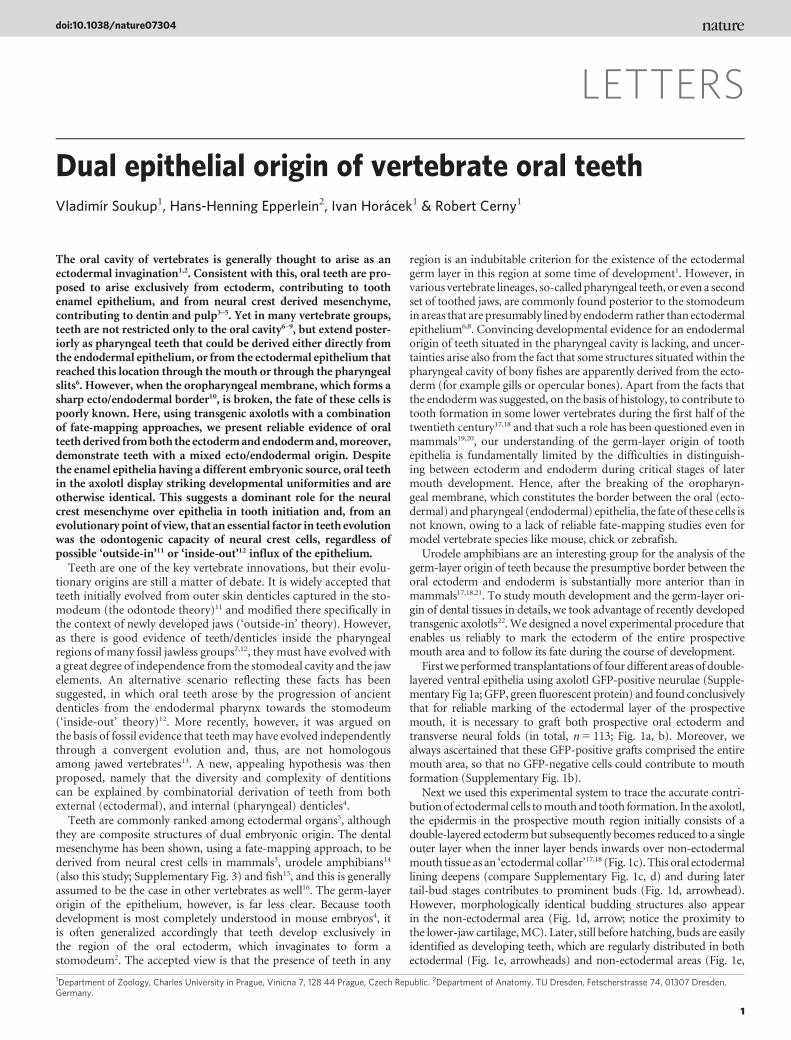
LETTERS
Dual epithelial origin of vertebrate oral teethVladimır Soukup1, Hans-Henning Epperlein2, Ivan Horacek1 & Robert Cerny1
The oral cavity of vertebrates is generally thought to arise as anectodermal invagination1,2. Consistent with this, oral teeth are pro-posed to arise exclusively from ectoderm, contributing to toothenamel epithelium, and from neural crest derived mesenchyme,contributing to dentin and pulp3–5. Yet in many vertebrate groups,teeth are not restricted only to the oral cavity6–9, but extend poster-iorly as pharyngeal teeth that could be derived either directly fromthe endodermal epithelium, or from the ectodermal epithelium thatreached this location through themouth or through the pharyngealslits6. However, when the oropharyngeal membrane, which forms asharp ecto/endodermal border10, is broken, the fate of these cells ispoorly known. Here, using transgenic axolotls with a combinationof fate-mapping approaches, we present reliable evidence of oralteethderived fromboth the ectodermandendodermand,moreover,demonstrate teeth with a mixed ecto/endodermal origin. Despitethe enamel epithelia having a different embryonic source, oral teethin the axolotl display striking developmental uniformities and areotherwise identical. This suggests a dominant role for the neuralcrest mesenchyme over epithelia in tooth initiation and, from anevolutionary point of view, that an essential factor in teeth evolutionwas the odontogenic capacity of neural crest cells, regardless ofpossible ‘outside-in’11 or ‘inside-out’12 influx of the epithelium.
Teeth are one of the key vertebrate innovations, but their evolu-tionary origins are still a matter of debate. It is widely accepted thatteeth initially evolved from outer skin denticles captured in the sto-modeum (the odontode theory)11 and modified there specifically inthe context of newly developed jaws (‘outside-in’ theory). However,as there is good evidence of teeth/denticles inside the pharyngealregions of many fossil jawless groups7,12, they must have evolved witha great degree of independence from the stomodeal cavity and the jawelements. An alternative scenario reflecting these facts has beensuggested, in which oral teeth arose by the progression of ancientdenticles from the endodermal pharynx towards the stomodeum(‘inside-out’ theory)12. More recently, however, it was argued onthe basis of fossil evidence that teethmay have evolved independentlythrough a convergent evolution and, thus, are not homologousamong jawed vertebrates13. A new, appealing hypothesis was thenproposed, namely that the diversity and complexity of dentitionscan be explained by combinatorial derivation of teeth from bothexternal (ectodermal), and internal (pharyngeal) denticles4.
Teeth are commonly ranked among ectodermal organs5, althoughthey are composite structures of dual embryonic origin. The dentalmesenchyme has been shown, using a fate-mapping approach, to bederived from neural crest cells in mammals3, urodele amphibians14
(also this study; Supplementary Fig. 3) and fish15, and this is generallyassumed to be the case in other vertebrates as well16. The germ-layerorigin of the epithelium, however, is far less clear. Because toothdevelopment is most completely understood in mouse embryos4, itis often generalized accordingly that teeth develop exclusively inthe region of the oral ectoderm, which invaginates to form astomodeum2. The accepted view is that the presence of teeth in any
region is an indubitable criterion for the existence of the ectodermalgerm layer in this region at some time of development1. However, invarious vertebrate lineages, so-calledpharyngeal teeth, or even a secondset of toothed jaws, are commonly found posterior to the stomodeumin areas that are presumably lined by endoderm rather than ectodermalepithelium6,8. Convincing developmental evidence for an endodermalorigin of teeth situated in the pharyngeal cavity is lacking, and uncer-tainties arise also from the fact that some structures situated within thepharyngeal cavity of bony fishes are apparently derived from the ecto-derm (for example gills or opercular bones). Apart from the facts thatthe endodermwas suggested, on the basis of histology, to contribute totooth formation in some lower vertebrates during the first half of thetwentieth century17,18 and that such a role has been questioned even inmammals19,20, our understanding of the germ-layer origin of toothepithelia is fundamentally limited by the difficulties in distinguish-ing between ectoderm and endoderm during critical stages of latermouth development. Hence, after the breaking of the oropharyn-geal membrane, which constitutes the border between the oral (ecto-dermal) andpharyngeal (endodermal) epithelia, the fate of these cells isnot known, owing to a lack of reliable fate-mapping studies even formodel vertebrate species like mouse, chick or zebrafish.
Urodele amphibians are an interesting group for the analysis of thegerm-layer origin of teeth because the presumptive border between theoral ectoderm and endoderm is substantially more anterior than inmammals17,18,21. To study mouth development and the germ-layer ori-gin of dental tissues in details, we took advantage of recently developedtransgenic axolotls22.We designed a novel experimental procedure thatenables us reliably to mark the ectoderm of the entire prospectivemouth area and to follow its fate during the course of development.
Firstweperformed transplantations of four different areas of double-layered ventral epithelia using axolotl GFP-positive neurulae (Supple-mentary Fig 1a; GFP, green fluorescent protein) and found conclusivelythat for reliable marking of the ectodermal layer of the prospectivemouth, it is necessary to graft both prospective oral ectoderm andtransverse neural folds (in total, n5 113; Fig. 1a, b). Moreover, wealways ascertained that these GFP-positive grafts comprised the entiremouth area, so that no GFP-negative cells could contribute to mouthformation (Supplementary Fig. 1b).
Next we used this experimental system to trace the accurate contri-butionof ectodermal cells tomouthand tooth formation. In the axolotl,the epidermis in the prospective mouth region initially consists of adouble-layered ectodermbut subsequently becomes reduced to a singleouter layer when the inner layer bends inwards over non-ectodermalmouth tissue as an ‘ectodermal collar’17,18 (Fig. 1c). This oral ectodermallining deepens (compare Supplementary Fig. 1c, d) and during latertail-bud stages contributes to prominent buds (Fig. 1d, arrowhead).However, morphologically identical budding structures also appearin the non-ectodermal area (Fig. 1d, arrow; notice the proximity tothe lower-jaw cartilage,MC). Later, still before hatching, buds are easilyidentified as developing teeth, which are regularly distributed in bothectodermal (Fig. 1e, arrowheads) and non-ectodermal areas (Fig. 1e,
1Department of Zoology, Charles University in Prague, Vinicna 7, 128 44 Prague, Czech Republic. 2Department of Anatomy, TU Dresden, Fetscherstrasse 74, 01307 Dresden,Germany.
doi:10.1038/nature07304
1

arrows). To confirm the identity of these structures as tooth buds, weused an antibody directed against calbindin (Sigma), a calcium-bindingprotein that specifically recognizes ameloblasts23. From sections whereboth GFP and immunostaining is visualized (Fig. 1f, g andSupplementary Fig. 2a, b), it is evident that the tooth primordia aredeveloping from both ectodermal and non-ectodermal epithelia.
To substantiate our finding that in the Mexican axolotl some oralteeth develop from non-ectodermal epithelia, we invented a double-labelling approach using which cells of both oral ectoderm and fore-gut endoderm can be reliablymarked andmapped (Fig. 2a). First, at aneurula stage, the double-layered prospective oral epithelium (fromthe same area as in the previous experiment) was extirpated. Theexposed endodermal layer was then focally injected using the lipo-philic dye DiI (Molecular Probes). Next a GFP-positive graft com-prising the entire prospective oral ectoderm (as above) wastransplanted orthotopically to wild-type host embryos. In thisapproach (n5 91), the entire prospective ectoderm of the oral areawas marked with a green fluorescent dye and some of the foregutendodermal cells, expected to contribute to tooth buds, were labelledwith a red fluorescent dye.
Using this double-fate-mapping approach, we obtained strongsupport for our previous conclusions that the axolotl possesses oralteeth with an epithelial lining of non-ectodermal origin. Specifically,dye injected into the foregut endoderm at the neurula stage wasfound in oral tooth germs and later in developing teeth (Fig. 2b–d,h and Supplementary Fig. 4), as well as, notably, in the epitheliumsituated between GFP-positive ectodermal epithelia (Fig. 2c).Moreover, alongside the contact zone between the ecto- and endo-dermal oral epithelia, we found tooth germs that consistently dem-onstrate a mixed contribution from both ecto- and endodermal cellsto their enamel epithelia (Fig. 2e–g and Supplementary Fig. 5). Onthe basis of our combined tracing approaches, we conclude that onthe upper jaw the enamel epithelia of the premaxillary/maxillaryteeth are always ectodermal, whereas the enamel epithelia of thevomero-palatal teeth are derived from the ectoderm, endoderm or
b
f
Stage 14
Oral cavityMouth
GFP ECT + fibronectinGFP ECT + fibronectin GFP ECT + fibronectinGFP ECT + fibronectin
GFP ECT transplant
Mouth
Stage 36 Stage 39
n = 113
Stage 43
Stage 43
2 h after operation
MA MC
Tr
Tr
a
e
f
dc
GFP ECT + calbindin GFP ECT + calbindinGFP ECT + calbindin GFP ECT + calbinding
Figure 1 | Ectoderm contribution to mouth and tooth formation in theMexican axolotl. a, An experimental scheme with the prospective oralectoderm (ECT) transplanted from a GFP-positive donor to a host embryo,ventral view (END, endoderm). b, An embryo 2 h after operation.c–g, Paramedial sections, head to the left, showing a contribution of the oralECT (green) to mouth and tooth formation. DAPI (blue) stains cell nuclei;fibronectin (red in c, d) marks cell and tissue borders. Initially the oral ECT(green) inflexes as a stomodeal collar (c). Then prominent tooth budsdevelop in ECT areas (arrowheads) as well as in non-ECT areas (arrows;d, e). Tooth buds, identified using anti-calbindin (red), develop within ECTareas (arrowheads) as well as non-ECT areas (arrows; f, g). Tr, trabecula;MA, mandibular arch; MC, Meckel’s cartilage. c, d, Vibratome 100-mmsections; e–g, cryostat 20-mm sections. Scale bars, 50 mm.
(3) GFP ECT transplantation
ECT, green; END, red;n = 91
(2) DiI END injection
(1) Oral ECT extirpation
c
f
h
MC
MCMC
GFP + calbindin GFP + calbindin
GFP + calbindin
GFP + DiI
DiI
GFP + DiI
GFP + calbindin
GFP + calbindin
GFP + DiI GFP + DiI
GFP + calbindin
GFP + DiI
DiI
GFP + DiI
Vomeral tooth
Splenial tooth
Dentary+splenial teeth
d
e
g
Vomeral tooth
Splenial teeth
Dentary+splenial teeth
a
b
Figure 2 | Endoderm contribution to tooth formation. a, Sketch of double-fate-mapping experiment: following extirpation of the prospective oral ECT(1), DiI was injected into foregut END (2) and the prospective oral ECT(GFP-positive) was transplanted orthotopically (3). b–h, Paramedial(cryostat 20-mm) sections, head to the left, showing a contribution of the oralECT (green) and mouth END (red in b–d, h) to tooth formation. DAPI(blue) stains cell nuclei, calbindin (red in e–g) marks tooth buds. Arrowspoint to END teeth; arrowheads to ECT teeth. e–g, Details of teeth of mixedorigin. h, A confocal image, inset in g, showing the END (DiI, red)contribution to splenial tooth germ. Scale bars, 25 mm.
LETTERS NATURE
2

from a mixed source, according to their position (Fig. 3a). On thelower jaw, dentary teeth are basically ectodermal and splenial endo-dermal; however, there are teeth of mixed origin situated on theanterior parts of these fields (Fig. 3a).
Next a quantitative screening was performed in which all teethwere counted and their respective germ-layer origins determined atfour different stages and based on 26 embryos from the double-labelling experiment (Fig. 4). This analysis revealed that of 1,137teeth, 374 were derived from ectoderm, 598 from endoderm, and155 were of mixed ecto/endodermal origin. We note that duringthe course of development, the proportion of ectoderm-derived teethslightly increases as teeth located on the premaxillary and maxillarybones, which are purely ectodermal, develop very late. Thus, in theaverage embryo (analysed at stage 45, when the mouth opens andanimals start to eat), of 82 teeth 29 were of ectoderm, 42 were ofendoderm and 11 were of mixed epithelial origin (SupplementaryTables 1–4). Non-epithelial derivatives, such as tooth dentin andpapillae, were derived from neural crest mesenchyme (from the tri-geminal neural crest stream; Supplementary Fig. 3). All quantitativeand statistical analyses were strongly significant (SupplementaryTables 1–4) and constitute robust support that our data are notbiased by any technical problems.
Previous theories have identified the ectodermal border in themouth as being central to tooth positioning11 (Fig. 3b, upper row).However, in the Mexican axolotl, the oral ectoderm does not form atrue stomodeum17,18. Instead, only an inner ectodermal layer bendsinwards as a stomodeal collar over the dense endodermal rod, whichblocks the prospective mouth at early stages of development (Fig. 3b,
lower row). Because of this positioning, the collar cells develop intothe basal cells, and the outer cells of the endodermal rod develop intothe apical cells of the oral epithelium during the course of mouthopening (Fig. 1e and Supplementary Fig. 2a–d; summarized inFig. 3b, lower row). The endodermal cells of the mouth, as part ofthe epithelial lining, are consequently found also on the outer surfaceof the mouth (Fig. 1e and Supplementary Fig. 2b–d, arrowheads;summarized in Fig. 3b, lower row). Thus, in the axolotl, the posteriorpart of the oral cavity is lined with the endodermal epithelium,whereas the anterior part is lined with an epithelium of double origin(Fig. 3b and Supplementary Fig. 2b). This provides reliable docu-mentation of an oral endodermal epithelial lining that reaches outsidethemouth, and, also, of an oral epithelium originating from two germlayers. A considerable number of reports onmouth development havebeen published, but, as underlined by our results, there is still a needfor detailed fate-mapping approaches in studies of dynamic interac-tions of cells and tissues derived from different germ layers.
Progressing from recent vivid discussions on the subject4,6,8,24, ourdata present reliable evidence of oral teeth of endodermal origin invertebrates. We speculate that oral teeth of endodermal origin mightform in all animalswith oral endoderm, that is, in urodele andprobablyalso lungfish species, andmaybe even in some frogs25, where themouthdevelops froma structure similar to the stomodeal collar.However, as apossible interdigitation of cells fromboth epithelial tissue layers duringmouth formation has not been fate-mapped for any vertebrate species,and some reports indicate that foregut endodermmay stretch more to
eECT
Upper jaw:Pre/max, premaxillary/maxillary
Vom-pal, vomero-palatal
Lower jaw:Den, dentarySpl, splenial
Tooth field origin:ECT / END
END
n
MC
Mouth formationEarly tailbud
A generalvertebrate
scheme
A schemeof axolotl
development
Teeth development
Den
Spl-Pal
Vom-
Pre/MaxPre/Max
TR
MC
TR
MC
PQPQ
Stomodeum ECT teeth
Stomodeal collar ECT + END teeth
b
a
Figure 3 | Germ-layer origin and morphogenesis of teeth of the Mexicanaxolotl. a, A sketch of the germ-layer origin of teeth in the Mexican axolotl.ECT teeth, green; END teeth, red; teeth of mixed origin, red–green (thecolouring in the key is a qualitative guide to the ratio of the components ineach tooth field). Cartilages visualized using alcian blue. b, Comparativedevelopmental morphogenesis of the mouth region and the germ-layerorigin of teeth of vertebrate (upper row) and an axolotl (lower row) embryo.In the majority of vertebrates, the mouth develops from a stomodeum withteeth distributed in invaginated ECT. In contrast, in urodeles the mouthdevelops from a stomodeal collar with an oral epithelium either of a dualorigin, with teeth of ECT or END, or of a mixed origin. PQ, palatoquadrate;TR, trabecula cranii; n, nose; e, eye.
44-45
Tooth field
Num
ber o
f tee
th
Premaxillary/maxillary
10
20
30
10
20
30
10
20
30
10
20
Stage 39
Stage 40–41
Stage 42–43
Stage 44–45
30
Dentary Vomero-palatal Splenial
Figure 4 | A quantitative screening of teeth fromdifferent tooth fieldswithrespect to their germ-layer origin. n5 1,137 teeth: 374 ECT (green), 598END (red), 155 ECT–END mixture (yellow). Data based on 26 animals,visualized at four stages: four animals at stage 39, 14 animals at stage 40–41,six animals at stage 42–43, two animals at stage 44–45. For details andoriginal data, see Supplementary Tables 1–4.
NATURE LETTERS
3

the anterior than hitherto believed8,19–21, we speculate that oral teeth ofendodermal originmight present amore common feature in vertebrateoral development than previously assumed.
Whereas the classical ‘outside-in’ theory implies that teeth wereinitially derived from the oral ectodermal layer11, the ‘inside-out’ the-ory strongly suggests that they were derived from the endodermallayer12, and this derivation is believed to impart differences to denticles,teeth or dentition in terms of shape and complexity4,12,24. However, thedual origin of enamel epithelia in otherwise morphologically identicalaxolotl oral tooth primordia (as regards complexity, shape, position,timing and morpho-differentiation of teeth), together with studiesillustrating deep shared molecular similarities between oral (suppo-sedly ectodermal) and pharyngeal (supposedly endodermal)teeth15,26,27 imply that ‘ectodermal’ and ‘endodermal’ teethdonotdifferessentially. It is beyond the scope of this study to identify the plesio-morphic germ-layer origin of tooth epithelium. However, our resultsclearly demonstrate that the germ-layer origin of epithelium intowhich themesenchyme cells come into contact does not affect the finalproduct of the odontogenic cascade.Mesenchymecells can thus appar-ently interact with a host of epithelial cells, forming teeth/denticleswhen in the stomodeum, in the pharyngeal cavity or on the skin sur-face. All this suggests that themajor agent of dental development is theneural crest mesenchyme rather than the epithelium, the role of whichin tooth patterning5,12,28 and even in tooth initiation29 may be lessfundamental than commonly believed. It therefore seems most likelythat all teeth of extant vertebrates—or, more precisely, the devel-opmentalmachinery producing them—have evolvedonly once, some-where in the oropharynx, driven by a neural crest signal.
METHODS SUMMARYEmbryos. Embryos of the Mexican axolotl (Ambystoma mexicanum) wereobtained, reared and staged as previously described30. GFP embryoswere obtainedfromtheMax-Planck-InstituteofMolecularCellBiology andGenetics inDresden,Germany, and were developed in the laboratory of E. Tanaka22.Operations and injections.GFP ectodermal transplantations were performed assketched in Fig. 1a (n5 113). At first, however, transplantations of four differentareas of double-layered ventral epithelia were performed (Supplementary Fig.1a) to define the entire ectodermal layer of the prospective mouth.The double-labelling approach by which cells of both oral ectoderm and
foregut endoderm were marked and mapped (as sketched in Fig. 2a (n5 91))includes extirpation of the double-layered prospective oral epithelia (from thesame area as in the previous experiment), focal injection of a CellTracker CM-DiI (Molecular Probes) into the exposed endodermal layer and, lastly, the ortho-topic transplantation of a GFP-positive prospective oral ectoderm (as above)into wild-type host embryos.Sectioning and immunostaining. Axolotl embryos were anaesthetized usingMS-222 (Sigma), fixed in 4% paraformaldehyde in phosphate buffered salineand sectioned using a Vibratome 1000 sectioning system (Ted Pella) or aCM3050 cryostat (Leica). Sections were counterstained using anti-fibronectinantibody (Dako) to visualize tissue borders, with 4,6-diamidino-2-phenylindole(DAPI) to mark cell nuclei, or with anti-calbindin antibody (Sigma), whichspecifically recognizes ameloblasts23.Image acquisition. Separate fluorescence images were captured using anOlympus BX51 microscope with a SPOT RT camera, or the Olympus CellR
IX81 with a Hamamatsu Photonics Orca camera, merged and optimized usingSpot and Adobe Photoshop software.
Full Methods and any associated references are available in the online version ofthe paper at www.nature.com/nature.
Received 9 January; accepted 29 July 2008.Published online XX 2008.
1. Romer, A. S. The Vertebrate Body (Saunders, 1962).2. Kardong, K. V.Vertebrates. Comparative Anatomy, Function, Evolution (Brown, 1995).3. Chai, Y. et al. Fate of the mammalian cranial neural crest during tooth and
mandibular morphogenesis. Development 127, 1671–1679 (2000).4. Tucker, A. & Sharpe, P. The cutting-edge of mammalian development: how the
embryo makes teeth. Nature Rev. Genet. 5, 499–508 (2004).5. Pispa, J. & Thesleff, I. Mechanisms of ectodermal organogenesis. Dev. Biol. 262,
195–205 (2003).
6. Stock, D.W. The genetic basis of modularity in the development and evolution ofthe vertebrate dentition. Phil. Trans. R. Soc. Lond. B 356, 1633–1653 (2001).
7. Janvier, P. Early Vertebrates (Oxford Univ. Press, 1996).8. Huysseune, A., Van der Heyden, C., Verreijdt, L., Wautier, K. & Van Damme, N.
Fish dentitions as paradigms for odontogenic questions. Connect. Tissue Res. 43,98–102 (2002).
9. Sire, J. Y., Davit-Beal, T., Delgado, S., Van Der Heyden, C. & Huysseune, A. First-generation teeth in nonmammalian lineages: evidence for a conserved ancestralcharacter? Microsc. Res. Tech. 59, 408–434 (2002).
10. Dickinson, A. J. & Sive, H. Development of the primary mouth in Xenopus laevis.Dev. Biol. 295, 700–713 (2006).
11. Reif, W.-E. Evolution of dermal skeleton and dentition in vertebrates: theodontode-regulation theory. Evol. Biol. 15, 287–368 (1982).
12. Smith, M. M. & Coates, M. I. in Major Events in Early Vertebrate Evolution (ed.Ahlberg, P. E.) 223–240 (Taylor and Francis, 2001).
13. Smith, M. M. & Johanson, Z. Separate evolutionary origins of teeth from evidencein fossil jawed vertebrates. Science 299, 1235–1236 (2003).
14. Chibon, P. Analyse experimentale de la regionalisation et des capacitesmorphogenetiques de la crete neurale chez l’Amphibien Urodele Pleurodeleswaltlii Michah. Mem. Soc. Zool. Fr. 36, 1–107 (1966).
15. Jackman,W. R., Draper, B.W. & Stock, D.W. Fgf signaling is required for zebrafishtooth development. Dev. Biol. 274, 139–157 (2004).
16. Hall, B. K. The Neural Crest in Development and Evolution (Springer, 1999).17. de Beer, G. R. The differentiation of neural crest cells into visceral cartilages and
odontoblasts inAmblystoma, and re-examination of the germ-layer theory. Proc. R.Soc. Lond. B 134, 377–398 (1947).
18. Adams, A. E. An experimental study of the development of the mouth in theamphibian embryo. J. Exp. Zool. 40, 311–379 (1924).
19. Imai, H., Osumi, N. & Eto, K. Contribution of foregut endoderm to tooth initiationof mandibular incisor in rat embryos. Eur. J. Oral Sci. 106 (Suppl. 1), 19–23 (1998).
20. Sharpe, P. T. Homeobox genes and orofacial development. Connect. Tissue Res. 32,17–25 (1995).
21. Barlow, L. A. & Northcutt, R. G. Embryonic origin of amphibian taste buds. Dev.Biol. 169, 273–285 (1995).
22. Sobkow, L., Epperlein, H.H., Herklotz, S., Straube,W. L. & Tanaka, E.M.A germlineGFP transgenic axolotl and its use to track cell fate: dual origin of the finmesenchyme during development and the fate of blood cells during regeneration.Dev. Biol. 290, 386–397 (2006).
23. Barlow, L. A. &Northcutt, R. G. Taste buds develop autonomously from endodermwithout induction by cephalic neural crest or paraxial mesoderm. Development124, 949–957 (1997).
24. Smith, M. M. & Coates, M. I. Evolutionary origins of the vertebrate dentition:phylogenetic patterns and developmental evolution. Eur. J. Oral Sci. 106 (Suppl. 1),482–500 (1998).
25. Reiss, J. O. Early development of chondrocranium in the tailed frog Ascaphus truei(Amphibia: Anura): implications for anuran palatoquadrate homologies. J.Morphol. 231, 63–100 (1997).
26. Debiais-Thibaud, M. et al. Development of oral and pharyngeal teeth in themedaka (Oryzias latipes): comparison ofmorphology and expression of eve1 gene.J. Exp. Zool. B Mol. Dev. Evol. 308, 693–708 (2007).
27. Fraser, G. J., Graham, A. & Smith, M. M. Conserved deployment of genes duringodontogenesis across osteichthyans. Proc. R. Soc. Lond. B 271, 2311–2317 (2004).
28. Smith, M. M. Vertebrate dentitions at the origin of jaws: when and how patternevolved. Evol. Dev. 5, 394–413 (2003).
29. Mitsiadis, T. A., Caton, J. & Cobourne, M. Waking-up the sleeping beauty:recovery of the ancestral bird odontogenic program. J. Exp. Zool. B Mol. Dev. Evol.306, 227–233 (2006).
30. Cerny, R. et al. Combined intrinsic and extrinsic influences pattern cranial neuralcrest migration and pharyngeal arch morphogenesis in axolotl. Dev. Biol. 266,252–269 (2004).
Supplementary Information is linked to the online version of the paper atwww.nature.com/nature.
AcknowledgementsWe wish to thank E. Tanaka and H. Andreas for supplying uswith GFP axolotl embryos, L. Mchedlishvili for technical advice and O. Sebesta forhelp with confocal microscopy. We are grateful to M. Bronner–Fraser, R. Ericsson,D. Meulemans Medeiros, L. Olsson, P. D. Polly, D. W. Stock and A. Tucker forcomments on the manuscript. Grants from the Ministry of Youth, Education andSport of the Czech Republic (MSMT 0021620828), SMWK (Dresden) and COSTAction B23 ‘Oral facial development and regeneration’ are gratefully acknowledged.
Author Contributions Tissue grafting was carried out in the laboratory of H.-H.E.,who also helped with initial experiments. V.S. made transplantations andperformed sectioning and image analyses as his Master’s thesis under thesupervision of R.C., who also designed the initial experiments and wrote themanuscript. R.C. and I.H. planned the study and interpreted the results, I.H. madestatistical analyses and helped in writing the manuscript, and all authors discussedthe results and commented on the manuscript.
Author Information Reprints and permissions information is available atwww.nature.com/reprints. Correspondence and requests for materials should beaddressed to R.C. ([email protected]).
LETTERS NATURE
4

METHODSEmbryos. Embryos of the Mexican axolotl (A. mexicanum) were obtained,reared and staged as previously described30,31. GFP embryos were obtained fromthe Max-Planck-Institute of Molecular Cell Biology and Genetics in Dresden,Germany, and were spawned from a b-actin promoter-driven GFP germ-linetransgenic animal that had been produced by plasmid injection22. Embryos werekept in tap water, and before being used for transplantations and injections,embryos were washed thoroughly with tap water and sterile Steinberg solutioncontaining antibiotics (Antibiotic-Antimycotic, Gibco) and then decapsulatedmanually.Operations and injections.GFP ectodermal transplantations were performed assketched in Fig. 1a (n5 113). Operations were performed under sterile condi-tions using tungsten needles in an agar dish containing 1M Steinberg solutionplus antibiotics.We designed an experimental procedure that enabled us tomarkthe ectoderm of the entire prospective mouth area reliably and to follow its fateduring the course of development. First we performed transplantations of fourdifferent areas of double-layered ventral epithelia from GFP-positive to hostneurulae (Supplementary Fig. 1a; numbers of animals used for each operationare indicated there) and found conclusively that for reliable marking of theectodermal layer of the prospective mouth it is necessary to graft both prospect-ive oral ectoderm and a transverse neural fold (Fig. 1a, b). Using this type oftransplantation, wewere able to follow the fate of the entire ectodermal layer thattranslocates into the mouth, and, therefore, in this way all ectoderm-derivedteeth became GFP-positive. We always ascertained, however, that these GFPgrafts comprised the entire mouth area, so that no GFP-negative cells couldcontribute to mouth formation (Supplementary Fig. 1b).Next we invented a double-labelling approach by which cells of both oral
ectoderm and foregut endoderm can be reliably marked and mapped (Fig. 2a).First, at a neurula stage, double-layered prospective oral epithelia (from the samearea as in the previous experiment) were extirpated. The exposed endodermallayer was then focally injected using the lipophilic dye DiI (Molecular Probes),dissolved in absolute ethanol to a concentration of 1mgml21 and further dilutedin nine parts of 10% sucrose in water just before injection. Then a graft from aGFP-positive neurula comprising the entire prospective oral ectoderm (asabove) was transplanted orthotopically to wild-type host embryos. In thisapproach (n5 91), the entire prospective ectoderm of the oral area was markedgreen (GFP) and some of foregut endodermal cells, expected to contribute totooth buds, were labelled red (DiI).Neural crest transplantations. Trigeminal neural crest cells were transplantedfrom GFP-positive to wild-type embryos at the neurula stage as described indetail elswhere32.
31. Epperlein, H. H.,Meulemans, D., Bronner-Fraser,M., Steinbeisser, H. & Selleck,M.A. Analysis of cranial neural crest migratory pathways in axolotl using cellmarkers and transplantation. Development 127, 2751–2761 (2000).
32. Cerny, R. et al.Developmental origins and evolution of jaws: new interpretation of‘‘maxillary’’ and ‘‘mandibular’’. Dev. Biol. 276, 225–236 (2004).
doi:10.1038/nature07304