Embed Size (px)
Citation preview
International Journal of Oral Health and Medical Research | ISSN 2395-7387 | MAY-JUNE 2018 | VOL 5 | ISSUE 1 57
REVIEW ARTICLE Roy G et al.: Oral Leukoplakia
Correspondence to: Dr. Gigi Roy, Department of Oral Medicine and
Radiology, Mar Baselios Dental College, Kothamangalam, Kerala, India Contact Us: www.ijohmr.com
Oral Leukoplakia: An Insight Gigi Roy1, Anu Vijayan2, Shamji Shajahan3, Anuja S4, Rashmi Elizabeth Mathen5
Leukoplakia is the most common oral white lesion that is classified under potentially malignant disorder affecting oral
mucosa. It is significant as it has a high risk of malignant transformation. This article reviews the epidemiology,
etiology and pathogenesis, clinical features and clinical variants, diagnosis, differential diagnosis, malignant potential,
histopathological features, and treatment. KEYWORDS: Potentially Malignant Disorder, Smoking, Malignant, Dysplasia, Leukoplakia, Oral Cancer
asasss Earlier clinical presentations of the oral cavity that are
recognized as precancerous were classified into two broad
groups like lesions and conditions1, with the following
definitions: a precancerous lesion is a morphologically
altered tissue in which oral cancer is more likely to occur
than in its apparently normal counterpart, and a
precancerous condition is a generalized state associated
with a significantly increased risk of cancer. But now all
clinical presentations that carry a risk of cancer come
under the term ‗potentially malignant disorders‘ to reflect
their widespread anatomical distribution.2
Leukoplakia (leukos meaning white; plakia meaning
patch) is the most common potentially malignant disorder
affecting the oral mucosa. The term leukoplakia was
coined by Schwimmer in 1877. It has been defined as ―a
predominantly white lesion of the oral mucosa that
cannot be characterized as any other definable lesion, not
associated with any physical or chemical causative agent
except the use of tobacco‖.3
Warnakulasuriya et al.in 2007 defined Leukoplakia
should be used to recognize white plaques of
questionable risk having excluded (other) known diseases
or disorders that carry no increased risk for cancer2.
According to Warnakulasuriya et al. , the new concept of
OL shall acknowledge white lesions with a questionable
risk of being an OL, being excluded any other
pathologies or known disorders which do not present
potential malignant risks such as candidiasis, lupus
erythematosus, lichen planus, hairy leukoplakia, frictional
keratosis, nicotinic stomatitis, and leukoedema.4
Global prevalence of oral leukoplakia range from 0.5% to
3.46%, and of the rates of carcinomatous transformation
of oral leukoplakia from 0.7% to 2.9%.5 Oral leukoplakia
is more prevalent in India where persons smoke and
practice the habit of tobacco and areca nut chewing more
than elsewhere6. Prevalence in India is 0.2-4.9 percent.
6,7
Oral leukoplakia is usually seen in middle aged people,
and its prevalence is higher with age. About 90% of oral
leukoplakias are associated with the use of tobacco/areca
nut and remaining 10% are idiopathic. Males are more
often affected than females probably owing to the greater
prevalence of tobacco use by males. The buccal mucosa
is affected in 25% of cases, the mandibular gingiva in
20%, the tongue in 10%, the floor of the mouth in 10%,
and other oral sites account for the remainder.6
Tobacco: The etiology of OL is considered
multifactorial, but smoking is appreciated to be a
frequently involved factor. It is much more common
among smokers than among nonsmokers8. Either in
smoke or smokeless (chew) form is the main etiologic
factor. Smoke/ Smokeless form release carcinogens that
either bind to epithelial DNAs and cause damage to DNA
causing mutation and resulting in dysplasia or malignant
transformation of a cell or highly reactive radicals are
formed which damage cell membrane, DNA
fragmentation, tissue damage and alter cellular
antioxidant defense system and causes keratinocyte
stimulation resulting in hyperkeratinization. Heat from
smoke and frictional irritation from chew form cause
keratinocyte stimulation as a protective response causing
hyperkeratinization9.
Alcohol- Individuals with leukoplakia generally consume
alcohol and tobacco together. Consuming alcohol alone
was not associated with development of leukoplakia. But
it was found to have some synergistic effect with tobacco
in the development of both leukoplakia and oral cancer3.
Mechanical trauma- Continuous trauma in the form of
chronic cheek biting, ill-fitting dentures, etc. have also
found to be causative factors for leukoplakia.3
Sanguinaria- Herbal extract sanguinaria which is used in
mouthwashes and toothpastes was found to develop
How to cite this article: Roy G, Vijayan A, Shajahan S, Anuja S, Mathen RE. Oral Leukoplakia: An Insight. Int J Oral Health Med Res 2018;5(1):57-61.
INTRODUCTION
1,3,4,5-Post Graduate, Department of Oral Medicine and Radiology, Mar Baselios Dental College, Kothamangalam, Ernakulam(Dist), Kerala, India. 2-Senior Lecturer,Department of Oral Medicine and Radiology, Mar Baselios Dental College, Kothamangalam, Ernakulam(Dist), Kerala, India.
ABSTRACT
EPIDEMIOLOGY
ETIOLOGY AND PATHOGENESIS
International Journal of Oral Health and Medical Research | ISSN 2395-7387 | MAY-JUNE 2018 | VOL 5 | ISSUE 1 58
REVIEW ARTICLE Roy G et al.: Oral Leukoplakia
leukoplakia (Sanguinaria-associated keratosis). Even after
stopping usage of this product, the lesion did not subside.
The commonest site was maxillary vestibule and alveolar
mucosa.3
Candida causing leukoplakia- Candida releases
nitrosamine specific proto-oncogene and also efficiently
converts ethanol into carcinogenic acetaldehyde in
alcohol drinker. This has a synergistic effect with tobacco
which increases dysplasia or leukoplakia. Studies show
that even after elimination of surface mycosis after
administration of antifungals, the leukoplakia persisted.
Malignant transformation of candida infected
leukoplakias was high, suggesting candida association as
a significant risk factor for oncogenesis.9
Papilloma virus- The role of Human Papilloma Virus
(HPV) in the etiology as well as oncogenesis of oral
leukoplakia have been under extensive molecular biology
and virology studies. HPV type 16 was isolated in oral
leukoplakias and carcinomas. In Proliferative Verrucous
leukoplakia (PVL), HPV 16 and 18 were demonstrated.3
Epstein barr virus (EBV)- The role of EBV in oral
leukoplakias was not found in any of the studies even
though it was found to be associated with etiology of oral
squamous cell carcinomas. If there is any role of EBV in
oral leukoplakias studies carrying out on a larger sample
may help us. 3
Leukoplakia is seen in middle age and older age with a
peak incidence in fifth to seventh decades. It is more
prevalent in males than females. Generally, it is seen as
gray, white or yellowish white in color. OL present on the
floor of the mouth, soft palate, and tongue are considered
as high-risk lesions, while, they may be considered as
low malignant risk when occurs in other areas4. Common
sites are buccal mucosa and commissures, followed by
lips, tongue, palate, alveolar ridge, floor of mouth, soft
palate and gingiva.7
Axell et al.,19849
1. Etiological
Tobacco-associated leukoplakia
Idiopathic leukoplakia
2. Clinically
Homogenous
Non homogenous
Erosive
Nodular
Verrucous
Bouquot Je & Whitaker10
Phase 1- Thin or Preleukoplakia
Phase 2- Homogenous/ Fissured/ Thick leukoplakia
Phase 3- Granular/ Nodular/ Verruciform/ Rough
leukoplakia
Phase 4- Erythroleukoplakia/ Speckled/ Non homogenous
leukoplakia
Pindborg11
Homogeneous leukoplakia
Non homogeneous leukoplakia
Bailoor and Nagesh 12
Speckled leukoplakia and non speckled leukoplakia
Homogenous, Ulcerative, Speckled
Reversible / irreversible
WHO (1980)11
Homogeneous
Smooth
Furrowed(Fissured)
Ulcerated
Non Homogeneous
Nodulospeckled
Preleukoplakia usually begins as thin, grey or greyish
white plaque that may appear somewhat translucent.9,10
Homogenous leukoplakia is thick smooth whitish with
cracked mud appearance and leathery consistency. These
are asymptomatic with a very low risk of malignancy.9,10
Granular/ Nodular/ Verruciform/ Rough leukoplakia are more severe form with surface irregularities like
nodular or granular or pointed papillary projections.9,10
Non- homogenous leukoplakia consists of erythematous
area seen on the whitish plaque. It is suggestive of further
progression of lesion towards malignancy. Patients
complain of pain, itching and discomfort.9,10
.
Proliferative verrucous leukoplakia is a subtype of
verrucous leukoplakia characterized by an aggressive
evolution, a multifocal appearance, resistance to
treatment, higher degree of recurrence and a high rate of
malignant transformation8. Hansen et al. (1985) first
described PVL as a distinct clinical form of OL. WHO
described the high rate of malignant transformation of
PVL. It is multifocal progressive lesions, commonly seen
in women. The most affected area was the lower gingival,
tongue, buccal mucosa, and alveolar ridge.3,13
There are four stages that have been described in the
development of PVL, initially as a simple hyperkeratosis
without epithelial dysplasia, followed by verrucous
hyperplasia, verrucous carcinoma, and finally
conventional carcinoma.14,15,16
Ghazali et al.,17
suggested
the following criteria for the diagnosis of PVL:
1. The lesion should start as homogeneous leukoplakia
with histopathological findings of dysplasia
2. Later in it should show verrucous areas
3. From single lesion, it should progress to multiple
lesions at the same or different site
4. It should progress later into different histological
stages.
5. It should show recurrence after treatment
Table 1 shows comparison of proliferative verrucous
leukoplakia and leukoplakia.18
CLINICAL FEATURES
CLINICAL VARIANTS
International Journal of Oral Health and Medical Research | ISSN 2395-7387 | MAY-JUNE 2018 | VOL 5 | ISSUE 1 59
REVIEW ARTICLE Roy G et al.: Oral Leukoplakia
Oral hairy leukoplakia (OHL): OHL is a white lesion
related to Epstein-Barr virus (EBV). It is usually
associated with AIDS. OHL is seen on lateral border of
the tongue, rarely on the buccal mucosa, with slightly
raised and corrugated hairy surface. These lesions are
also white in colour, cannot be scraped off and
asymptomatic like leukoplakias. As its etiological factor
is EBV, OHL must not be considered as a variant of
leukoplakia.3 Typically unilateral or bilateral, adherent,
slightly elevated whitish or gray patches. Principally
located mainly on lateral margins, dorsum, or ventrum of
the tongue Occasionally observed over the floor of the
mouth, palate, or oropharynx. Usually asymptomatic.19
Idiopathic leukoplakia: It is a rare potentially malignant
lesion with an increased risk of malignant transformation
as compared to the tobacco associated form. It is usually
found on the tongue. It can also develop on the gingiva.
Van der Waal et al., reported that idiopathic leukoplakia
have an incidence of 36%.20
On the basis of history and clinical examination, a
diagnosis of leukoplakia is made. To confirm the
diagnosis, a biopsy is done, so that proper treatment can
be planned. An incisional biopsy should be performed in
large lesions including some adjacent normal tissue and
an excisional biopsy should be performed in small
lesions. The main significance of incisional biopsy in
large lesions is to detect the dysplasia, grade of dysplasia
if present, as dysplasia, carcinoma in situ or invasive
carcinoma cannot be predicted clinically. Incisional
biopsy is done if the lesion is large in size, inaccessible
sites, multiple sites, and if the lesion is non homogenous.
It also helps in excluding other definable white lesions.
The site of the biopsy should be from symptomatic area
of the lesion. It should be taken from red or indurated
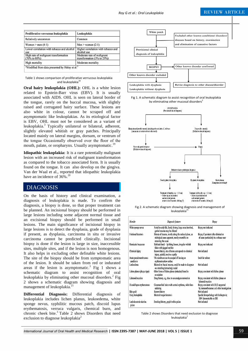
areas if the lesion is asymptomatic.3 Fig 1 shows a
schematic diagram to assist recognition of oral
leukoplakia by eliminating other mucosal disorders.2 Fig
2 shows a schematic diagram showing diagnosis and
management of leukoplakia.21
Differential Diagnosis: Differential diagnosis of
leukoplakia includes lichen planus, leukoedema, white
sponge nevus, syphilitic mucous patch, discoid lupus
erythematosis, verruca vulgaris, chemical burn, and
chronic cheek bite.3
Table 2 shows Disorders that need
exclusion to diagnose leukoplakia2
Table 1 shows comparison of proliferative verrucous leukoplakia and leukoplakia18
DIAGNOSIS
Fig 1. A schematic diagram to assist recognition of oral leukoplakia by eliminating other mucosal disorders2
Fig 2. A schematic diagram showing diagnosis and management of leukoplakia21
Table 2 shows Disorders that need exclusion to diagnose leukoplakia2

International Journal of Oral Health and Medical Research | ISSN 2395-7387 | MAY-JUNE 2018 | VOL 5 | ISSUE 1 60
REVIEW ARTICLE Roy G et al.: Oral Leukoplakia
Warnakulasuriya et al. were listed as a risk for malignant
transformation in PMD.22,2
1. Female gender
2. Long duration of leukoplakia
3. Leukoplakia in non-smokers (idiopathic leukoplakia)
4. Location on the tongue and/or floor of the mouth
5. Size >200 mm2
6. Non-homogeneous type
7. Presence of Candida albicans
8. Presence of epithelial dysplasia.
Fig 3 Clinical presentations of leukoplakia, progressing
from a low-risk malignant transformation potential on the
left to a high-risk potential on the right.11
Malignant
transformation of oral leukoplakias has been reported in
the range of 1%-20% over 1- 30 years.23
In some lesions epithelial dysplasia may be seen and may
range from mild to severe, based on its presence
leukoplakia is of two types dysplastic and non dysplastic.
Sublingual keratosis are bilateral and possess a parallel
corrugated wrinkled surface texture. This is called Ebbing
tide.
Table 3 shows histological changes in leukoplakia8. Table
4 shows Van der Waal et al., (2000) OLEP Classification
and Staging System3. Table 5 shows Leukoplakia
classification (WHO 1980).3
Conservative treatment includes3,9,24
Elimination of habit
Advise to take green tea
Enameloplasty to smoothen sharp teeth and
replacement of faulty restorations to avoid trauma
Nystatin therapy in case of candidal leukoplakia
Carotenoids and retinoids
Lycopene
Topical bleomycin
Photodynamic therapy
Gene therapy- Newer treatment in which synthetic
antisense oligonucleotides complementary to the
start codons of human papilloma virus type 18E6 and
E7 genes can significantly inhibit growth invitro of
oral carcinoma cell lines.
Surgical treatment includes3,9,24
Scalpel surgery is the treatment of choice and mostly
performed procedure. Scar formation is the main
disadvantage.
Cryosurgery- Application of extreme cold with liquid
nitrogen has been successfully used in treatment of
leukoplakia,
MALIGNANT POTENTIAL
Fig 3. Clinical presentations of leukoplakia, progressing from a low-risk malignant transformation potential on the left to a high-risk potentiai
on the right.23
HISTOPATHOLOGICAL
FEATURES
Table 3 shows histological changes in leukoplakia8.
Table 4 Van der Waal et al., (2000) OLEP Classification and Staging System3
Table 5 shows Leukoplakia classification (WHO 1980)3
MANAGEMENT
International Journal of Oral Health and Medical Research | ISSN 2395-7387 | MAY-JUNE 2018 | VOL 5 | ISSUE 1 61
REVIEW ARTICLE Roy G et al.: Oral Leukoplakia
Laser surgery- The main advantage of Co2 laser
therapy are excellent healing, lack of postoperative
complications like bleeding and low recurrence rates
which is superior to other forms of treatment.
Oral leukoplakia is the most common potentially
malignant disorder. It is important in the early diagnosis
of leukoplakia when it is usually asymptomatic and it is
simple to remove possible factors involved in its etiology
-smoking, thus reducing the rate of malignant
transformation.
1. World Health Organization. Report of a meeting of
investigators on the histological definition of precancerous
lesions. Geneva: World Health Organization, 1973,
Can⁄731
2. Warnakulasuriya S, Johnson N W, van der Waal.
Nomenclature and classification of potentially malignant
disorders of the oral mucosa. J Oral Pathol Med. 2007; 36:
575–80
3. Abidullah M, Kiran G, Kavitha G, Swetha R, Shilpa R.
Leuloplakia - review of a potentially malignant disorder. J
Clin Diagnos Res. 2014;8(8): ZE01-ZE04
4. Adriana Spinola Ribeiro et al. A review of the nonsurgical
treatment of oral leukoplakia. Int J Dent. 2010: 1-10
5. S. Petti. Pooled estimate of world leukoplakia prevalence:
a systematic review. Oral Oncol. 2003;39(8):770–780,
2003
6. Feller L, Lemmer J. Oral leukoplakia as it relates to HPV
infection: a review. Int J Dent. 2012: 1-7
7. Blaggana A, Blaggana V, Vohra P. Oral leukoplakia: a
therapeutic challenge-an update. J Inno Dent. 2011;1(2)
8. Parlatescua I, Gheorghea C, Coculescub E, Tovarua S.
Oral Leukoplakia – An Update. MAEDICA – J Clin Med.
2014; 9(1): 88-93
9. Shivhare P. Textbook of oral medicine and oral radiology:
202-209.
10. Bouquot J E, Bryan Whitaker B. Oral leukoplakia—
Rationale for diagnosis and prognosis of its clinical
subtypes or "phases". Quintessence Int. 1994;25: 133-140.
11. Neville BW, Damm DD, Allen CM, Bouquot JE. Oral and
Maxillofacial pathology. 2nd ed. Philadelphia W B
Saunders. 2002.p. 218-21.
12. Bailoor DN, Nagesh KS (2005). Fundamentals of oral
medicine and radiology. chapter 12. First edition. Jaypee
brothers medical publishers. New Delhi.
13. Bagan J, Scully C, Jimenez Y, Martorell M. Proliferative
verrucous leukoplakia: a concise update. Oral Diseases.
2010; 16: 328–332
14. Hansen LS, Olsen JA, Silverman S Jr. Proliferative
verrucous leukoplakia. A longterm study of thirty patients.
Oral Surg Oral Med Oral Pathol.1985;60:5285-98.
15. Gandolfo S, Castellani R, Pentenero M. Proliferative
verrucous leukoplakia: A potentially malignant disorder
involving periodontal sites. J Periodontol. 2009;80: 274-
81.
16. Bagan JV, et al. Proliferative verrucous leukoplakia: high
incidence of gingival squamous cell carcinoma. J Oral
Pathol Med. 2003;32:379-82.
17. Ghazali N, Bakri MM, Zain RB. Aggressive, multifocal
oral verrucous leukoplakia: Proliferative verrucous
leukoplakia or not? J Oral Pathol Med. 2003;32:383–92.
18. Thomas P S, Robert B B, William H S. Proliferative
verrucous leukoplakia: An aggressive form of oral
leukoplakia. J Dent Hyg. 2004;78(3): 1-6
19. Dimitris Triantos, Stephen R. Porter, Crispian Scully,
Chong Gee Teo. Oral Hairy Leukoplakia:
Clinicopathologic Features, Pathogenesis, Diagnosis, and
Clinical Significance. Clin Inf Dis.1997;25:1392–6
20. Shesha Prasad , Ramakrishna T, Anuradha Pai, Sujatha D.
Idiopathic Leukoplakia- Report of a Rare Case and
Review. J Clin Diag Res. 2015; 9(3): ZD11-ZD12
21. Pedro Diz et al. Oral leukoplakia and erythroplakia: a
protocol for diagnosis and management. EAOM -
Diagnostic and therapeutic protocols: 1-8
22. Kayalvizhi E B, Lakshman V L, Sitra G, Yoga S, Kanmani
R, Megalai N. Oral leukoplakia: A review and its update. J
Med Radiol Pathol Surg. 2016; 2: 18–22
23. Glick M. Burkets oral medicine. 12th edition. 2015: 100-
103
24. Ongole R. Textbook of oral medicine, oral diagnosis and
oral radiology. 2nd edition. 2016: 137-146
CONCLUSION
REFERENCES
Source of Support: Nil
Conflict of Interest: Nil