Embed Size (px)
Citation preview

� Classification:
� Hereditary � Oral Epithelial Naevus
� Pachyonychia Congenita
� Dyskeratosis Congenita
� Tylosis
� Hereditary Benign Intraepithelial Dyskeratosis
� Follicular Keratosis
� Leukoedema
� Traumatic � Mechanical (Frictional Keratosis)
� Chemical
� Thermal
���� Infective � Candidosis
� Acute Pseudomembranous
� Chronic Hyperplastic
� Chronic Mucocutaneous
� Syphilitic Leukoplakia
� Hairy Leukoplakia
� Idiopathic � Leukoplakia
� Dermatological � Lichen Planus
� Lupus Erythematosus
Neoplastic � Carcinoma-in-situ
� Squamous cell carcinoma

Oral Epithelial Naevus (White Sponge Naevus):
Autosomal D
Clin:
� Asymptomatic
� Whitish, soft, translucent & irregularly thickened
� Usually bilateral (all OM)
� Border
� Other sites
Hist:
� Acanthosis
� Moderate-marked Hyperparakeratosis
� Marked intracellular oedema (prickle & parakeratin)
� � Dysplasia
� � Inflammation in LP
Pachyonychia Congenita:
AD
Extreme thickening of nails (≈ birth)
Palmoplantar hyperkeratosis & hyperhidrosis
White patches on D or LB of tongue or buccal m
♦ Hist ≈ WSN

Dyskeratosis Congenita:
? Mode of inheritance
Pigmentation of skin
Dystrophic nails
Destructive periodontitis
Hyperkeratosis of oral & other MMS: premalignant
Tylosis:
AD
Palmoplantar hyperkeratosis
Esophageal Ca in later life
± Oral hyperkeratosis
Hereditary Benign Intraepithelial Dyskeratosis:
AD (North Carolina)
Conjunctivitis
Oral white folds and plaques
Hist: acanthosis & premature keratinization
Follicular Keratosis (Darier’s disease):
AD & sporadic
Face, trunk, ears & scalp: heavily k papules (coalesce & infected)
Orally: Small whitish papules on K mucosa
Hist:
� Hyperk
� Suprabasal clefts
� Corps ronds & grains

Leukoedema:
Variation of normal
90% of blacks
Site: BM bilaterally (⇓LB of tongue)
Clin:
� Asymptomatic
� Diffuse, translucent, grayish-white, filmy appearance
� Stretching
Hist:
� Mild parak & acanthosis
� Intracytoplasmic fluid & glycogen
� Normal LP
Traumatic Keratosis::::
A) Mechanical:
• Frictional Keratosis
• Prolonged mild abrasion
• Sharp tooth, restoration, biting, denture
• Clin:
� Dense white patch w rough surface
� Cheek biting
• Dx:
� Cause
� Size & shape
� Resolve when cause removed
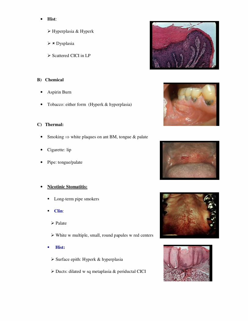
• Hist:
� Hyperplasia & Hyperk
� � Dysplasia
� Scattered CICI in LP
B) Chemical
• Aspirin Burn
• Tobacco: either form (Hyperk & hyperplasia)
C) Thermal:
• Smoking ⇒ white plaques on ant BM, tongue & palate
• Cigarette: lip
• Pipe: tongue/palate
• Nicotinic Stomatitis:
� Long-term pipe smokers
� Clin:
� Palate
� White w multiple, small, round papules w red centers
� Hist:
� Surface epith: Hyperk & hyperplasia
� Ducts: dilated w sq metaplasia & periductal CICI

◙ Idiopathic white lesions:
� Leukoplakia:
� Definition: a predominantly white patch that cannot be characterized as any other definable
lesion.
� Epidemiology:< 4%, M>F, older, site
� Clinically:
� Size
� Colour
� Homogenous: plaque-like ± surface variations
� Non-homogenous: speckled, ulcerated, nodular, warty
� Erythroplakia: A bright-red patch on the OM which cannot be characterized clinically or
pathologically as being due to any other condition. Homogenous or speckled
� Aetiology: Unknown, incriminated factors
1. Tobacco
2. Alcohol
3. Candida
4. Viruses: HPV 16

5. Epithelial atrophy:
� Iron (Sideropenic dysphagia, Patterson-Kelly, Plummer-Vinson Syndrome)
� Oral submucous fibrosis
� Tertiary Syphilis
� Vit
� Hist: no specific histological features
� Hyperk or hyperparak or both
� Hyperplasia or atrophy
�� Melanin pigment in basal epithelium ± melanin incontinence
� CICI in LP
� ± Dysplasia:
� Nuclear & cellular pleomorphism � � N/C ratio
� Nuclear hyperchromatism � � & abnormal mitosis
� Disturbed polarity of basal cells � Basal cell hyperplasia
� Drop-shaped rete pegs � Disturbed maturation
� Deep cell keratinization � Loss of intercellular adherence
���� Dysplasia: mild, moderate, severe
���� Homogenous leukoplakia: 10%
���� Non- homogenous leukoplakia: 50%
���� Erythroplakia: 50% Ca or Ca-in-situ, majority of the rest: severe dysplasia

Prognosis:
� Unpredictable (0.3-18%) over prolonged periods
� Risk factors:
� Non-smokers � Family history
� Advanced age � F
�Non-homogenous � Sublingual area
� Duration � Enlargement or ∆ in character
� Dysplasia
◙ Dermatological:
���� Lichen Planus:
� CID of skin & mms affecting ≈ 1%
� 30-60 years of age, 60% F
� 40% skin & oral; 35% skin; 25% oral
� Clin:
� Skin:
Clusters of raised purplish papules pruritic papules 2-3 m
Wickham’s Striae
Koebner phenomenon
Location
Nails
Duration
� Oral lesions:
Most frequent site
Other sites
Least frequent

Distribution
Clinically:
� Reticular:
Lacework, Striae of Wickham
Asymptomatic
Site
� Plaque-like:
≈ Leukoplakia
Asymptomatic
Site
� Papular:
Small white papules that may coalesce, asymptomatic
� Atrophic:
≈ Erythroplakia often with striae
Gingiva, desquamative gingivitis, symptomatic
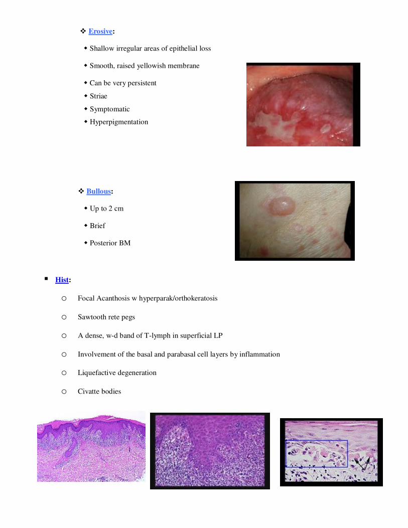
� Erosive:
Shallow irregular areas of epithelial loss
Smooth, raised yellowish membrane
Can be very persistent
Striae
Symptomatic
Hyperpigmentation
� Bullous:
Up to 2 cm
Brief
Posterior BM
� Hist:
o Focal Acanthosis w hyperparak/orthokeratosis
o Sawtooth rete pegs
o A dense, w-d band of T-lymph in superficial LP
o Involvement of the basal and parabasal cell layers by inflammation
o Liquefactive degeneration
o Civatte bodies

� Prognosis:
� 0.5-2.5 % over 5-year period, erosive
� Aetiology: unknown
� Genetic predisposition
� Infective agents
� Systemic disease: DM, Hypertension, U colitis, liver disease & GVHD
� Lichenoid reaction: Drugs (antimalarial, gold, methyldopa, NSAI) & amalgam
� Tobacco
� Vitamin ↓
� Psychiatric disorders
� Immunopathogenesis
� Pathogenesis:
� Langerhan’s cells
� ? Ag similar to antigens on kerationocytes of pts w certain MHC Ags
� Processed by Langerhan’s cells and presented activate production of CD8
���� Lupus Erythromatosus:
� A C.T disease with two main forms:
1. Chronic discoid LE:
� Face, scalp & ears
� Scaly red patches ± butterfly pattern
� Oral lesions in ≈ 50%
� Cheeks
� Vermillion border
� Discoid area of erythema

w white keratotic border ± radiating striae
� Hist:
� Ortho/parak epith
� Hyperplasia/atrophy
� Keratin plugging
� Subepith & deep perivascular lymphocytes
� ± Liquefactive degeneration
� DIF: granular linear deposits of IGg, C3 & fibrinogen in BM (Lupus band)
2. Systemic LE:
� Most common
� Kidney
� Arthritis, heart & lung involvement, anemia, vasculitis, rash
� Fatigue, malaise, fever, psychosis, lymphadenopathy
� Oral lesions in ≈ 20%, more severe erythematous patches/BM
� Aetiology: genetic, autoimmune