Embed Size (px)
Citation preview
3/21/16
1
Cardiac Physiology using: Positron Emission Tomography
(PET)
-What it can do
-How it works
Steve Bacharach (NIH, UCSF)
How is PET (and conv. Nuc med) different?
• Mammography, CT, US, (MRI): – Gives image of morphology (anatomy)
• PET (Positron Emission Tomography): – Image of physiology – Images of Biochemical Function
Nuclear Imaging • Uses a tracer to follow biochemical Reactions • In Nuc imaging
– Attach a radioactive element (a “tracer”) to the biochemical
• “Label” the biochemical – Often 99mTc, or 131,123I or with PET, other isotopes – Inject it (or swallow or breath it) – Image the radiation emitted (with Gamma Camera or
PET camera) – Sensitive so am’t injected is so small it does NOT
influence physiology
Goal of Nuclear Imaging
• Trace fate of biochemical compounds
– Static image of their distribution in organ(s)
– Set of dynamic images: images as a function of time • Uptake by organ/tissue • Metabolism • clearance
Problems with conventional Gamma Camera (non PET) nuc imaging
• Tc (etc) not naturally present – Usually must chelate it (e.g. DTPA, etc)
• Labeled biochemical -> not exactly same behaviour as unlabeled form
• Gamma Camera is Sensitive, but not as sensitive as we’d like – Conventional nuc imaging
• Still many 100s of times more sensitive than MRI
• Can’t measure absolute amounts of tracer – only relative
Two kinds of images of cardiac “function”
• Biochemical function
• Mechanical function – Fraction of blood pumped at each beat – Track edges of LV with time
• Compute volume from area of each slice
– OR (with nuc) directly measure blood volume with time
3/21/16
2
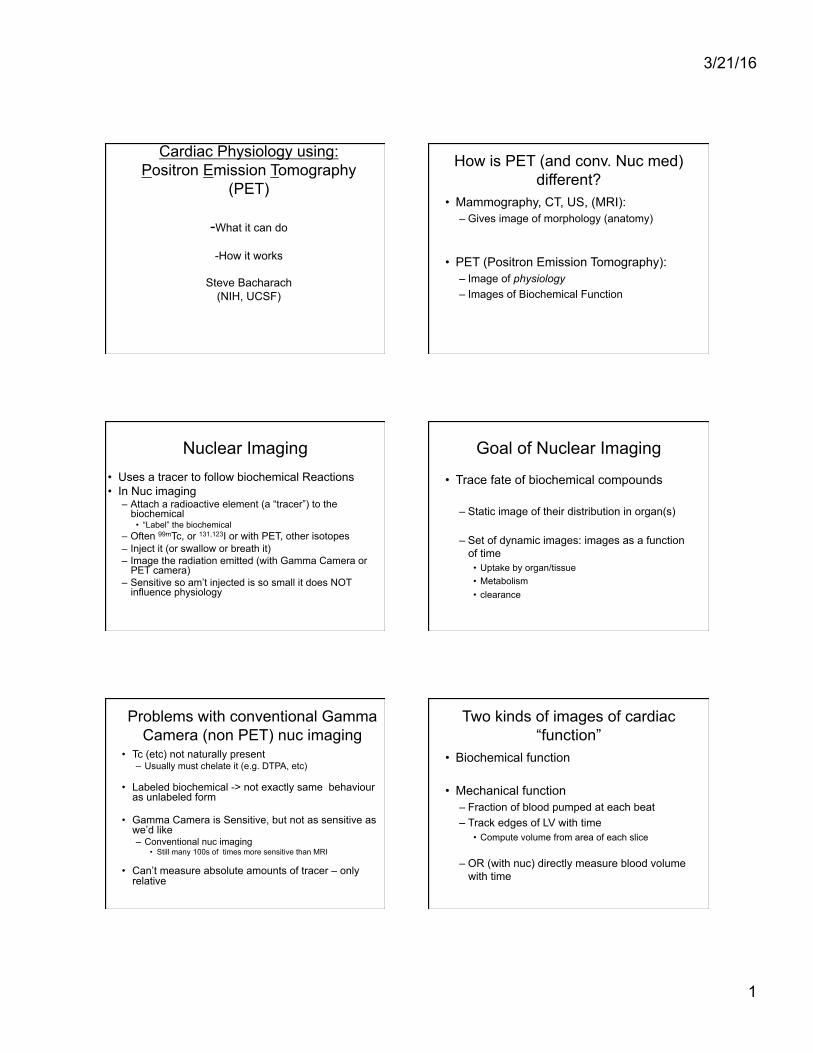
Mechanical Function by Gated Blood Pool (MUGA)
• Label blood with tracer – Many ways to do this (RBCs or even albumin)
• Am’t radn emitted Prop to Blood Volume – #photons emitted prop to blood volume
• Draw contour including LV (doesn’t have to be that accurate)
• Plot #photons detected vs time
• No need to make it tomographic! So only 1 contour total, or 1 per time point.
ES ED
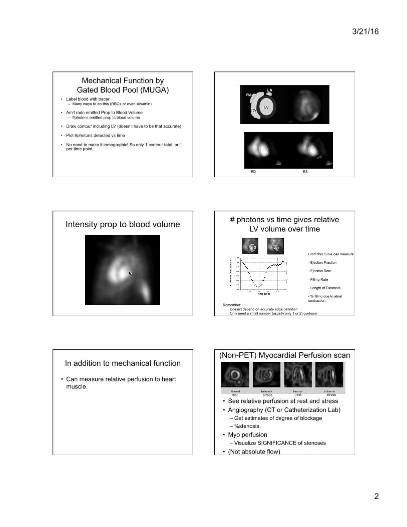
Intensity prop to blood volume # photons vs time gives relative LV volume over time
From this curve can measure
- Ejection Fraction
- Ejection Rate
- Filling Rate
- Length of Diastasis
- % filling due to atrial contraction
Remember: Doesn’t depend on accurate edge definition Only need a small number (usually only 1 or 2) contours
In addition to mechanical function
• Can measure relative perfusion to heart muscle.
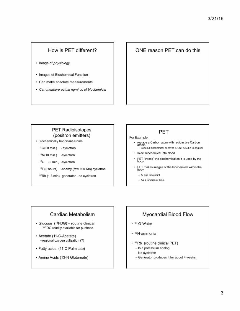
(Non-PET) Myocardial Perfusion scan
• See relative perfusion at rest and stress • Angiography (CT or Catheterization Lab)
– Get estimates of degree of blockage – %stenosis
• Myo perfusion – Visualize SIGNIFICANCE of stenoses
• (Not absolute flow)
rest stress rest stress
3/21/16
3
How is PET different?
• Image of physiology
• Images of Biochemical Function
• Can make absolute measurements
• Can measure actual ngm/ cc of biochemical
ONE reason PET can do this
PET Radioisotopes (positron emitters)
• Biochemically Important Atoms
11C (20 min.) - cyclotron
13N (10 min.) -cyclotron
15O (2 min.) -cyclotron
18F (2 hours) -nearby (few 100 Km) cyclotron
82Rb (1.3 min) -generator - no cyclotron
PET
• replace a Carbon atom with radioactive Carbon atoms – Labeled biochemical behaves IDENTICALLY to original
• Inject biochemical into blood
• PET “traces” the biochemical as it is used by the body.
• PET makes images of the biochemical within the body
– At one time point
– As a function of time.
For Example:
Cardiac Metabolism
• Glucose (18FDG) – routine clinical – 18FDG readily available for puchase
• Acetate (11-C-Acetate) – regional oxygen utilization (?)
• Fatty acids (11-C Palmitate)
• Amino Acids (13-N Glutamate)
Myocardial Blood Flow
• 15 O-Water
• 13N-ammonia
• 82Rb (routine clinical PET) – Is a potassium analog – No cyclotron – Generator produces it for about 4 weeks.
3/21/16
4
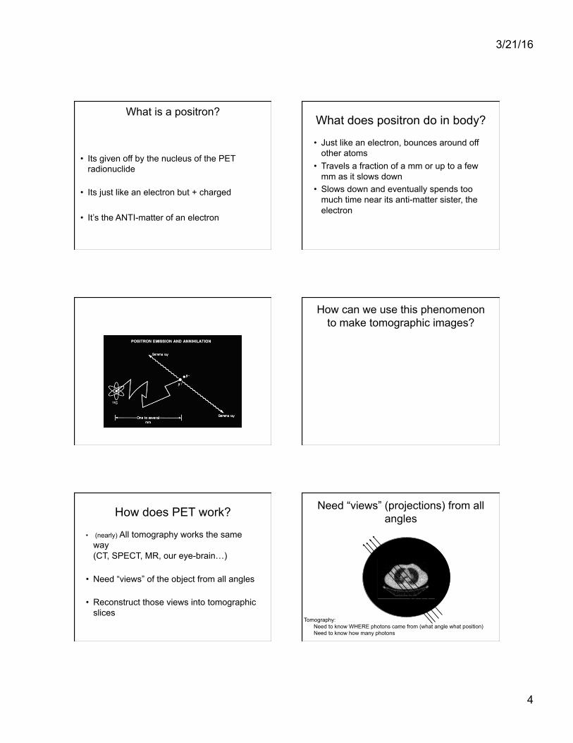
What is a positron?
• Its given off by the nucleus of the PET radionuclide
• Its just like an electron but + charged
• It’s the ANTI-matter of an electron
What does positron do in body?
• Just like an electron, bounces around off other atoms
• Travels a fraction of a mm or up to a few mm as it slows down
• Slows down and eventually spends too much time near its anti-matter sister, the electron
How can we use this phenomenon to make tomographic images?
How does PET work?
• (nearly) All tomography works the same way (CT, SPECT, MR, our eye-brain…)
• Need “views” of the object from all angles
• Reconstruct those views into tomographic slices
Need “views” (projections) from all angles
Tomography: Need to know WHERE photons came from (what angle what position) Need to know how many photons
3/21/16
5
CT scanner
• Need to know WHERE photons came from • Along line between detector and Xray Tube
X-Ray Tube
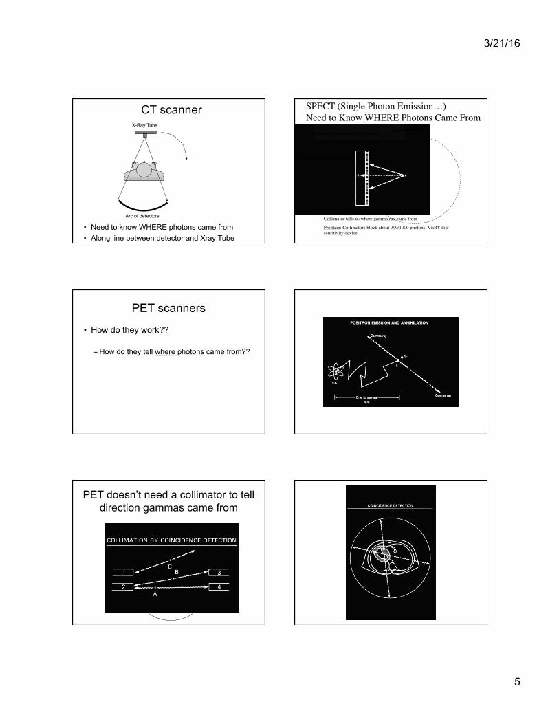
Arc of detectors Collimator tells us where gamma ray came from
Problem: Collimators block about 999/1000 photons. VERY low sensitivity device.
SPECT (Single Photon Emission…)���Need to Know WHERE Photons Came From
Use a collimator
Rotate scanner around patient
PET scanners
• How do they work??
– How do they tell where photons came from??
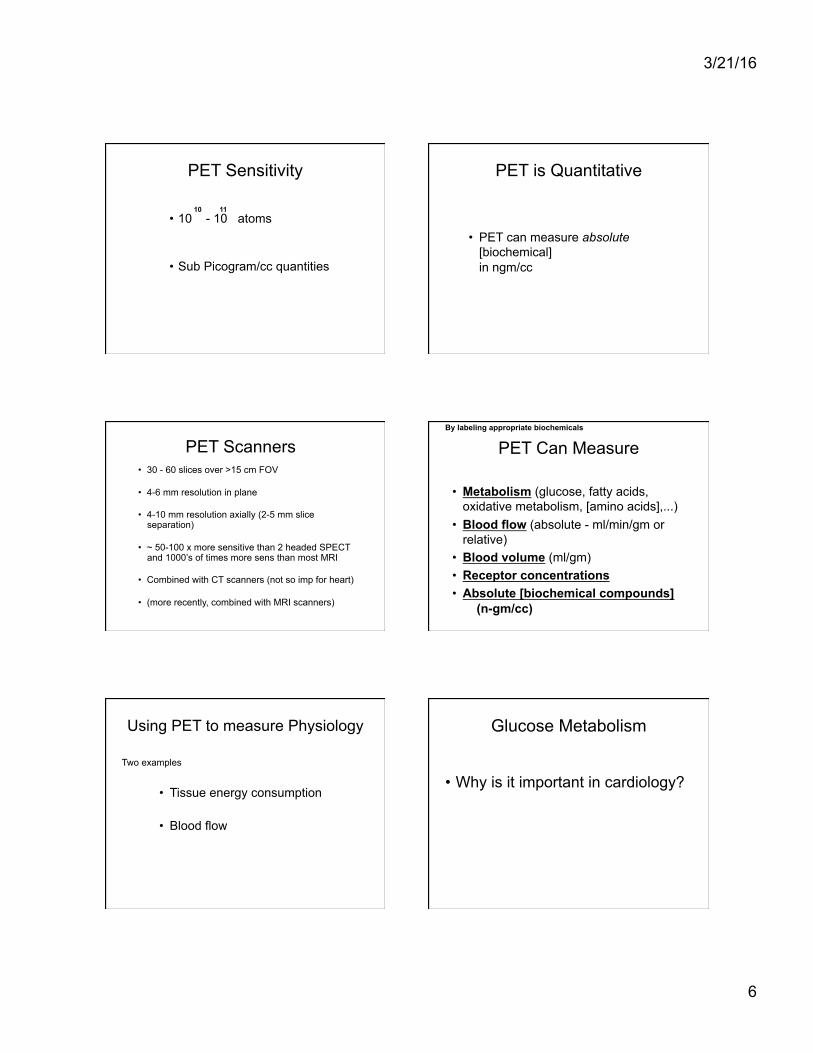
PET doesn’t need a collimator to tell direction gammas came from
3/21/16
6
PET Sensitivity
• 10 - 10 atoms
• Sub Picogram/cc quantities
10 11
PET is Quantitative
• PET can measure absolute [biochemical] in ngm/cc
PET Scanners • 30 - 60 slices over >15 cm FOV
• 4-6 mm resolution in plane
• 4-10 mm resolution axially (2-5 mm slice separation)
• ~ 50-100 x more sensitive than 2 headed SPECT and 1000’s of times more sens than most MRI
• Combined with CT scanners (not so imp for heart)
• (more recently, combined with MRI scanners)
PET Can Measure
• Metabolism (glucose, fatty acids, oxidative metabolism, [amino acids],...)
• Blood flow (absolute - ml/min/gm or relative)
• Blood volume (ml/gm) • Receptor concentrations • Absolute [biochemical compounds]
(n-gm/cc)
By labeling appropriate biochemicals
Using PET to measure Physiology
• Tissue energy consumption
• Blood flow
Two examples
Glucose Metabolism
• Why is it important in cardiology?
3/21/16
7
Aerobic vs anaerobic glucose metabolism
Glucose -----> Glucose 6P -------> Pyruvate
Needs a little ATP
Gives off net 2 ATP
NO OXYGEN NEEDED
Pyruvate ----> CO2 + Water + Lots of ATP (~34) OXYGEN NEEDED
Note: Just Bacharach’s version of biochemistry - don’t trust details
(Lactate)
Metabolism of Glucose (one molecule)
• 1st steps (called “glycolysis”) – Don’t require oxygen – Produce 2 ATP’s of energy – DOES require “spark” of energy
• 2nd steps – DO require lots of oxygen – Produces 34 more ATP’s of energy
• Therefore: – Very LITTLE energy produced/molecule w/o oxygen – LOTS of energy producec/molecule with oxygen
• A cell needs certain amount of E – If there is Oxygen -> don’t need much glucose – If no oxygen -> need LOTS of glucose
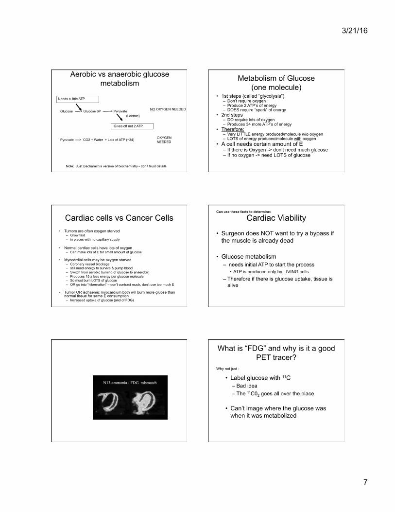
Cardiac cells vs Cancer Cells • Tumors are often oxygen starved
– Grow fast – in places with no capillary supply
• Normal cardiac cells have lots of oxygen – Can make lots of E for small amount of glucose
• Myocardial cells may be oxygen starved – Coronary vessel blockage – still need energy to survive & pump blood – Switch from aerobic burning of glucose to anaerobic – Produces 15 x less energy per glucose molecule – So must burn LOTS of glucose – OR go into “hibernation” – don’t contract much, don’t use too much E
• Tumor OR ischaemic myocardium both will burn more gluose than normal tissue for same E consumption – Increased uptake of glucose (and of FDG)
Cardiac Viability
• Surgeon does NOT want to try a bypass if the muscle is already dead
• Glucose metabolism – needs initial ATP to start the process
• ATP is produced only by LIVING cells – Therefore if there is glucose uptake, tissue is
alive
Can use these facts to determine:
What is “FDG” and why is it a good PET tracer?
• Label glucose with 11C – Bad idea – The 11C02 goes all over the place
• Can’t image where the glucose was when it was metabolized
Why not just :
3/21/16
8
18F-Fluoro-Deoxy-Glucose (FDG)
OH
H
OH
OH OH
18F
O ‘Molecule of the Century’
- Henry Wagner
Patient injected activity: 10 mCi ~ 1 nano-gram (6.4*10-12 moles = 3.8*1012 molecules)
How Cells “burn” Glucose and F-DeoxyGlucose
FDG Static Images (5 mCi, “3-D” mode)
GE Advance, 5 mCi FDG, 28 min post inj., 30 min scan
4.25mm/slice
TO get absolute flows/metabolic rates Need Physiologic/mathematical
model • Physiologist/physician/physicist work together
• Make model of what happens to biochemical – It may depend on blood flow, metabolism, etc
• Take PET data over time (wash-in/out)
• Use model to measure absolute quantities of blood flow, metabolism….
Some tracers
• Not metabolized at all
• Have a simple model
• Water is good example (H215O) – can be
used to measure blood flow

3/21/16
9
In this simple model:
• tracer washes out exponentially with time • exp constant = flow
Other Models • Let you measure absolute myocardial blood
flow from:
– 13NH3 or 15O-water (requires a cyclotron)
– 82Rb (Routine clinical -no need for cyclotron)
• Absolute metabolic rates (e.g. of glucose)
• Other physiologic parameters from a variety of labeled biochemicals
82Rb at Stress and Rest
• DiCarli et al • In Obese women
– Sens: 95% – Spec: 90%
Absolute flow from 13NH3
3/21/16
10
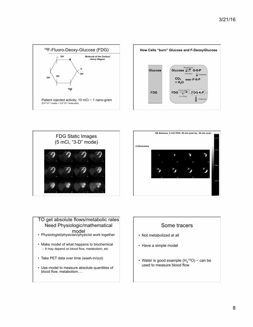
Physiologic Imaging with PET (1 Slice of 35)
Glucose Metabolic rate Blood Volume Image (ml blood/gm tissue)
Blood Flow Image (ml/min/gm)
Avg. tumor blood flow = 3.6ml/min/gm
Small vessel
Tumor
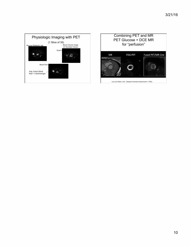
Combining PET and MR PET Glucose + DCE MR
for “perfusion”
Lau et al Wash. Univ. Delayed Contrast enhancement + FDG)