Embed Size (px)
Citation preview
NTNU Det medisinske fakultet
White Matter Abnormalities seen on Diffusion Tensor Imaging Relate to Neuroimpairments in VLBW
Children at 15 Years of Age
Jon Skranes1, Torgil R Vangberg2, Arild Kristoffersen2, Siri Kulseng1, Marit Indredavik3, Kari Anne I. Evensen1, Marit Martinussen2, David Tuch5, Anders Dale5, Olav Haraldseth2, Torstein Vik4 and Ann Mari Brubakk1
1Dep of Laboratory Medicine, Children’s and Woman’s Health; 2Dep of Circulation and Medical Imaging; 3Dep of Neuroscience; 4Dep of Public Health and General Practice, Faculty of Medicine, Norwegian University of Science and Technology, Trondheim, Norway; 5Martinos Center, MGH, Boston, USA
NTNU Det medisinske fakultet
BACKGROUND • VLBW (birth weight < 1500 grams) children are at increased risk of neuroimpairments caused by perinatal injury like
periventricular leukomalacia (PVL). • Brain DTI may detect subtle changes in white matter microstructure
that is not seen on conventional MRI.
OBJECTIVE• To compare cerebral DTI findings with the results from extensive psychiatric, behavioural, cognitive and motor assessments in VLBW adolescents at age 15.• We hypothesized that reduced fractional anisotropy in specific areas of white matter was related with abnormal test results.
NTNU Det medisinske fakultet
MATERIAL• 34 VLBW children • 49 non-SGA term controls • Examined at 15 years of age in a population based study
METHODS• Movement-ABC • Developmental test of Visual-Motor Integration (VMI) with
supplementary tests: Visual perception and Motor coordination • Psychiatric interview (Kaufman-SADS), ADHD rating scale• Subtests from WISC-III: Arithmetic, Vocabulary, Picture arrangement,
Block design
NTNU Det medisinske fakultet
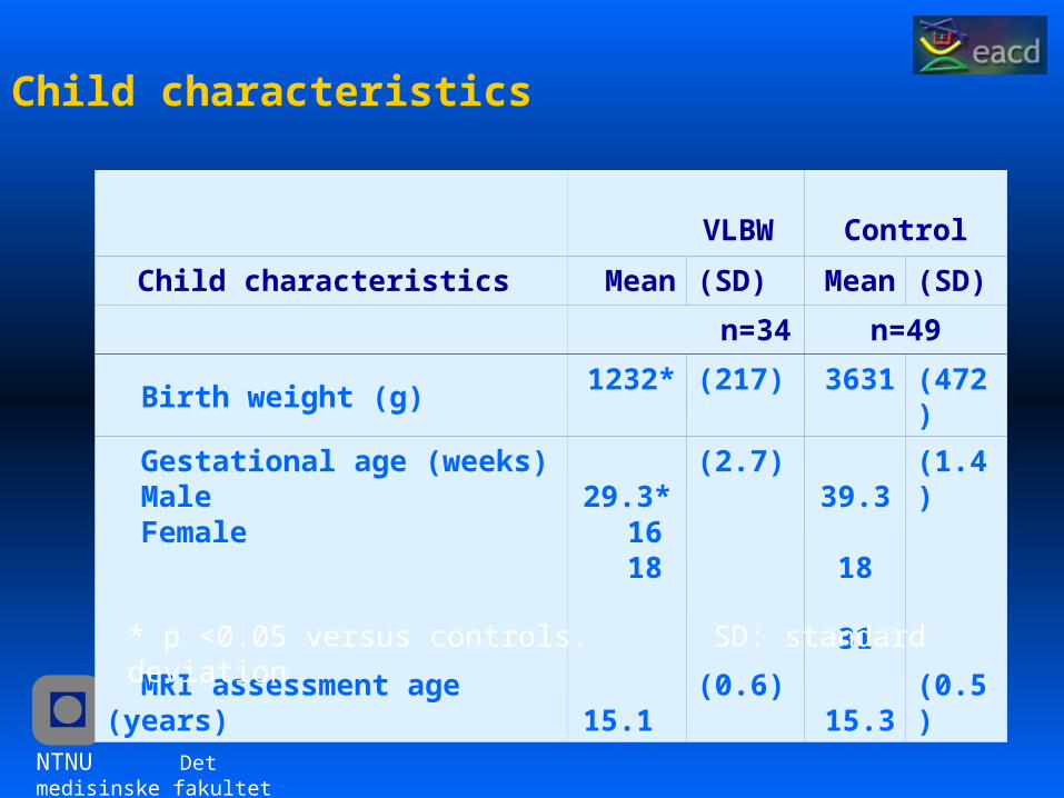
Child characteristics
VLBW Control
Child characteristics Mean (SD) Mean (SD)
n=34 n=49
Birth weight (g) 1232* (217) 3631 (472)
Gestational age (weeks) Male Female
29.3* 16 18
(2.7)
39.3 18 31
(1.4)
MRI assessment age (years) 15.1 (0.6) 15.3 (0.5)
* p <0.05 versus controls. SD: standard deviation
NTNU Det medisinske fakultet
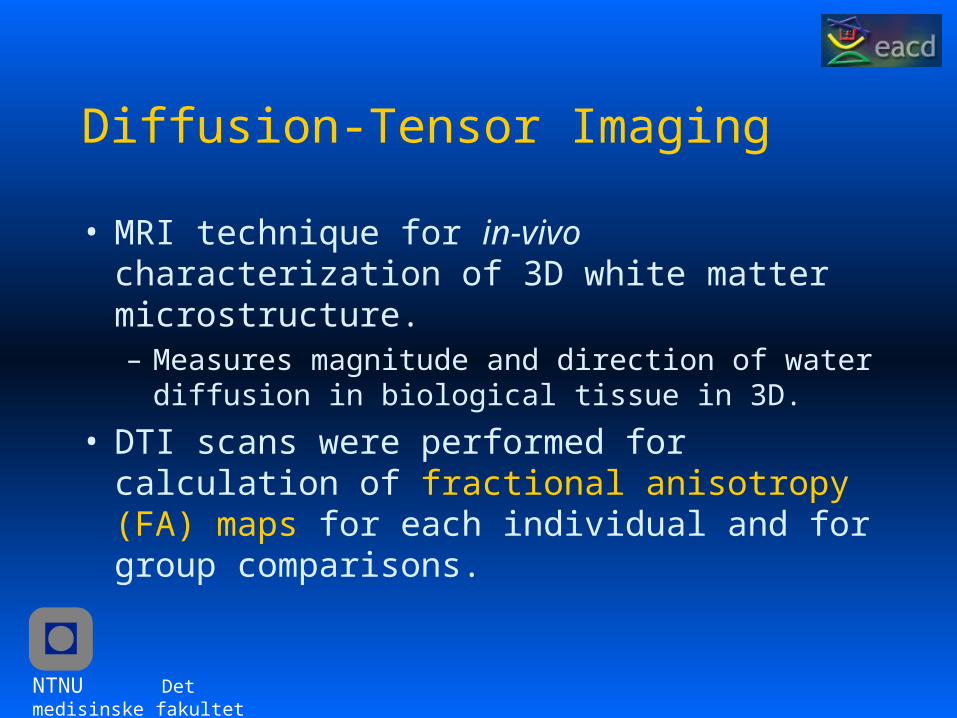
Diffusion-Tensor Imaging
• MRI technique for in-vivo characterization of 3D white matter microstructure. – Measures magnitude and direction of water diffusion in
biological tissue in 3D.• DTI scans were performed for calculation of fractional
anisotropy (FA) maps for each individual and for group comparisons.
NTNU Det medisinske fakultet
METHODS cont.
• Fractional Anisotropy (FA): The extent to which diffusion is directionally restricted, i.e. anisotropic. Scalar measure: 0 to 1
• For isotropic diffusion (λ1 = λ2 = λ3), FA is zero, and in the case where there is a strongly preferred direction of diffusion (λ1 >> λ2 = λ3), FA approaches one.
• Mean FA values for anatomical areas of significant differences between VLBW and controls were identified and then compared with clinical data for the VLBW adolescents (low performers vs. normal performers).
NTNU Det medisinske fakultet
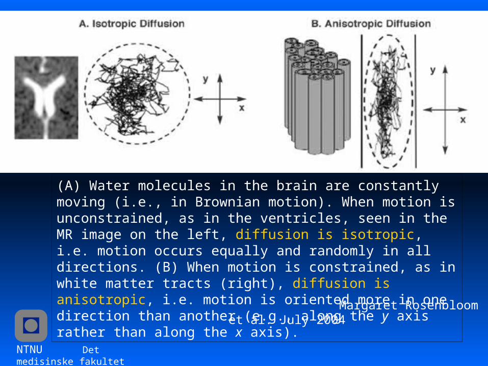
(A) Water molecules in the brain are constantly moving (i.e., in Brownian motion). When motion is unconstrained, as in the ventricles, seen in the MR image on the left, diffusion is isotropic, i.e. motion occurs equally and randomly in all directions. (B) When motion is constrained, as in white matter tracts (right), diffusion is anisotropic, i.e. motion is oriented more in one direction than another (e.g., along the y axis rather than along the x axis).
Margaret Rosenbloom et al. July 2004
NTNU Det medisinske fakultet
Image Acquisition
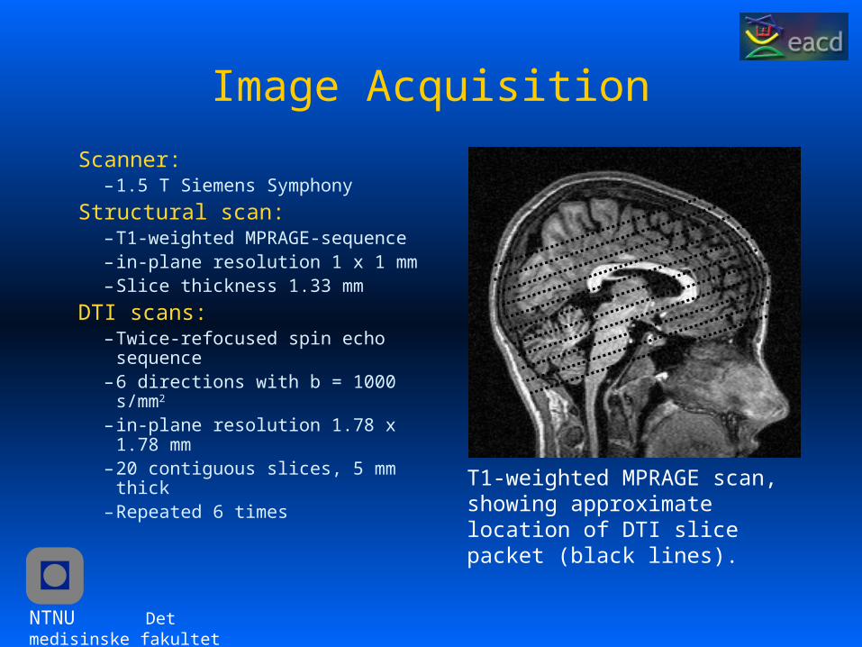
Scanner: – 1.5 T Siemens Symphony
Structural scan: – T1-weighted MPRAGE-sequence– in-plane resolution 1 x 1 mm– Slice thickness 1.33 mm
DTI scans:– Twice-refocused spin echo sequence– 6 directions with b = 1000 s/mm2
– in-plane resolution 1.78 x 1.78 mm – 20 contiguous slices, 5 mm thick– Repeated 6 times
T1-weighted MPRAGE scan,showing approximate location of DTI slice packet (black lines).
NTNU Det medisinske fakultet
Diffusion Tensor Calculation
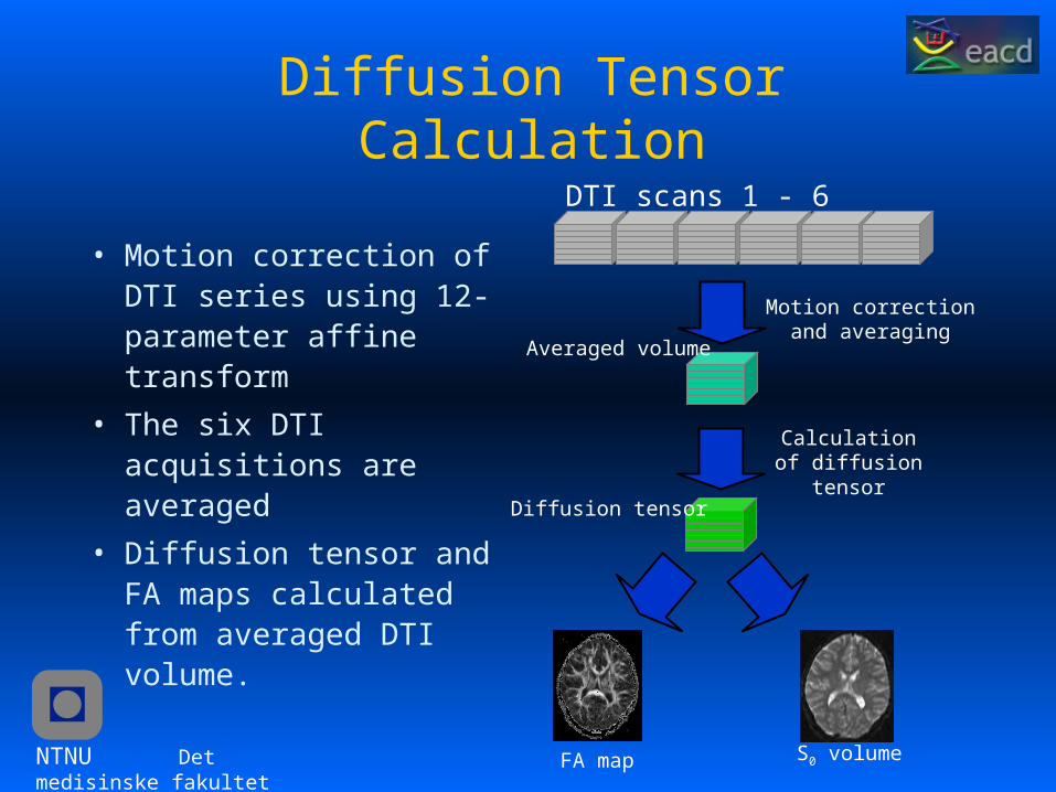
• Motion correction of DTI series using 12-parameter affine transform
• The six DTI acquisitions are averaged
• Diffusion tensor and FA maps calculated from averaged DTI volume.
DTI scans 1 - 6
Motion correction and averaging
Averaged volume
Diffusion tensor
Calculation of diffusion tensor
FA map S0 volume
NTNU Det medisinske fakultet
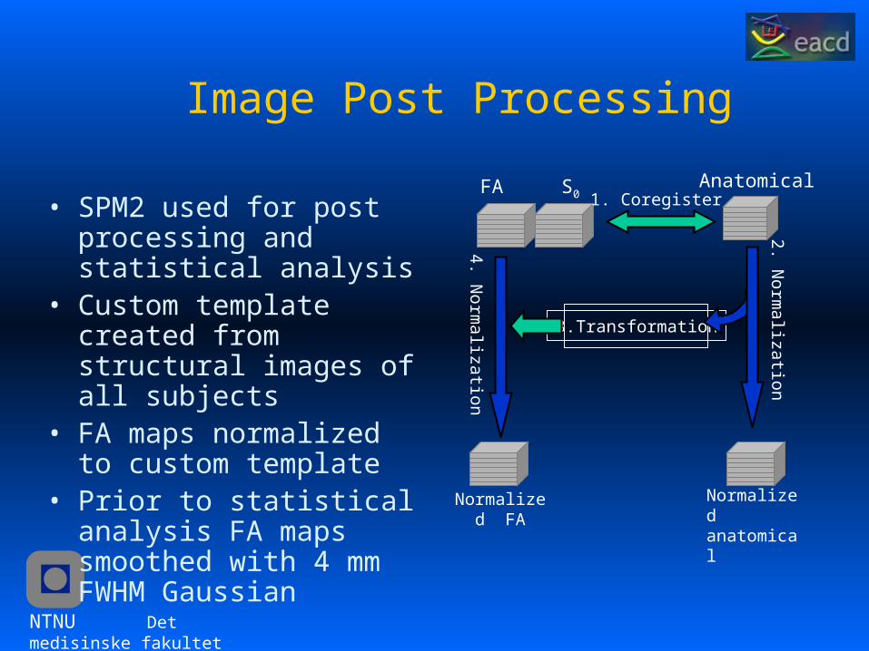
Image Post Processing
• SPM2 used for post processing and statistical analysis
• Custom template created from structural images of all subjects
• FA maps normalized to custom template
• Prior to statistical analysis FA maps smoothed with 4 mm FWHM Gaussian
FA S0Anatomical
3.Transformation
1. Coregister
Normalizedanatomical
2. Norm
alization
Normalized FA
4. Norm
alization
NTNU Det medisinske fakultet
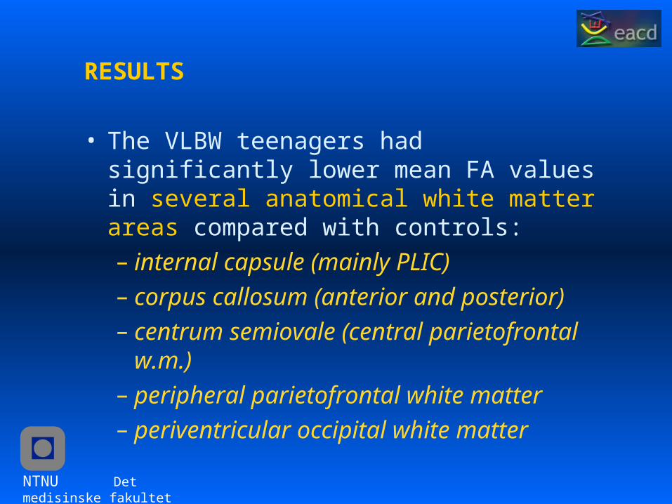
RESULTS
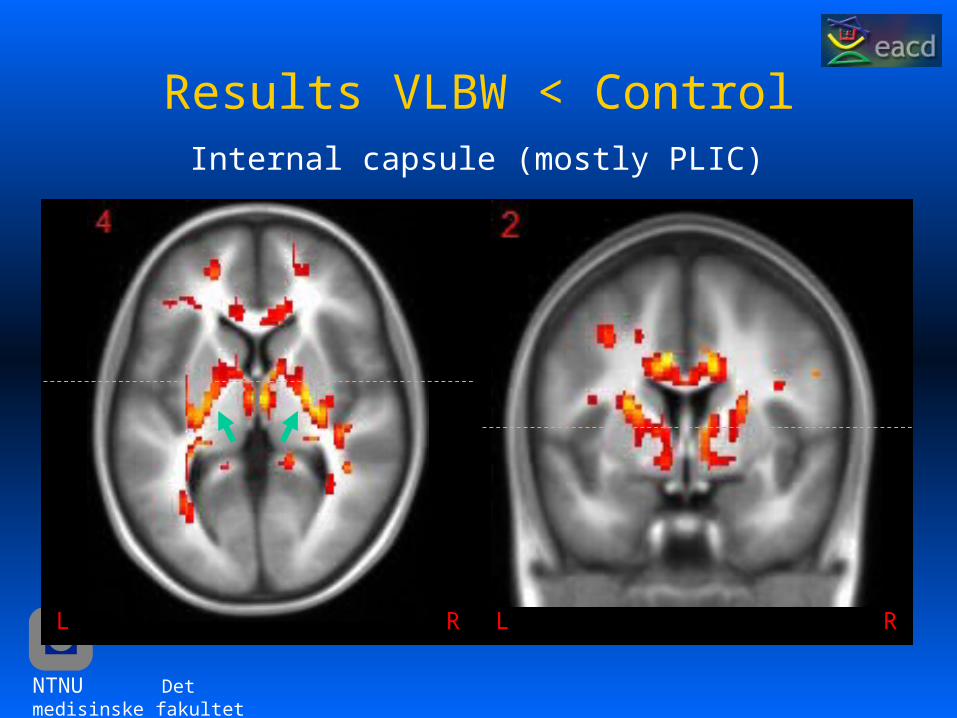
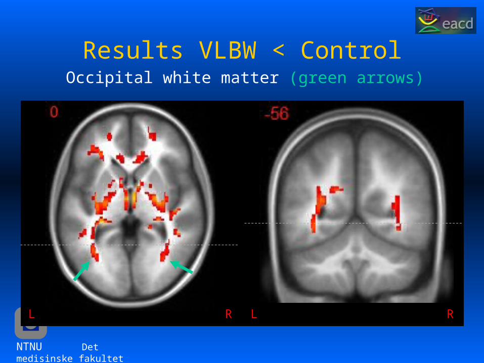
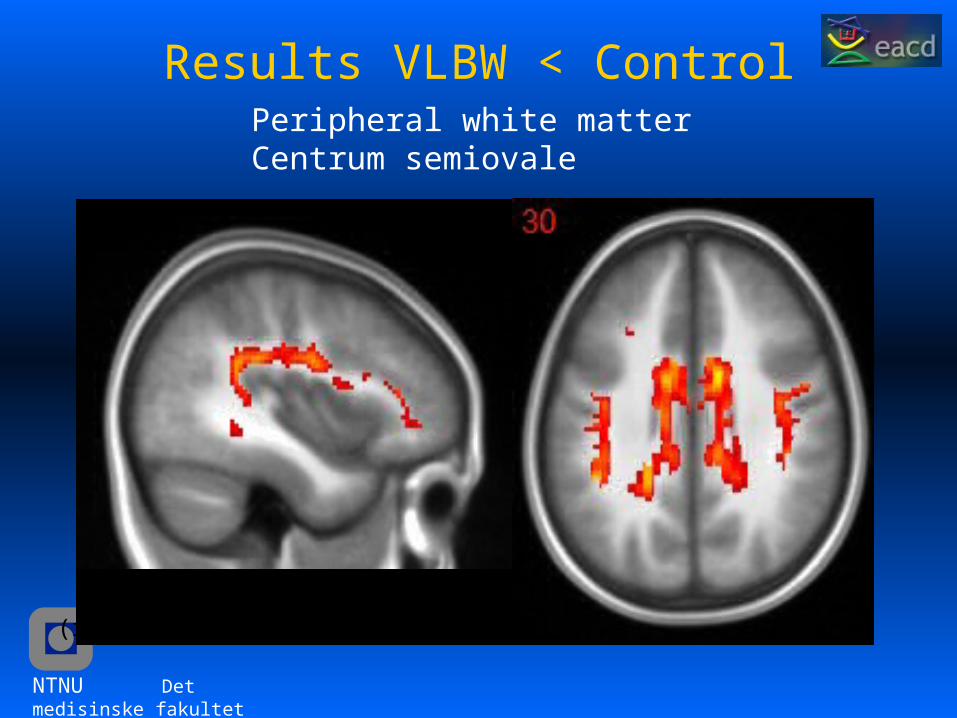
• The VLBW teenagers had significantly lower mean FA values in several anatomical white matter areas compared with controls: – internal capsule (mainly PLIC) – corpus callosum (anterior and posterior) – centrum semiovale (central parietofrontal w.m.) – peripheral parietofrontal white matter – periventricular occipital white matter
NTNU Det medisinske fakultet
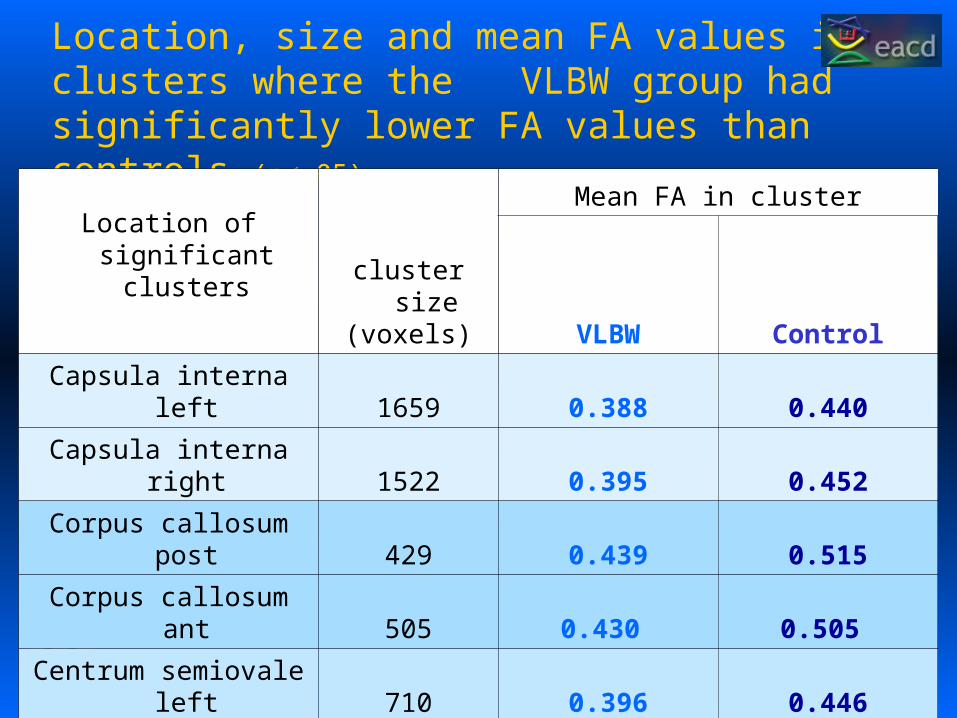
Location, size and mean FA values in clusters where the VLBW group had significantly lower FA values than controls (p<.05).
Location of significant
clusters cluster size(voxels)
Mean FA in cluster
VLBW Control
Capsula interna left 1659 0.388 0.440
Capsula interna right 1522 0.395 0.452
Corpus callosum post 429 0.439 0.515
Corpus callosum ant 505 0.430 0.505
Centrum semiovale left 710 0.396 0.446
Centrum semiovale right 395 0.406 0.457
Peripheral wm left 235 0.382 0.428
Peripheral wm right 144 0.371 0.415
Occipital wm left 212 0.414 0.469
NTNU Det medisinske fakultet
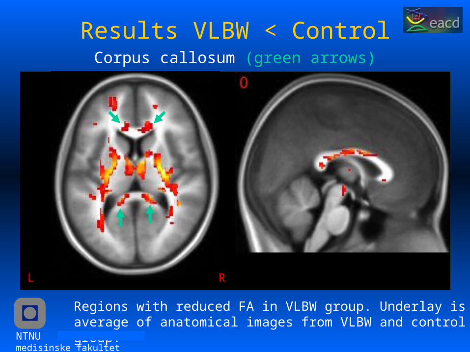
Results VLBW < ControlCorpus callosum (green arrows)
RL
Regions with reduced FA in VLBW group. Underlay is average of anatomical images from VLBW and control group.
NTNU Det medisinske fakultet
Results VLBW < ControlInternal capsule (mostly PLIC)
RL RL
NTNU Det medisinske fakultet
Results VLBW < ControlOccipital white matter (green arrows)
RL RL
NTNU Det medisinske fakultet
Results VLBW < Control
(right side)
Peripheral white matterCentrum semiovale
NTNU Det medisinske fakultet
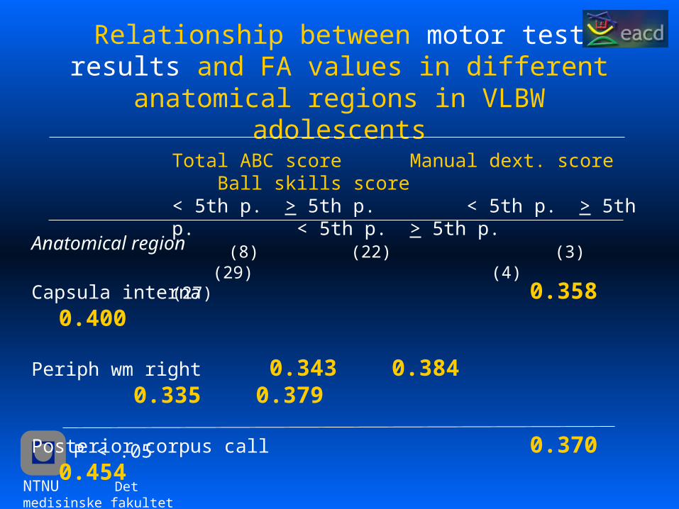
Relationship between motor test results and FA values in different anatomical regions in VLBW adolescents
Total ABC score Manual dext. score Ball skills score< 5th p. > 5th p. < 5th p. > 5th p. < 5th p. > 5th p. (8) (22) (3) (29) (4) (27)
Anatomical region
Capsula interna 0.358 0.400
Periph wm right 0.343 0.384 0.335 0.379
Posterior corpus call 0.370 0.454
P < .05
NTNU Det medisinske fakultet
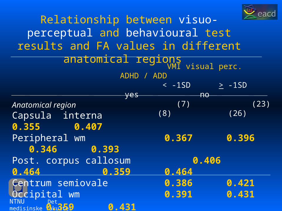
Relationship between visuo-perceptual and behavioural test results and FA values in different anatomical regions
VMI visual perc. ADHD / ADD < -1SD > -1SD yes no (7) (23) (8) (26)
Anatomical region
Capsula interna 0.355 0.407 Peripheral wm 0.367 0.396 0.346 0.393Post. corpus callosum 0.406 0.464 0.359 0.464Centrum semiovale 0.386 0.421Occipital wm 0.391 0.431 0.359 0.431
NTNU Det medisinske fakultet
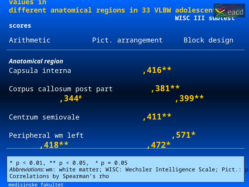
Correlations between WISC III subtest scores and FA values in different anatomical regions in 33 VLBW adolescents
WISC III subtest scores
Arithmetic Pict. arrangement Block design Anatomical region
Capsula interna ,416**
Corpus callosum post part ,381** ,344# ,399**
Centrum semiovale ,411**
Peripheral wm left ,571* ,418** ,472*
Occipital wm left ,388**
* p < 0.01, ** p < 0.05, # p = 0.05Abbreviations: wm: white matter; WISC: Wechsler Intelligence Scale; Pict.: pictureCorrelations by Spearman’s rho
NTNU Det medisinske fakultet
Discussion• The VLBW group has reduced anisotropy in several wm
areas which are known to be affected by premature birth (PVL, asphyxia)
• periventricular occipital white matter• centrum semiovale• corpus callosum• posterior limb of internal capsule
• The reduced anisotropy may be due to:• reduced myelination• fiber disorganization• fewer axons
NTNU Det medisinske fakultet
Conclusions / Speculations
• Low FA values in specific brain areas seem to relate to motor, cognitive, perceptual and behavioural impairments in VLBW adolescents.
• The neuroimpairments may be due to disturbed white matter microstructure and connectivity of commissural and association fibres between different cortical areas.
• The disrupted connectivity may influence neuronal networks and thereby neurodevelopment and higher cognitive functions in VLBW adolescents.
NTNU Det medisinske fakultet