Embed Size (px)
Citation preview

RESEARCH ARTICLE Open Access
Novel mutations in ALDH1A3 associatedwith autosomal recessive anophthalmia/microphthalmia, and review of theliteratureSiying Lin1, Gaurav V. Harlalka1, Abdul Hameed2, Hadia Moattar Reham3, Muhammad Yasin3, Noor Muhammad3,Saadullah Khan3, Emma L. Baple1, Andrew H. Crosby1 and Shamim Saleha3*
Abstract
Background: Autosomal recessive anophthalmia and microphthalmia are rare developmental eye defectsoccurring during early fetal development. Syndromic and non-syndromic forms of anophthalmia andmicrophthalmia demonstrate extensive genetic and allelic heterogeneity. To date, disease mutations have beenidentified in 29 causative genes associated with anophthalmia and microphthalmia, with autosomal dominant,autosomal recessive and X-linked inheritance patterns described. Biallelic ALDH1A3 gene variants are the leadinggenetic causes of autosomal recessive anophthalmia and microphthalmia in countries with frequent parentalconsanguinity.
Methods: This study describes genetic investigations in two consanguineous Pakistani families with a total ofseven affected individuals with bilateral non-syndromic clinical anophthalmia.
Results: Using whole exome and Sanger sequencing, we identified two novel homozygous ALDH1A3 sequencevariants as likely responsible for the condition in each family; missense mutation [NM_000693.3:c.1240G > C, p.Gly414Arg; Chr15:101447332G > C (GRCh37)] in exon 11 (family 1), and, a frameshift mutation [NM_000693.3:c.172dup, p.Glu58Glyfs*5; Chr15:101425544dup (GRCh37)] in exon 2 predicted to result in protein truncation(family 2).
Conclusions: This study expands the molecular spectrum of pathogenic ALDH1A3 variants associated withanophthalmia and microphthalmia, and provides further insight of the key role of the ALDH1A3 in human eyedevelopment.
Keywords: Autosomal recessive anophthalmia and microphthalmia, ALDH1A3 gene, Mutations, Variants, Exomesequencing, Consanguineous families
BackgroundAnophthalmia and microphthalmia are severe congenitaldevelopmental defects of the eye. In the clinical context,anophthalmia refers to complete absence of the globe inthe orbit, whilst microphthalmia refers to the presence ofa small globe within the orbit. Both anophthalmia andmicrophthalmia are more commonly bilateral, although
they can also present unilaterally. These are relatively raredefects, occurring with an estimated combined incidenceof 1 in 10,000 live births [1]. Anophthalmia and micro-phthalmia can occur as isolated malformations, or as partof a syndrome. Both syndromic and non-syndromic formsof anophthalmia and microphthalmia have been associ-ated with autosomal recessive, autosomal dominant andX-linked patterns of inheritance, and display extensivegenetic heterogeneity [2]. Mutations in numerous genesincluding RAX, PAX6, SOX2, OTX2,VSX2, RARB, BMP7,BCOR, BMP4, FOXE3, STRA6, SMOC1, SHH, SNX3,
* Correspondence: [email protected] of Biotechnology and Genetic Engineering, Kohat University ofScience and Technology (KUST), Kohat, Khyber Pakhtunkhwa 26000, PakistanFull list of author information is available at the end of the article
© The Author(s). 2018 Open Access This article is distributed under the terms of the Creative Commons Attribution 4.0International License (http://creativecommons.org/licenses/by/4.0/), which permits unrestricted use, distribution, andreproduction in any medium, provided you give appropriate credit to the original author(s) and the source, provide a link tothe Creative Commons license, and indicate if changes were made. The Creative Commons Public Domain Dedication waiver(http://creativecommons.org/publicdomain/zero/1.0/) applies to the data made available in this article, unless otherwise stated.
Lin et al. BMC Medical Genetics (2018) 19:160 https://doi.org/10.1186/s12881-018-0678-6

MFRP, PRSS56, GDF3, GDF6, TENM3, C12orf57, YAP1,ABCB6, ATOH7, VAX1, NDP, ALDH1A3 and SMARCA4have all been described in association with microphthal-mia, and some, including RAX, PAX6, SOX2, OTX2,RARB, BMP7, BCOR, BMP4, FOXE3, STRA6, SMOC1,GDF6 and ALDH1A3 have also been described in associ-ation with anophthalmia [3–5]. SOX2 mutations are themajor single-gene cause of anophthalmia and microphthal-mia, accounting for ~ 10–15% of all cases [6]. Mutations inother genes have been shown to account for another ~ 25%of cases of anophthalmia and microphthalmia [7]. In up to50–60% of cases however, the underlying genetic cause re-mains undetermined [2, 8].Mutations in the aldehyde dehydrogenase 1 family, mem-
ber A3 (ALDH1A3) gene have been found in associationwith autosomal recessive anophthalmia and microphthal-mia in individuals of different ethnicities. Notably, muta-tions of this gene appear to be the major cause of theseconditions in consanguineous families of Pakistani origin[3, 6]. The ALDH1A3 gene encodes a NAD-dependentaldehyde dehydrogenase, which is among one of threeretinaldehyde dehydrogenases (the others being ALDH1A1and ALDH1A2) that play a key role in the biosynthesis ofretinoic acid from retinaldehyde. Retinoic acid functionsas a ligand for DNA-binding retinoid receptors thatdirectly regulate transcription of specific target genes inthe retinoic-acid signaling pathway in vertebrates [9], andpromotes neuronal differentiation in the embryonicnervous system [10]. It also has an important function inthe normal early embryonic development of ocular andnasal regions [11].In this study, we identified homozygous novel missense
and frameshift sequence alterations in ALDH1A3 thatsegregate with the disease phenotype in consanguineousPakistani families with isolated anophthalmia, and discussour findings alongside existing literature in this area.
MethodsAscertainment of familyThis study was approved by the ethical committee, KohatUniversity of Science and Technology (KUST; Pakistan),and families were subsequently recruited. Informed writ-ten consent was obtained from parents, and consent wasobtained on behalf of their children. A consanguineousfamily, extending over four generations and comprising of4 living affected and 12 unaffected members, was re-cruited from the Khyber Pakhtunkhwa region of Pakistan.All the affected individuals were in the fourth generation.Another consanguineous family, extending over two gener-ations with three affected and five unaffected members,was also recruited from same region of Pakistan. Bloodsamples were collected from affected and unaffected indi-viduals, and all affected individuals were clinically evaluated
by an ophthalmologist for obtaining medical and family his-tories and clinical assessment.
Genetic studiesFollowing informed consent, genomic DNA from theblood samples was extracted using the ReliaPrep™ kit(Blood gDNA Miniprep System, Promega) according tothe manufacturer’s protocol. To identify the causativegene, whole-exome sequencing was performed on a singleaffected individual in each family (subject IV: 7 in family 1and II: 1 in family 2, Fig. 1) to develop a profile of variantsnot present in publicly available databases and raresequence variants. Coding regions were captured byHiSeq2000 using paired-end (2 × 100) protocol at a meancoverage depth of 30X at the Otogenetics Corporation(Norcross, GA, USA). The Agilent SureSelect Human AllExonV4 (51 Mb) enrichment kit was used for exomeenrichment. The sequence reads were aligned to thehuman genome reference sequence [hg19] and read align-ment, variant calling, and annotation were performed byDNAnexus (DNAnexus Inc., Mountain View, CA; https://dnanexus.com).Allele-specific primers were designed using Primer3 web
software (primer sequences are available upon request) toevaluate segregation of variants via dideoxy sequencing.Polymerase chain reaction (PCR) was undertaken for allrecruited family members using allele-specific primersfollowing standard conditions, with products sequencedby Source BioScience LifeSciences (https://www.sourcebioscience.com/). Pathogenicity of the identified missensesequence variation in the ALDH1A3 gene was also analyzedusing PolyPhen-2 (http://genetics.bwh.harvard.edu/pph2/),PROVEAN (http://provean.jcvi.org/index.php) and SIFT(http://provean.jcvi.org/index.php) specialized predictionsoftware. To compare and correlate the ALDH1A3 gene var-iants with the phenotype, all reported variants were retrievedfrom HGMD (http://www.hgmd.cf.ac.uk/ac/search.php),OMIM (https://www.ncbi.nlm.nih.gov/omim/) and PubMed(https://www.ncbi.nlm.nih.gov/pubmed/) databases.
ResultsSubjectsPedigree analysis of recruited consanguineous Pakistanifamilies suggested an autosomal recessive inheritance ofthe disease in these families (Fig. 1). In total, seven livingaffected individuals with normal intelligence as well as 13healthy individuals including parents and siblings fromboth families were investigated. The four affected individ-uals IV:3, IV:5, IV:7 and IV:8 were 13, 18, 14 and 12 yearsof age in first family, while three affected individuals II:1,II:4 and II:8 were 19, 10, 4 years of age in second familyrespectively at the time of examination. On the basis ofbasic clinical ophthalmic assessment, bilateral isolated an-ophthalmia was the major finding in all affected members
Lin et al. BMC Medical Genetics (2018) 19:160 Page 2 of 8

of the investigated families. No other neurological andbehavioral features were observed in affected individuals.
Genetic findingsThe exome data was first inspected to exclude previouslydescribed variants in genes known to cause ocular disease.Variants were then assessed and filtered for rare,non-synonymous exonic or splice variants, with a popula-tion frequency of < 0.01 in control databases (includingthe Genome Aggregation Database; gnomAD, the ExomeAggregation Consortium; ExAC, and the 1000 GenomesProject). A single candidate novel homozygous missensevariant [NM_000693.3:c.1240G >C; Chr15:101447332G >C (GRCh37)] was identified in exon 11 of ALDH1A3(Fig. 1) in first family. This variant leads to a substitutionof glycine with arginine and at the evolutionary conservedposition 414 (p.Gly414Arg) according to the UCSC Hu-man Genome (GRCh37/ hg19) and Ensemble databases(Additional file 1 A). The p.Gly414Arg variant is not listedin the Genome Aggregation Database (gnomAD). In silicoanalysis of p.Gly414Arg using PolyPhen-2, PROVEANand SIFT predicted it to be damaging or deleterious with ascore of 1.000, − 7.559 and 3.25 respectively (Additional file 1B). In family 2, a novel variant [NM_000693.3:c.172dup;Chr15:101425544dup (GRCh37)] was identified in exon 2
of the ALDH1A3 gene (Fig. 1). This single base pair duplica-tion is predicted to result in a frameshift followed by apremature stop codon (p. Glu58Glyfs*5). The variants infamilies 1 and 2 segregate as expected for an autosomalrecessive condition in each family, and both variants aresummarized in Table 1 alongside all other reporteddisease-associated ALDH1A3 variants; a schematic represen-tation of each mutation in ALDH1A3 is also shown in Fig. 2.
DiscussionThe ALDH1A3 (NG_012254.1) gene comprises 13 exonsspanning ~ 36 kb on chromosome 15 (15q26.3), encoding a512-amino acid NAD-dependent aldehyde dehydrogenaselocalized in the cytoplasm, nucleus, endoplasmic reticulumand mitochondria [12]. Structural analysis reveals thatALDH1A3 shares high structural homology with othertypes of aldehyde dehydrogenases. ALDH1A3 assembles asa tetramer, however, each of its monomeric units isindependently able to oxidize retinaldehyde into retinoicacid using NAD as a cofactor. Each monomeric unit foldsinto 13 α-helices, 19 β-sheets and the connecting loops,arranged into three functional domains: the NAD-bindingdomains (L20-D149 and I171–G282), the catalytic domain(G283–M482), and the C-terminal oligomerization do-mains (K150–P170 and S483–L507), [11].
A(i) A(ii)
B(ii) C(ii)B(i) C(i)
Fig. 1 A Pedigrees of the Pakistani families investigated, and genetic findings. A(i) and A(ii). Family 1 (Ai) and family 2 (Aii) pedigrees showingsegregation of the variants identified in each case. B(i) and B(ii). Photographs of two affected individuals in both families with non-syndromicclinical anophthalmia C(i) and C(ii). Sequence chromatograms showing wild-type alongside ALDH1A3 [NM_000693.3:c.1240G > C, p.Gly414Arg;Chr15:101447332G > C (GRCh37)] in exon 11 (family 1), and, a frameshift mutation [NM_000693.3:c.172dup, p.Glu58Glyfs*5; Chr15:101425544dup(GRCh37)] variants
Lin et al. BMC Medical Genetics (2018) 19:160 Page 3 of 8
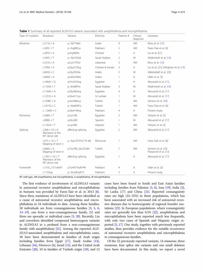
The first evidence of involvement of ALDH1A3 variantsin autosomal recessive anophthalmia and microphthalmiain humans was provided by Fares-Taie et al. in 2013 [6].Since then, mutations of ALDH1A3 have been identified asa cause of autosomal recessive anophthalmia and micro-phthalmia in 54 individuals to date. Among these families,50 individuals are from consanguineous families [3, 5, 6,13–19], one from a non-consanguineous family, [5] andthree are sporadic or individual cases [5, 20]. Recently, Liuand coworkers identified compound heterozygous variantsin ALDH1A3 in a proband from a non-consanguineousfamily with anophthalmia [21]. Among the reported ALD-H1A3-associated anophthalmia and microphthalmia cases,30 have been demonstrated in families of Arab originincluding families from Egypt [17], Saudi Arabia [14],Lebanan [16], Morocco [6], Israel [15], and the United ArabEmirates [20], 10 in families of Turkish origin [18], and 12
cases have been found in South and East Asian familiesincluding families from Pakistan [3, 6], Iran [19], India [3],Sri Lanka [17] and China [21]. Reported consanguinityrates are high (22–55%) in these populations, which hasbeen associated with an increased risk of autosomal reces-sive diseases due to homozygosity of regional founder mu-tations [22]. In European populations, where consanguinityrates are generally less than 0.5% [22], anophthalmia andmicrophthalmia have been reported much less frequently,with only two cases of Spanish and Hispanic origin re-ported [5, 17]. Our study, together with previously reportedstudies, thus provides evidence for the notable occurrenceof autosomal recessive anophthalmia and microphthalmiain consanguineous families.Of the 22 previously reported variants, 14 missense, three
nonsense, four splice site variants and one small deletionhave been documented. In this study, we report a novel
Table 1 Summary of all reported ALDH1A3 variants associated with anophthalmia and microphthalmia
Type of mutation Mutations Variants Ethnicity Patients # Clinicaldiagnosis
Literature
Missense c.211G > A p. Val71Met Israeli 9 AM Mory et al. [15]
c.265C > T p. Arg89Cys Pakistani 2 AM Fares-Taie et al. [6]
c.287G > A p.Arg96His Chinese 1 A Liu et al. [21]
c.434C > T p. Ala145Val Saudi Arabian 2 M Aldahmesh et al. [14]
c.521G > A p.Cys174Tyr Lebanese 3 AM Roos et al. [16]
c.709G > A p.Gly237Arg Chinese & Iranian 3 A Liu et al. [21]; Dehghani et al. [19]
c.845G > C p.Gly282Ala Arabic 2 M Alabdullatif et al. [20]
c.964G > A p.Val322Met Indian 1 A Ullah et al. [3]
c.1064C > G p.Pro355Arg Egyptian 1 A Abouzeid et al. [17].
c.1105A > T p. Ile369Pro Saudi Arabian 3 M Aldahmesh et al. [14]
c.1144G > A p.Gly382Arg Egyptian 4 A Abouzeid et al. [17]
c.1231G > A p.Glu411Lys Sri Lankan 1 M Abouzeid et al. [17]
c.1398C > A p.Asn466Lys Turkish 2 AM Semerci et al. [18]
c.1477G > C p. Ala493Pro Turkish 1 AM Fares-Taie et al. [6]
c. 1240G > C p.Gly414Arg Pakistani 4 A Present study
Nonsense c.568A > T p.Lys190 Egyptian 2 AM Yahyavi et al. [5]
c.898G > T p.Glu300 Spanish 1 M Abouzeid et al. [17]
c.1165A > T p.Lys389 Hispanic 1 AM Yahyavi et al. [5]
Splicing c.204 + 1G > AAlteration of theWT donor site
affecting splicing Egyptian 2 AM Abouzeid et al. [17]
c.475 + 1G > TSkipping of exon 5
p. Asp159-Pro179 del Moroccan 1 AM Fares-Taie et al. [6]
c.666G > ASkipping of exon 6
p.Trp180_Glu222del Turkish 7 AM Semerci et al. [18]Plaisancié et al. [13]
c.1391 + 1G > TAlteration of theWT donor site
affecting splicing Egyptian 1 A Abouzeid et al. [17]
Frameshift c.1310_1311delAT p.Tyr437Trpfs*44 Pakistani 4 A Ullah et al. [3]
c.172dup p. Glu58Glyfs*5 Pakistani 3 A Present study
WT wild type, AM anophthalmia and microphthalmia, A anophthalmia, M microphthalmia
Lin et al. BMC Medical Genetics (2018) 19:160 Page 4 of 8

missense mutation (c.1240G >C; p.(Gly414Arg)) inALDH1A3 in a consanguineous four generations family ofPakistani origin. This Gly414Arg substitution affects ahighly conserved residue across model organisms includinghumans. This variant, as with the previously documentedmissense ALDH1A3 variants Val322Met, Ile369Pro,Gly382Arg, Pro355Arg, Glu411Lys and Asn466Lys [3, 14,17, 18], is presumed to be located in the functionally im-portant catalytic domain that governs substrate specificity.Missense variants in the ALDH1A3 catalytic domain arethought to result in an aberrant tertiary structure with
abnormal protein folding, leading to subsequent proteindegradation and loss of function, and the novel variantidentified in this study is believed to cause disease via asimilar mechanism. Two nonsense variants, p.Lys389* andp.Glu300* have also been identified in the catalyticdomain of ALDH1A3, resulting in the predictedtruncation of the protein product due to mRNA targeteddegradation [5, 17]. A single frameshift deletion variantp.Tyr437Trpfs*44 has also been reported in this domain,also predicted to cause loss of function of ALDH1A3 vianonsense-mediated decay [3].
Fig. 2 ALDH1A3 gene mutations associated with anopthalmia and microphthalmia. a Schematic representation of exons of the ALDH1A3 genehighlighting the positions of all disease causing mutations identified to date. b Domains of predicted protein product as described by Morettiand colleagues [11], highlighting the positions of all disease associated variants identified to date. Discrete color pattern of variants shows typeof phenotype (red: anophthalmia, blue: micophthalmia and a combination of red and blue: both anophthalmia and microphthlamia
Lin et al. BMC Medical Genetics (2018) 19:160 Page 5 of 8

In the oligomerization domain of ALDH1A3, a singlemissense variant Ala493Pro has been identified, and isexpected to hamper the specific activity of the ALDH1A3tetramer due to the introduction of a helix kink that leadsto an incorrect position of the two beta sheets relative toeach other within the oligomerization domain at proteinlevel [6]. Fares-Taie et al. [6] found homozygosity for ac.475 + 1G > T splice site mutation in the ALDH1A3 genethat was predicted to abolish the splice-donor site ofintron 4, with an in-frame skipping of exon 5 expected.This would cause a deletion of critical amino acid residues(Asp159-Pro179) in both the oligomerization domain(Asp159-Pro170) and in the NAD-binding domain(Ile171- Pro179) of ALDH1A3 at protein level, presum-ably affecting both its oligomerization and binding orcatalytic abilities. Abouzeid et al. [17] found homozygosityfor a c.1391 + 1G > T splice site mutation in the ALDH1A3gene that causes alteration of the wild type donor site(http://www.umd.be/HSF3/ or http://krainer01.cshl.edu/cgi-bin/tools/ESE3/esefinder.cgi), (Table 1), and is there-fore predicted to affect interaction with core spliceosomeproteins resulting in non-functional ALDH1A3 proteinproduction.Variants in the NAD-binding domain of ALDH1A3 also
result in loss of function. The ALDH1A3 variant allelesidentified in the NAD-binding domain, important for tetra-mer stabilization include Val71Met, Arg89Cys, Arg96His,Ala145Val, Cys174Tyr, Gly237Arg and Gly282Ala [6, 14–16, 19–21]. In the present study, a further novel variant(p.Glu58Glyfs*5) was identified in the NAD-binding do-main. These variants may impact on tetramer stability, withthe newly synthesized unstable proteins predicted to be un-stable and therefore subjected to proteasome-dependentdegradation in the cells [6, 21]. The Cys174Tyr, Lys190*,Gly237Arg and Gly282Ala variants are located at the footof the NAD-binding domain (Ile171- Gly282). Variants inthis region are important, may directly affect NAD bindingby altering the conformation of ALDH1A3 in NAD bindingpockets [11], leading to proteasome degradation [5, 21]. Ahomozygous splice site mutation (c.204 + 1G >A) wasfound by Abouzeid et al. [17] in the head of NAD bindingdomain of the ALDH1A3 protein that was predicted to leadto an improperly spliced product by affecting the donorsplice site of intron 2. Another homozygous splice sitemutation c.666G >A was detected by Semerci et al. [18] inthe foot of the NAD-binding domain of the ALDH1A3protein that was shown to cause an inframe deletion of 43amino acids (Trp180_Glu222del) at the foot of this domain.These splice site mutations are also likely to affect thetetrameric stability or conformation in NAD bindingpockets that are a prerequisite for the normal function ofthe ALDH1A3 protein.ALDH1A3-associated anophthalmia and microphthalmia,
is frequently reported in association with other ocular and
extra ocular anomalies, such as the presence of shorteyelids, blepharophimosis and reduced palpebral fissures [5,13, 17, 21], entropion [5], conjunctival symblepharon [17],conjunctival discoloration [17], large eyebrows andsynophrys [17, 18], coloboma [5, 14, 16, 17, 20], hypoplasiaof the optic tracts and chiasm [5, 6, 15, 17, 18], hypoplasticextra ocular muscles [15, 18], high arched palate [17],refractive errors including both myopia and hyperopia [14,16], and esotropia [14]. There is a high variability observedin the phenotypic expression of dysmorphic or extra ocularfeatures associated with anophthalmia and microphthalmia,even in individuals with the same ALDH1A3 geneticvariants [13, 18, 19]. Mild hypoplasia of the vermis (variantof Dandy-Walker malformation), as well as pulmonarystenosis and atrial septal defect, have also been reported inassociation with ALDH1A3-associated anophthalmia andmicrophthalmia [6, 18]. As these extra ocular findings haveonly been reported in a single individual, it remains unclearif these features are associated with the ALDH1A3 muta-tion, or occur due to a separate genetic disorder. Occasion-ally, patients with ALDH1A3-associated anophthalmia andmicrophthalmia are also reported to have neurocognitive orbehavioral features including intellectual disability, develop-mental delay and autism [6, 14, 16, 18]. However, thisassociation is controversial due to the wide interfamilialvariability in the neurocognitive or behavioral outcomes[14, 16, 18], and the important impact of visual impairmentduring development [23, 24]. In addition, intellectualdisabilities due to other genetic disorders may be morecommon in populations with high consanguinity [25].It has previously been suggested that the difference in
phenotype between microphthalmia and anophthalmia maybe the result of residual ALDH1A3 activity [17]. However, areview of all known disease-causing mutations inALDH1A3 (Fig. 2 and Table 1) does not seem to supportthis hypothesis, with no consistent correlation between aparticular phenotype (anophthalmia or microphthalmia)and the nature of variation (missense, nonsense, frameshiftor splice variants) or the protein domain affected (NAD--binding domain, catalytic domain or oligomerization do-main). This may partly be due to difficulty in distinguishingbetween anophthalmia from severe microphthalmia inroutine clinical practice. True congenital anophthalmia canonly be diagnosed radiologically or histologically, and mostpublished cases of clinical anophthalmia probably includecases of severe microphthalmia, where residual oculartissue may have been present in the orbit despite externalappearances of an absent globe [1].There is a wide phenotypic variation in ALDH1A3-asso-
ciated ocular disease. Individuals with the same ALDH1A3variant can display both anophthalmia and microphthalmiain different eyes [5, 17, 20], and affected individuals withthe same mutation within the same family have been foundto have clinical phenotypes of differing severity [5, 13, 16–
Lin et al. BMC Medical Genetics (2018) 19:160 Page 6 of 8

18]. Epidemiological studies have predicted the contribu-tion of both genetic and environmental factors in thepathogenesis of congenital eye defects including anophthal-mia and microphthalmia [26], and the wide phenotypicspectrum seen may result from the impact of other factorssuch as modifying genes or environmental influences affect-ing the ALDH1A3-associated eye disease phenotype. Fur-ther studies would be useful to define this interaction andelucidate underlying pathways.Determining the underlying diagnosis in patients with
anophthalmia and microphthalmia is often challengingdue to the genetic heterogeneity of the disorder and thewide variation in phenotypic expression. Taken together,these factors makes it extremely difficult to establish anaccurate diagnosis based on clinical presentation alone.This problem is particularly significant in developingcountries such as Pakistan, where many families reside inhighly remote and rural regions with limited access tohealthcare and ophthalmic services. There is also limitedavailability of specific and expensive radiological investiga-tions such as ocular ultrasound or magnetic resonanceimaging which are required in cases of clinical anophthal-mia to definitively differentiate between true congenitalanophthalmia and severe microphthalmia. This has im-portant prognostic implications, as anophthalmia is morefrequently associated with a wide range of systemic anom-alies including developmental intracranial and hemifacialanomalies, and as such carries a poorer prognosis thanmicrophthalmia [27]. The application of modern genomictechnologies in our families enabled an accurate moleculardiagnosis of ALDH1A3-associated anophthalmia/ micro-phthalmia to be established and has facilitated informedgenetic counselling. Although extraocular features havebeen reported in association with ALDH1A3-associatedocular disease, these are uncommon, and the associationsare controversial, providing a relatively good prognosis foraffected families when compared to other known causesof anophthalmia.
ConclusionsIn summary, our results add to the molecular spectrum ofautosomal recessive microphthalmia and anophthalmia ingeneral and of Pakistan in particular. The identification ofa novel variants in ALDH1A3 in the present study consoli-dates the key role of this gene in autosomal recessiveanophthalmia and microphthalmia, contributes to theexpanding spectrum of disease-causing ALDH1A3 genevariants, and emphasizes the key function of ALDH1A3 inhuman eye development. Given that ALDH1A3 genemutations appear to be the most common cause ofanophthalmia and microphthalmia in consanguineousfamilies [3, 6, 15–21], screening for variants in this genebefore exome analysis in populations with high rates ofconsanguinity should be considered.
Additional file
Additional file 1: A. Conservation analysis: Multiple alignments of thepartial amino acid sequences of ALDH1A3 in a variety of vertebrate andnon-vertebrate species, show stringent conservation of Glycine at position414. B. In silico analysis of the p.Gly414Arg amino acid substitution identifiedin exon 11 of the ALDH1A3 gene. (DOCX 526 kb)
AcknowledgementsThe authors would like to thank the patients and their family members fortheir participation in this study.
FundingThis research was supported by the Kohat University of Science andTechnology, Kohat, Pakistan and RILD Wellcome Wolfson Centre (Level 4),Royal Devon and Exeter NHS Foundation Trust, UK.
Availability of data and materialsThe datasets used and/or analysed during the current study available fromthe corresponding author on reasonable request.
Authors’ contributionsClinical data collection, collation, and analysis: SL, ELB, HMR, MY and SS;Genetic testing and data analysis: SL, GVH, ELB, AHC, NM, SK and SS;Manuscript writing and revision: SL, GVH, ELB, AHC, AH and SS; Studysupervision and coordination: ELB, AHC, and SS. All authors read andapproved the final manuscript.
Ethics approval and consent to participateThis study was approved by the ethical committee, Kohat University of Scienceand Technology (KUST; Pakistan) and the study was carried out in accordancewith the principles outlined in the Declaration of Helsinki (1964). The familieswere subsequently recruited. Informed written consent was obtained fromparents, and consent was obtained on behalf of their children.
Consent for publicationConsent for publication was obtained from the parents of patientsinvestigated in this study, including personal information, clinical detailsand images of patients.
Competing interestsThe authors declare that they have no competing interests.
Publisher’s NoteSpringer Nature remains neutral with regard to jurisdictional claims inpublished maps and institutional affiliations.
Author details1Medical Research, RILD Wellcome Wolfson Centre (Level 4), Royal Devonand Exeter NHS Foundation Trust, Exeter, Devon EX2 5DW, UK. 2Institute ofBiomedical and Genetic Engineering (IBGE), Islamabad 44000, Pakistan.3Department of Biotechnology and Genetic Engineering, Kohat University ofScience and Technology (KUST), Kohat, Khyber Pakhtunkhwa 26000, Pakistan.
Received: 18 May 2018 Accepted: 2 September 2018
References1. Verma AS, FitzPatrick DR. Anophthalmia and microphthalmia. Orphanet J
Rare Dis. 2007;2:47. https://doi.org/10.1186/1750-1172-2-47.2. Bardakjian TM, Schneider A. The genetics of anophthalmia and
microphthalmia. Curr Opin Ophthalmol. 2011;22(5):309–13. https://doi.org/10.1097/ICU.0b013e328349b004.
3. Ullah E, Nadeem Saqib MA, Sajid S, Shah N, Zubair M, Khan MA, et al.Genetic analysis of consanguineous families presenting withcongenital ocular defects. Exp Eye Res. 2016;146:163–71. https://doi.org/10.1016/j.exer.2016.03.014.
Lin et al. BMC Medical Genetics (2018) 19:160 Page 7 of 8

4. Williamson KA, FitzPatrick DR. The genetic architecture of microphthalmia,anophthalmia and coloboma. Eur J Med Genet. 2014;57(8):369–80. https://doi.org/10.1016/j.ejmg.2014.05.002.
5. Yahyavi M, Abouzeid H, Gawdat G, de Preux AS, Xiao T, Bardakjian T, et al.ALDH1A3 loss of function causes bilateral anophthalmia/ microphthalmiaand hypoplasia of the optic nerve and optic chiasm. Hum Mol Genet. 2013;22(16):3250–8. https://doi.org/10.1093/hmg/ddt179.
6. Fares-Taie L, Gerber S, Chassaing N, Clayton-Smith J, Hanein S, Silva E, et al.ALDH1A3 mutations cause recessive anophthalmia and microphthalmia. AmJ Hum Genet. 2013;92(2):265–70. https://doi.org/10.1016/j.ajhg.2012.12.003.
7. Ragge NK, Subak-Sharpe ID, Collin JR. A practical guide to the managementof anophthalmia and microphthalmia. Eye (Lond). 2007;21(10):1290–300.https://doi.org/10.1038/sj.eye.6702858.
8. Jimenez NL, Flannick J, Yahyavi M, Li J, Bardakjian T, Tonkin L, et al. Targeted‘next-generation’ sequencing in anophthalmia and microphthalmia patientsconfirms SOX2, OTX2 and FOXE3 mutations. BMC Med Genet. 2011;12:172.https://doi.org/10.1186/1471-2350-12-172.
9. Kumar S, Sandell LL, Trainor PA, Koentgen F, Duester G. Alcohol andaldehyde dehydrogenases: retinoid metabolic effects in mouseknockout models. Biochim Biophys Acta. 2012;1821(1):198–205. https://doi.org/10.1016/j.bbalip.2011.04.004.
10. Xu J, Wang H, Liang T, Cai X, Rao X, Huang Z, Sheng G. Retinoic acidpromotes neural conversion of mouse embryonic stem cell inadherent monoculture. Mol Biol Rep. 2012;39(2):789–95. https://doi.org/10.1007/s11033-011-0800-8.
11. Moretti A, Li J, Donini S, Sobol RW, Rizzi M, Garavaglia S. Crystal structure ofhuman aldehyde dehydrogenase 1A3 complexed with NAD+ and retinoicacid. Sci Rep. 2016;6:35710. https://doi.org/10.1038/srep35710.
12. Braun T, Bober E, Singh S, Agarwal DP, Goedde HW. Evidence for a signalpeptide at the amino-terminal end of human mitochondrial aldehydedehydrogenase. FEBS Lett. 1987;215(2):233–6.
13. Plaisancié J, Brémond-Gignac D, Demeer B, Gaston V, Verloes A, Fares-Taie L,et al. Incomplete penetrance of biallelic ALDH1A3 mutations. Eur J MedGenet. 2016;59(4):215–8. https://doi.org/10.1016/j.ejmg.2016.02.004.
14. Aldahmesh MA, Khan AO, Hijazi H, Alkuraya FS. Mutations in ALDH1A3 causemicrophthalmia. Clin Genet. 2013;84(2):128–31. https://doi.org/10.1111/cge.12184.
15. Mory A, Ruiz FX, Dagan E, Yakovtseva EA, Kurolap A, Parés X, et al. Amissense mutation in ALDH1A3 causes isolated microphthalmia/anophthalmia in nine individuals from an inbred Muslim kindred. Eur J HumGenet. 2014;22(3):419–22. https://doi.org/10.1038/ejhg.2013.157.
16. Roos L, Fang M, Dali C, Jensen H, Christoffersen N, Wu B, et al. Ahomozygous mutation in a consanguineous family consolidates the role ofALDH1A3 in autosomal recessive microphthalmia. Clin Genet. 2014;86(3):276–81. https://doi.org/10.1111/cge.12277.
17. Abouzeid H, Favez T, Schmid A, Agosti C, Youssef M, Marzouk I, et al.Mutations in ALDH1A3 represent a frequent cause of microphthalmia/anophthalmia in consanguineous families. Hum Mutat. 2014;35(8):949–53.https://doi.org/10.1002/humu.22580.
18. Semerci CN, Kalay E, Yıldırım C, Dinçer T, Olmez A, Toraman B, et al. Novelsplice-site and missense mutations in the ALDH1A3 gene underlyingautosomal recessive anophthalmia/microphthalmia. Br J Ophthalmol. 2014;98(6):832–40. https://doi.org/10.1136/bjophthalmol-2013-304058.
19. Dehghani M, Dehghan Tezerjani M, Metanat Z, Vahidi Mehrjardi MY. ANovel Missense Mutation in the ALDH13 Gene Causes Anophthalmia inTwo Unrelated Iranian Consanguineous Families. Int J Mol Cell Med. 2017;6(2):131–4. https://doi.org/10.22088/acadpub.BUMS.6.2.7.
20. Alabdullatif MA, Al Dhaibani MA, Khassawneh MY, El-Hattab AW.Chromosomal microarray in a highly consanguineous population:diagnosticyield, utility of regions of homozygosity, and novel mutations.Clin Genet. 2017;91(4):616–22. https://doi.org/10.1111/cge.12872 .
21. Liu Y, Lu Y, Liu S, Liao S. Novel compound heterozygous mutations of ALDH1A3contribute to anophthalmia in a non-consanguineous Chinese family. Genet MolBiol. 2017;40(2):430–5. https://doi.org/10.1590/1678-4685-GMB-2016-0120.
22. Bittles AH. A community genetics perspective on consanguineous marriage.Community Genet. 2008;11(6):324–30. https://doi.org/10.1159/000133304.
23. Brambring M, Asbrock D. Validity of false belief tasks in blind children. J AutismDev Disord. 2010;40:1471–84. https://doi.org/10.1007/s10803-010-1002-2.
24. Brown R, Hobson RP, Lee A, Stevenson J. Are there “autistic-like” features incongenitally blind children? J Child Psychol Psychiatry. 1997;38:693–703.
25. Musante L, Ropers HH. Genetics of recessive cognitive disorders. TrendsGenet. 2014;30:32–9. https://doi.org/10.1016/j.tig.2013.09.008.
26. Hornby SJ, Ward SJ, Gilbert CE, Dandona L, Foster A, Jones RB.Environmental risk factors in congenital malformations of the eye. Ann TropPaediatr. 2002;22:67–77. https://doi.org/10.1179/027249302125000193.
27. Schittkowski MP, Guthoff RF. Systemic and ophthalmological anomalies incongenital anophthalmic or microphthalmia patients. Br J Ophthalmol.2010;94:487–93. https://doi.org/10.1136/bjo.2009.163436.
Lin et al. BMC Medical Genetics (2018) 19:160 Page 8 of 8