Embed Size (px)
Citation preview
1
Novel, highly specific N-demethylases enable bacteria to live on caffeine and 1
related purine alkaloids 2
3 4 5 Ryan M. Summers1,2,4, Tai Man Louie1,2,4, Chi-Li Yu1,2, Lokesh Gakhar3, Kailin C. Louie1, and 6
Mani Subramanian1,2,* 7
8 9 10 1Department of Chemical and Biochemical Engineering, University of Iowa, 4133 Seamans 11 Center, Iowa City, IA 52242, U.S.A. 12
13 2The Center for Biocatalysis and Bioprocessing, University of Iowa, 2501 Crosspark Rd., Suite 14 C-100, Coralville, IA 52241, U.S.A. 15
16 3Protein Crystallography Facility, University of Iowa, 4-680 Bowen Science Building, Iowa 17 City, IA 52242, U.S.A. 18
19 4These authors contributed equally to this work. 20 21 *Correspondence should be addressed to Mani Subramanian ([email protected]) 22 23 24 25
26
Running Title: Methylxanthine N-demethylase genes of P. putida CBB5. 27
28
29
Copyright © 2012, American Society for Microbiology. All Rights Reserved.J. Bacteriol. doi:10.1128/JB.06637-11 JB Accepts, published online ahead of print on 10 February 2012
on July 21, 2018 by guesthttp://jb.asm
.org/D
ownloaded from
2
ABSTRACT 30
The molecular basis for the ability of bacteria to live on caffeine as sole carbon and 31
nitrogen source is unknown. Pseudomonas putida CBB5, which grows on several purine 32
alkaloids, metabolizes caffeine and related methylxanthines via sequential N-demethylation to 33
xanthine. Metabolism of caffeine by CBB5 was previously attributed to one broad-specificity 34
methylxanthine N-demethylase composed of two subunits, NdmA and NdmB. Here, we report 35
that NdmA and NdmB are actually two independent Rieske non-heme iron monooxygenases 36
with N1- and N3-specific N-demethylation activity, respectively. Activity for both enzymes is 37
dependent on electron transfer from NADH via a redox-center-dense Rieske reductase, NdmD. 38
NdmD itself is a novel protein with one Rieske [2Fe-2S] cluster, one plant-type [2Fe-2S] cluster, 39
and one FMN per enzyme. All ndm genes are located in a 13.2-kb genomic DNA fragment 40
which also contained a formaldehyde dehydrogenase. NdmA, NdmB, and NdmD were cloned as 41
His6-fusion genes, expressed in E. coli, and purified using a Ni-NTA column. NdmA-His6 plus 42
His6-NdmD catalyzed N1-demethylation of caffeine, theophylline, paraxanthine, and 1-43
methylxanthine to theobromine, 3-methylxanthine, 7-methylxanthine, and xanthine, respectively. 44
NdmB-His6 plus His6-NdmD catalyzed N3-demethylation of theobromine, 3-methylxanthine, 45
caffeine, and theophylline to 7-methylxanthine, xanthine, paraxanthine, and 1-methylxanthine, 46
respectively. One formaldehyde was produced from each methyl group removed. Activity of an 47
N7-specific N-demethylase, NdmC, has been confirmed biochemically. This is the first report of 48
bacterial N-demethylase genes that enable bacteria to live on caffeine. These genes represent a 49
new class of Rieske oxygenases and have the potential to produce biofuels, animal feed, and 50
pharmaceuticals from coffee and tea waste. 51
52
53
on July 21, 2018 by guesthttp://jb.asm
.org/D
ownloaded from
3
INTRODUCTION 54
Many natural products and xenobiotic compounds contain N-linked methyl groups. A 55
search of the Combined Chemical Dictionary database (http://ccd.chemnetbase.com) identified 56
19,091 compounds out of approximately 500,000 entries that contain at least one N-methyl 57
group. N-Demethylations of many of these compounds by members of cytochrome P450s, 58
flavoenzymes, and 2-ketoglutarate-dependent non-heme iron oxygenases families are critical 59
biological processes in living organisms (1, 12, 17, 27, 31). These processes include 60
detoxification of drugs and xenobiotic compounds, regulation of chromatin dynamics and gene 61
transcription, and repair of alkylation damages in purine and pyrimidine bases in nucleic acids. 62
Members of all aforementioned enzyme families also catalyze O-demethylation reactions (14). 63
Bacteria have evolved highly specific Rieske [2Fe-2S] domain-containing O-demethylases that 64
belong to the Rieske oxygenase (RO) family for the degradation of methoxybenzoates (5, 16). 65
However, to the best of our knowledge, there is no description of N-demethylation by ROs. 66
Caffeine (1,3,7-trimethylxanthine) and related N-methylated xanthines are purine 67
alkaloids that are extensively used as psychoactive substances and food ingredients by humans. 68
Humans metabolize caffeine mainly via N-demethylation catalyzed by the hepatic cytochrome 69
P450s 1A2 and 2E1 (2). Various bacteria have been reported to metabolize caffeine and related 70
methylxanthines by N-demethylation. However, nothing is known about the genes involved (8), 71
although this topic has recently attracted popular press 72
(http://www.uiowa.edu/~biocat/datafiles/CBB5/CBB5%20Articles.pdf). The nature of bacterial 73
N-demethylases has remained elusive, since they have been reported as unstable to conventional 74
purification procedures (8). We recently isolated a caffeine-degrading bacterium, Pseudomonas 75
putida CBB5, from soil by enrichment on caffeine as the sole source of carbon and nitrogen (34). 76
on July 21, 2018 by guesthttp://jb.asm
.org/D
ownloaded from
4
CBB5 is unique because it completely N-demethylates caffeine and all related methylxanthines, 77
including theophylline (1,3-dimethylxanthine), which is rarely metabolized by bacteria, to 78
xanthine. 79
A novel methylxanthine N-demethylase (Ndm) with broad substrate specificity was 80
purified from CBB5 (30). Ndm was characterized as a soluble oxygenase composed of two 81
subunits, NdmA and NdmB, with apparent molecular masses of 40 and 35 kDa, respectively. 82
The N-demethylation activity of Ndm was dependent on a specific reductase present in CBB5, 83
which oxidized NAD(P)H and presumably transferred electrons to Ndm for N-demethylation. 84
Ndm was hypothesized to be a RO based on its reductase dependence, stimulation of activity by 85
exogenous Fe2+, UV/visible absorption spectrum, utilization of oxygen as a co-substrate, and 86
homology of the N-terminal amino acid sequences of NdmA and NdmB to two hypothetical 87
ROs. The oxygenase components of all crystallized ROs are either in α3 or α3β3 configurations, 88
with the α subunit serving as the catalytic subunit and the β subunit serving a structural purpose 89
(11). Molecular masses of the α and β subunits are typically 40-50 kDa and 20 kDa, 90
respectively. The fact that both NdmA and NdmB, inseparable by several chromatographic 91
steps, are similar in size to the α subunits of ROs led us to hypothesize that they could be 92
individual N-demethylating ROs with different properties that co-purified from CBB5. Here, we 93
report cloning of a 13.2-kb gene cluster from CBB5. Genes encoding NdmA, NdmB, and the 94
reductase (NdmD) were identified within this gene cluster. Functional expression of these genes 95
in E. coli and biochemical characterization of the recombinant enzymes substantiated NdmA and 96
NdmB as individual ROs with highly specific N1- and N3-demethylation activity, respectively, 97
on methylxanthines. NdmD was absolutely required for the activities of NdmA and NdmB. 98
99
on July 21, 2018 by guesthttp://jb.asm
.org/D
ownloaded from
5
MATERIALS AND METHODS 100
Chemicals. Caffeine, theophylline, theobromine, paraxanthine, 1-methylxanthine, 3-101
methylxanthine, 7-methylxanthine, xanthine, ammonium acetate, acetic acid, 2,4-pentanedione, 102
and bovine cytochrome c were purchased from Sigma-Aldrich (St. Louis, MO). Tryptone, yeast 103
extract, soytone, and agar were obtained from Becton, Dickinson and Company (Sparks, MD). 104
NADH, isopropyl β-D-thiogalactopyranoside (IPTG), 5-bromo-4-chloro-3-indolyl β-D-105
galactopyranoside (X-gal), and Tris Base were obtained from RPI Corp. (Mt. Prospect, IL). 106
Restriction enzymes were purchased from New England Biolabs (Ipswich, MA). PfuUltra DNA 107
polymerase (Stratagene, Santa Clara, CA), Taq DNA polymerase, and Phusion HF polymerase 108
(both from New England Biolabs) were used in various PCR reactions as indicated. PCR 109
primers were purchased from Integrated DNA Technologies (Coralville, IA). High-pressure 110
liquid chromatography (HPLC)-grade methanol (J.T. Baker, Phillipsberg, NJ) was used in all 111
chromatographic studies. 112
PCR amplification of ndmA- and ndmB-containing genomic DNA fragments plus 113
flanking regions from the CBB5 genome. The procedures used to create two genomic DNA 114
libraries and generate eight overlapping PCR fragments that spanned 13.2-kb of CBB5 genome 115
are reported in detail in Supplementary Methods. Analyses of open reading frames (ORFs) were 116
performed manually with the help of GeneMark.hmm for prokaryotes (23), FGENSB 117
(http://linux1.softberry.com), and GLIMMER (9). 118
Cloning and heterologous expression of ndmA, ndmB, and ndmD. Forward primer 119
pET-ndmA-F and reverse primer pET-ndmA-R2 (Supplementary Table 1S) were used for PCR 120
amplification of ndmA from CBB5 genomic DNA using PfuUltra DNA polymerase with a 121
thermal profile of 30 s at 95°C, 30 s at 58°C, and 60 s at 72°C for 30 cycles. The PCR product 122
on July 21, 2018 by guesthttp://jb.asm
.org/D
ownloaded from
6
was digested with NdeI and EcoRI and then ligated into the plasmid pET32a previously digested 123
with NdeI and EcoRI, producing plasmid pET-ndmA. A site-directed mutagenesis procedure 124
was carried out using the procedure described in the QuikChange II Site-Directed Mutagenesis 125
kit (Stratagene) to remove the ndmA stop codon and fuse the His6-tag on pET32a to the 3′ end of 126
ndmA. PCR primers ndmA-Histag-F and ndmA-Histag-R (Supplementary Table 1S) were used 127
in this site-directed mutagenesis procedure, and the resultant plasmid was designated as pET-128
ndmA-His. 129
ndmB was cloned into pET32a as a C-terminal His-tag fusion gene using the overlap 130
extension PCR procedure described by Bryksin and Matsumura (6). Chimeric primers OE_PCR-131
F2 and OE_PCR-R (Supplementary Table 1S) were used in PCR to amplify ndmB from CBB5 132
genomic DNA using Taq DNA polymerase, with a thermal profile of 30 s at 94°C, 30 s at 60°C, 133
and 45 s at 72°C for 30 cycles. The 1.1-kb PCR product was gel-purified and used as a mega 134
primer in a second round of PCR with 3 ng of pET32a as template and Phusion HF DNA 135
polymerase. The thermal profile was 10 s at 98°C, 30 s at 60°C, and 3.5 min at 72°C for 20 136
cycles. After completion of PCR, 20 units of DpnI was directly added to the PCR and incubated 137
at 37°C for 1 hour. The reaction was then electroporated into electrocompetent E. cloni®10G 138
cells (Lucigen, Middleton, WI), and plasmid pET32-ndmB-His was recovered. 139
Forward primer ndmD-F-NdeI and reverse primer ndmD-R-HindIII (Supplementary 140
Table 1S) were designed to amplify ndmD from CBB5 genomic DNA using Taq DNA 141
polymerase with a thermal profile of 30 s at 95°C, 30 s at 55°C, and 90 s at 72°C for five cycles, 142
followed by 30 s at 95°C, 30 s at 60°C, and 90 s at 72°C for 30 cycles. The PCR product was 143
cloned into the pGEMT-easy vector, resulting in plasmid pTA-ndmD. The cloned ndmD was 144
on July 21, 2018 by guesthttp://jb.asm
.org/D
ownloaded from
7
then released from pTA-ndmD by digestion with NdeI plus EcoRI and ligated to pET28a which 145
was previously digested with NdeI and EcoRI, resulting in plasmid pET28-His-ndmD. 146
DNA sequencing of pET28-ndmA-His, pET28-ndmB-His, and pET28-His-ndmD 147
confirmed PCR amplification did not introduce any mutation into ndmA, ndmB, and ndmD. 148
Plasmid pET32-ndmA-His, pET32-ndmB-His, and pET28-His-ndmD were individually 149
transformed into E. coli BL21(DE3) for over-production of recombinant proteins. Expression of 150
ndmA-His and ndmB-His was carried out in the same manner. The cells were grown in LB broth 151
with 100 µg·mL-1 ampicillin at 37°C with agitation at 250 rpm. When the cell density reached an 152
OD600 of 0.5, sterile iron(III) chloride was added to the culture at a final concentration of 10 μM, 153
and the culture was shifted to 18°C for incubation. IPTG at a final concentration of 0.1 mM (for 154
ndmA) or 1 mM (for ndmB) was added to induce gene expression when the OD600 reached 0.8-155
1.0. Induced cells were incubated at 18°C for 18 h and harvested by centrifugation. Cells were 156
stored at -80°C prior to lysis. 157
Expression of His-ndmD was carried out in similar manner, with minor modifications. 158
Cells were grown in Terrific Broth with 30 μg·mL-1 kanamycin at 37°C with agitation at 250 159
rpm. When the cell density reached an OD600 of 0.5, sterile FeCl3 and ethanol were added to the 160
culture at final concentrations of 10 μM and 0.1% (v/v), respectively, and the culture was shifted 161
to incubation at 18°C. When the OD600 of the culture reached 0.8, IPTG was added to a final 162
concentration of 0.2 mM. The culture was incubated at 18°C for 18 h and harvested by 163
centrifugation. Cells were stored at -80°C prior to lysis. 164
Purification of His-tagged NdmA, NdmB, and NdmD. About 5.2 g frozen cells 165
containing NdmA-His6 and 4.2 g cells containing NdmB-His6 were thawed and each suspended 166
to a final volume of 30 mL in 25 mM potassium phosphate (KPi) buffer (pH 7) containing 10 167
on July 21, 2018 by guesthttp://jb.asm
.org/D
ownloaded from
8
mM imidazole and 300 mM NaCl. Additionally, 40.3 g frozen cells containing His6-NdmD were 168
suspended to 100 mL in the same buffer. Cells were lysed by passing twice through a chilled 169
French press at 138 MPa. The lysates were centrifuged at 30,000 × g for 20 min and the 170
supernatants were saved as cell extracts for purification of NdmA-His6, NdmB-His6, and His6-171
NdmD. 172
All enzyme purification was performed at 4°C using an automated fast protein liquid 173
chromatography system (ÄKTA FPLC system, Amersham Pharmacia Biotech). Cell extracts 174
containing soluble enzyme were purified on a 40-mL (bed volume) Ni-NTA column (GE 175
Healthcare) at a flow rate of 5 mL·min-1. The column was pre-equilibrated in binding buffer 176
consisting of 300 mM NaCl and 10 mM imidazole in 25 mM KPi buffer (pH 7). Thirty mL of 177
cell extracts containing NdmA-His6 or NdmB-His6 and 80 mL cell extract containing His6-178
NdmD were passed through the Ni-NTA column to allow for binding of His-tagged proteins. 179
Unbound protein was washed from the column with 200 mL binding buffer. Bound protein was 180
then eluted with 120 mL elution buffer consisting of 300 mM NaCl and 250 mM imidazole in 25 181
mM KPi buffer (pH 7) and concentrated using Amicon ultrafiltration units (MWCO 30,000). 182
Each concentrated enzyme solution was dialyzed (MWCO 10,000) at 4°C against 2 L 50 mM 183
KPi buffer (pH 7.5) with 5% (v/v) glycerol and 1 mM DTT (KPGD buffer) with 4 changes of 184
dialysis buffer within 24 hours to remove imidazole. All purified enzymes were stored short-185
term on ice and at -80°C for long term storage. 186
Preparation of NdmC-enriched fraction. P. putida CBB5 was grown in M9 mineral 187
salts medium (26) supplemented with 0.4% soytone and 0.25% caffeine at 30°C with 250 rpm 188
rotary shaking. About 12.5 g (wet weight) CBB5 was suspended in 25 mL 50 mM KPi buffer 189
(pH 7.5) with 10 μg·mL-1 DNaseI. Cells were broken using a French press as described above. 190
on July 21, 2018 by guesthttp://jb.asm
.org/D
ownloaded from
9
Unbroken cells and debris were removed from the lysate by centrifugation (16,000 × g for 10 191
min at 4°C), and the supernatant was designated as the cell extract. 192
The partially purified fraction containing NdmC and NdmD activity (previously 193
designated as Ccr) was prepared by separation on DEAE Sepharose and Phenyl Sepharose as 194
described previously (30). The Ccr fraction, which eluted from Phenyl Sepharose under 0.25 to 195
0 M ammonium sulfate, was washed twice with 60 mL 50 mM KPGD buffer and concentrated to 196
1 mL using Amicon ultrafiltration units with MWCO 10,000. This concentrated Ccr fraction 197
was loaded onto a 5 mL Q Sepharose column (GE Healthcare) pre-equilibrated in KPGD buffer. 198
After washing unbound proteins from the column with 15 mL equilibration buffer, bound 199
proteins were eluted with a 15-mL linear gradient of KCl (0 to 0.1 M) followed by a 180-mL 200
linear gradient of KCl (0.1 to 0.4 M) in KPGD buffer. NdmC and NdmD activities co-eluted 201
from the Q Sepharose column and were concentrated to 2 mL using Amicon 10,000 MWCO 202
ultrafiltration units. 203
The Q Sepharose-purified fraction was washed three times with 30 mL 5 mM KPGD (pH 204
6.0) and loaded onto a 15 mL hydroxyapatite column (BioRad). The unbound protein was 205
washed from the column with 15 mL equilibration buffer, and bound protein was eluted from the 206
column with a 180-mL linear gradient of KPGD (5 to 250 mM). The NdmC and NdmD 207
activities co-eluted under 200 mM KPGD and were concentrated to 1 mL with Amicon 10,000 208
MWCO ultrafiltration units. 209
Molecular masses of purified proteins were estimated under denaturing conditions by 210
PAGE on 10% Bis-Tris gels with MOPS running buffer (Invitrogen). Gels were stained for 211
viewing with GelCode Blue Safe Protein Stain (Thermo Fisher Scientific, Waltham, MA). 212
on July 21, 2018 by guesthttp://jb.asm
.org/D
ownloaded from
10
Enzyme activity assays. NADH:cytochrome c oxidoreductase activity was determined 213
as described by Ueda et al.(32). A typical 1-ml reaction in 50 mM KPi buffer (pH 7.5) contained 214
300 µM NADH, 87 µM bovine cytochrome c (type III; Sigma), and 1.8 µg of partially purified 215
reductase from CBB5 or 0.2 µg of purified His6-NdmD. The activity was determined by 216
monitoring the increase in absorbance at 550 nm due to reduction of cytochrome c at 30°C. An 217
extinction coefficient of 21,000 M-1 cm-1 for reduced minus oxidized cytochrome c was used for 218
quantitating the activity. One unit of activity was defined as one µmol of cytochrome c reduced 219
per min. 220
Methylxanthine N-demethylase activity assay contained, in 1-ml total volume, 0.5 mM 221
methylxanthine, 0.5 mM NADH, 50 µM Fe(NH4)2(SO4)2, and an appropriate amount of NdmA-222
His6 or NdmB-His6 (7.4 μg -2.5 mg protein, depending on substrate specificity) in 50 mM KPi 223
buffer (pH 7.5). Approximately 4 U of partially purified reductase (Ccr), prepared as described 224
previously (30), or 59 U of purified His6-NdmD was added to the reaction mixture. Catalase 225
from bovine liver (4,000 U) was also added to reactions containing His6-NdmD. The reaction 226
mixture was incubated at 30°C with 300 rpm shaking on an incubating microplate shaker (VWR, 227
Radnor, PA). Periodically, a small aliquot was sampled from the reaction mixture and mixed 228
with an equal volume of acetonitrile for quantifying concentrations of methylxanthines and N-229
demethylated products by HPLC. One unit of N-demethylase activity was defined as the 230
consumption of 1 μmole methylxanthine per minute. 231
Determination of kinetic parameters. Apparent kinetic parameters of NdmA-His6 and 232
NdmB-His6 were determined by measuring the initial rate of disappearance (vo) of 233
methylxanthines in 50 mM KPi buffer (pH 7.5) at 30°C. The initial substrate concentrations 234
([S]) used in these experiments were from 25 to 500 µM. Substrates were incubated with His6-235
on July 21, 2018 by guesthttp://jb.asm
.org/D
ownloaded from
11
NdmD plus either NdmA-His6 or NdmB-His6 under standard conditions for 15 min as described 236
previously (30). 237
Determination of oxygen requirement. Oxygen consumption by NdmA-His6 and 238
NdmB-His6 during N-demethylation of caffeine and theobromine, respectively, were determined 239
in a closed reaction vessel equipped with a Clarke-type oxygen electrode (Digitial Model 10, 240
Rank Brothers Ltd., Cambridge, England). The electrode was calibrated by using glucose 241
oxidase (Sigma) and glucose for consumption of oxygen. Enzyme activity assay was performed 242
at 30°C in a total volume of 1.2 mL of air-saturated 50 mM KPi buffer (pH 7.5) with about 150 243
μM caffeine (or theobromine), 200 μM NADH, 50 μM Fe(NH4)2(SO4)2, 4,000 U catalase, 295 μg 244
NdmA-His6 or 627 μg NdmB-His6, and 49 U His6-NdmD. The reaction was initiated by adding 245
NdmA-His6 (or NdmB-His6) plus His6-NdmD after equilibration of all other reaction 246
components for 5 min. After 5.5 min, a 120-µL aliquot was withdrawn from the reaction, 247
immediately mixed with an equal volume of acetonitrile to stop the enzyme reaction, and 248
analyzed for N-demethylated product by HPLC. Background oxygen consumption was 249
quantitated in control reactions containing all reaction components except the methylxanthine 250
substrate. 251
Formaldehyde determination. Production of formaldehyde during N-demethylation of 252
caffeine by NdmA-His6 and theobromine by NdmB-His6 was determined by derivatizing 253
formaldehyde with Nash reagent prepared by the method of Jones et al. (18). Standards were 254
prepared with known concentrations of formaldehyde added to control enzyme reaction mixtures 255
without the methylxanthine substrates. 256
Analytical procedures. Identification and quantification of methylxanthines and their 257
metabolites were conducted with a Shimadzu LC-20AT HPLC system equipped with a SPD-258
on July 21, 2018 by guesthttp://jb.asm
.org/D
ownloaded from
12
M20A photodiode array detector and a Hypersil BDS C18 column (4.6 by 125 mm) as described 259
previously (34). For analysis of 3,5-diacetyl-1,4-dihydrolutidine, methanol-water-acetic acid 260
(30:20:0.5, v/v/v) was used as an isocratic mobile phase at a flow rate of 0.5 ml min-1. For 261
analysis of flavin at 450 nm, 100 μL of His6-NdmD was added to 900 μL methanol and heated at 262
95°C for 15 min. The heated mixture was cooled to room temperature, concentrated to 50 μL, 263
and mixed with 50 μL acetonitrile prior to analysis of flavin by HPLC. The retention time of the 264
flavin present in His6-NdmD was compared to those of standards of FAD and FMN. Protein 265
concentration was determined by the Bradford method (4) using bovine serum albumin as the 266
standard with a dye reagent purchased from Bio-Rad. Iron content in NdmA-His6 and NdmB-267
His6 was determined by ICP-MS. An aliquot of purified NdmA-His6 and NdmB-His6 was mixed 268
with equal volume of trace metal-free, ultrapure concentrated nitric acid. The mixture was 269
heated at 160oC for 1 h to breakdown all organic materials. The acid digest was then diluted 270
appropriately with ultrapure water for quantification of iron by a Thermo X-series II ICP-MS 271
system at the Department of Geosciences, University of Iowa. The ICP-MS system was 272
calibrated with high purity iron standard solution. Bovine cytochrome c was used as positive 273
control. Acid-labile sulfur content in enzyme was determined colorimetrically using the N,N-274
dimethyl-p-phenylenediamine assay (29). The N-terminal amino acid sequences of the three 275
major protein bands in the purified protein fraction containing NdmC and NdmD activities were 276
determined at the Protein Facility, Iowa State University, Ames, Iowa, USA. 277
Homology modeling. The program MODELLER version 9.10 (25) was used to generate 278
10 models each of NdmA and NdmB. A ClustalW (21) alignment of NdmA and NdmB with the 279
DdmC (dicamba O-demethylase) sequence and the 1.75 Å DdmC structure [Protein Data Bank 280
(PDB) ID 3GKE] (10) as template were used as input to MODELLER. The resulting models 281
on July 21, 2018 by guesthttp://jb.asm
.org/D
ownloaded from
13
were superimposed with the 2.1 Å dicamba-bound DdmC structure (PDB ID 3GL2) (10) and 282
analyzed in PyMOL 1.4.1 (Schrödinger, LLC). A caffeine molecule was modeled by hand in the 283
position of the dicamba from 3GL2. 284
Nucleotide sequence accession numbers. The nucleotide sequences of ndmA (accession 285
no. JQ061127), ndmB (JQ061128), and ndmD (JQ061130) have been deposited at the GenBank 286
nucleotide sequence database. 287
288
RESULTS 289
Cloning of an ndm gene cluster from CBB5. Degenerate PCR primers were designed 290
from the N-terminal amino acid sequences of NdmA and NdmB (30) and the conserved protein 291
and nucleotide sequences in two hypothetical ROs: cdm, a putative caffeine demethylase gene 292
(19) and a hypothetical protein in Janthinobacterium sp. Marseille (mma_0224, GenBank 293
accession no. YP_001351914). Using these PCR primers, we amplified two PCR products 294
(fragment a and fragment d, Fig. 1a) from CBB5 genomic DNA, each containing an incomplete 295
open-reading-frame (ORF) designated as ndmA and ndmB. In combination with PCR primers 296
designed from the nucleotide sequences of fragments a and d and the plasmid backbone of two 297
size-fractionated genomic libraries of CBB5, we used a nested-PCR approach and successfully 298
amplified DNA regions flanking ndmA and ndmB (Supplementary Methods). Ultimately, eight 299
overlapping PCR products were amplified from the CBB5 genome, covering 13.2-kb of DNA 300
(Fig. 1a). Computational analysis of this genomic region identified ten complete ORFs, 301
designated as orf1-7, ndmA, ndmB, and ndmD, plus the 5′ end of an incomplete ORF designated 302
as orf8 (directly downstream of ndmD). 303
on July 21, 2018 by guesthttp://jb.asm
.org/D
ownloaded from
14
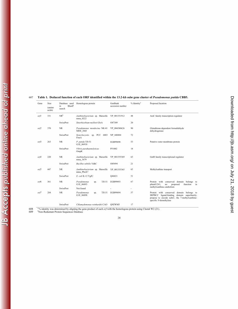
Based on sequence homologies, functions were proposed for orf1-7 (Table 1). The 304
theoretical N-terminal protein sequences of ndmA and ndmB completely matched the N-terminal 305
protein sequences previously obtained from purified NdmA and NdmB proteins. ndmA encoded 306
a 40.2-kDa protein consisting of 351 amino acids, consistent with the estimated molecular mass 307
of 40 kDa previously estimated for NdmA by SDS-PAGE. ndmB encoded a 40.9-kDa protein 308
with 355 amino acids, which was larger in size than the previously estimated molecular mass of 309
35-kDa for purified NdmB by SDS-PAGE (30). Consistent with the SDS-PAGE results, when 310
ndmB was expressed as a C-terminal His-tagged protein in E. coli, its apparent molecular mass 311
estimated by SDS-PAGE was also smaller than the theoretical MW deduced from the gene 312
sequence (Supplementary Fig. 1Sa). The protein sequences encoded by ndmA and ndmB were 313
used as queries for BLASTP search in the GenBank database. Both sequences were homologous 314
to the catalytic α subunits of different ROs. Conserved sequences for a Rieske [2Fe-2S] domain 315
(CXHX16CX2H) and a non-heme Fe(II) domain [(E/D)X2HX4H] were identified in the 316
theoretical protein sequences of both ndmA and ndmB. Furthermore, ndmA and ndmB 317
transcribed divergently from each other (Fig. 1a), indicating they are not part of the same 318
transcriptional unit. All of these support the hypothesis that NdmA and NdmB could 319
individually function as N-demethylating ROs. 320
A typical reductase gene for ROs was not found directly next to either ndmA or ndmB. 321
However, ndmD was theorized to encode a 65-kDa RO reductase that coupled NADH to ndmA 322
and ndmB. The NdmD protein sequence had a conserved Rieske-type [2Fe-2S] domain at its N-323
terminal half (Fig. 1b). Additionally, a flavin-binding domain, an NADH-binding domain, and a 324
plant-type [2Fe-2S] domain were identified at its C-terminal half, similar to FNRc-type 325
reductases of ROs (20). Some ROs are 3-component systems requiring a reductase and a 326
on July 21, 2018 by guesthttp://jb.asm
.org/D
ownloaded from
15
ferredoxin for electron transfer to oxygenase components. In these 3-component ROs, the iron-327
sulfur clusters in the ferredoxins are either the Rieske [2Fe-2S] type or [3Fe-4S] type (20). We 328
hypothesized that ndmD represented a unique gene fusion of a ferredoxin gene and a reductase 329
gene into a single ORF and encoded a functional reductase that specifically coupled to NdmA 330
and/or NdmB. 331
Functional expression and characterization of ndmA, ndmB, and ndmD gene 332
products. ndmA, ndmB, and ndmD were individually expressed as His-tagged fusion proteins in 333
E. coli BL21(DE3), and the recombinant proteins were purified using nickel-affinity 334
chromatography (Supplementary Fig. 2Sa). Purified His6-NdmD contained 4 atoms of iron, 4 335
acid-labile sulfur atoms, and one molecule of FMN per His6-NdmD monomer. The flavin 336
prosthetic group in His6-NdmD was released by boiling, indicating that it is not covalently 337
bound. The iron, acid-labile sulfur, and flavin content in His6-NdmD were in agreement with the 338
presence of a Rieske [2Fe-2S] domain, a plant type [2Fe-2S] domain, and a flavin-binding 339
domain predicted by ndmD gene sequence. His6-NdmD oxidized NADH and reduced 340
cytochrome c concomitantly, similar to several RO reductases (15, 28, 33). However, His6-341
NdmD could not N-demethylate caffeine or any related methylxanthine in the presence or 342
absence of NADH and Fe2+. 343
Purified NdmA-His6 and NdmB-His6 (Supplementary Fig. 2Sa) both contained 344
approximately 2 moles of acid-labile sulfur and 2 moles of iron per mole of enzyme monomer. 345
The iron content is lower than the expected value of 3 Fe per α subunit for ROs, which is 346
probably due to dissociation of non-heme Fe from proteins during purification. UV/visible 347
absorption spectra of oxidized NdmA-His6 and NdmB-His6 (Supplementary Fig. 2Sb,c) are 348
similar to other well-characterized ROs, with absorption maxima at 319, 453, and 553 nm and 349
on July 21, 2018 by guesthttp://jb.asm
.org/D
ownloaded from
16
320, 434, and 550 nm, respectively. Purified NdmA-His6 and NdmB-His6 could neither oxidize 350
NADH nor reduce cytochrome c. Additionally, neither of them could N-demethylate caffeine or 351
any related methylxanthine in the presence or absence of NADH and Fe2+. However, when 352
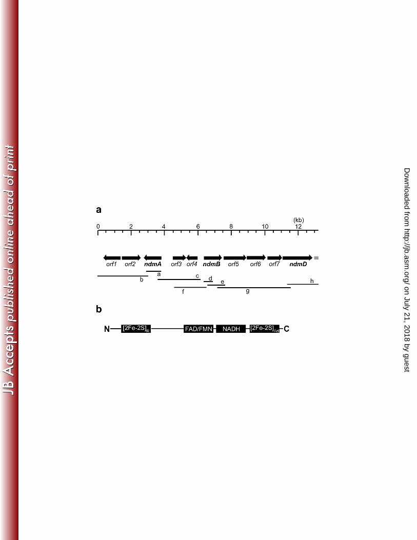
NdmA-His6 was incubated with His6-NdmD, caffeine, NADH, and exogenous Fe2+, caffeine was 353
stoichiometrically N1-demethylated to theobromine (3,7-dimethylxanthine) and formaldehyde 354
(Fig. 2a). Incubation of NdmB-His6 with His6-NdmD, theobromine, NADH, and Fe2+ resulted in 355
stoichiometric N3-demethylation of theobromine to 7-methylxanthine and formaldehyde (Fig. 356
2b). One O2 is consumed for the removal of each N-methyl group from the methylxanthine 357
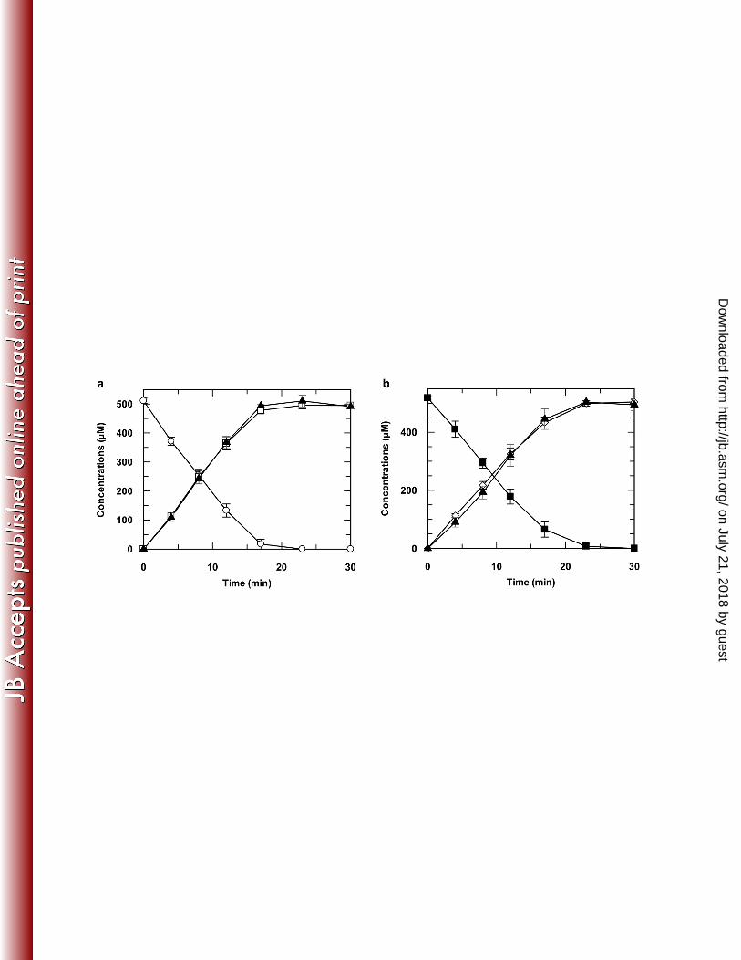
substrates by either NdmA-His6 or NdmB-His6 (Fig. 3). 358
The functions of NdmA and NdmB as position-specific methylxanthine N-demethylases 359
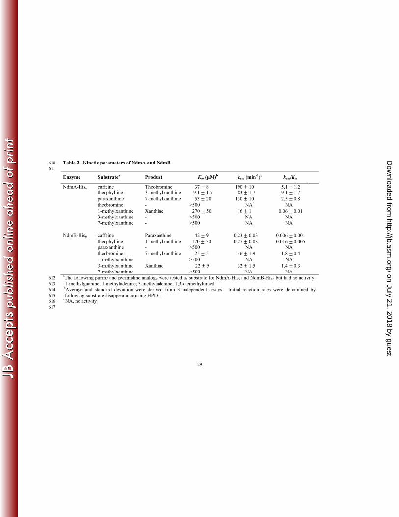
were further supported by the steady-state kinetic parameters of these two enzymes (Table 2). 360
Theobromine was the preferred substrate for NdmB-His6, with the highest kcat/KM value of 1.8 ± 361
0.4 min-1μM-1, followed closely by 3-methylxanthine. The catalytic efficiencies of NdmB-His6 362
for methylxanthines containing an N1-methyl group were 102-103 times lower than those of 363
theobromine or 3-methylxanthine. NdmB-His6 had no activity on paraxanthine, 1-364
methylxanthine, or 7-methylxanthine. Clearly, NdmB-His6 was highly specific for N3-linked 365
methyl groups of methylxanthines. In contrast, theophylline was the preferred substrate for 366
NdmA-His6, followed by caffeine and paraxanthine. NdmA-His6 had low activity on 1-367
methylxanthine and was inactive on theobromine, 3-methylxanthine, and 7-methylxanthine. 368
Thus, NdmA-His6 catalyzed the demethylation only at the N1-position of methylxanthines. 369
Various methylated purine and pyrimidine analogs were not N-demethylated by NdmA-His6 and 370
NdmB-His6, suggesting both enzymes are unlikely to be broad-specificity purine demethylases 371
involved in nucleic acid repair (1). 372
on July 21, 2018 by guesthttp://jb.asm
.org/D
ownloaded from
17
Identification of NdmC, a 7-methylxanthine-specific N-demethylase, in CBB5. 373
Previously, when purified Ndm (containing both NdmA and NdmB) was coupled with a partially 374
purified reductase fraction (designated as Ccr) from CBB5, caffeine was completely N-375
demethylated to xanthine (30). This result indicated the presence of an N7-demethylase activity 376
in CBB5, which was not associated with NdmA, NdmB, or NdmD. We have now confirmed that 377
this N7-demethylase activity, designated as NdmC, was co-purifying with NdmD in previous 378
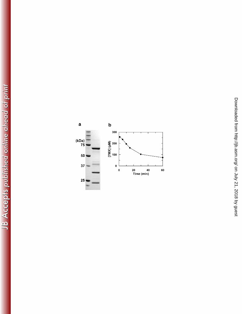
experiments. As shown in Fig. 4a, a highly enriched protein fraction with NdmC activity 379
contained 3 major protein bands when analyzed by SDS-PAGE. N-terminal protein sequences 380
were determined for all 3 bands. The N-terminal protein sequence of the 67-kDa band was 381
identical to the ndmD gene product. Furthermore, the N-terminal protein sequences of the 32-382
kDa and 22-kDa bands were identical to those encoded by orf7 and the incomplete ORF orf8, 383
respectively. This highly enriched NdmC fraction specifically N7-demethylated 7-384
methylxanthine to xanthine at the same rates observed in reactions containing active NdmA-His6 385
or NdmB-His6 (Fig. 4b). Caffeine, paraxanthine, and theobromine were not N-demethylated by 386
this fraction, indicating 7-methylxanthine was the sole substrate for NdmC. 387
388
DISCUSSION 389
In this report, we have identified and characterized the genes of P. putida CBB5 390
responsible for N-demethylation of caffeine and related methylxanthines, which is the essential 391
first step for assimilating the carbon and nitrogen in caffeine. ndmA and ndmB respectively 392
encode N1- and N3-specific methylxanthine demethylases (Table 2), allowing caffeine to be 393
metabolized to 7-methylxanthine. The consumption of one O2 by either NdmA or NdmB to 394
remove one N-linked methyl group from methylxanthines as formaldehyde suggests NdmA and 395
on July 21, 2018 by guesthttp://jb.asm
.org/D
ownloaded from
18
NdmB are monooxygenases. NdmA and NdmB activities are dependent on a reductase 396
component, which is encoded by ndmD as illustrated in this report. NdmD oxidizes NADH and 397
transfers electrons to NdmA and NdmB, which catalyze the N-demethylation reactions. 398
Previously, we purified a methylxanthine N-demethylase, Ndm, from P. putida CBB5. 399
Ndm was composed of both NdmA and NdmB, which were not separable by several 400
chromatographic steps (30). Therefore, we concluded NdmA and NdmB were two subunits of a 401
single broad-specific N-demethylase, Ndm. In this report, conclusive evidence has been 402
provided that NdmA and NdmB are individual N-demethylases with unique substrate 403
specificities. The physical properties (shape, surface charge density, MW, etc.) of NdmA and 404
NdmB appear to be very similar, resulting in their previous co-purification. Based on the 405
substrate specificities of NdmA and NdmB, we propose the N-demethylation of caffeine by 406
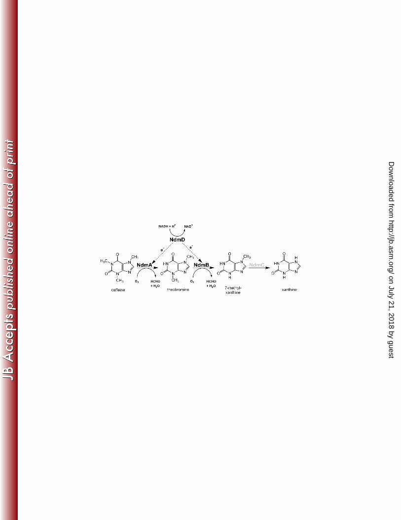
CBB5 occurs primarily in the sequence depicted in Fig. 5. The N1-methyl group is first removed 407
from caffeine by NdmA, forming theobromine, which is the preferred substrate of NdmB. 408
NdmB then removes the N3-methyl group, producing 7-methylxanthine. 409
The gene sequences of ndmA and ndmB support our previous hypothesis of Ndm as a RO. 410
Conserved sequences for a Rieske [2Fe-2S] domain and a mononuclear non-heme Fe(II) domain, 411
which is most likely the site of oxidation, are identified in the protein sequences deduced from 412
ndmA and ndmB. The dependency of NdmA and NdmB on the reductase component (NdmD), 413
UV/visible absorption spectra, utilization of oxygen as a co-substrate, and stimulation of NdmA 414
and NdmB activity by exogenous Fe2+ are characteristics commonly found among ROs. 415
Traditionally, ROs have been classified according to their electron transport components (3). 416
However, Gibson and Parales (13) have demonstrated in a phylogenetic analysis that the 417
catalytic α subunit of ROs clustered into four major groups according to the substrates utilized by 418
on July 21, 2018 by guesthttp://jb.asm
.org/D
ownloaded from
19
the ROs. We extended this analysis and constructed a rootless phylogenetic tree including 419
NdmA, NdmB, plus 64 well-characterized ROs (Supplementary Fig. 2S). 420
NdmA and NdmB clustered with Cdm and the hypothetical RO in Janthinobacterium sp. 421
Marseille (mma_0224) in a distinct clade. Cdm, being 89% identical to NdmA, is likely to be a 422
caffeine N-demethylase. The function of mma_0224 is not clear, as it is 53% and 58% identical 423
to NdmA or NdmB, respectively. The nearest-neighbor clades to NdmA and NdmB contain ROs 424
that catalyze O-demethylation or cleavage of C-N or C-O bonds. Some of them are 425
monooxygenases (e.g. DdmC and VanA), while others are dioxygenases (such as CarAa). The 426
homology between these enzymes and either NdmA (9-17% identity) or NdmB (8-15% identity) 427
is minimal and restricted to the N-terminal regions of these proteins where the Rieske [2Fe-2S] 428
domain is located. The low similarity between NdmA/NdmB and other ROs is not surprising 429
considering the diversity of specific substrates used by these enzymes and the fact that the 430
specific substrate binding site of ROs is predominantly at the C-terminal portion of RO α 431
subunits (11). Our phylogenetic analysis did not support monophylogeny between NdmA/NdmB 432
with the majority of ROs, which hydroxylate aromatic ring substrates. It is likely that divergent 433
evolution of ROs resulted in two groups, one for catalyzing aromatic ring hydroxylations and 434
one for C-O/C-N bond-cleaving reactions. 435
When Ndm was purified previously from P. putida CBB5, Ndm activity was assayed 436
using a partially purified reductase component isolated from CBB5 (30). Under that assay 437
condition, we observed complete N-demethylation of caffeine to xanthine. NdmA and NdmB 438
present in the Ndm accounted for the N1- and N3-demethylation activity, respectively. However, 439
neither NdmA nor NdmB could be responsible for N-demethylation of 7-methylxanthine to 440
xanthine, since 7-methylxanthine is not a substrate for either enzyme (Table 2). Here, we have 441
on July 21, 2018 by guesthttp://jb.asm
.org/D
ownloaded from
20
identified the 7-methylxanthine-specific N-demethylation activity, designated as NdmC. NdmC 442
was found to co-purify with NdmD (Fig. 4a). The highly enriched reductase fraction contained 443
NdmD plus two additional major protein bands. From their respective N-terminal protein 444
sequences, the genes encoding these two proteins were identified as orf7 and orf8 in the ndm 445
gene cluster (Fig 1a). Although orf8 is an incomplete ORF, its partial theoretical protein 446
sequence displayed significant similarity to various glutathione-S-transferases. Meanwhile, the 447
theoretical protein sequence deduced from orf7 showed significant identity to proteins with a 448
conserved domain belonging to the SRPBCC ligand-binding domain superfamily. Included in 449
this superfamily are the C-terminal catalytic domains of aromatic ring-hydroxylating RO α 450
subunits. Interestingly, conserved sequence of a Rieske [2Fe-2S] domain was not present in the 451
deduced protein sequence of orf7. However, conserved sequence of a mononuclear non-heme 452
Fe(II) domain, which is usually the catalytic site for ROs, was identified in orf7. We currently 453
hypothesize orf7 encodes NdmC, the N-demethylase specific for 7-methylxanthine. Expression 454
of this protein to confirm N7-demethylase activity has proven difficult due to formation of 455
inclusion bodies under various cloning and growth conditions. 456
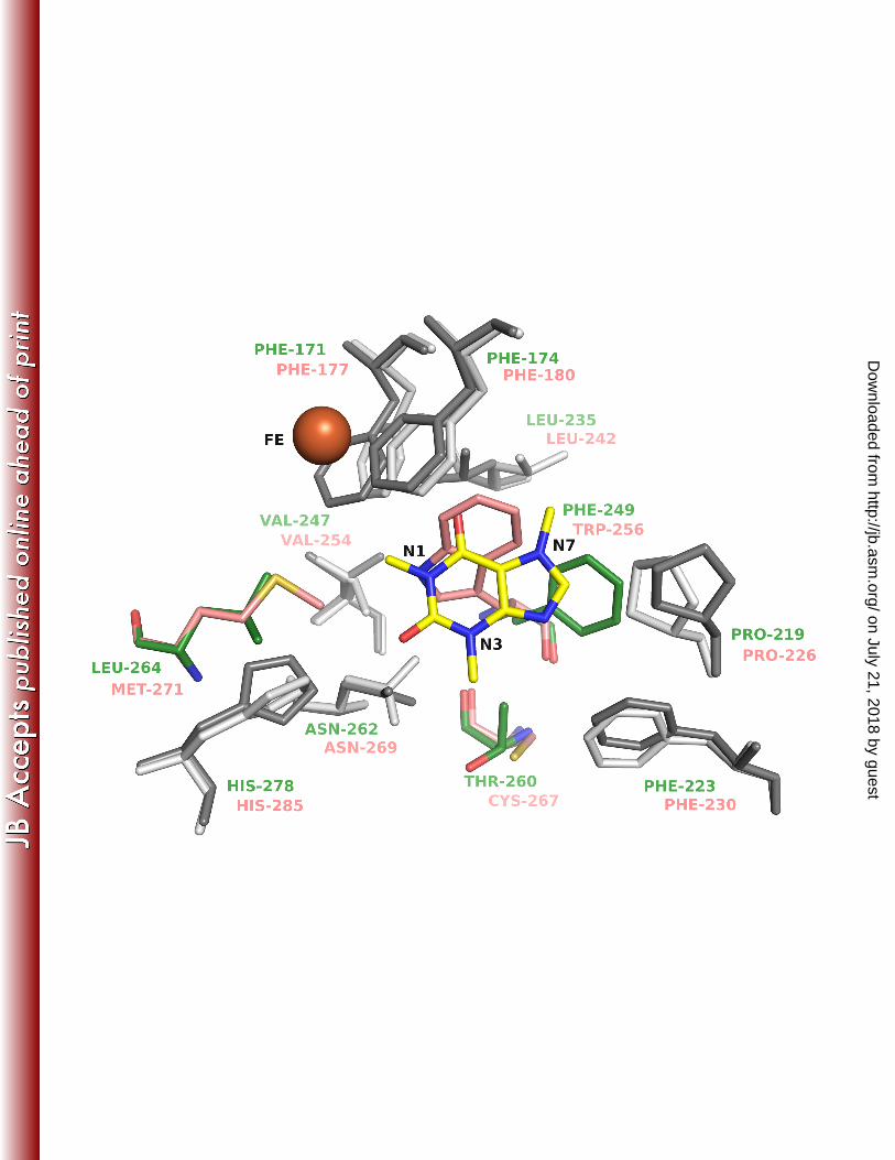
DdmC (dicamba O-demethylase) is one of the few ROs with available crystal structure 457
(10) (PDB ID 3GKE) that are evolutionary related to NdmA and NdmB, and was also the top 458
match in a BLAST search against the PDB for NdmA and NdmB sequences. NdmA and NdmB 459
share only about 40% similarity (~ 20% identity) with DdmC across approximately 350 residues. 460
Therefore, NdmA and NdmB homology models were generated using DdmC as a structural 461
template (Fig. 6). A comparison of the active sites in the NdmA and NdmB homology models 462
indicates that the pocket can readily accommodate caffeine next to the non-heme iron for 463
demethylation. Most of the residues lining the active site pocket in NdmA and NdmB 464
on July 21, 2018 by guesthttp://jb.asm
.org/D
ownloaded from
21
superimpose very well across the ten models generated for each (Fig. 6). There are only 3 465
residues in the binding pocket that are not conserved between NdmA (Phe 249, Thr 260 and 466
Leu264) and NdmB (Trp256, Cys267 and Met271). The most notable non-conserved pair is 467
Phe249 in NdmA and Trp 256 in NdmB. Both residues are close to the catalytic non-heme iron. 468
Trp256 was found to explore multiple conformations in the various homology models generated, 469
all of which altered the shape of the cavity near the catalytic site significantly. It seems steric 470
effects due to the shape and size of the residue at this position in NdmA and NdmB may be one 471
of the contributing factors that alter the binding orientation of the ligand affecting substrate 472
specificity. Similar effects of bound ligand orientation on substrate specificity and product 473
selectivity have been previously discussed for ROs (11). Any further structural features that 474
explain the specificity difference between NdmA and NdmB must await crystal structure 475
elucidation, which is in progress. 476
In summary, we propose that utilization of caffeine by CBB5 occurs via N-demethylation 477
in a preferential, ordered sequence (Fig. 5). This ordered N-demethylation of caffeine is 478
catalyzed by NdmA and NdmB at the N1- and N3- positions, respectively, of various 479
methylxanthines. Both NdmA and NdmB are monooxygenases; one O2 is consumed per N-480
methyl group removed as formaldehyde. NdmD appears to be the sole reductase for transfer of 481
electrons from NADH to NdmA, NdmB, and possibly NdmC, for oxygen activation and N-482
demethylation to formaldehyde. Although we have established enzymologically that NdmC 483
catalyzes N7-demethylation of 7-methylxanthine, the gene correlation has not yet been 484
established. This discovery of highly specific methylxanthine N-demethylases has shed light on 485
the long-standing question of how bacteria are able to use caffeine and other methylxanthines as 486
on July 21, 2018 by guesthttp://jb.asm
.org/D
ownloaded from
22
sole carbon and nitrogen source. CBB5 is able to use xanthine, formaldehyde, and formate, 487
which are liberated from caffeine and other methylxanthines, for growth (data not shown). 488
NdmA, NdmB, and NdmC could have broad applications in bioremediation of 489
environments contaminated by caffeine and related methylxanthines, particularly in countries 490
with large coffee- and tea-processing industries. These genes could also find utility in detecting 491
caffeine, a marker for human activities in wastewater streams (7), or in converting the enormous 492
waste generated via manufacturing of coffee and tea to animal feed and feedstocks for fuels and 493
fine chemicals (24). Last, but not least, these genes could prove useful in production of 494
pharmaceutically-useful modified xanthine analogs, which are currently being synthesized by 495
challenging multi-step processes (22). The assignment of NdmA and NdmB to the RO enzyme 496
family broadens our understanding on the enzymatic mechanism for N-demethylation reactions, 497
as we generally have less knowledge regarding demethylases than methylating enzymes (14). 498
This first report of soluble bacterial RO N-demethylases and their complete gene sequences will 499
certainly stimulate the discovery of new N-demethylases involved in the degradation of many 500
natural products and xenobiotics. 501
502
ACKNOWLEDGEMENTS 503
This research was supported by the University of Iowa Research Funds. We also thank 504
Dr. David Peate and Jay Thompson in the Department of Geoscience for their assistance in the 505
ICP-MS experiment. 506
507
on July 21, 2018 by guesthttp://jb.asm
.org/D
ownloaded from
23
REFERENCES 508
1. Aas, P. A., M. Otterlei, P. O. Falnes, C. B. Vagbo, F. Skorpen, M. Akbari, O. 509
Sundheim, M. Bjoras, G. Slupphaug, E. Seeberg, and H. E. Krokan. 2003. Human and 510
bacterial oxidative demethylases repair alkylation damage in both RNA and DNA. Nature 511
421:859-863. 512
2. Arnaud, M. J. 2011. Pharmacokinetics and metabolism of natural methylxanthines in 513
animal and man, p. 33-91. In B. B. Fredholm (ed.), Handbook of experimental 514
pharmacology, vol. 200. Springer-Verlag, Berlin-Heidelberg. 515
3. Batie, C. J., D. P. Ballou, and C. C. Correll. 1991. Phthalate dioxygenase reductase and 516
related flavin-iron-sulfur containing electron transferases, p. 546-566. In F. Müller (ed.), 517
Chemistry and biochemistry of flavoenzymes, vol. 3. CRC Press, Boca Raton, Fla. 518
4. Bradford, M. M. 1976. A rapid and sensitive method for the quantitation of microgram 519
quantities of protein utilizing the principle of protein-dye binding. Anal. Biochem. 72:248-520
254. 521
5. Brunel, F., and J. Davidson. 1988. Cloning and sequencing of Pseudomonas genes 522
encoding vanillate demethylase. J. Bacteriol. 170:4924-4930. 523
6. Bryksin, A. V., and I. Matsumura. 2010. Overlap extension PCR cloning: a simple and 524
reliable way to create recombinant plasmids. Biotechniques 48:463-465. 525
7. Buerge, I. J., T. Poiger, M. D. Muller, and H. R. Buser. 2006. Combined sewer 526
overflows to surface waters detected by the anthropogenic marker caffeine. Environ. Sci. 527
Technol. 40:4096-4102. 528
8. Dash, S. S., and S. N. Gummadi. 2006. Catabolic pathways and biotechnological 529
applications of microbial caffeine degradation. Biotechnol. Lett. 28:1993-2002. 530
on July 21, 2018 by guesthttp://jb.asm
.org/D
ownloaded from
24
9. Delcher, A. L., D. Harmon, S. Kasif, O. White, and S. L. Salzberg. 1999. Improved 531
microbial gene identification with GLIMMER. Nucleic Acid Res. 27:4636-4641. 532
10. Dumitru, R., W. Z. Wang, D. P. Weeks, and M. A. Wilson. 2009. Crystal structure of 533
dicamba monooxygenase: a Rieske nonheme oxygenase that catalyzes oxidative 534
demethylation. J. Mol. Biol. 392:498-510. 535
11. Ferraro, D. J., L. Gakhar, and S. Ramaswamy. 2005. Rieske business: structure-536
function of Rieske non-heme oxygenases. Biochem. Biophys. Res. Commun. 338:175-190. 537
12. Gerken, T., C. A. Girard, Y. C. Tung, C. J. Webby, V. Saudek, K. S. Hewitson, G. S. 538
Yeo, M. A. McDonough, S. Cunliffe, L. A. McNeill, J. Galvanovskis, P. Rorsman, P. 539
Robins, X. Prieur, A. P. Coll, M. Ma, Z. Jovanovic, I. S. Farooqi, B. Sedgwick, I. 540
Barroso, T. Lindahl, C. P. Ponting, F. M. Ashcroft, S. O'Rahilly, and C. J. Schofield. 541
2007. The obesity-associated FTO gene encodes a 2-oxoglutarate-dependent nucleic acid 542
demethylase. Science 318:1469-1472. 543
13. Gibson, D. T., and R. E. Parales. 2000. Aromatic hydrocarbon dioxygenases in 544
environmental biotechnology. Curr. Opin. Biotech. 11:236-243. 545
14. Hagel, J. M., and P. J. Facchini. 2010. Biochemistry and occurrence of O-demethylation 546
in plant metabolism. Front. Physiol. 1:14. 547
15. Haigler, B. E., and D. T. Gibson. 1990. Purifcation and properties of NADH-548
ferredoxinNAP reductase, a component of naphthalene dioxygenase from Pseudomonas sp. 549
strain NCIB 9816. J. Bacteriol. 172:457-464. 550
16. Herman, P. L., M. Behrens, S. Chakraborty, B. M. Chrastil, J. Barycki, and D. P. 551
Weeks. 2005. A three-component dicamba O-demethylase from Pseudomonas maltophilia, 552
on July 21, 2018 by guesthttp://jb.asm
.org/D
ownloaded from
25
strain DI-6: gene isolation, characterization, and heterologous expression. J. Biol. Chem. 553
280:24759-24767. 554
17. Hollenberg, P. F. 1992. Mechanisms of cytochrome P450 and peroxidase-catalyzed 555
xenobiotic metabolism. FASEB J. 6:686-694. 556
18. Jones, S. B., C. M. Terry, T. E. Lister, and D. C. Johnson. 1999. Determination of 557
submicromolar concentrations of formaldehyde by liquid chromatography. Anal. Chem. 558
71:4030-4033. 559
19. Koide, Y., S. Nakane, and Y. Imai. 1996. Caffeine demethylate gene-containing DNA 560
fragment and microbial process for producing 3-methyl-7-alkylxanthine. United States 561
patent US5550041. 562
20. Kweon, O., S. J. Kim, S. Baek, J. C. Chae, M. D. Adjei, D. H. Baek, Y. C. Kim, and C. 563
E. Cerniglia. 2008. A new classification system for bacterial Rieske non-heme iron 564
aromatic ring-hydroxylating oxygenases. BMC Biochem. 9:11. 565
21. Larkin, M. A., G. Blackshields, N. P. Brown, R. Chenna, P. A. McGettigan, H. 566
McWilliam, F. Valentin, I. M. Wallace, A. Wilm, R. Lopez, J. D. Thompson, T. J. 567
Gibson, and D. G. Higgins. 2007. Clustal W and Clustal X version 2.0. Bioinformatics 568
23:2947-2948. 569
22. Liu, G., P. S. M. M. Reddy, J. R. Barber, S. C. Ng, and Y. F. Zhou. 2010. Synthesis of 570
novel 3,7-Dihydro-purine-2,6-dione derivatives. Synthetic Commun. 40:1418-1436. 571
23. Lukashin, A. V., and M. Borodovsky. 1998. GeneMark.hmm: new solutions for gene 572
finding. Nucleic Acid Res. 26:1107-1115. 573
on July 21, 2018 by guesthttp://jb.asm
.org/D
ownloaded from
26
24. Mussatto, S. I., E. M. S. Machado, S. Martins, and J. A. Teixeira. 2011. Production, 574
composition, and application of coffee and its industrial residues. Food Bioprocess. Tech. 575
4:661-672. 576
25. Sali, A., and T. L. Blundell. 1993. Comparative protein modelling by satisfaction of 577
spatial restraints. J. Mol. Biol. 234:779-815. 578
26. Sambrook, J., E. F. Fritsch, and T. Maniatis. 1989. Molecular cloning: a laboratory 579
manual, 2nd ed. Cold Spring Harbor laboratory Press, Cold Spring Harbor, N. Y. 580
27. Shi, Y., F. Lan, C. Matson, P. Mulligan, J. R. Whetstine, P. A. Cole, and R. A. Casero. 581
2004. Histone demethylation mediated by the nuclear amine oxidase homolog LSD1. Cell 582
119:941-953. 583
28. Subramanian, V., T. Liu, W. Yeh, and D. T. Gibson. 1979. Toluene dioxygenase: 584
purification of an iron-sulfur protein by affinity chromatography. Biochem. Biophys. Res. 585
Commun. 91:1131-1139. 586
29. Suhara, K., S. Takemori, M. Katagiri, K. Wada, and H. Kobayashi. 1975. Estimation 587
of labile sulfide in iron-sulfur proteins. Anal. Biochem. 68:632-636. 588
30. Summers, R. M., T. M. Louie, C. L. Yu, and M. Subramanian. 2011. Characterization 589
of a broad-specificity non-haem iron N-demethylase from Pseudomonas putida CBB5 590
capable of utilizing several purine alkaloids as sole carbon and nitrogen source. 591
Microbiology 157:583-592. 592
31. Tsukada, Y., J. Fang, H. Erdjument-Bromage, M.E.Warren, C. H. Borchers, P. 593
Tempst, and Y. Zhang. 2006. Histone demethylation by a family of JmjC domain-594
containing proteins. Nature 439:811-816. 595
on July 21, 2018 by guesthttp://jb.asm
.org/D
ownloaded from
27
32. Ueda, T., E. T. Lode, and M. J. Coon. 1972. Enzymatic ω-oxidation. VI. Isolation of 596
homogenous reduced diphosphopyridine nucleotide-rubredoxin reductase. J. Biol. Chem. 597
247:2109-2116. 598
33. Yu, C. L., W. Liu, D. J. Ferraro, E. N. Brown, J. V. Parales, S. Ramaswamy, G. J. 599
Zylstra, D. T. Gibson, and R. E. Parales. 2007. Purification, characterization, and 600
crystallization of the components of a biphenyl dioxygenase system from Sphingobium 601
yanoikuyae B1. J. Ind. Microbiol. Biotechnol. 34:311-324. 602
34. Yu, C. L., T. M. Louie, R. Summers, Y. Kale, S. Gopishetty, and M. Subramanian. 603
2009. Two distinct pathways for metabolism of theophylline and caffeine are coexpressed 604
in Pseudomonas putida CBB5. J. Bacteriol. 191:4624-4632. 605
606
on July 21, 2018 by guesthttp://jb.asm
.org/D
ownloaded from
28
Table 1. Deduced function of each ORF identified within the 13.2-kb ndm gene cluster of Pseudomonas putida CBB5. 607
Gene Size
(amino acids)
Database used in BlastP search
Homologous protein GenBank accession number
% Identitya Proposed fucntion
orf1 331 NRb Janthinobacterium sp. Marseille mma_0222
YP_001351912 48 AraC family transcription regulator
SwissProt Sinorhizobium meliloti GlxA O87389 20
orf2 370 NR Pseudomonas mendocina NK-01 MDS_2843
YP_004380626 90 Glutathione-dependent formaldehyde dehydrogenase
SwissProt Synechocystis. sp. PCC 6803 FrmA
NP_440484 72
orf3 263 NR P. putida TJI-51 G1E_06918
EGB99698
53 Putative outer membrane protein
SwissProt Vibrio parahaemolyticus OmpK
P51002 18
orf4 220 NR Janthinobacterium sp. Marseille mma_3679
YP_001355369 63 GntR family transcriptional regulator
SwissProt Bacillus subtilis YdhC O05494 21
orf5 447 NR Janthinobacterium sp. Marseille mma_PbuX7
YP_001355365 65 Methylxathine transport
SwissProt E. coli K-12 YgfU Q46821 51
orf6 361 NR Pseudomonas sp. TJI-51 G1E_06893
EGB99693 67 Protein with conserved domain belongs to pfam01261; no proposed function in methylxanthine catabolism
SwissProt Not found - -
orf7 284 NR Pseudomonas sp. TJI-51 G1E_06898
EGB99694 57 Protein with conserved domain belongs to SRPBCC ligand-binding domain superfamily; propose to encode ndmC, the 7-methylxanthine-specific N-demethylase
SwissProt Chlamydomonas reinhardtii CAO Q9ZWM5 17
a % identity was determined by aligning the gene product of each orf with the homologous protein using Clustal W2 (21). 608 bNon-Reduntant Protein Sequences Database 609
on July 21, 2018 by guesthttp://jb.asm
.org/D
ownloaded from
29
Table 2. Kinetic parameters of NdmA and NdmB 610 611
Enzyme Substratea Product Km (µM)b kcat (min-1)b kcat/Km 1 1
NdmA-His6 caffeine Theobromine 37 8 190 10 5.1 1.2 theophylline 3-methylxanthine 9.1 1.7 83 1.7 9.1 1.7 paraxanthine 7-methylxanthine 53 20 130 10 2.5 0.8 theobromine - >500 NAc NA 1-methylxanthine Xanthine 270 50 16 1 0.06 0.01 3-methylxanthine - >500 NA NA 7-methylxanthine - >500 NA NA NdmB-His6 caffeine Paraxanthine 42 9 0.23 0.03 0.006 0.001 theophylline 1-methylxanthine 170 50 0.27 0.03 0.016 0.005 paraxanthine - >500 NA NA theobromine 7-methylxanthine 25 5 46 1.9 1.8 0.4 1-methylxanthine - >500 NA NA 3-methylxanthine Xanthine 22 5 32 1.5 1.4 0.3 7-methylxanthine - >500 NA NA aThe following purine and pyrimidine analogs were tested as substrate for NdmA-His6 and NdmB-His6 but had no activity: 612 1-methylguanine, 1-methyladenine, 3-methyladenine, 1,3-diemethyluracil. 613
bAverage and standard deviation were derived from 3 independent assays. Initial reaction rates were determined by 614 following substrate disappearance using HPLC. 615
c NA, no activity 616 617
on July 21, 2018 by guesthttp://jb.asm
.org/D
ownloaded from
30
FIGURE LEGENDS 618
Figure 1. (a) Organization of ndmABD and orf1-7 in the 13.2-kb gene cluster in P. putida 619
CBB5. Black arrows indicate the position and orientation of each orf. The grey box directly 620
downstream of ndmD represents the 5′ end of a partial ORF, orf8. Black lines (labeled a to h) 621
represent the 8 overlapping PCR products used to assemble this map. (b) Schematic 622
organization of conserved domains identified in the deduced protein sequence of ndmD. 623
Designations: [2Fe-2S]R, Rieske [2Fe-2S] domain; [FAD/FMN], flavin adenine dinucleotide or 624
flavin mononucleotide binding domain; [NADH], reduced nicotinamide adenine dinucleotide 625
binding domain; [2Fe-2S]Fd, plant-type ferredoxin [2Fe-2S] domain. 626
627
Figure 2. Stoichiometric N-demethylation of methylxanthines by NdmA-His6 and NdmB-His6. 628
(a) NdmA-His6 N-demethylated 510 ± 10 µM caffeine () to 500 ± 10 μM theobromine (), 629
resulting in production of 510 ± 20 μM formaldehyde (), or one mole of formaldehyde 630
produced per mole of theobromine formed. (b) NdmB-His6 N-demethylated 510 ± 10 μM 631
theobromine () to 500 ± 10 μM 7-methylxanthine () and 500 ± 3 μM formaldehyde (). 632
Concentrations reported were means of triplicates with standard deviation. 633
634
Figure 3. Consumption of O2 by NdmA-His6 and NdmB-His6 during N-demethylation of 635
caffeine and theobromine, respectively. N-Demethylation of 150 ± 3 µM caffeine to 140 ± 3 μM 636
theobromine by NdmA-His6 with consumption of 130 ± 20 μM O2 () or 140 ± 20 µM 637
theobromine to 140 ± 9 μM 7-methylxanthine by NdmB-His6 with consumption of 130 ± 10 μM 638
O2 () are shown. Background oxygen consumption in an enzyme reaction with either NdmA-639
on July 21, 2018 by guesthttp://jb.asm
.org/D
ownloaded from
31
His6 or NdmB-His6 but without methylxanthine is also shown (). Concentrations reported 640
were means of triplicates with standard deviations. 641
642
Figure 4. (a) A SDS-PAGE gel and (b) N7-demethylation of 7-methylxanthine by a highly 643
enriched enzyme preparation containing NdmC and NdmD activities. Three major protein bands 644
with estimated molecular masses of 67, 32, and 22 kDa were present in this highly enriched 645
enzyme preparation. 646
647
Figure 5. Proposed sequential N-demethylation of caffeine by Pseudomonas putida CBB5, 648
based on catalytic efficiencies of NdmA-His6 and NdmB-His6. N-demethylation of caffeine is 649
initated at the N1-position by NdmA, forming theobromine. NdmB then catalyzes removal of the 650
N-linked methyl group at the N3 position, producing 7-methylxanthine. Oxygen is the co-651
substrate for NdmA and NdmB. NdmD couples with NdmA and NdmB by transferring 652
electrons from NADH to NdmA and NdmB for oxygen activation. Each methyl group removed 653
results in the formation of one formaldehyde. NdmC is proposed to be specific for N-654
demethylation of 7-methylxanthine to xanthine. 655
656
Figure 6. Active site model of NdmA and NdmB: Residues lining the active site cavity of 657
NdmA and NdmB homology models are shown as sticks. Residues conserved between NdmA 658
and NdmB are colored light grey and dark grey respectively. Non-conserved residues are colored 659
green (NdmA) and salmon (NdmB). Caffeine (yellow sticks) is modeled into the active site at the 660
position of dicamba in DdmC (pdb ID 3GL2) (10). The non-heme iron from the dicamba 661
structure is shown as an orange sphere. 662
on July 21, 2018 by guesthttp://jb.asm
.org/D
ownloaded from