Embed Size (px)
Citation preview
Normal ECG: Rate and Rhythm
Read chapters 4 and 22
ECG Interpretation*Standardization
Rate
1. RR interval
2. Heart rate
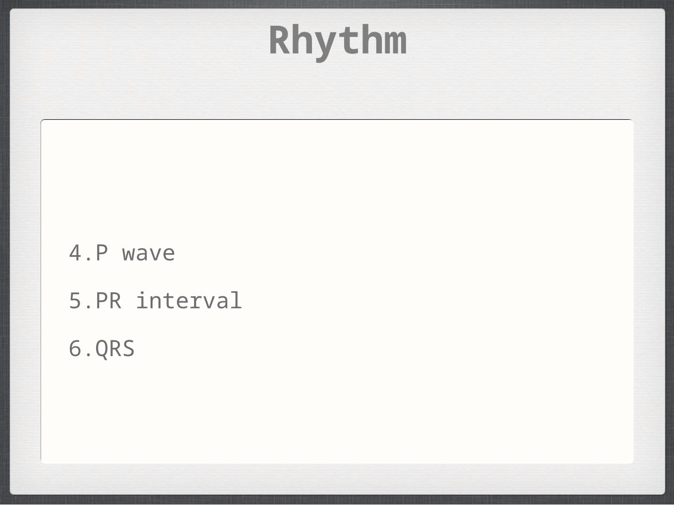
Rhythm
3. PP interval
4. P wave
width, height, shape, etc.
5. PR interval
6. QRS
width (and height)
axis
R wave progression
abnormal Q waves
ST segment
T waves
QT interval
U waves*See Chapter 22
ECG Interpretation
Univ. of Wisconsin Medical School
• http://www.fammed.wisc.edu/pcc/ecg/
The Normal ECG
Normal = normal sinus rhythm
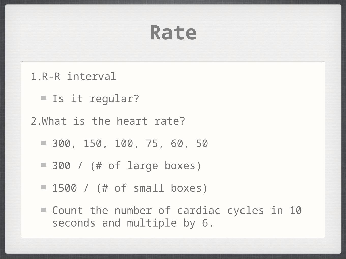
Rate
1.R-R interval
Is it regular?
2.What is the heart rate?
300, 150, 100, 75, 60, 50
300 / (# of large boxes)
1500 / (# of small boxes)
Count the number of cardiac cycles in 10 seconds and multiple by 6.
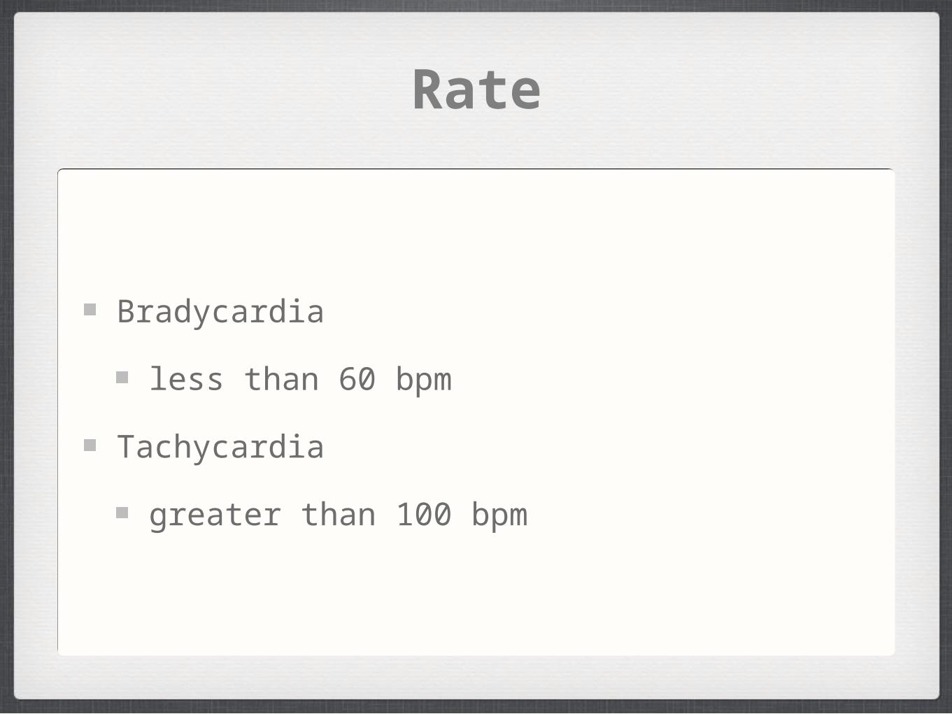
Rate
Bradycardia
less than 60 bpm
Tachycardia
greater than 100 bpm
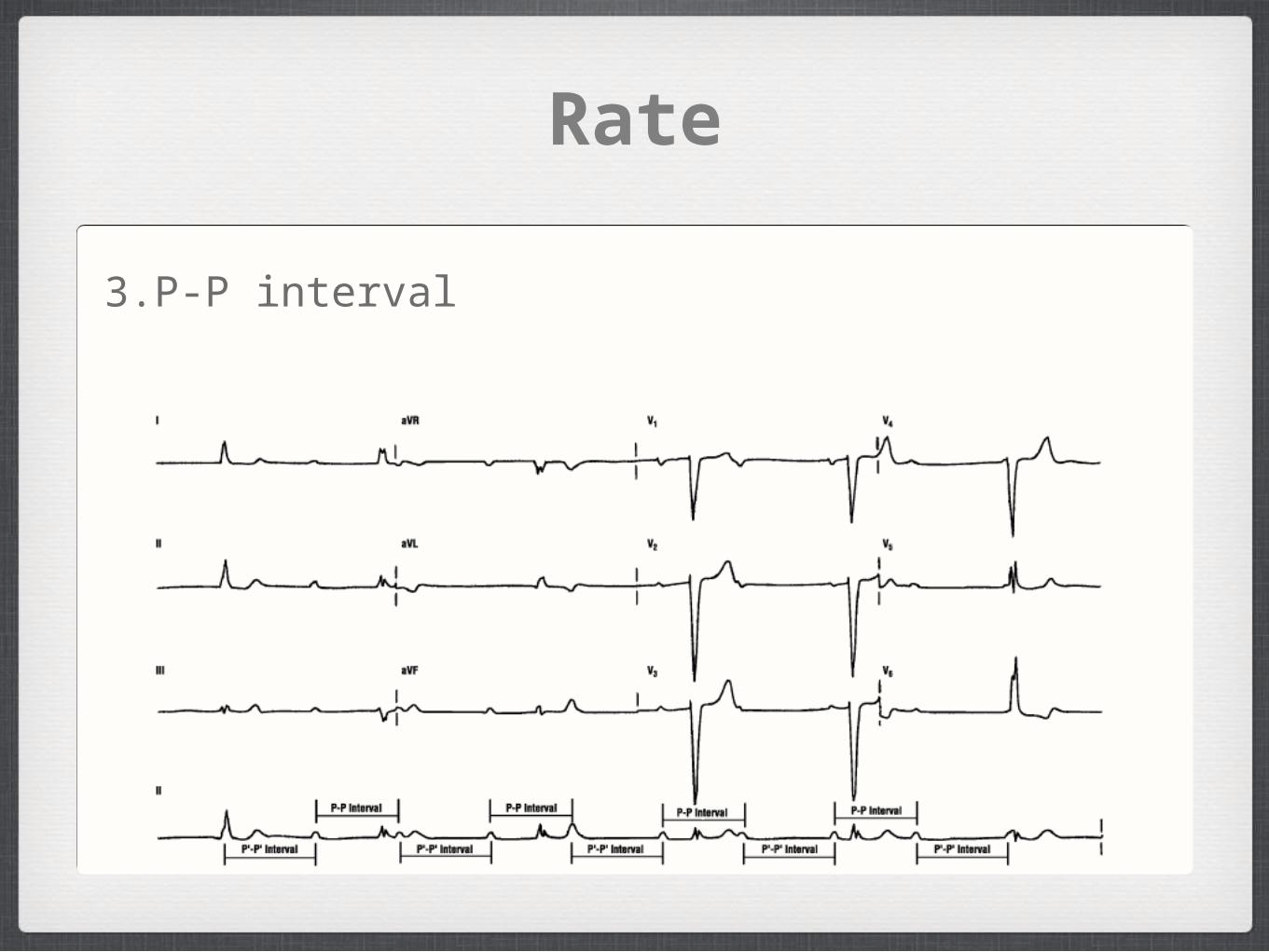
Rate
3.P-P interval
Rhythm
4.P wave
5.PR interval
6.QRS
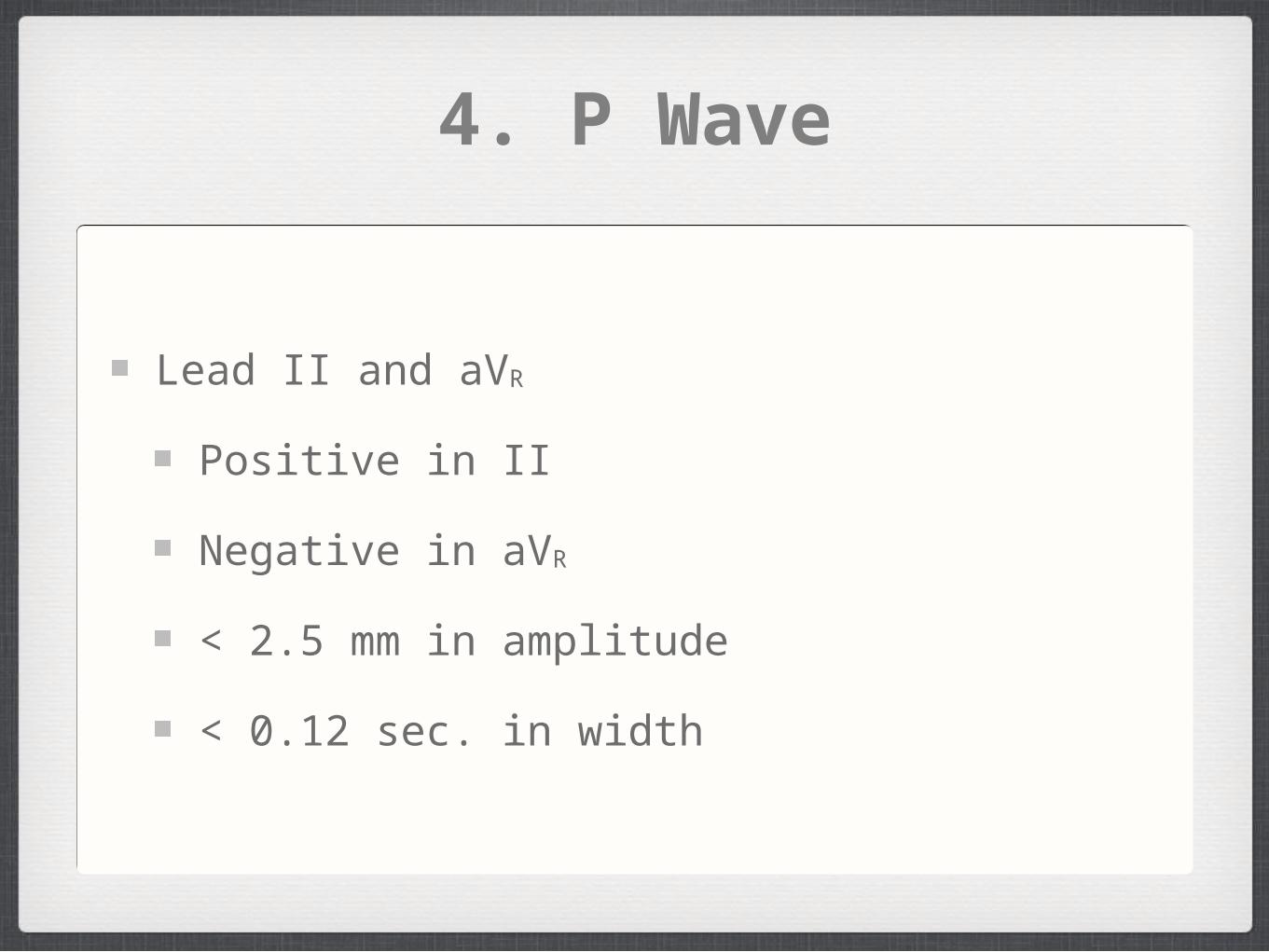
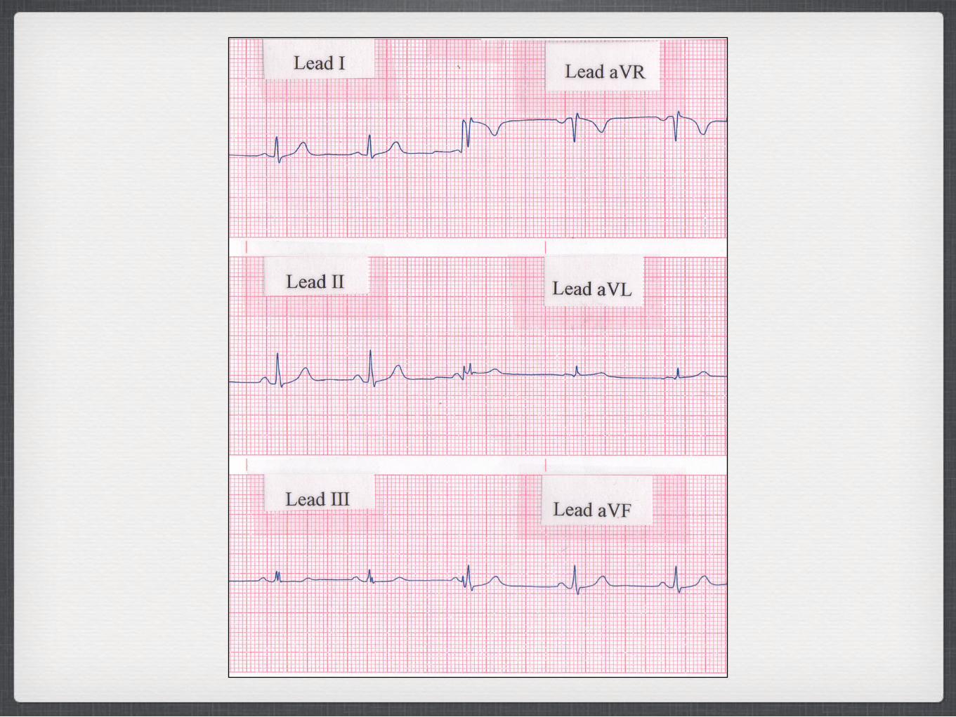
4. P Wave
Lead II and aVR
Positive in II
Negative in aVR
< 2.5 mm in amplitude
< 0.12 sec. in width
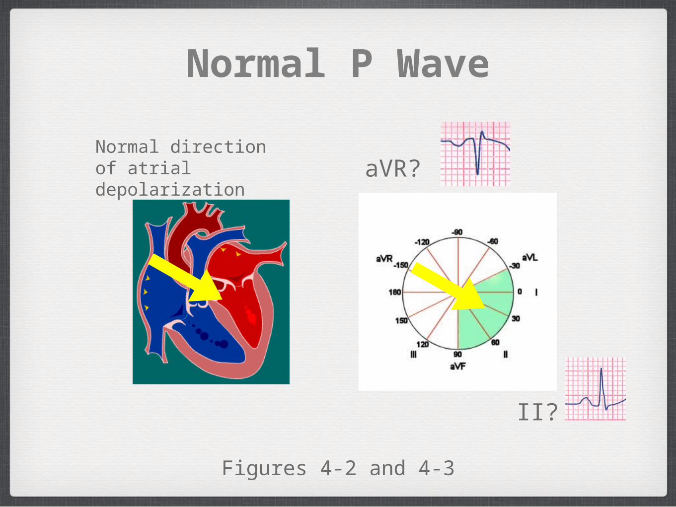
Normal P Wave
aVR?
II?
Figures 4-2 and 4-3
Normal direction of atrial depolarization
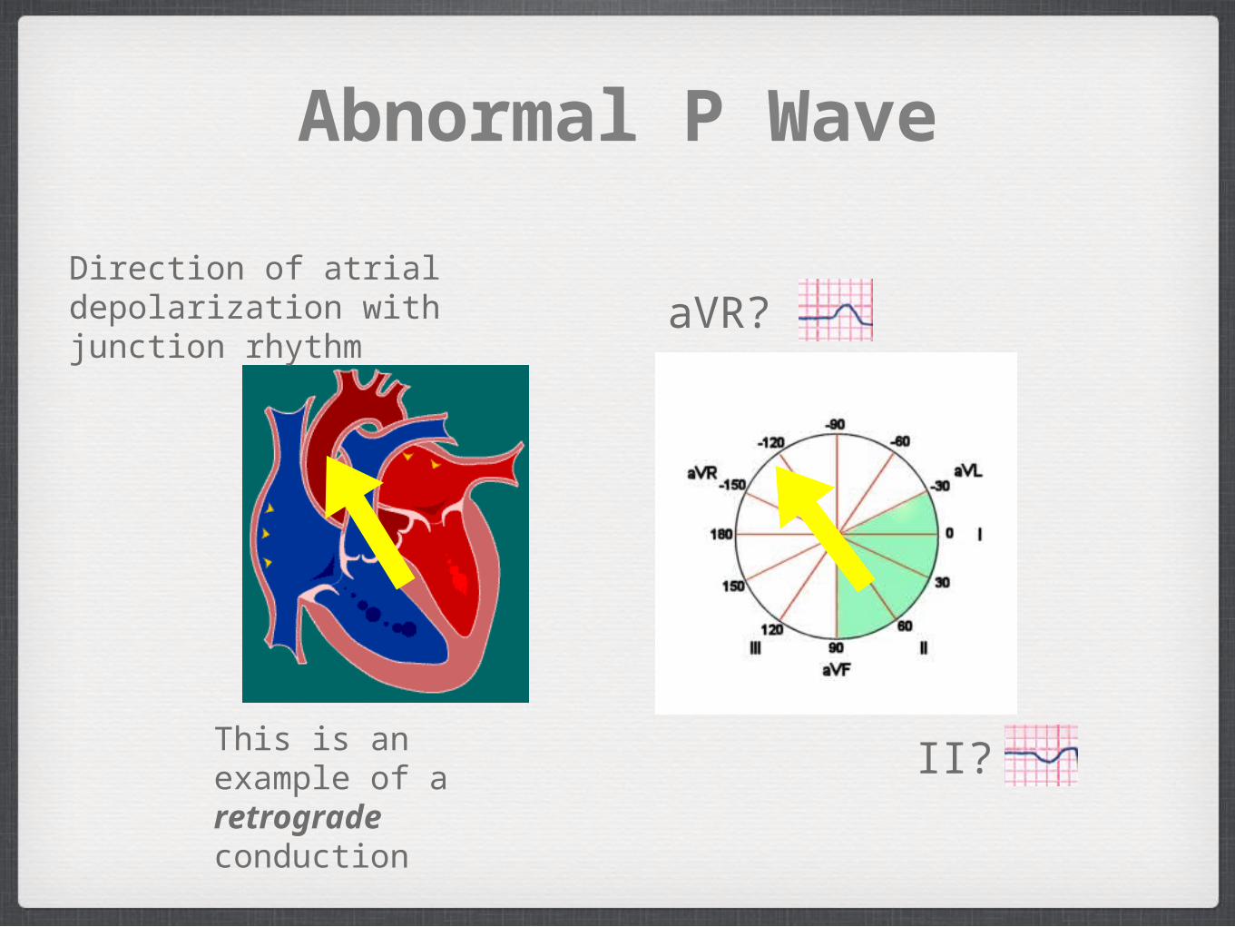
Abnormal P Wave
aVR?
II?
Direction of atrial depolarization with junction rhythm
This is an example of a retrograde conduction
P wave
The same direction as QRS
Only one P wave in front of QRS
Do all the P waves look alike?

5. PR interval
0.12 - 0.20 seconds
6. QRS Complex
What is the width? (less than 0.10 seconds)
Do all the QRS waves in the same lead look alike?
R wave progressionAxisAbnormal Q waves (infarction)

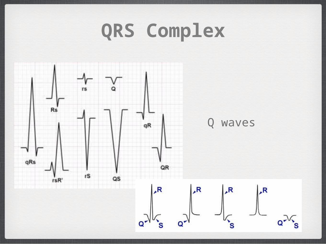
QRS Complex
Q waves
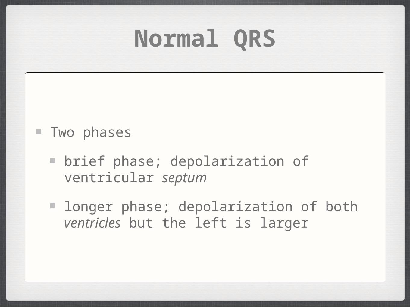
Normal QRS
Two phases
brief phase; depolarization of ventricular septum
longer phase; depolarization of both ventricles but the left is larger
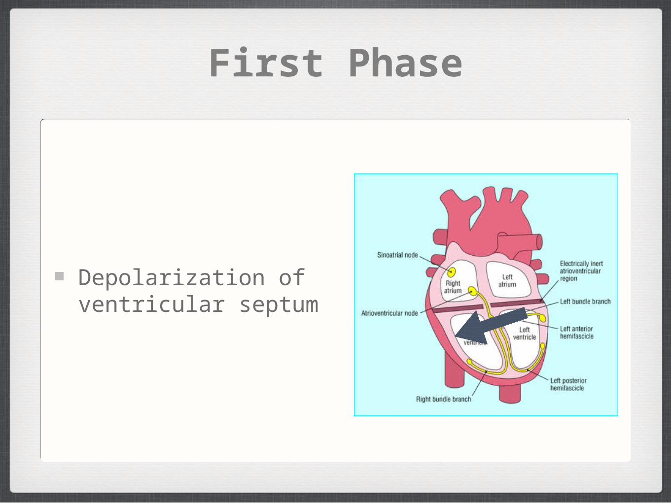
First Phase
Depolarization of ventricular septum
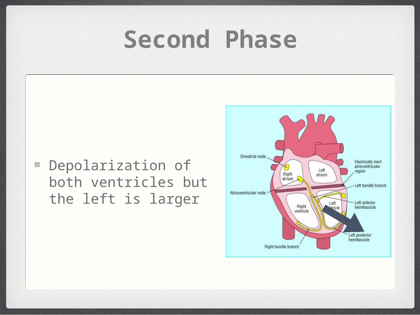
Second Phase
Depolarization of both ventricles but the left is larger
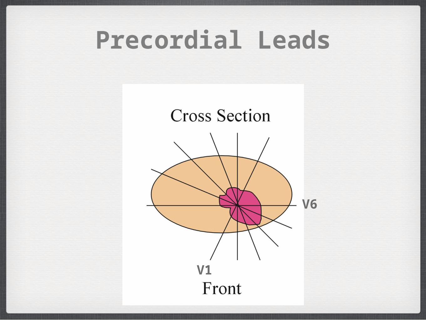
Precordial Leads
V1
V6
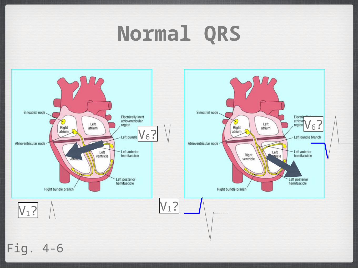
Normal QRS
V1? V1?
V6?V6?
Fig. 4-6
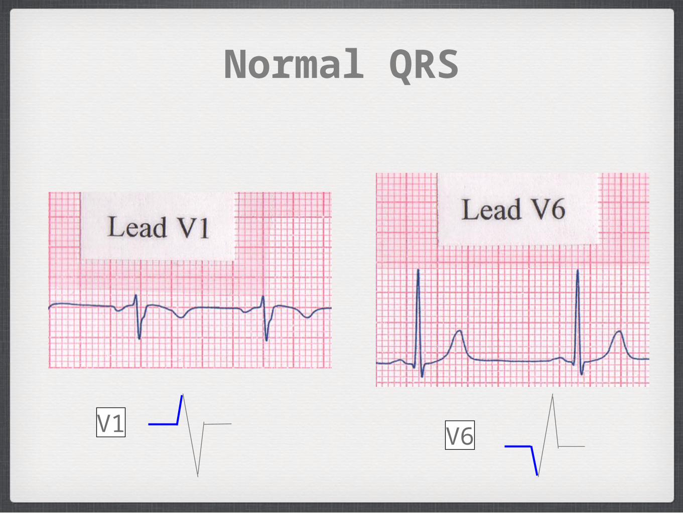
Normal QRS
V1 V6
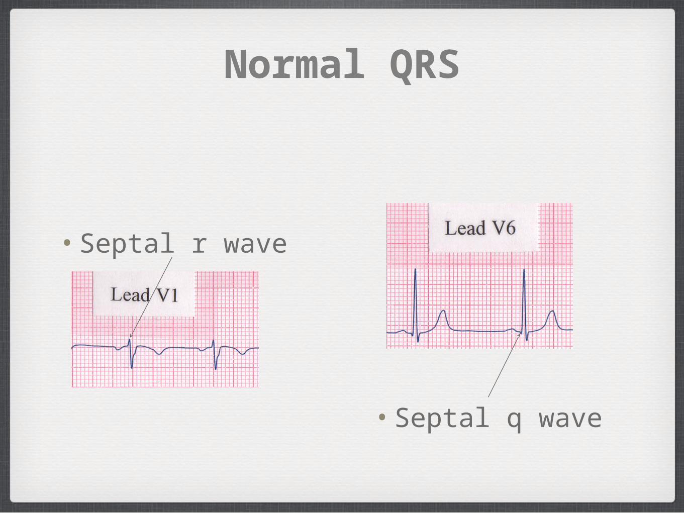
Normal QRS
• Septal r wave
• Septal q wave
6. QRS Complex
R wave progression
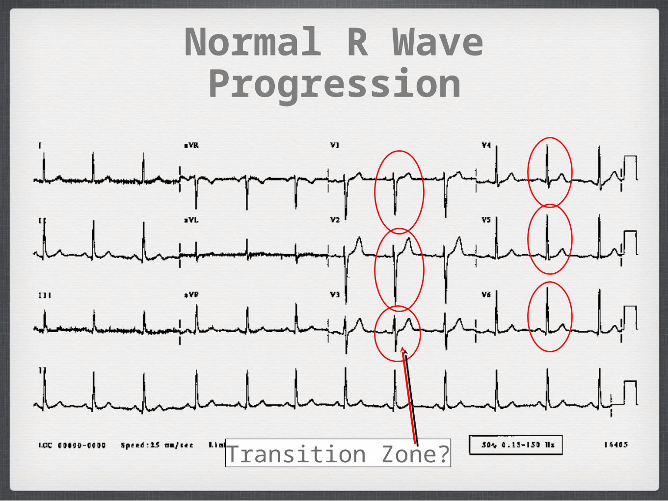
Normal R Wave Progression
Transition Zone?
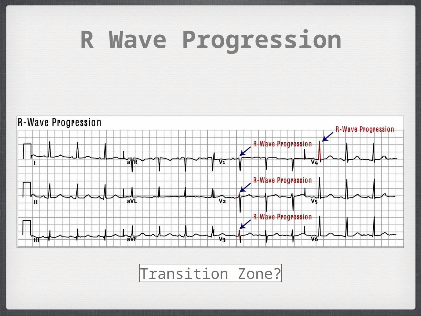
R Wave Progression
Transition Zone?
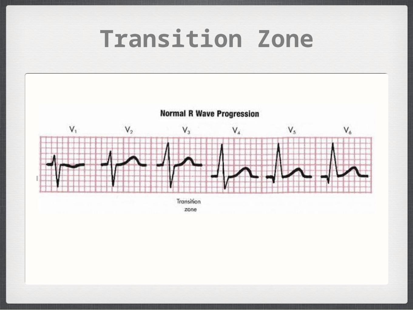
Transition Zone
Figure 4-7
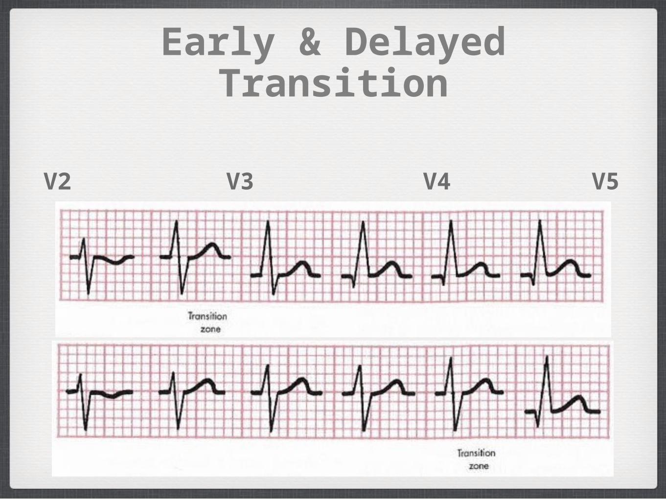
Early & Delayed Transition
• Figure 4-7
V1 V2 V3 V4 V5 V6
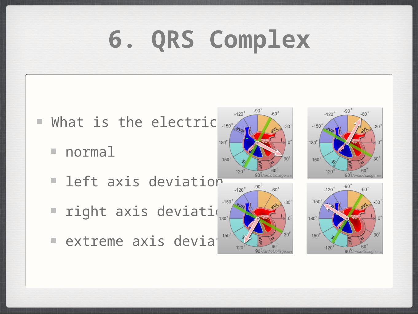
6. QRS Complex
What is the electrical axis?
normal
left axis deviation
right axis deviation
extreme axis deviation
7. St Segment
ST segment elevation or depression
(see chapters 8 & 9)
8. T Wave
Normally positive where QRS wave is positive
V3- V6 and II, but negative in aVR
Abnormally tall T waves

Practice
ECG Library
http://www.ecglibrary.com/ecghome.html
ECG: The Art of Interpretation
http://www.12leadecg.com/full/
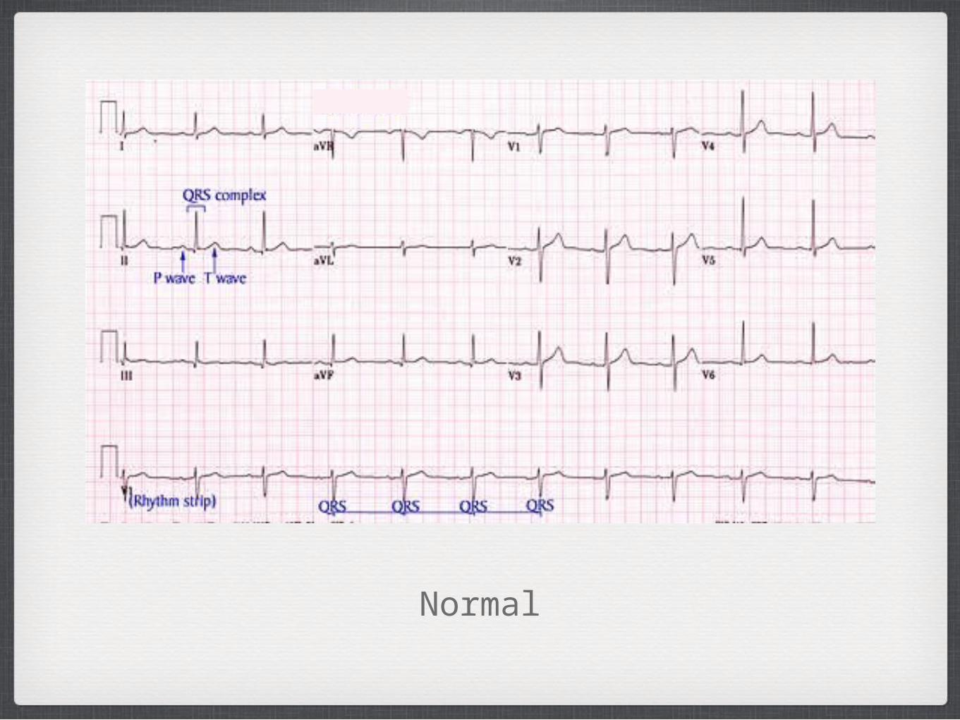
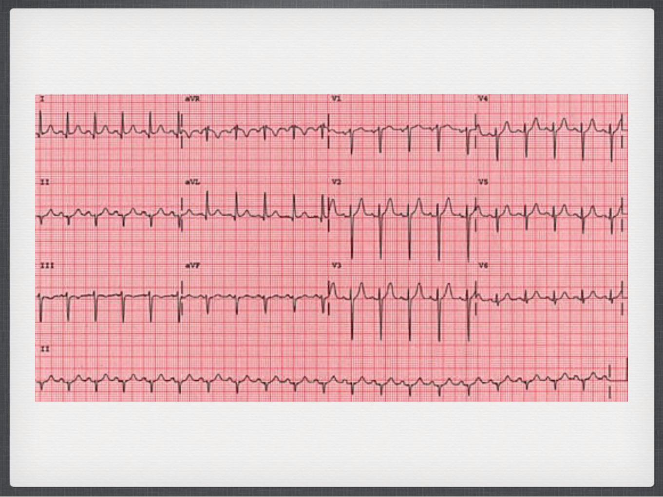
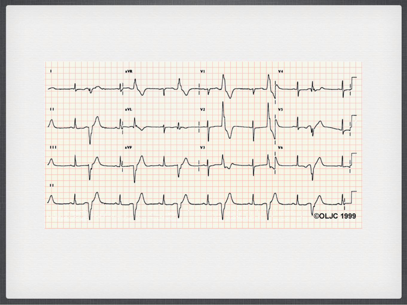
Normal
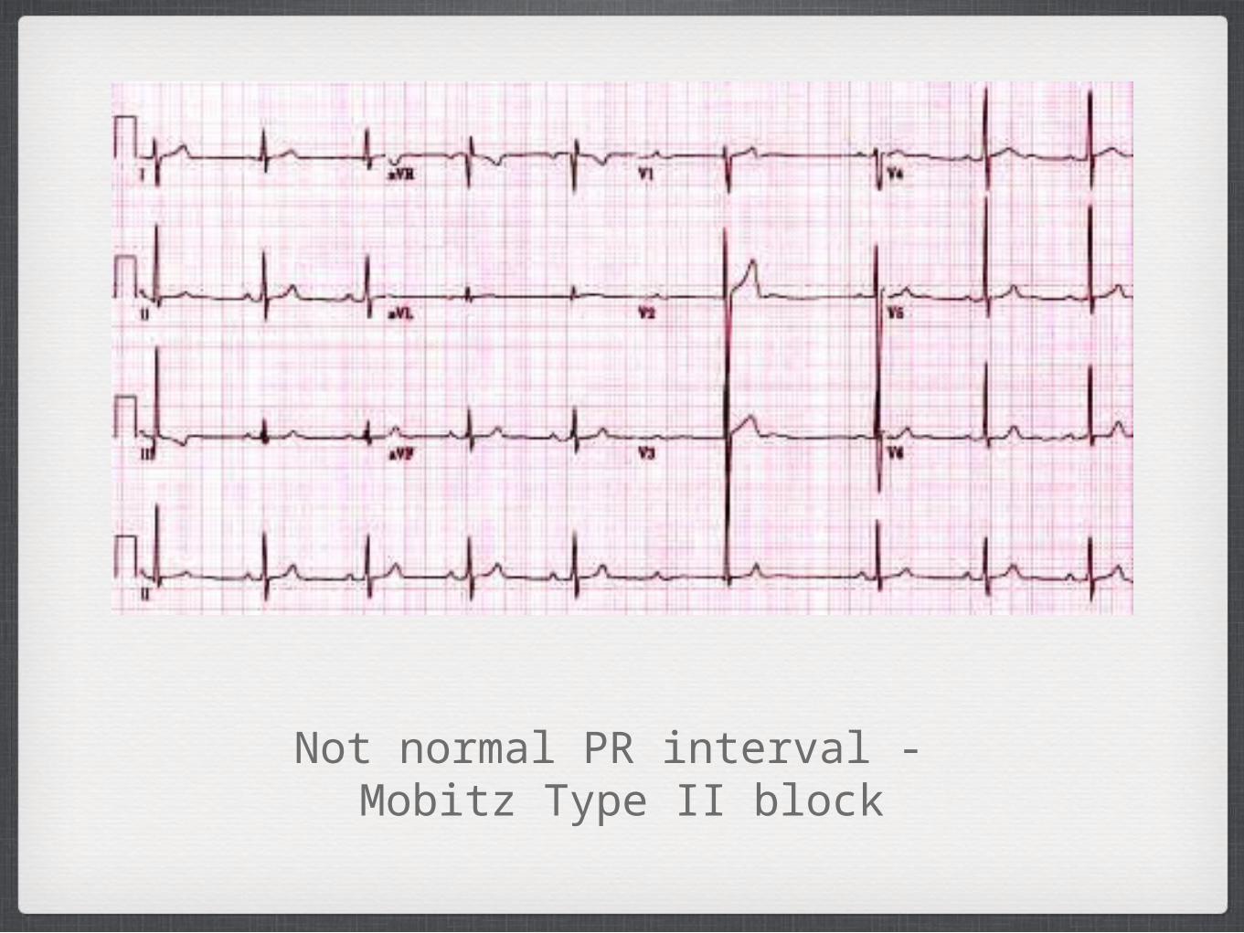
Not normal PR interval - Mobitz Type II block
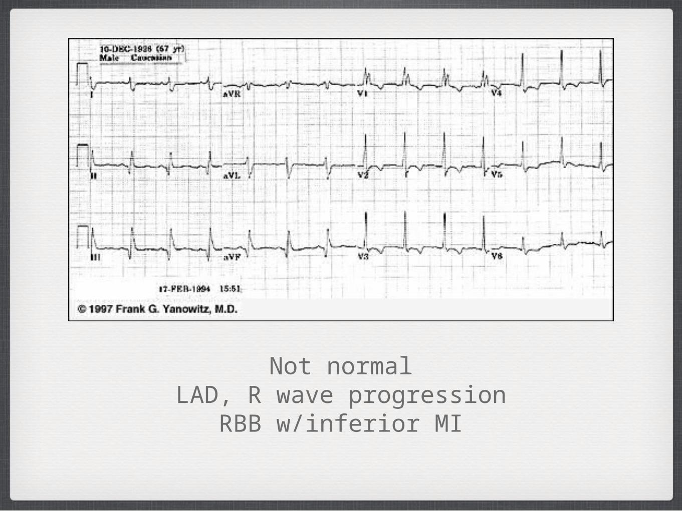
Not normalLAD, R wave progression
RBB w/inferior MI

Not normal -First degree block, left atrial enlargement, left
bundle branch block, & inferior MI
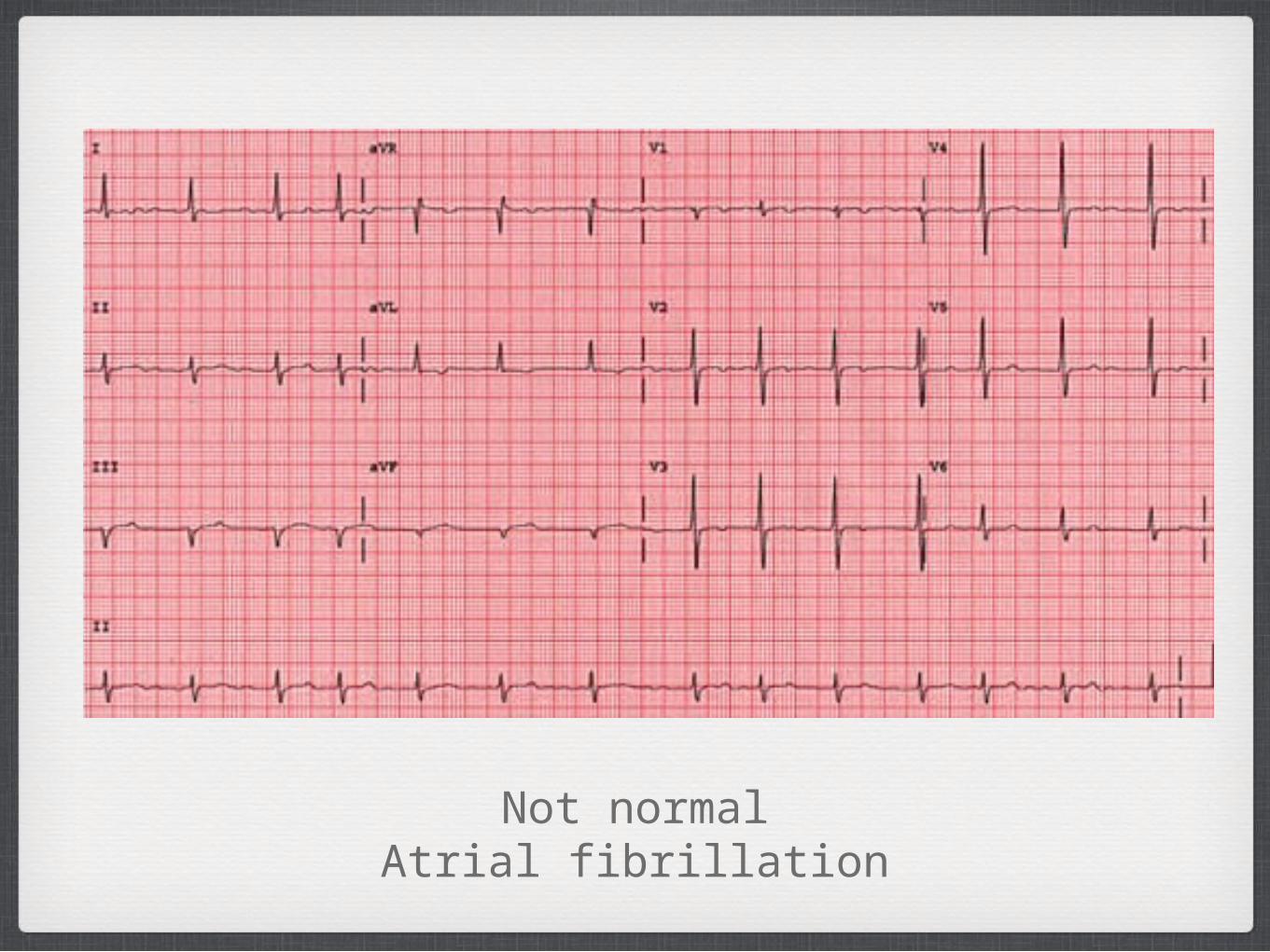
Not normalAtrial fibrillation

Normal
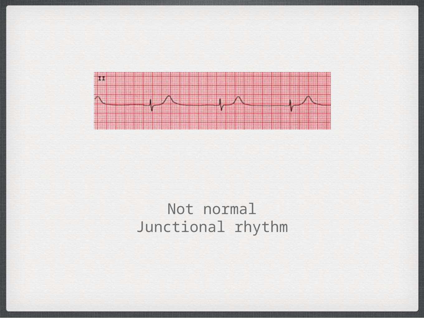
Not normalJunctional rhythm
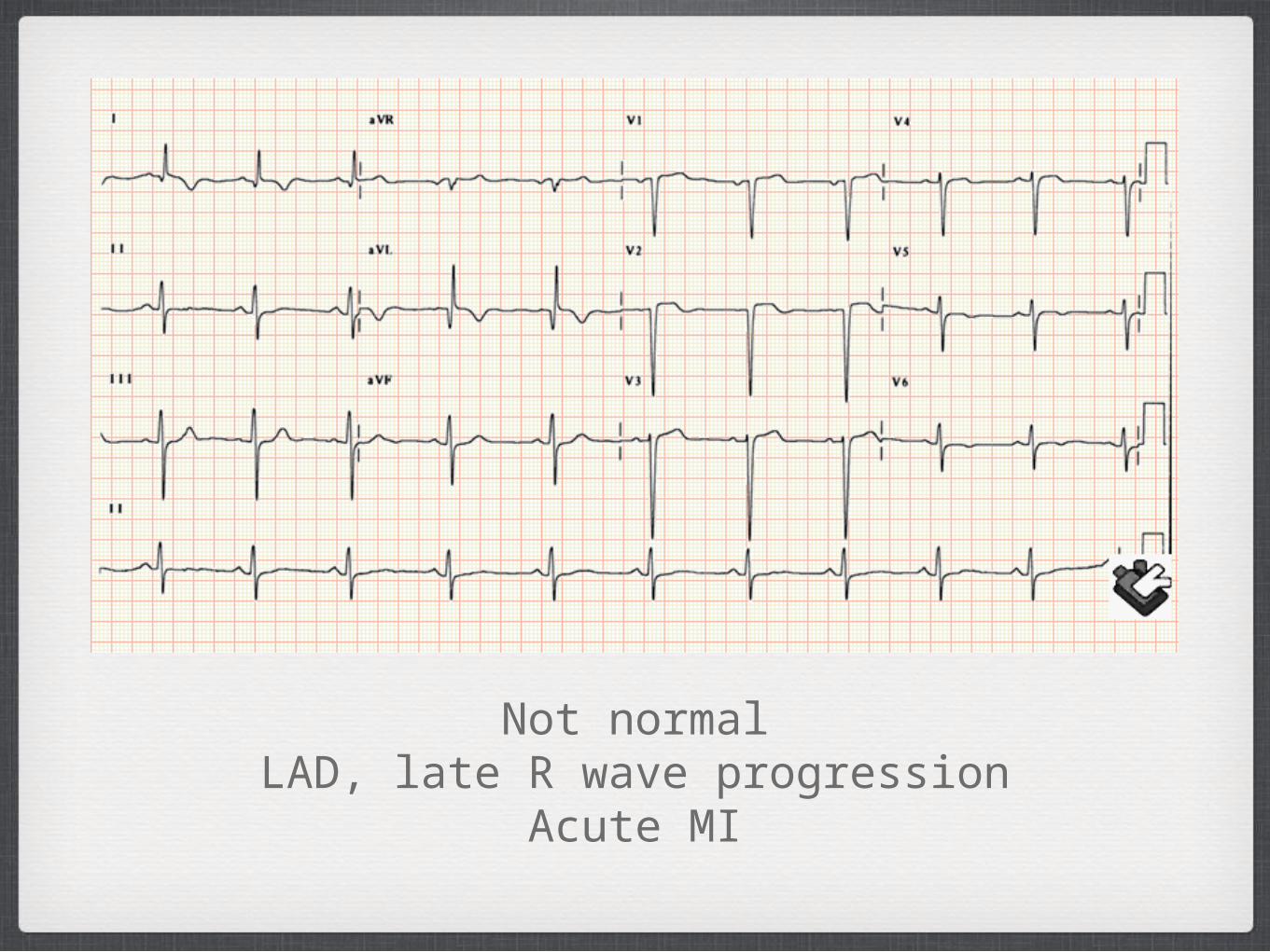
Not normalLAD, late R wave progression
Acute MI
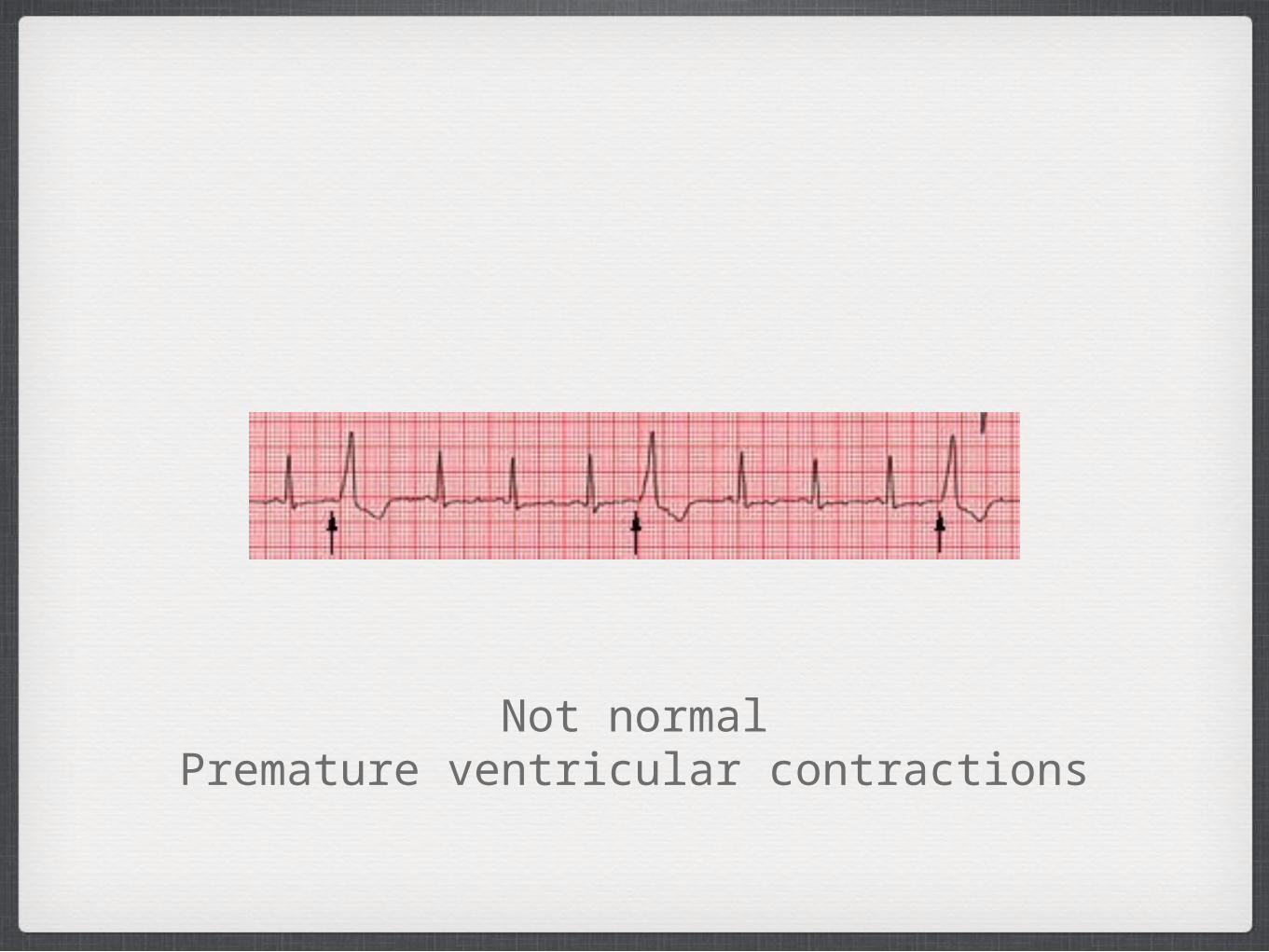
Not normalPremature ventricular contractions

Not normal
Ventricular tachycardia: note fast rate and wide bizarre QRS.
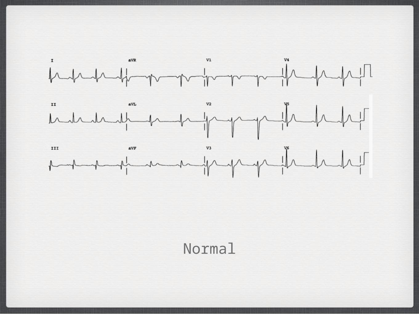
Normal

Not normal
Second degree AV block - type II

Not normalRAD, R wave progression
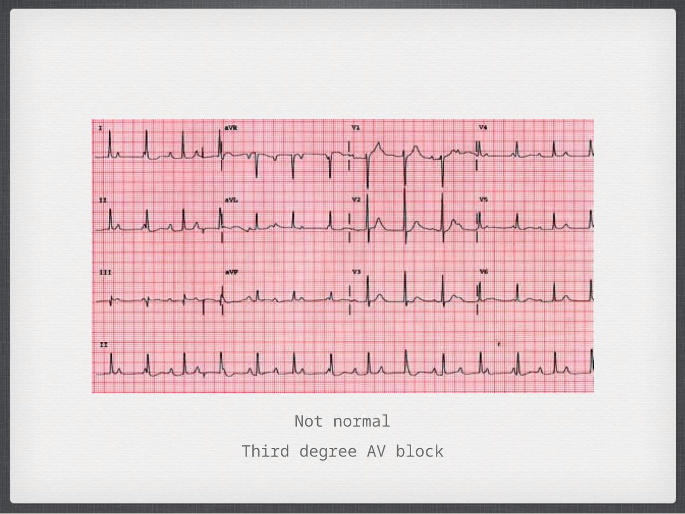
Not normal
Third degree AV block
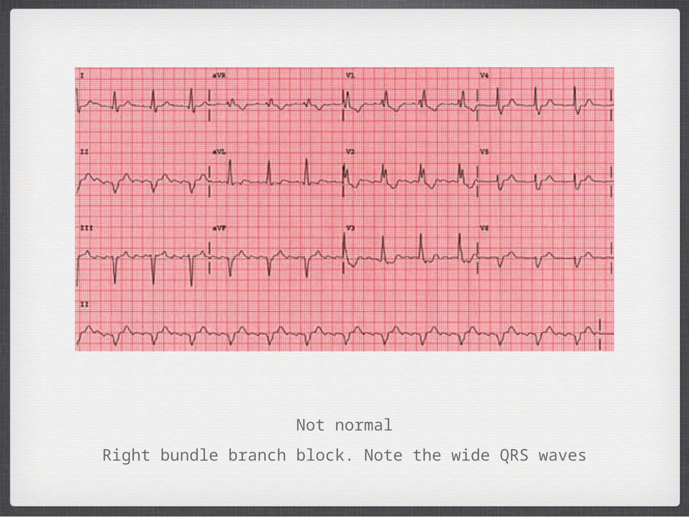
Not normal
Right bundle branch block. Note the wide QRS waves
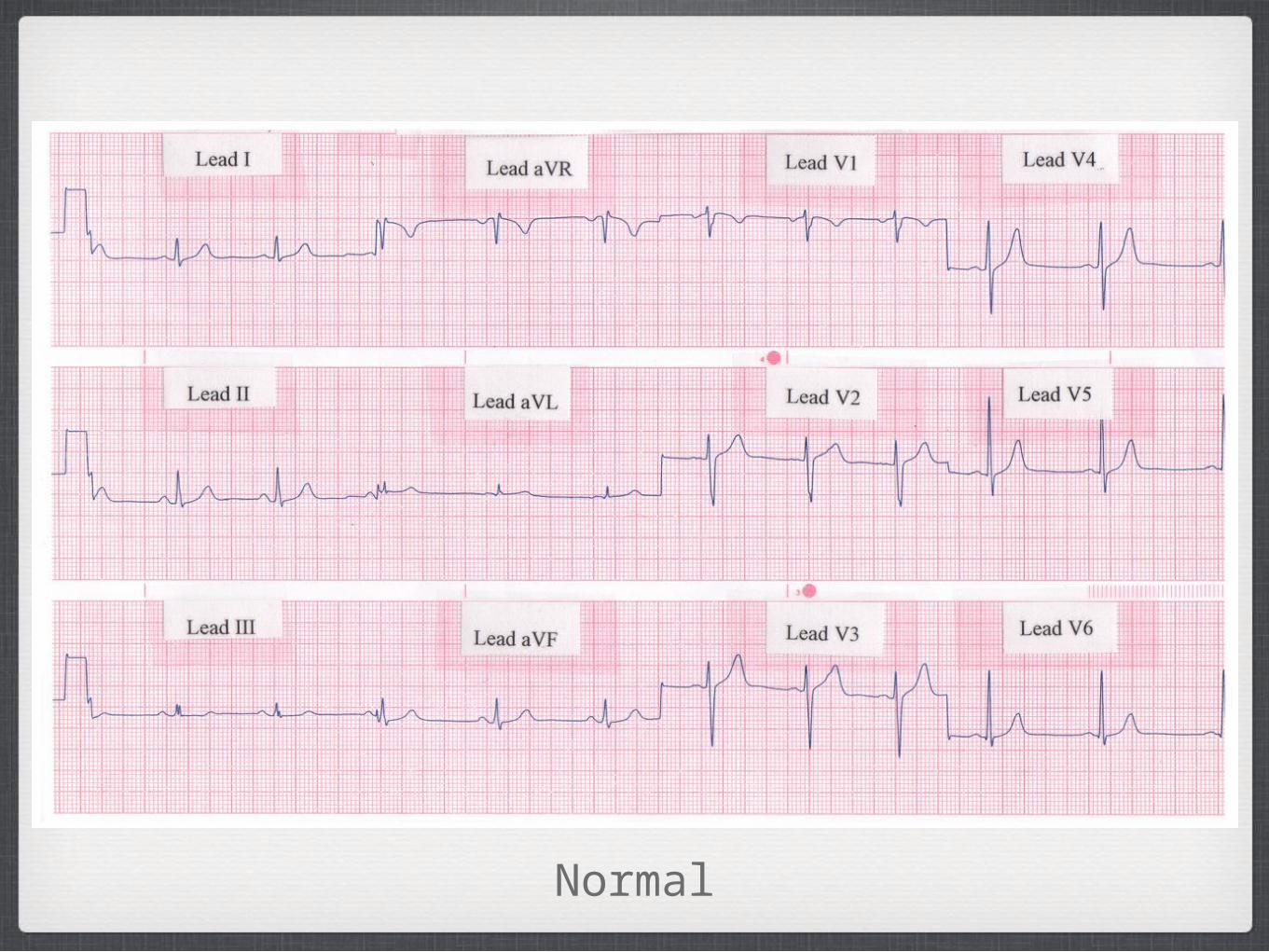
Normal
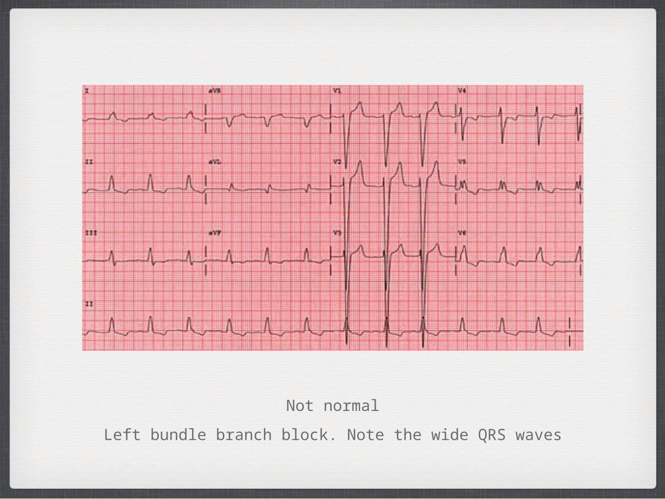
Not normal
Left bundle branch block. Note the wide QRS waves
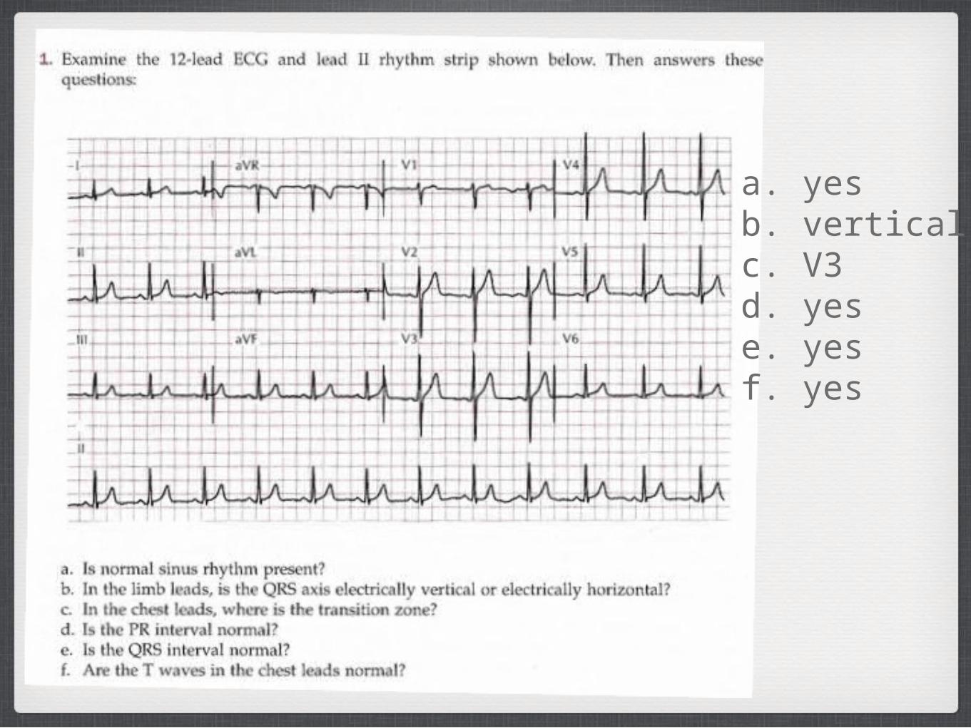
a. yesb. verticalc. V3d. yese. yesf. yes
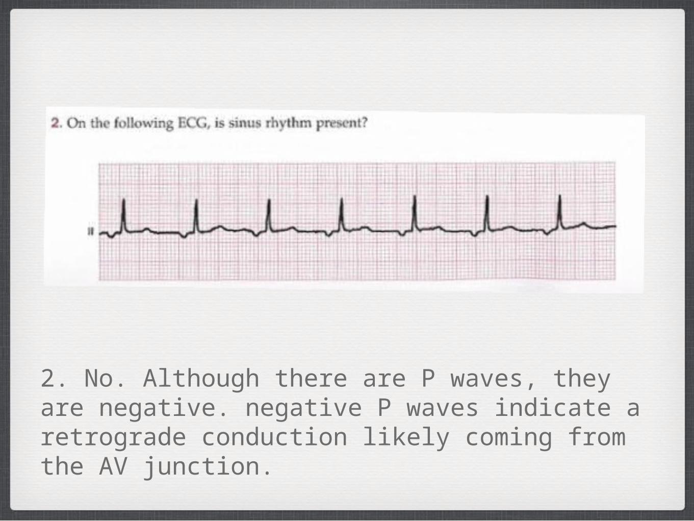
2. No. Although there are P waves, they are negative. negative P waves indicate a retrograde conduction likely coming from the AV junction.