Embed Size (px)
Citation preview
NONO couples the circadian clock to the cell cycleElzbieta Kowalskaa, Juergen A. Rippergerb, Dominik C. Hoeggerc, Pascal Brueggera, Thorsten Buchd, Thomas Birchlerd,Anke Muellere, Urs Albrechtb, Claudio Contaldoc, and Steven A. Browna,1
aInstitute of Pharmacology and Toxicology and dInstitute of Experimental Immunology, University of Zurich, 8057 Zurich, Switzerland; bDivision ofBiochemistry, Department of Medicine, University of Fribourg, 1700 Fribourg, Switzerland; cDivision of Plastic and Reconstructive Surgery, Departmentof Surgery, University Hospital Zurich, 8006 Zurich, Switzerland; and eLaboratory of Chronobiology, Institute of Medical Immunology, CharitéUniversitätsmedizin, 10117 Berlin, Germany
This Feature Article is part of a series identified by the Editorial Board as reporting findings of exceptional significance.
Edited by Michael W. Young, The Rockefeller University, New York, NY, and approved November 26, 2012 (received for review August 2, 2012)
Mammalian circadian clocks restrict cell proliferation to definedtime windows, but the mechanism and consequences of this in-terrelationship are not fully understood. Previouslywe identified themultifunctional nuclear protein NONO as a partner of circadian PE-RIOD (PER) proteins. Here we show that it also conveys circadiangating to the cell cycle, a connection surprisingly important forwound healing in mice. Specifically, although fibroblasts fromNONO-deficientmice showedapproximatelynormal circadian cycles,they displayed elevated cell doubling and lower cellular senescence.At a molecular level, NONO bound to the p16-Ink4A cell cycle check-point gene and potentiated its circadian activation in a PER protein-dependent fashion. Loss of either NONO or PER abolished this acti-vation and circadian expression of p16-Ink4A and eliminated circa-dian cell cycle gating. In vivo, lack of NONO resulted in defectivewound repair. Because wound healing defects were also seen inmultiple circadian clock-deficientmouse lines, our results thereforesuggest that coupling of the cell cycle to the circadian clock viaNONO may be useful to segregate in temporal fashion cell prolif-eration from tissue organization.
keratinocyte | p54nrb | RNA-binding protein | paraspeckle protein
The circadian clock adapts organisms to their daily surround-ings both behaviorally and physiologically. In animals, not
only are complex behaviors such as sleep and mood governed bythis oscillator, but also different body functions such as digestion,circulation, and respiration (1). The basic mechanism of thisclock is cell-autonomous in all studied species possessing a cir-cadian clock. In mammals, individual clocks in most cells aresynchronized by a brain “master clock” in the suprachiasmaticnucleus of the hypothalamus to orchestrate all rhythmic physi-ology (2). On a cellular level, circadian physiology extends even toprocesses such as proliferation (3–7), apoptosis (8), and DNAdamage repair (6, 9), which are thought to play important roles incancer control (8, 10).In individual cells, the circadian clock mechanism consists of
oscillating feedback loops of transcription of “core” oscillator genesand posttranslational modifications of their protein products thatregulate protein stability, activity, and/or localization. For example,in mammals the transcription of periods (Per) and cryptochomes(Cry) are activated by BMAL1:CLOCK heterodimers at cis-acting elements called E-boxes, and their protein products formcomplexes that repress their own transcription (11). We origi-nally identified the RNA-binding protein NONO (also calledp54nrb) biochemically as a new member of this circadian tran-scriptional repressor complex in mice, and mutation of itsorthologNonA in flies resulted in severe attenuation of circadianrhythmicity (12). However, apart from its interaction with thiscircadian repressor complex, NONO’s mechanism of actionwithin the clock remains unknown.The mechanism of the cell cycle has been reviewed extensively
elsewhere (13, 14). Rather than having a fixed duration, its periodis tightly regulated by cellular processes via checkpoint proteinsthat gate cell cycle progression. The circadian clock is one such
regulatory process, and it has been shown that the circadian clockcan directly time cell division (3, 4), although not necessarily in allcell types (15, 16). The exact mechanism of this regulation is atthe moment only partially understood and may involve circadianregulation of cell cycle checkpoint genes like wee1 (3) and p21-Waf1 (17), or interaction of clock factors with CSK-homologouskinase 1/2 (CHK1/2) proteins (18, 19). In addition, a limitednumber of accessory factors have been assigned roles in bothprocesses, including the mammalian Timeless protein (TIM),which interacts with both the circadian CRY2 protein and the cellcycle CHK1 protein (18), and p54nrb/NONO, which interacts withproteins involved in DNA damage repair (20, 21).To understand the role of the NONO protein in the circadian
clock and its relationship to the cell cycle, we created NONO-de-ficient mice. Although these mice showed only minor circadiandefects (22), circadian gating by the cell cycle was lost in fibroblastsfrom these mice, allowing us to probe both the mechanism and thephysiological relevance of the connection between the two pro-cesses. Our investigations not only uncover a unique role for NONOin the circadian transcriptional regulation of the p16-Ink4A cell cyclecheckpoint gene, but also suggest that circadian gating of the cellcycle by NONO plays an unsuspected role in tissue regeneration.
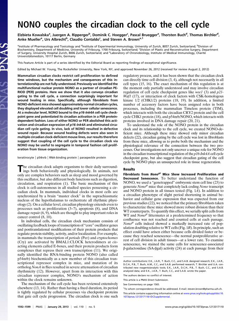
ResultsFibroblasts from Nonogt Mice Show Increased Proliferation andDecreased Senescence. To better understand the function ofNONO, we used a “genetrapped” embryonic stem cell line (23) togenerate Nonogt mice that completely lack coding Nono transcriptand NONO protein in all tissues tested (Fig. 1A). In addition toa circadian phenotype of slight period shortening in animal be-havior and cellular gene expression that was expected from ourprevious studies (22), we noticed that the primary fibroblasts takento characterize these mice showed more robust division than theirWT counterparts. To quantify this effect, we serially split cells fromWT and Nonogt littermates at a predetermined frequency so thatconfluence was not reached and counted cells at each passage.Nonogt cells indeed showed a markedly increased rate of pop-ulation doubling relative toWTcells (Fig. 1B). In principle, such aneffect could have arisen either because cells divided faster or be-cause they reached senescence—the normal postproliferative ar-rest of cell division in adult tissues—at a lower rate. To examinesenescence, we stained the same cells for senescence-associatedβ-galactosidase (SA-βgal) activity (24) at each passage from their
Author contributions: E.K., J.A.R., T. Buch, C.C., and S.A.B. designed research; E.K., J.A.R.,D.C.H., P.B., T. Buch, A.M., C.C., and S.A.B. performed research; T. Birchler and U.A. con-tributed new reagents/analytic tools; E.K., J.A.R., D.C.H., P.B., T. Buch, C.C., and S.A.B.analyzed data; and E.K., J.A.R., T. Buch, C.C., and S.A.B. wrote the paper.
The authors declare no conflict of interest.
This article is a PNAS Direct Submission.
See Commentary on page 1565.1To whom correspondence should be addressed. E-mail: [email protected].
This article contains supporting information online at www.pnas.org/lookup/suppl/doi:10.1073/pnas.1213317110/-/DCSupplemental.
1592–1599 | PNAS | January 29, 2013 | vol. 110 | no. 5 www.pnas.org/cgi/doi/10.1073/pnas.1213317110
initial isolation until their complete senescence. Nonogt cellsexhibited a roughly twofold decreased proportion of senescentcells at every passage (Fig. 1C).If Nonogt cells had reduced senescence rather than an in-
creased division rate, then fewer cells should remain nondividingin cultures of equivalent age. We tested this hypothesis by stainingdividing Nonogt and WT cells from the same passage with thepermanent cytoplasmic stain CFSE (carboxyfluorescein diacetate,succinimidyl ester) and then determining dye content by flowcytometry 4 d later. This dye remains trapped within the cells butis diluted with each cytokinesis. Hence it provides a quantitativeanalysis of the percentage of a cell population that has divided(25). According to this experiment, all Nonogt cells had divided atleast once, whereas 40% of the WT cells had not divided (Fig.
1D). Reintroduction of NONO into primary Nonogt fibroblastsvia lentiviral transduction slowed division and increased senes-cence, and addition of NONO to WT cells slowed division evenfurther (Fig. 1E), confirming the dose-dependent role of NONOin restraining cell proliferation and pointing to a probable rolefor this protein in the cell cycle.
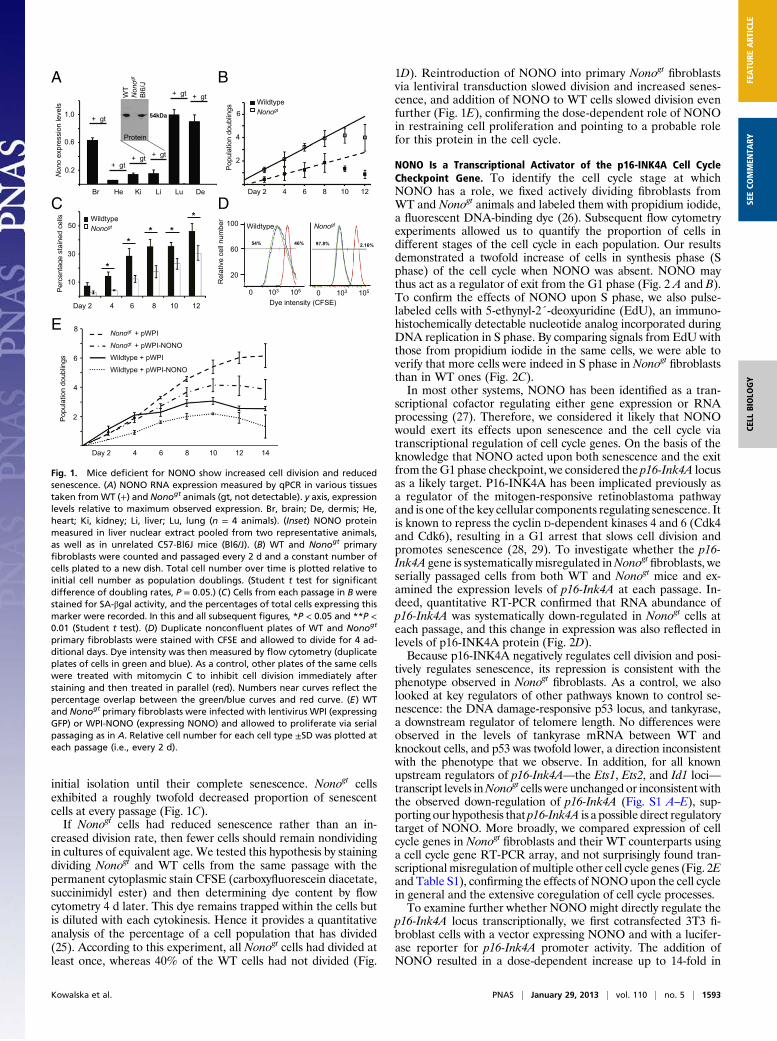
NONO Is a Transcriptional Activator of the p16-INK4A Cell CycleCheckpoint Gene. To identify the cell cycle stage at whichNONO has a role, we fixed actively dividing fibroblasts fromWT and Nonogt animals and labeled them with propidium iodide,a fluorescent DNA-binding dye (26). Subsequent flow cytometryexperiments allowed us to quantify the proportion of cells indifferent stages of the cell cycle in each population. Our resultsdemonstrated a twofold increase of cells in synthesis phase (Sphase) of the cell cycle when NONO was absent. NONO maythus act as a regulator of exit from the G1 phase (Fig. 2 A and B).To confirm the effects of NONO upon S phase, we also pulse-labeled cells with 5-ethynyl-2´-deoxyuridine (EdU), an immuno-histochemically detectable nucleotide analog incorporated duringDNA replication in S phase. By comparing signals from EdU withthose from propidium iodide in the same cells, we were able toverify that more cells were indeed in S phase in Nonogt fibroblaststhan in WT ones (Fig. 2C).In most other systems, NONO has been identified as a tran-
scriptional cofactor regulating either gene expression or RNAprocessing (27). Therefore, we considered it likely that NONOwould exert its effects upon senescence and the cell cycle viatranscriptional regulation of cell cycle genes. On the basis of theknowledge that NONO acted upon both senescence and the exitfrom theG1 phase checkpoint, we considered the p16-Ink4A locusas a likely target. P16-INK4A has been implicated previously asa regulator of the mitogen-responsive retinoblastoma pathwayand is one of the key cellular components regulating senescence. Itis known to repress the cyclin D-dependent kinases 4 and 6 (Cdk4and Cdk6), resulting in a G1 arrest that slows cell division andpromotes senescence (28, 29). To investigate whether the p16-Ink4A gene is systematically misregulated inNonogt fibroblasts, weserially passaged cells from both WT and Nonogt mice and ex-amined the expression levels of p16-Ink4A at each passage. In-deed, quantitative RT-PCR confirmed that RNA abundance ofp16-Ink4A was systematically down-regulated in Nonogt cells ateach passage, and this change in expression was also reflected inlevels of p16-INK4A protein (Fig. 2D).Because p16-INK4A negatively regulates cell division and posi-
tively regulates senescence, its repression is consistent with thephenotype observed in Nonogt fibroblasts. As a control, we alsolooked at key regulators of other pathways known to control se-nescence: the DNA damage-responsive p53 locus, and tankyrase,a downstream regulator of telomere length. No differences wereobserved in the levels of tankyrase mRNA between WT andknockout cells, and p53 was twofold lower, a direction inconsistentwith the phenotype that we observe. In addition, for all knownupstream regulators of p16-Ink4A—the Ets1, Ets2, and Id1 loci—transcript levels inNonogt cells were unchanged or inconsistent withthe observed down-regulation of p16-Ink4A (Fig. S1 A–E), sup-porting our hypothesis that p16-Ink4A is a possible direct regulatorytarget of NONO. More broadly, we compared expression of cellcycle genes in Nonogt fibroblasts and their WT counterparts usinga cell cycle gene RT-PCR array, and not surprisingly found tran-scriptional misregulation ofmultiple other cell cycle genes (Fig. 2Eand Table S1), confirming the effects of NONOupon the cell cyclein general and the extensive coregulation of cell cycle processes.To examine further whether NONO might directly regulate the
p16-Ink4A locus transcriptionally, we first cotransfected 3T3 fi-broblast cells with a vector expressing NONO and with a lucifer-ase reporter for p16-Ink4A promoter activity. The addition ofNONO resulted in a dose-dependent increase up to 14-fold in
Br He Ki Li Lu De
WT
Non
ogt
Bl6
/J
54kDa
Protein
A
Wildtype Nonogt
B
100
60
20
0 102 103 104 105
Dye intensity (CFSE)0 102 103 104 105
54% 46% 97.8% 2.16%
*
** *
*R
elat
ive
cell
num
berWildtype
Nonogt
WildtypeNonogt
Non
oex
pres
sion
leve
ls
1.0
0.6
0.2
Per
cent
age
stai
ned
cells
10
30
50
0 103 105 0 103 105
Day 2 4 6 8 10 12
2
4
6
8
Popu
latio
n do
ublin
gs
Popu
latio
n do
ublin
gs
Day 2 4 6 8 10 12 14
Day 2 4 6 8 10 12
6
4
2
+ gt
+ gt+ gt + gt
+ gt + gt
Nonogt + pWPI
Nonogt + pWPI-NONO
Wildtype + pWPI
Wildtype + pWPI-NONO
E
DC
Fig. 1. Mice deficient for NONO show increased cell division and reducedsenescence. (A) NONO RNA expression measured by qPCR in various tissuestaken fromWT (+) and Nonogt animals (gt, not detectable). y axis, expressionlevels relative to maximum observed expression. Br, brain; De, dermis; He,heart; Ki, kidney; Li, liver; Lu, lung (n = 4 animals). (Inset) NONO proteinmeasured in liver nuclear extract pooled from two representative animals,as well as in unrelated C57-Bl6J mice (Bl6/J). (B) WT and Nonogt primaryfibroblasts were counted and passaged every 2 d and a constant number ofcells plated to a new dish. Total cell number over time is plotted relative toinitial cell number as population doublings. (Student t test for significantdifference of doubling rates, P = 0.05.) (C) Cells from each passage in B werestained for SA-βgal activity, and the percentages of total cells expressing thismarker were recorded. In this and all subsequent figures, *P < 0.05 and **P <0.01 (Student t test). (D) Duplicate nonconfluent plates of WT and Nonogt
primary fibroblasts were stained with CFSE and allowed to divide for 4 ad-ditional days. Dye intensity was then measured by flow cytometry (duplicateplates of cells in green and blue). As a control, other plates of the same cellswere treated with mitomycin C to inhibit cell division immediately afterstaining and then treated in parallel (red). Numbers near curves reflect thepercentage overlap between the green/blue curves and red curve. (E) WTand Nonogt primary fibroblasts were infected with lentivirus WPI (expressingGFP) or WPI-NONO (expressing NONO) and allowed to proliferate via serialpassaging as in A. Relative cell number for each cell type ±SD was plotted ateach passage (i.e., every 2 d).
Kowalska et al. PNAS | January 29, 2013 | vol. 110 | no. 5 | 1593
CELL
BIOLO
GY
FEATU
REART
ICLE
SEECO
MMEN
TARY
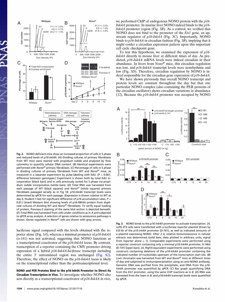
luciferase signal compared with the levels obtained with the re-porter alone (Fig. 3A), whereas a minimal promoter of p16-Ink4A(D-141) was not activated, suggesting that NONO functions asa transcriptional coactivator of the p16-Ink4A locus. By contrast,transcription of a reporter containing the CMV promoter drivingexpression of a hybrid p16-Ink4A-luciferase transcript includingthe entire 3′ untranslated region was unchanged (Fig. S2).Therefore, the effect of NONO on the p16-Ink4A locus is likelyon the transcriptional rather than the posttranscriptional level.
NONO and PER Proteins Bind to the p16-Ink4A Promoter to Direct ItsCircadian Transcription in Vivo. To investigate whether NONO alsoacts directly as a transcriptional coactivator of p16-Ink4A in vivo,
we performed ChIP of endogenous NONO protein with the p16-Ink4A promoter. In murine liver NONO indeed binds to the p16-Ink4A promoter region (Fig. 3B). As a control, we verified thatNONO does not bind to the promoter of the Ets1 gene, an up-stream regulator of p16-Ink4A (Fig. 3C). Importantly, NONObinds to p16-Ink4A in circadian fashion (Fig. 3B), implying that itmight confer a circadian expression pattern upon this importantcell cycle checkpoint gene.To test this hypothesis, we examined the expression of p16-
Ink4A directly in mouse liver at different times of day. As pre-dicted, p16-Ink4A mRNA levels were indeed circadian in theirabundance. In livers from Nonogt mice, this circadian regulationwas lost, and p16-Ink4A transcript levels were nonrhythmic andlow (Fig. 3D). Therefore, circadian regulation by NONO is in-deed responsible for the circadian gene expression of p16-Ink4A.We have shown previously that overall NONO transcript and
protein levels are constant throughout the day but that oneparticular NONO complex (also containing the PER proteins ofthe circadian oscillator) shows circadian variations in abundance(12). Because the p16-Ink4A promoter was occupied by NONO
BA
Dye intensity (PI)
G1 71.1 +/- 3.0% S 4.8 +/- 0.1%
G2/M 22.2 +/- 1.3%
0 50K 100K 150K 200K0
Cel
lnum
ber
2000
1500
1000
500
Dye intensity (PI)
G1 61.3 +/- 3.0%S 8.8 +/- 1.0%
G2/M 27.6 +/- 3.5%
0 0 50K 100K 150K 200K
4000
3000
2000
1000
Day 2 4 6 8 10 12
WildtypeNonogt
p16-
Ink4
A e
xpre
ssio
n
2
14
6
10
Wildtype Nonogt
DC
E
Wildtype Nonogt
Wildtype Nonogt
P16-INK4A
Ponceau S
Retinoblastomapathway
DNA damagepathway
TelomerepathwayTelomere
shortening
ATMATR
DNA damage
mdm2 p53
p21cip1
Cyclin ECdk2
Oncogenes
ArfP16-Ink4A
Reactive oxygenSpecies (ROS)Mitogens
Cyclin DCdk4/6
Rb
Cel
lnum
ber
40
30
20
10Perc
enta
ge o
f cel
ls
Total EdU incorporationS-phase enrichment
Fig. 2. NONO-deficient mice show an increased proportion of cells in S phaseand reduced levels of p16-Ink4A. (A) Dividing cultures of primary fibroblastsfrom WT mice were stained with propidium iodide and analyzed by flowcytometry to quantify cellular DNA content. (B) Identical experiments wereperformed with Nonogt primary fibroblasts. (C) Percentage of cells in S phasein dividing cultures of primary fibroblasts from WT and Nonogt mice, asmeasured in a separate experiment by pulse-labeling with EdU. (P < 0.001,difference between genotypes.) Experiment is shown both by total EdU in-corporation (black bars) and in cells previously sorted for S phase via propi-dium iodide incorporation (white bars). (D) Total RNA was harvested fromeach passage of WT (black squares) and Nonogt (white squares) primaryfibroblasts passaged serially as in Fig 1B. p16-Ink4A transcript levels weredetermined by qPCR for each passage. (Expression is shown relative to WT atday 0, Student t test for significant difference of p16 accumulation rates, P =0.01.) (Inset) Western blot showing levels of p16-INK4A protein from dupli-cate cultures of dividing WT and Nonogt
fibroblasts. To verify equal loadingof protein, Ponceau-S staining of the same blot section is depicted beneath.(E) Total RNA was harvested from cells under conditions as in A and subjectedto qPCR array analysis. A selection of genes relative to senescence pathways isshown. Genes regulated in Nonogt cells are shown with gray arrows.
A
added amount of NONO (in ng)
p16-Ink4A full length promoter p16-Ink4A D-141
C +10 +20 +50 +100 +150 C +10 +50 +100
Luci
fera
seac
tivity
2
6
10
14
18
****
B
**
CT0 4 8 12 16 20
Per
cent
of i
nput
0.08
0.06
0.04
0.02
WildtypeNonogt
CT0 4 8 12 16 20
0.08
0.06
0.04
0.02
WildtypeNonogt
p16-Ink4A promoter Ets1 promoterC
D*
**WildtypeNonogt
p16-
Ink4
A e
xpre
ssio
n
CT0 4 8 12 16 20
3
2
1
Construct used
1.0
0.8
0.6
0.4
0.2
Luci
fera
seac
tivity
** **
Fig. 3. NONO binds to the p16-Ink4A promoter to activate transcription. (A,Left) 3T3 cells were transfected with a luciferase reporter plasmid driven by0.8 kb of the p16-Ink4A promoter (D-761), as well as indicated amounts ofa plasmid expressing NONO. After 2 d, relative bioluminescence in cellularextracts was determined (solid bars; data plotted in arbitrary units, signalfrom reporter alone = 1). Comparable experiments were performed usinga reporter construct containing only a minimal p16-Ink4A promoter, 0.14kb(D-141) (open bars). (A, Right) equivalent experiments were performed usingconstructs containing deletions of the p16-Ink4A promoter containing theindicated number of nucleotides upstream of the transcription start site. (B)Liver chromatin was harvested from WT and Nonogt mice at different timesof day and subjected to immunoprecipitation using an anti-NONO antibody.Genomic DNA was purified from the precipitate, and DNA from the p16-Ink4A promoter was quantified by qPCR. (C) Bar graph quantifying DNAfrom the Ets1 promoter, using the same ChIP reactions as in B. (D) RNA washarvested from the livers in B, and p16-Ink4A transcript levels were quantifiedby qPCR.
1594 | www.pnas.org/cgi/doi/10.1073/pnas.1213317110 Kowalska et al.
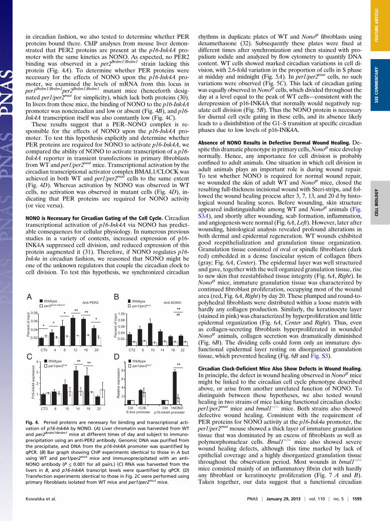
in circadian fashion, we also tested to determine whether PERproteins bound there. ChIP analyses from mouse liver demon-strated that PER2 proteins are present at the p16-Ink4A pro-moter with the same kinetics as NONO. As expected, no PER2binding was observed in a per2Brdm1/Brdm1 strain lacking thisprotein (Fig. 4A). To determine whether PER proteins werenecessary for the effects of NONO upon the p16-Ink4A pro-moter, we examined the levels of mRNA from this locus inper1Brdm1/Brdm1per2Brdm1/Brdm1 mutant mice (henceforth desig-nated per1/per2mut for simplicity), which lack both proteins (30).In livers from these mice, the binding of NONO to the p16-Ink4Apromoter was noncircadian and low or absent (Fig. 4B), and p16-Ink4A transcription itself was also constantly low (Fig. 4C).These results suggest that a PER–NONO complex is re-
sponsible for the effects of NONO upon the p16-Ink4A pro-moter. To test this hypothesis explicitly and determine whetherPER proteins are required for NONO to activate p16-Ink4A, wecompared the ability of NONO to activate transcription of a p16-Ink4A reporter in transient transfections in primary fibroblastsfromWT and per1/per2mut mice. Transcriptional activation by thecircadian transcriptional activator complex BMAL1/CLOCK wasachieved in both WT and per1/per2mut cells to the same extent(Fig. 4D). Whereas activation by NONO was observed in WTcells, no activation was observed in mutant cells (Fig. 4D), in-dicating that PER proteins are required for NONO activity(or vice versa).
NONO is Necessary for Circadian Gating of the Cell Cycle. Circadiantranscriptional activation of p16-Ink4A via NONO has predict-able consequences for cellular physiology. In numerous previousstudies in a variety of contexts, increased expression of p16-INK4A suppressed cell division, and reduced expression of thisprotein augmented it (31). Therefore, if NONO regulates p16-Ink4a in circadian fashion, we reasoned that NONO might beone of the unknown regulators that couple the circadian clock tocell division. To test this hypothesis, we synchronized circadian
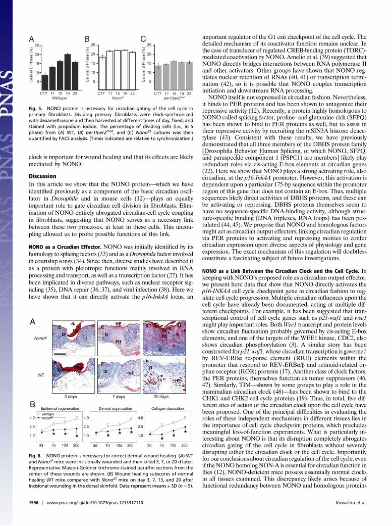
rhythms in duplicate plates of WT and Nonogt fibroblasts usingdexamethasone (32). Subsequently these plates were fixed atdifferent times after synchronization and then stained with pro-pidium iodide and analyzed by flow cytometry to quantify DNAcontent. WT cells showed marked circadian variations in cell di-vision, with 2.6-fold variation in the proportion of cells in S phaseat midday and midnight (Fig. 5A). In per1/per2mut cells, no suchvariations were observed (Fig. 5C). This lack of circadian gatingwas equally observed inNonogt cells, which divided throughout theday at a level equal to the peak of WT cells—consistent with thederepression of p16-INK4A that normally would negatively reg-ulate cell division (Fig. 5B). Thus the NONO protein is necessaryfor diurnal cell cycle gating in these cells, and its absence likelyleads to a disinhibition of the G1–S transition at specific circadianphases due to low levels of p16-INK4A.
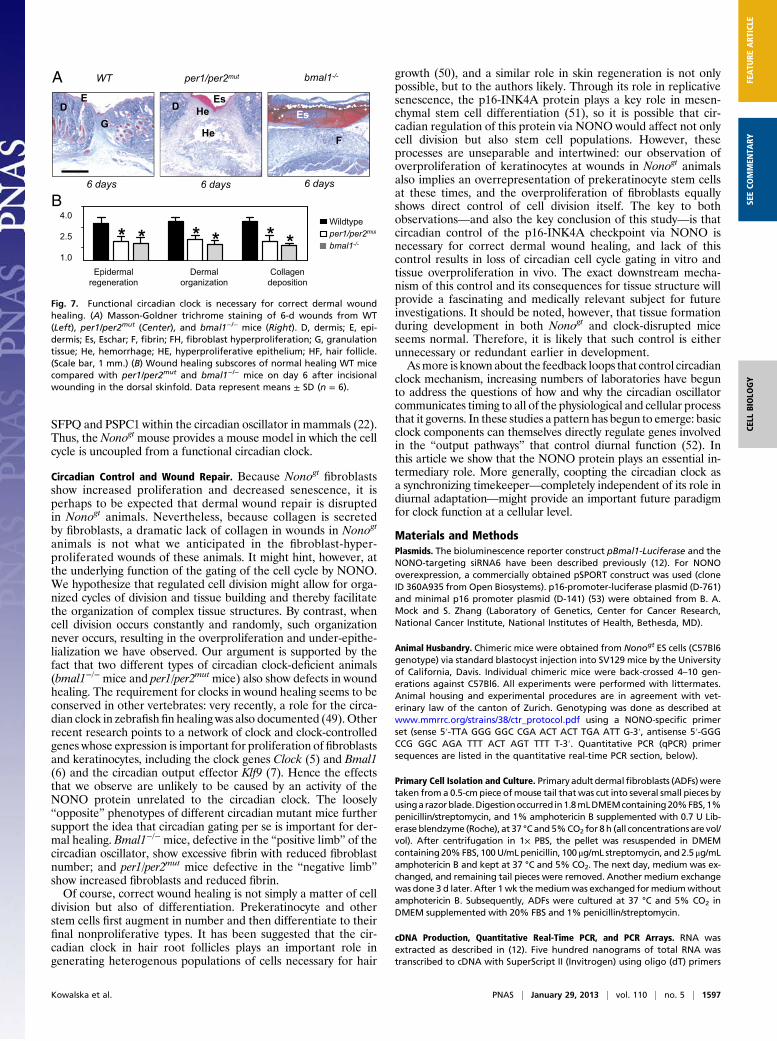
Absence of NONO Results in Defective Dermal Wound Healing. De-spite this dramatic phenotype in primary cells,Nonogtmice developnormally. Hence, any importance for cell division is probablyconfined to adult animals. One situation in which cell division inadult animals plays an important role is during wound repair.To test whether NONO is required for normal wound repair,we wounded the skin of adult WT and Nonogt mice, closed theresulting full-thickness incisional wound with Steri-strips, and fol-lowed the wound healing process after 3, 7, 13, and 20 d by histo-logical wound healing scores. Before wounding, skin structureappeared indistinguishable among WT and Nonogt animals (Fig.S3A), and shortly after wounding, scab formation, inflammation,and angiogenesis were normal (Fig. 6A, Left). However, later afterwounding, histological analysis revealed profound alterations inboth dermal and epidermal regeneration. WT wounds exhibitedgood reepithelialization and granulation tissue organization.Granulation tissue consisted of oval or spindle fibroblasts (darkred) embedded in a dense fascicular system of collagen fibers(gray; Fig. 6A, Center). The epidermal layer was well structuredand gave, together with the well organized granulation tissue, riseto new skin that reestablished tissue integrity (Fig. 6A, Right). InNonogt mice, immature granulation tissue was characterized bycontinued fibroblast proliferation, occupying most of the woundarea (red, Fig. 6A, Right) by day 20. These plumped and round-to-polyhedral fibroblasts were distributed within a loose matrix withhardly any collagen production. Similarly, the keratinocyte layer(stained in pink) was characterized by hyperproliferation and littleepidermal organization (Fig. 6A, Center and Right). Thus, evenas collagen-secreting fibroblasts hyperproliferated in woundedNonogt animals, collagen secretion was dramatically diminished(Fig. 6B). The dividing cells could form only an immature dys-functional epidermal layer resting on disorganized granulationtissue, which prevented healing (Fig. 6B and Fig. S3).
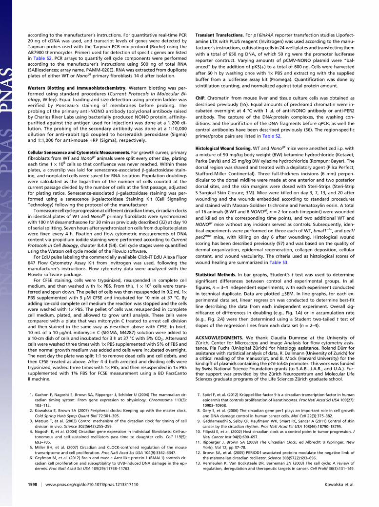
Circadian Clock-Deficient Mice Also Show Defects in Wound Healing.In principle, the defect in wound healing observed in Nonogt micemight be linked to the circadian cell cycle phenotype describedabove, or arise from another unrelated function of NONO. Todistinguish between these hypotheses, we also tested woundhealing in two strains of mice lacking functional circadian clocks:per1/per2mut mice and bmal1−/− mice. Both strains also showeddefective wound healing. Consistent with the requirement ofPER proteins for NONO activity at the p16-Ink4a promoter, theper1/per2mut mouse showed a thick layer of immature granulationtissue that was dominated by an excess of fibroblasts as well aspolymorphonuclear cells. Bmal1−/− mice also showed severewound healing defects, although this time marked by lack ofepithelial coverage and a highly disorganized granulation tissuethroughout the observation period. Most wounds in bmal1−/−
mice consisted mainly of an inflammatory fibrin clot with hardlyany fibroblast or keratinocyte proliferation (Fig. 7 A and B).Taken together, our data suggest that a functional circadian
A
D
E-box promoter p16-Ink4A promoter
Per
cent
of i
nput 0.08
0.06
0.04
0.02
*
**
*
* ***
CT0 4 8 12 16 20
Rel
ativ
eex
pres
sion
6
4
2
8
Ctrl +C/B Ctrl +NONO
Wildtypeper1/per2mut
BWildtypeper1/per2mut
Wildtypeper2Brdm1/Brdm1
Wildtypeper1/per2mut
CT2 6 10 14 18 22
CT2 6 10 14 18 22
Per
cent
of i
nput
0.08
0.06
0.04
0.02
1.00
ONON-itnA2REP-itnA
Wildtypeper1/per2mut
P16
-Ink4
A e
xpre
ssio
n
3
2
1
4 ** ** **
**all
C
Fig. 4. Period proteins are necessary for binding and transcriptional acti-vation of p16-Ink4A by NONO. (A) Liver chromatin was harvested from WTand per2Brdm1/Brdm1 mice at different times of day and subject to immuno-precipitation using an anti-PER2 antibody. Genomic DNA was purified fromthe precipitate, and DNA from the p16-Ink4A promoter was quantified byqPCR. (B) Bar graph showing ChIP experiments identical to those in A butusing WT and per1/per2mut mice and immunoprecipitated with an anti-NONO antibody (P ≤ 0.001 for all pairs.) (C) RNA was harvested from thelivers in B, and p16-Ink4A transcript levels were quantified by qPCR. (D)Transfection experiments identical to those in Fig. 2C were performed usingprimary fibroblasts isolated from WT mice and per1/per2mut mice.
Kowalska et al. PNAS | January 29, 2013 | vol. 110 | no. 5 | 1595
CELL
BIOLO
GY
FEATU
REART
ICLE
SEECO
MMEN
TARY
clock is important for wound healing and that its effects are likelymediated by NONO.
DiscussionIn this article we show that the NONO protein—which we haveidentified previously as a component of the basic circadian oscil-lator in Drosophila and in mouse cells (12)—plays an equallyimportant role to gate circadian cell division in fibroblasts. Elim-ination of NONO entirely abrogated circadian-cell cycle couplingin fibroblasts, suggesting that NONO serves as a necessary linkbetween these two processes, at least in these cells. This uncou-pling allowed us to probe possible functions of this link.
NONO as a Circadian Effector. NONO was initially identified by itshomology to splicing factors (33) and as aDrosophila factor involvedin courtship songs (34). Since then, diverse studies have described itas a protein with pleiotropic functions mainly involved in RNAprocessing and transport, as well as a transcription factor (27). It hasbeen implicated in diverse pathways, such as nuclear receptor sig-naling (35), DNA repair (36, 37), and viral infection (38). Here wehave shown that it can directly activate the p16-Ink4A locus, an
important regulator of the G1 exit checkpoint of the cell cycle. Thedetailed mechanism of its coactivator function remains unclear. Inthe case of transducer of regulated CREB-binding protein (TORC)-mediated coactivation by NONO,Amelio et al. (39) suggested thatNONO directly bridges interactions between RNA polymerase IIand other activators. Other groups have shown that NONO reg-ulates nuclear retention of RNAs (40, 41) or transcription termi-nation (42), so it is possible that NONO couples transcriptioninitiation and downstream RNA processing.NONO itself is not expressed in circadian fashion. Nevertheless,
it binds to PER proteins and has been shown to antagonize theirrepressive activity (12). Recently, a protein highly homologous toNONO called splicing factor, proline- and glutamine-rich (SFPQ)has been shown to bind to PER proteins as well, but to assist intheir repressive activity by recruiting the mSIN3A histone deace-tylase (43). Consistent with these results, we have previouslydemonstrated that all three members of the DBHS protein family[Drosophila Behavior Human Splicing, of which NONO, SFPQ,and paraspeckle component 1 (PSPC1) are members] likely playredundant roles via cis-acting E-box elements at circadian genes(22). Here we show that NONO plays a strong activating role, alsocircadian, at the p16-Ink4A promoter. However, this activation isdependent upon a particular 175-bp sequence within the promoterregion of this gene that does not contain an E-box. Thus, multiplesequences likely direct activities of DBHS proteins, and these canbe activating or repressing. DBHS proteins themselves seem tohave no sequence-specific DNA-binding activity, although struc-ture-specific binding (DNA triplexes, RNA loops) has been pos-tulated (44, 45). We propose that NONO and homologous factorsmight act as circadian output effectors, linking circadian regulationvia PER proteins to activating and repressing moities to confercircadian expression upon diverse aspects of physiology and geneexpression. The exact mechanism of this regulation will doubtlessconstitute a fascinating subject of future investigations.
NONO as a Link Between the Circadian Clock and the Cell Cycle. Inkeeping with NONO’s proposed role as a circadian output effector,we present here data that show that NONO directly activates thep16-INK4A cell cycle checkpoint gene in circadian fashion to reg-ulate cell cycle progression. Multiple circadian influences upon thecell cycle have already been documented, acting at multiple dif-ferent checkpoints. For example, it has been suggested that tran-scriptional control of cell cycle genes such as p21-waf1 and wee1might play important roles. BothWee1 transcript and protein levelsshow circadian fluctuation probably governed by cis-acting E-boxelements, and one of the targets of the WEE1 kinase, CDC2, alsoshows circadian phosphorylation (3). A similar story has beenconstructed for p21-waf1,whose circadian transcription is governedby REV-ERBα response element (RRE) elements within thepromoter that respond to REV-ERBα/β and retinoid-related or-phan receptor (ROR) proteins (17). Another class of clock factors,the PER proteins, themselves function as tumor suppressors (46,47). Similarly, TIM—shown by some groups to play a role in themammalian circadian clock (48)—has been shown to bind to theCHK1 and CHK2 cell cycle proteins (19). Thus, in total, five dif-ferent sites of action of the circadian clock upon the cell cycle havebeen proposed. One of the principal difficulties in evaluating theroles of these independent mechanisms in different tissues lies inthe importance of cell cycle checkpoint proteins, which precludesmeaningful loss-of-function experiments. What is particularly in-teresting about NONO is that its disruption completely abrogatescircadian gating of the cell cycle in fibroblasts without severelydisrupting either the circadian clock or the cell cycle. Importantlyfor our conclusions about circadian regulation of the cell cycle, evenif the NONOhomolog NON-A is essential for circadian function inflies (12), NONO-deficient mice possess essentially normal clocksin all tissues examined. This discrepancy likely arises because offunctional redundancy between NONO and homologous proteins
Wildtype
Cel
ls in
S P
hase
(%) 25
20
15
10
5
CT7 11 15 19 23 CT7 11 15 19 23 CT7 11 15 19 23
Cel
ls in
S P
hase
(%) 25
20
15
10
5 Cel
ls in
S P
hase
(%) 25
20
15
10
5
per1/per2mut
Nonogt
A B C
Fig. 5. NONO protein is necessary for circadian gating of the cell cycle inprimary fibroblasts. Dividing primary fibroblasts were clock-synchronizedwith dexamethasone and then harvested at different times of day, fixed, andstained with propidium iodide. The percentage of dividing cells (i.e., in Sphase) from (A) WT, (B) per1/per2mut, and (C) Nonogt cultures was thenquantified by FACS analysis. (Times indicated are relative to synchronization.)
DE
DE
D
E
DE
DE
DE
HEEs
Es
HEFH
HF
HF
Nonogt
WT
Es
3 days 7 days
A
20 days
BwildtypeNonogt
* * *3d 7d 13d 20d
* * *3d 7d 13d 20d
* * *
4.0
2.5
1.0
4.0
2.5
1.0
4.0
2.5
1.0
3d 7d 13d 20d
Epidermal regeneration Dermal organization Collagen deposition
3d 7d 13d 20d 3d 7d 13d 20d
Fig. 6. NONO protein is necessary for correct dermal wound healing. (A) WTand Nonogt mice were incisionally wounded and then killed 3, 7, or 20 d later.Representative Masson-Goldner trichrome-stained paraffin sections from thecenter of these wounds are shown. (B) Wound healing subscores of normalhealing WT mice compared with Nonogt mice on day 3, 7, 13, and 20 afterincisional wounding in the dorsal skinfold. Data represent means ± SD (n = 5).
1596 | www.pnas.org/cgi/doi/10.1073/pnas.1213317110 Kowalska et al.
SFPQ and PSPC1 within the circadian oscillator in mammals (22).Thus, the Nonogt mouse provides a mouse model in which the cellcycle is uncoupled from a functional circadian clock.
Circadian Control and Wound Repair. Because Nonogt fibroblastsshow increased proliferation and decreased senescence, it isperhaps to be expected that dermal wound repair is disruptedin Nonogt animals. Nevertheless, because collagen is secretedby fibroblasts, a dramatic lack of collagen in wounds in Nonogt
animals is not what we anticipated in the fibroblast-hyper-proliferated wounds of these animals. It might hint, however, atthe underlying function of the gating of the cell cycle by NONO.We hypothesize that regulated cell division might allow for orga-nized cycles of division and tissue building and thereby facilitatethe organization of complex tissue structures. By contrast, whencell division occurs constantly and randomly, such organizationnever occurs, resulting in the overproliferation and under-epithe-lialization we have observed. Our argument is supported by thefact that two different types of circadian clock-deficient animals(bmal1−/− mice and per1/per2mut mice) also show defects in woundhealing. The requirement for clocks in wound healing seems to beconserved in other vertebrates: very recently, a role for the circa-dian clock in zebrafish fin healing was also documented (49).Otherrecent research points to a network of clock and clock-controlledgenes whose expression is important for proliferation of fibroblastsand keratinocytes, including the clock genes Clock (5) and Bmal1(6) and the circadian output effector Klf9 (7). Hence the effectsthat we observe are unlikely to be caused by an activity of theNONO protein unrelated to the circadian clock. The loosely“opposite” phenotypes of different circadian mutant mice furthersupport the idea that circadian gating per se is important for der-mal healing. Bmal1−/− mice, defective in the “positive limb” of thecircadian oscillator, show excessive fibrin with reduced fibroblastnumber; and per1/per2mut mice defective in the “negative limb”show increased fibroblasts and reduced fibrin.Of course, correct wound healing is not simply a matter of cell
division but also of differentiation. Prekeratinocyte and otherstem cells first augment in number and then differentiate to theirfinal nonproliferative types. It has been suggested that the cir-cadian clock in hair root follicles plays an important role ingenerating heterogenous populations of cells necessary for hair
growth (50), and a similar role in skin regeneration is not onlypossible, but to the authors likely. Through its role in replicativesenescence, the p16-INK4A protein plays a key role in mesen-chymal stem cell differentiation (51), so it is possible that cir-cadian regulation of this protein via NONO would affect not onlycell division but also stem cell populations. However, theseprocesses are unseparable and intertwined: our observation ofoverproliferation of keratinocytes at wounds in Nonogt animalsalso implies an overrepresentation of prekeratinocyte stem cellsat these times, and the overproliferation of fibroblasts equallyshows direct control of cell division itself. The key to bothobservations—and also the key conclusion of this study—is thatcircadian control of the p16-INK4A checkpoint via NONO isnecessary for correct dermal wound healing, and lack of thiscontrol results in loss of circadian cell cycle gating in vitro andtissue overproliferation in vivo. The exact downstream mecha-nism of this control and its consequences for tissue structure willprovide a fascinating and medically relevant subject for futureinvestigations. It should be noted, however, that tissue formationduring development in both Nonogt and clock-disrupted miceseems normal. Therefore, it is likely that such control is eitherunnecessary or redundant earlier in development.Asmore is known about the feedback loops that control circadian
clock mechanism, increasing numbers of laboratories have begunto address the questions of how and why the circadian oscillatorcommunicates timing to all of the physiological and cellular processthat it governs. In these studies a pattern has begun to emerge: basicclock components can themselves directly regulate genes involvedin the “output pathways” that control diurnal function (52). Inthis article we show that the NONO protein plays an essential in-termediary role. More generally, coopting the circadian clock asa synchronizing timekeeper—completely independent of its role indiurnal adaptation—might provide an important future paradigmfor clock function at a cellular level.
Materials and MethodsPlasmids. The bioluminescence reporter construct pBmal1-Luciferase and theNONO-targeting siRNA6 have been described previously (12). For NONOoverexpression, a commercially obtained pSPORT construct was used (cloneID 360A935 from Open Biosystems). p16-promoter-luciferase plasmid (D-761)and minimal p16 promoter plasmid (D-141) (53) were obtained from B. A.Mock and S. Zhang (Laboratory of Genetics, Center for Cancer Research,National Cancer Institute, National Institutes of Health, Bethesda, MD).
Animal Husbandry. Chimeric mice were obtained from Nonogt ES cells (C57Bl6genotype) via standard blastocyst injection into SV129 mice by the Universityof California, Davis. Individual chimeric mice were back-crossed 4–10 gen-erations against C57Bl6. All experiments were performed with littermates.Animal housing and experimental procedures are in agreement with vet-erinary law of the canton of Zurich. Genotyping was done as described atwww.mmrrc.org/strains/38/ctr_protocol.pdf using a NONO-specific primerset (sense 5′-TTA GGG GGC CGA ACT ACT TGA ATT G-3′, antisense 5′-GGGCCG GGC AGA TTT ACT AGT TTT T-3′. Quantitative PCR (qPCR) primersequences are listed in the quantitative real-time PCR section, below).
Primary Cell Isolation and Culture. Primaryadult dermalfibroblasts (ADFs)weretaken from a 0.5-cmpiece ofmouse tail thatwas cut into several small pieces byusinga razorblade.Digestionoccurred in1.8mLDMEMcontaining20%FBS,1%penicillin/streptomycin, and 1% amphotericin B supplemented with 0.7 U Lib-erase blendzyme (Roche), at 37 °Cand 5%CO2 for 8h (all concentrations are vol/vol). After centrifugation in 1× PBS, the pellet was resuspended in DMEMcontaining20%FBS, 100U/mLpenicillin, 100 μg/mL streptomycin, and2.5 μg/mLamphotericin B and kept at 37 °C and 5% CO2. The next day, medium was ex-changed, and remaining tail pieces were removed. Another medium exchangewas done 3 d later. After 1wk themediumwas exchanged formediumwithoutamphotericin B. Subsequently, ADFs were cultured at 37 °C and 5% CO2 inDMEM supplemented with 20% FBS and 1% penicillin/streptomycin.
cDNA Production, Quantitative Real-Time PCR, and PCR Arrays. RNA wasextracted as described in (12). Five hundred nanograms of total RNA wastranscribed to cDNA with SuperScript II (Invitrogen) using oligo (dT) primers
AEs
Es
6 days 6 days 6 days
HeDDE
WT per1/per2mut bmal1-/-
HeF
G
B
Epidermalregeneration
Dermalorganization
Collagendeposition
4.0
2.5
1.0
Wildtypeper1/per2mut
bmal1-/-* * * * * *
Fig. 7. Functional circadian clock is necessary for correct dermal woundhealing. (A) Masson-Goldner trichrome staining of 6-d wounds from WT(Left), per1/per2mut (Center), and bmal1−/− mice (Right). D, dermis; E, epi-dermis; Es, Eschar; F, fibrin; FH, fibroblast hyperproliferation; G, granulationtissue; He, hemorrhage; HE, hyperproliferative epithelium; HF, hair follicle.(Scale bar, 1 mm.) (B) Wound healing subscores of normal healing WT micecompared with per1/per2mut and bmal1−/− mice on day 6 after incisionalwounding in the dorsal skinfold. Data represent means ± SD (n = 6).
Kowalska et al. PNAS | January 29, 2013 | vol. 110 | no. 5 | 1597
CELL
BIOLO
GY
FEATU
REART
ICLE
SEECO
MMEN
TARY
according to the manufacturer’s instructions. For quantitative real-time PCR20 ng of cDNA was used, and transcript levels of genes were detected byTaqman probes used with the Taqman PCR mix protocol (Roche) using theAB7900 thermocycler. Primers used for detection of specific genes are listedin Table S2. PCR arrays to quantify cell cycle components were performedaccording to the manufacturer’s instructions using 500 ng of total RNA(SABiosciences; array name, PAMM-020E). RNA was extracted from duplicateplates of either WT or Nonogt primary fibroblasts 14 d after isolation.
Western Blotting and Immunohistochemistry. Western blotting was per-formed using standard procedures (Current Protocols in Molecular Bi-ology, Wiley). Equal loading and size detection using protein ladder wasverified by Ponceau-S staining of membranes before probing. Theprobing of the primary anti-NONO antibody (polyclonal antibody raisedby Charles River Labs using bacterially produced NONO protein, affinity-purified against the antigen used for injection) was done at a 1:200 di-lution. The probing of the secondary antibody was done at a 1:10,000dilution for anti-rabbit IgG coupled to horseradish peroxidase (Sigma)and 1:1,000 for anti-mouse HRP (Sigma), respectively.
Cellular Senescence and Cytometric Measurements. For growth curves, primaryfibroblasts from WT and Nonogt animals were split every other day, platingeach time 1 × 10⁶ cells so that confluence was never reached. Within theseplates, a coverslip was laid for senescence-associated β-galactosidase stain-ing, and nonplated cells were saved for RNA isolation. Population doublingswere calculated as the logarithm of the number of cells counted at thecurrent passage divided by the number of cells at the first passage, adjustedfor plating ratios. Senescence-associated β-galactosidase staining was per-formed using a senescence β-galactosidase Staining Kit (Cell SignalingTechnology) following the protocol of the manufacturer.
Tomeasurecell cycleprogressionatdifferentcircadiantimes, circadianclocksin identical plates of WT and Nonogt primary fibroblasts were synchronizedwith 100 nM dexamethasone for 30 min as previously described (32) at day 10of serial splitting. Seven hours after synchronization cells from duplicate plateswere fixed every 4 h. Fixation and flow cytometric measurements of DNAcontent via propidium iodide staining were performed according to CurrentProtocols in Cell Biology, chapter 8.4.4 (54). Cell cycle stages were quantifiedusing the Watson cell cycle model of the FlowJo software.
For EdU pulse labeling the commercially available Click-iT EdU Alexa Fluor647 Flow Cytometry Assay Kit from Invitrogen was used, following themanufacturer’s instructions. Flow cytometry data were analyzed with theFlowJo software package.
For CFSE staining, cells were trypsinized, resuspended in complete cellmedium, and then washed with 1× PBS. From this, 1 × 10⁶ cells were trans-ferred and spun down. The pellet of cells was then resuspended in 0.2 mL 1×PBS supplemented with 5 μM CFSE and incubated for 10 min at 37 °C. Byadding ice-cold complete cell medium the reaction was stopped and the cellswere washed with 1× PBS. The pellet of cells was resuspended in completecell medium, plated, and allowed to grow until analysis. These cells werecompared with a plate that was mitomycin C treated to arrest cell divisionand then stained in the same way as described above with CFSE. In brief,10 mL of a 10 μg/mL mitomycin C (SIGMA, M4287) solution were added toa 10-cm dish of cells and incubated for 3 h at 37 °C with 5% CO2. Afterwardcells were washed three times with 1× PBS supplemented with 5% of FBS andthen normal growth medium was added and cells were incubated overnight.The next day the plate was split 1:1 to remove dead cells and cell debris, andthen CFSE treated as above. After 4 d both arrested and dividing cells weretrypsinized, washed three times with 1× PBS, and then resuspended in 1× PBSsupplemented with 1% FBS for FCSE measurement using a BD FacsCantoII machine.
Transient Transfections. For p16Ink4A reporter transfection studies Lipofect-amine LTX with PLUS reagent (Invitrogen) was used according to the manu-facturer’s instructions, cultivating cells in 24-well plates and transfecting themwith a total of 650 ng DNA, of which 50 ng were the promoter luciferasereporter construct. Varying amounts of pCMV-NONO plasmid were “bal-anced” by the addition of pKS(+) to a total of 600 ng. Cells were harvestedafter 60 h by washing once with 1× PBS and extracting with the suppliedbuffer from a luciferase assay kit (Promega). Quantification was done byscintillation counting, and normalized against total protein amount.
ChIP. Chromatin from mouse liver and tissue culture cells was obtained asdescribed previously (55). Equal amounts of precleared chromatin were in-cubated overnight at 4 °C with 1 μL of anti-NONO antibody or anti-PER2antibody. The capture of the DNA:protein complexes, the washing con-ditions, and the purifiction of the DNA fragments before qPCR, as well thecontrol antibodies have been described previously (56). The region-specificprimer/probe pairs are listed in Table S2.
Histological Wound Scoring. WT and Nonogt mice were anesthetized i.p. witha mixture of 90 mg/kg body weight (BW) ketamine hydrochloride (Ketavet;Parke Davis) and 25 mg/kg BW xylazine hydrochloride (Rompun; Bayer). Thedorsal region was shaved and treated with a depilatory agent (Pilca Perfect;Stafford-Miller Continental). Three full-thickness incisions (6 mm) perpen-dicular to the dorsal midline were made at one anterior and two posteriordorsal sites, and the skin margins were closed with Steri-Strips (Steri-StripS Surgical Skin Closure; 3M). Mice were killed on day 3, 7, 13, and 20 afterwounding and the wounds embedded according to standard proceduresand stained with Masson-Goldner trichrome and hematoxylin eosin. A totalof 16 animals (8 WT and 8 NONOgt, n = 2 for each timepoint) were woundedand killed on the corresponding time points, and two additional WT andNONOgt mice without any incisions served as controls. Subsequently, iden-tical experiments were performed on three each of WT, bmal1−/−, and per1/per2mut mice, with killing on day 6 after wounding. Histological woundscoring has been described previously (57) and was based on the quality ofdermal organization, epidermal regeneration, collagen deposition, cellularcontent, and wound vascularity. The criteria used as histological scores ofwound healing are summarized in Table S3.
Statistical Methods. In bar graphs, Student’s t test was used to determinesignificant differences between control and experimental groups. In allfigures, n = 3–4 independent experiments, with each experiment conductedin technical duplicate. Data are plotted ±SEM. In line graphs, for each ex-perimental data set, linear regression was conducted to determine best-fitline describing the data from each independent experiment. Overall sig-nificance of differences in doubling (e.g., Fig. 1A) or in accumulation rate(e.g., Fig. 2A) were then determined using a Student two-tailed t test ofslopes of the regression lines from each data set (n = 2–4).
ACKNOWLEDGMENTS. We thank Claudia Dumrese at the University ofZürich, Center for Microscopy and Image Analysis for flow cytometry assis-tance, Pia Fuchs (Unispital Zürich) for histology assistance, Roland Dürr forassistance with statistical analysis of data, R. Dallmann (University of Zurich) fora critical reading of the manuscript, and B. Mock (Harvard University) for thekind gift of plasmids containing the p16-Ink4a promoter. This work was fundedby Swiss National Science Foundation grants (to S.A.B., J.A.R., and U.A.). Fur-ther support was provided by the Zürich Neurozentrum and Molecular LifeSciences graduate programs of the Life Sciences Zürich graduate school.
1. Gachon F, Nagoshi E, Brown SA, Ripperger J, Schibler U (2004) The mammalian cir-cadian timing system: From gene expression to physiology. Chromosoma 113(3):103–112.
2. Kowalska E, Brown SA (2007) Peripheral clocks: Keeping up with the master clock.Cold Spring Harb Symp Quant Biol 72:301–305.
3. Matsuo T, et al. (2003) Control mechanism of the circadian clock for timing of celldivision in vivo. Science 302(5643):255–259.
4. Nagoshi E, et al. (2004) Circadian gene expression in individual fibroblasts: Cell-au-tonomous and self-sustained oscillators pass time to daughter cells. Cell 119(5):693–705.
5. Miller BH, et al. (2007) Circadian and CLOCK-controlled regulation of the mousetranscriptome and cell proliferation. Proc Natl Acad Sci USA 104(9):3342–3347.
6. Geyfman M, et al. (2012) Brain and muscle Arnt-like protein-1 (BMAL1) controls cir-cadian cell proliferation and susceptibility to UVB-induced DNA damage in the epi-dermis. Proc Natl Acad Sci USA 109(29):11758–11763.
7. Spörl F, et al. (2012) Krüppel-like factor 9 is a circadian transcription factor in humanepidermis that controls proliferation of keratinocytes. Proc Natl Acad Sci USA 109(27):10903–10908.
8. Gery S, et al. (2006) The circadian gene per1 plays an important role in cell growthand DNA damage control in human cancer cells. Mol Cell 22(3):375–382.
9. Gaddameedhi S, Selby CP, Kaufmann WK, Smart RC, Sancar A (2011) Control of skincancer by the circadian rhythm. Proc Natl Acad Sci USA 108(46):18790–18795.
10. Filipski E, et al. (2002) Host circadian clock as a control point in tumor progression. JNatl Cancer Inst 94(9):690–697.
11. Ripperger J, Brown SA (2009) The Circadian Clock, ed Albrecht U (Springer, NewYork), Vol 12, pp 37–78.
12. Brown SA, et al. (2005) PERIOD1-associated proteins modulate the negative limb ofthe mammalian circadian oscillator. Science 308(5722):693–696.
13. Vermeulen K, Van Bockstaele DR, Berneman ZN (2003) The cell cycle: A review ofregulation, deregulation and therapeutic targets in cancer. Cell Prolif 36(3):131–149.
1598 | www.pnas.org/cgi/doi/10.1073/pnas.1213317110 Kowalska et al.
14. Orford KW, Scadden DT (2008) Deconstructing stem cell self-renewal: Genetic insightsinto cell-cycle regulation. Nat Rev Genet 9(2):115–128.
15. Pendergast JS, Yeom M, Reyes BA, Ohmiya Y, Yamazaki S (2010) Disconnected cir-cadian and cell cycles in a tumor-driven cell line. Commun Integr Biol 3(6):536–539.
16. Yeom M, Pendergast JS, Ohmiya Y, Yamazaki S (2010) Circadian-independent cellmitosis in immortalized fibroblasts. Proc Natl Acad Sci USA 107(21):9665–9670.
17. Gréchez-Cassiau A, Rayet B, Guillaumond F, Teboul M, Delaunay F (2008) The circa-dian clock component BMAL1 is a critical regulator of p21WAF1/CIP1 expression andhepatocyte proliferation. J Biol Chem 283(8):4535–4542.
18. Unsal-Kaçmaz K, Mullen TE, Kaufmann WK, Sancar A (2005) Coupling of human cir-cadian and cell cycles by the timeless protein. Mol Cell Biol 25(8):3109–3116.
19. Yang X, Wood PA, Hrushesky WJ (2010) Mammalian TIMELESS is required for ATM-dependent CHK2 activation and G2/M checkpoint control. J Biol Chem 285(5):3030–3034.
20. Proteau A, et al. (2005) The multifunctional nuclear protein p54nrb is multi-phosphorylated in mitosis and interacts with the mitotic regulator Pin1. J Mol Biol 346(4):1163–1172.
21. Stier S, et al. (2005) Identification of p54(nrb) and the 14-3-3 Protein HS1 as TNF-al-pha-inducible genes related to cell cycle control and apoptosis in human arterialendothelial cells. J Biochem Mol Biol 38(4):447–456.
22. Kowalska E, et al. (2012) Distinct roles of DBHS family members in the circadiantranscriptional feedback loop. Mol Cell Biol 32(22):4585–4594.
23. Skarnes WC, et al.; International Gene Trap Consortium (2004) A public gene trapresource for mouse functional genomics. Nat Genet 36(6):543–544.
24. Litaker JR, et al. (1998) Expression profile of senescence-associated beta-galactosidaseand activation of telomerase in human ovarian surface epithelial cells undergoingimmortalization. Int J Oncol 13(5):951–956.
25. Lyons AB, Parish CR (1994) Determination of lymphocyte division by flow cytometry. JImmunol Methods 171(1):131–137.
26. Krishan A (1975) Rapid flow cytofluorometric analysis of mammalian cell cycle bypropidium iodide staining. J Cell Biol 66(1):188–193.
27. Shav-Tal Y, Zipori D (2002) PSF and p54(nrb)/NonO—multi-functional nuclear pro-teins. FEBS Lett 531(2):109–114.
28. Serrano M, Hannon GJ, Beach D (1993) A new regulatory motif in cell-cycle controlcausing specific inhibition of cyclin D/CDK4. Nature 366(6456):704–707.
29. Ohtani N, Yamakoshi K, Takahashi A, Hara E (2004) The p16INK4a-RB pathway:Molecular link between cellular senescence and tumor suppression. J Med Invest 51(3-4):146–153.
30. Zheng B, et al. (2001) Nonredundant roles of the mPer1 and mPer2 genes in themammalian circadian clock. Cell 105(5):683–694.
31. Huschtscha LI, Reddel RR (1999) p16(INK4a) and the control of cellular proliferativelife span. Carcinogenesis 20(6):921–926.
32. Balsalobre A, et al. (2000) Resetting of circadian time in peripheral tissues by gluco-corticoid signaling. Science 289(5488):2344–2347.
33. Dong B, Horowitz DS, Kobayashi R, Krainer AR (1993) Purification and cDNA cloningof HeLa cell p54nrb, a nuclear protein with two RNA recognition motifs and extensivehomology to human splicing factor PSF and Drosophila NONA/BJ6. Nucleic Acids Res21(17):4085–4092.
34. Rendahl KG, Jones KR, Kulkarni SJ, Bagully SH, Hall JC (1992) The dissonance mutationat the no-on-transient-A locus of D. melanogaster: Genetic control of courtship songand visual behaviors by a protein with putative RNA-binding motifs. J Neurosci 12(2):390–407.
35. Dong X, et al. (2009) p54nrb is a transcriptional corepressor of the progesterone re-ceptor that modulates transcription of the labor-associated gene, connexin 43 (Gja1).Mol Endocrinol 23(8):1147–1160.
36. Li S, et al. (2009) Involvement of p54(nrb), a PSF partner protein, in DNA double-strand break repair and radioresistance. Nucleic Acids Res 37(20):6746–6753.
37. Salton M, Lerenthal Y, Wang SY, Chen DJ, Shiloh Y (2010) Involvement of Matrin 3and SFPQ/NONO in the DNA damage response. Cell Cycle 9(8):1568–1576.
38. Zolotukhin AS, et al. (2003) PSF acts through the human immunodeficiency virus type1 mRNA instability elements to regulate virus expression. Mol Cell Biol 23(18):6618–6630.
39. Amelio AL, et al. (2007) A coactivator trap identifies NONO (p54nrb) as a componentof the cAMP-signaling pathway. Proc Natl Acad Sci USA 104(51):20314–20319.
40. Prasanth KV, et al. (2005) Regulating gene expression through RNA nuclear retention.Cell 123(2):249–263.
41. Zhang Z, Carmichael GG (2001) The fate of dsRNA in the nucleus: A p54(nrb)-con-taining complex mediates the nuclear retention of promiscuously A-to-I edited RNAs.Cell 106(4):465–475.
42. Kaneko S, Rozenblatt-Rosen O, Meyerson M, Manley JL (2007) The multifunctionalprotein p54nrb/PSF recruits the exonuclease XRN2 to facilitate pre-mRNA 3′ pro-cessing and transcription termination. Genes Dev 21(14):1779–1789.
43. Duong HA, Robles MS, Knutti D, Weitz CJ (2011) A molecular mechanism for circadianclock negative feedback. Science 332(6036):1436–1439.
44. Nelson LD, et al. (2012) Triplex DNA-binding proteins are associated with clinicaloutcomes revealed by proteomic measurements in patients with colorectal cancer.Mol Cancer 11(1):38.
45. Peng R, et al. (2002) PSF and p54nrb bind a conserved stem in U5 snRNA. RNA 8(10):1334–1347.
46. Gery S, Koeffler HP (2010) Circadian rhythms and cancer. Cell Cycle 9(6):1097–1103.47. Fu L, Pelicano H, Liu J, Huang P, Lee C (2002) The circadian gene Period2 plays an
important role in tumor suppression and DNA damage response in vivo. Cell 111(1):41–50.
48. Barnes JW, et al. (2003) Requirement of mammalian Timeless for circadian rhyth-micity. Science 302(5644):439–442.
49. Idda ML, et al. (2012) Circadian timing of injury-induced cell proliferation in zebrafish.PLoS One 7(3):e34203.
50. Janich P, et al. (2011) The circadian molecular clock creates epidermal stem cell het-erogeneity. Nature 480(7376):209–214.
51. Li H, et al. (2009) The Ink4/Arf locus is a barrier for iPS cell reprogramming. Nature 460(7259):1136–1139.
52. Ripperger JA, Shearman LP, Reppert SM, Schibler U (2000) CLOCK, an essentialpacemaker component, controls expression of the circadian transcription factor DBP.Genes Dev 14(6):679–689.
53. Zhang S, et al. (2003) p16 INK4a gene promoter variation and differential binding ofa repressor, the ras-responsive zinc-finger transcription factor, RREB. Oncogene 22(15):2285–2295.
54. Darzynkiewicz Z, Juan G, Bedner E (2001) Determining cell cycle stages by flow cy-tometry. Current Protocols in Cell Biology, eds Bonifacino JS, Dasso M, Harford JB,Lippincott-Schwartz J, Yamada KM (John Wiley & Sons, New York), pp 8.4.1–8.4.18.
55. Ripperger JA, Schibler U (2006) Rhythmic CLOCK-BMAL1 binding to multiple E-boxmotifs drives circadian Dbp transcription and chromatin transitions. Nat Genet 38(3):369–374.
56. Schmutz I, Ripperger JA, Baeriswyl-Aebischer S, Albrecht U (2010) The mammalianclock component PERIOD2 coordinates circadian output by interaction with nuclearreceptors. Genes Dev 24(4):345–357.
57. Altavilla D, et al. (2001) Inhibition of lipid peroxidation restores impaired vascularendothelial growth factor expression and stimulates wound healing and angiogen-esis in the genetically diabetic mouse. Diabetes 50(3):667–674.
Kowalska et al. PNAS | January 29, 2013 | vol. 110 | no. 5 | 1599
CELL
BIOLO
GY
FEATU
REART
ICLE
SEECO
MMEN
TARY