Embed Size (px)
Citation preview
Structure of the heterodimer of human NONO andparaspeckle protein component 1 and analysis of itsrole in subnuclear body formationDaniel M. Passona,1, Mihwa Leea,1, Oliver Rackhamb, Will A. Stanleya, Agata Sadowskab, Aleksandra Filipovskab,Archa H. Foxb, and Charles S. Bonda,2
aSchool of Chemistry and Biochemistry and bWestern Australian Institute for Medical Research, Centre for Medical Research, University of Western Australia,Crawley, Western Australia 6009, Australia
Edited by Brian W. Matthews, University of Oregon, Eugene, OR, and approved February 17, 2012 (received for review December 16, 2011)
Proteins of the Drosophila behavior/human splicing (DBHS) familyinclude mammalian SFPQ (PSF), NONO (p54nrb), PSPC1, and inverte-brate NONA and Hrp65. DBHS proteins are predominately nuclear,and are involved in transcriptional and posttranscriptional gene reg-ulatory functions as well as DNA repair. DBHS proteins influencea wide gamut of biological processes, including the regulation ofcircadian rhythm, carcinogenesis, and progression of cancer. Addi-tionally, mammalian DBHS proteins associate with the architecturallong noncoding RNA NEAT1 (Menε/β) to form paraspeckles, subnu-clear bodies that alter gene expression via the nuclear retention ofRNA.Herewedescribe the crystal structure of theheterodimer of themultidomain conserved region of the DBHS proteins, PSPC1 andNONO. These proteins form an extensively intertwined dimer, con-sistent with the observation that the different DBHS proteins aretypically copurified from mammalian cells, and suggesting that theyact as obligate heterodimers. The PSPC1/NONO heterodimer hasa right-handed antiparallel coiled-coil that positions two of fourRNA recognition motif domains in an unprecedented arrangementon either side of a 20-Å channel. This configuration is supported bya protein:protein interaction involving the NONA/paraspeckle do-main, which is characteristic of the DBHS family. By examining vari-ous mutants and truncations in cell culture, we find that DBHSproteins require anadditional antiparallel coiled-coil emanating fromeither end of the dimer for paraspeckle subnuclear body formation.These results suggest that paraspeckles may potentially formthroughself-associationofDBHSdimers into higher-order structures.
gene regulation | nuclear organization | protein structure |RNA-binding proteins
The Drosophila behavior/human splicing (DBHS) proteins havebeen described as multifunctional nuclear proteins, implicated
in subnuclear body formation; transcription initiation; coactivationand corepression; constitutive and alternative splicing; transcrip-tional termination; and DNA repair (1) and contributing to circa-dian rhythm regulation (2, 3), tumor suppression (4), andDrosophila behavior (5). Mammalian genomes encode threeparalogous DBHS proteins: NONO, SFPQ, and PSPC1, whereasinvertebrates encode one or two. NONO and SFPQ are highlyabundant, expressed in a variety of cell lines and tissues (1). Incontrast, PSPC1 ismore selectively expressed, found at low levels inHeLa cells, but expressed at equal levels with NONO and SFPQ insertoli cells of the testis, acting as a coactivator of transcription (6).Mammalian DBHS proteins are essential for the formation
of subnuclear paraspeckles (7), where they interact with a long-noncoding RNA, NEAT1 (8–10), and influence its spatial ar-rangement (11). Paraspeckle formation is also dependent upontranscription of NEAT1, which acts as a structural scaffold, nu-cleating the bodies at least in part by recruiting DBHS proteins(12, 13). DBHS proteins functioning within paraspeckles bind toadenosine-to-inosine edited inverted-repeat hairpins in the 3′UTR of specific mRNAs (13–15). This type of RNA is retainedin the nucleus and sequestered in paraspeckles (16).
All DBHS proteins have a core conserved region of ∼300 aa.Their N- and C-terminal extensions differ giving total lengths of400 (NONO) to >700 aa (SFPQ), with this extra polypeptideconsisting primarily of polyproline/glutamine-rich low-complex-ity regions and functional sequences including nuclear localiza-tion signals. The DBHS core consists of clearly defined structuraldomains: two tandem RNA recognition motif (RRM) domainsand a 100-aa segment of predicted coiled-coil (Fig. 1A and Fig.S1)—a putative protein:protein interaction motif. Between thesecond RRM and the coiled-coil lies a previously uncharac-terized 52-aa conserved domain, NONA/paraspeckle (NOPS),which is definitive of the DBHS family. DBHS proteins formhomo- and heterodimers via their core domains (17, 18) andhave been shown to interact with nucleic acids through theirRRM domains (1). Though RRM1 of DBHS proteins is ca-nonical (containing four aromatic residues at conserved positionsthat are typically essential for RNA binding) (19), RRM2 is con-sidered noncanonical: three of these conserved residues aresubstituted to Thr, Lys, and Ile, implying that either the RRM2sdo not bind RNA, or that they bind it in an unexpected manner.Nevertheless, the residues at these positions within both RRMsare important for localizing DBHS proteins to paraspeckles (18).To shed light on how DBHS proteins mediate their specific
functions in their many facets of gene regulation via protein:protein and protein:RNA interaction domains, we determinedthe structure of a heterodimer formed by the homologous con-served regions of PSPC1 and NONO. We have used this to guidefurther mutagenesis and fluorescence microscopy experiments togain insight into the requirements for assembly of paraspeckles.
Results and DiscussionPseudosymmetric Structure of a Truncated PSPC1/NONO Heterodimer.The structure of the human PSPC1/NONO heterodimer encom-passes four domains: RRM1, RRM2, NOPS, and coiled-coil. Wepreviously showed that recombinant coexpression of these trun-cated proteins in Escherichia coli resulted in soluble heterodimericcomplex (18), and full details of crystallization and X-ray datacollection are provided elsewhere (20). Crystals prepared using
Author contributions: D.M.P., M.L., O.R., W.A.S., A.F., A.H.F., and C.S.B. designed research;D.M.P., M.L., O.R., W.A.S., A.S., A.F., A.H.F., and C.S.B. performed research; D.M.P., M.L.,O.R., A.F., A.H.F., and C.S.B. analyzed data; and D.M.P., M.L., O.R., A.F., A.H.F., and C.S.B.wrote the paper.
The authors declare no conflict of interest.
This article is a PNAS Direct Submission.
Data deposition: The crystallography, atomic coordinates, and structure factors reportedin this paper have been deposited in the Protein Data Bank, www.pdb.org (PDB ID code3SDE).1D.M.P. and M.L. contributed equally to this work.2To whom correspondence should be addressed. E-mail: [email protected].
This article contains supporting information online at www.pnas.org/lookup/suppl/doi:10.1073/pnas.1120792109/-/DCSupplemental.
4846–4850 | PNAS | March 27, 2012 | vol. 109 | no. 13 www.pnas.org/cgi/doi/10.1073/pnas.1120792109
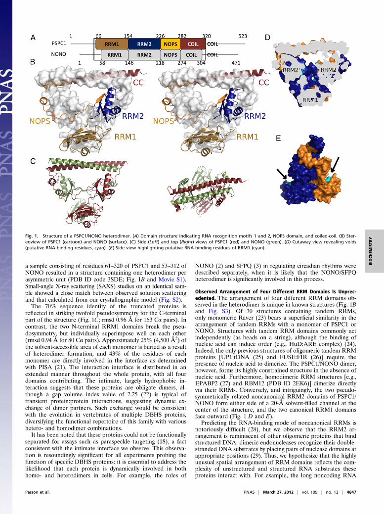
a sample consisting of residues 61–320 of PSPC1 and 53–312 ofNONO resulted in a structure containing one heterodimer perasymmetric unit (PDB ID code 3SDE; Fig. 1B and Movie S1).Small-angle X-ray scattering (SAXS) studies on an identical sam-ple showed a close match between observed solution scatteringand that calculated from our crystallographic model (Fig. S2).The 70% sequence identity of the truncated proteins is
reflected in striking twofold pseudosymmetry for the C-terminalpart of the structure (Fig. 1C; rmsd 0.96 Å for 163 Cα pairs). Incontrast, the two N-terminal RRM1 domains break the pseu-dosymmetry, but individually superimpose well on each other(rmsd 0.94 Å for 80 Cα pairs). Approximately 25% (4,500 Å2) ofthe solvent-accessible area of each monomer is buried as a resultof heterodimer formation, and 43% of the residues of eachmonomer are directly involved in the interface as determinedwith PISA (21). The interaction interface is distributed in anextended manner throughout the whole protein, with all fourdomains contributing. The intimate, largely hydrophobic in-teraction suggests that these proteins are obligate dimers, al-though a gap volume index value of 2.25 (22) is typical oftransient protein:protein interactions, suggesting dynamic ex-change of dimer partners. Such exchange would be consistentwith the evolution in vertebrates of multiple DBHS proteins,diversifying the functional repertoire of this family with varioushetero- and homodimer combinations.It has been noted that these proteins could not be functionally
separated for assays such as paraspeckle targeting (18), a factconsistent with the intimate interface we observe. This observa-tion is resoundingly significant for all experiments probing thefunction of specific DBHS proteins: it is essential to address thelikelihood that each protein is dynamically involved in bothhomo- and heterodimers in cells. For example, the roles of
NONO (2) and SFPQ (3) in regulating circadian rhythms weredescribed separately, when it is likely that the NONO/SFPQheterodimer is significantly involved in this process.
Observed Arrangement of Four Different RRM Domains Is Unprec-edented. The arrangement of four different RRM domains ob-served in the heterodimer is unique in known structures (Fig. 1Band Fig. S3). Of 30 structures containing tandem RRMs,only monomeric Raver (23) bears a superficial similarity in thearrangement of tandem RRMs with a monomer of PSPC1 orNONO. Structures with tandem RRM domains commonly actindependently (as beads on a string), although the binding ofnucleic acid can induce order (e.g., HuD:ARE complex) (24).Indeed, the only previous structures of oligomeric tandem RRMproteins [UP1:tDNA (25) and FUSE:FIR (26)] require thepresence of nucleic acid to dimerize. The PSPC1/NONO dimer,however, forms its highly constrained structure in the absence ofnucleic acid. Furthermore, homodimeric RRM structures [e.g.,EPABP2 (27) and RBM12 (PDB ID 2EK6)] dimerize directlyvia their RRMs. Conversely, and intriguingly, the two pseudo-symmetrically related noncanonical RRM2 domains of PSPC1/NONO form either side of a 20-Å solvent-filled channel at thecenter of the structure, and the two canonical RRM1 domainsface outward (Fig. 1 D and E).Predicting the RNA-binding mode of noncanonical RRMs is
notoriously difficult (28), but we observe that the RRM2 ar-rangement is reminiscent of other oligomeric proteins that bindstructured DNA: dimeric endonucleases recognize their double-stranded DNA substrates by placing pairs of nuclease domains atappropriate positions (29). Thus, we hypothesize that the highlyunusual spatial arrangement of RRM domains reflects the com-plexity of unstructured and structured RNA substrates theseproteins interact with. For example, the long noncoding RNA
Fig. 1. Structure of a PSPC1/NONO heterodimer. (A) Domain structure indicating RNA recognition motifs 1 and 2, NOPS domain, and coiled-coil. (B) Ster-eoview of PSPC1 (cartoon) and NONO (surface). (C) Side (Left) and top (Right) views of PSPC1 (red) and NONO (green). (D) Cutaway view revealing voids(putative RNA-binding residues, cyan). (E) Side view highlighting putative RNA-binding residues of RRM1 (cyan).
Passon et al. PNAS | March 27, 2012 | vol. 109 | no. 13 | 4847
BIOCH
EMISTR
Y
(lncRNA) NEAT1 contains regions of secondary structure, andinverted Alu repeats form hairpins depending on the extent ofA-to-I editing. The twofold pseudosymmetric arrangement ofRRMs in the heterodimer may be able to accommodate thisduplex nucleic acid.
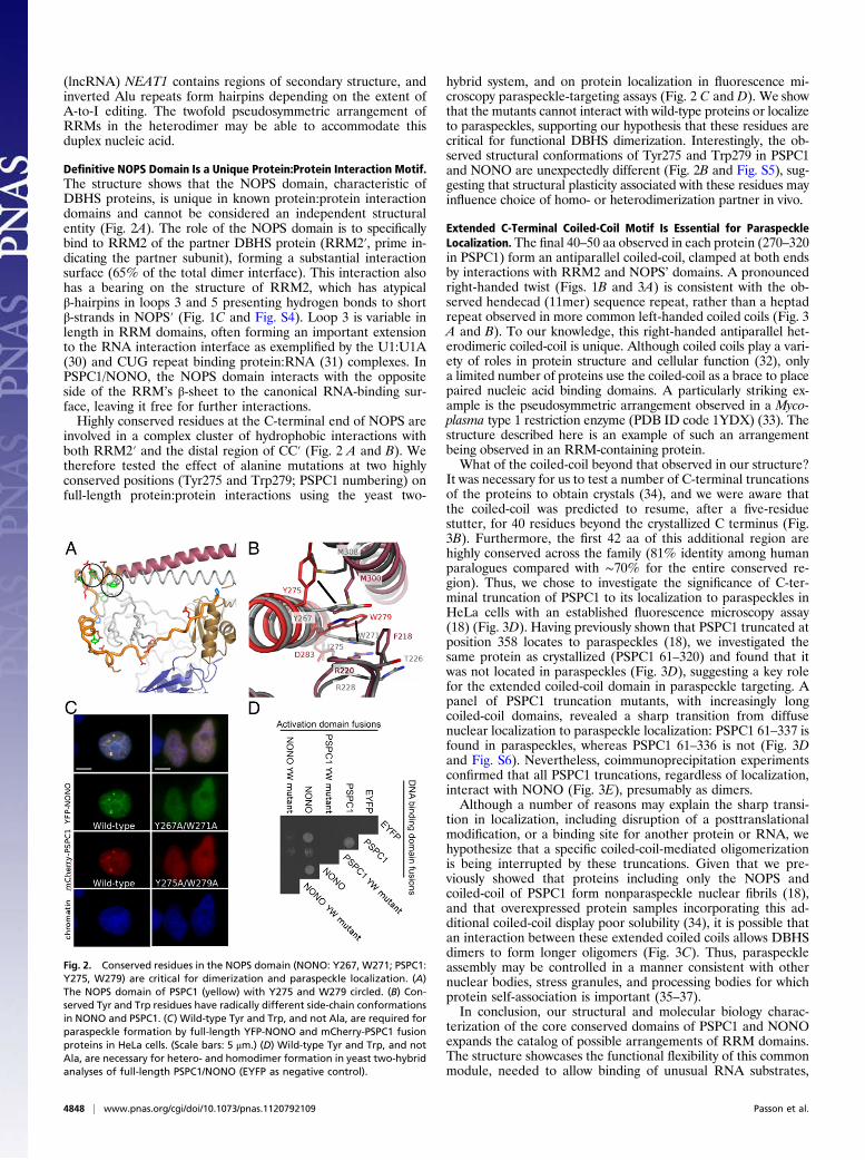
Definitive NOPS Domain Is a Unique Protein:Protein Interaction Motif.The structure shows that the NOPS domain, characteristic ofDBHS proteins, is unique in known protein:protein interactiondomains and cannot be considered an independent structuralentity (Fig. 2A). The role of the NOPS domain is to specificallybind to RRM2 of the partner DBHS protein (RRM2′, prime in-dicating the partner subunit), forming a substantial interactionsurface (65% of the total dimer interface). This interaction alsohas a bearing on the structure of RRM2, which has atypicalβ-hairpins in loops 3 and 5 presenting hydrogen bonds to shortβ-strands in NOPS′ (Fig. 1C and Fig. S4). Loop 3 is variable inlength in RRM domains, often forming an important extensionto the RNA interaction interface as exemplified by the U1:U1A(30) and CUG repeat binding protein:RNA (31) complexes. InPSPC1/NONO, the NOPS domain interacts with the oppositeside of the RRM’s β-sheet to the canonical RNA-binding sur-face, leaving it free for further interactions.Highly conserved residues at the C-terminal end of NOPS are
involved in a complex cluster of hydrophobic interactions withboth RRM2′ and the distal region of CC′ (Fig. 2 A and B). Wetherefore tested the effect of alanine mutations at two highlyconserved positions (Tyr275 and Trp279; PSPC1 numbering) onfull-length protein:protein interactions using the yeast two-
hybrid system, and on protein localization in fluorescence mi-croscopy paraspeckle-targeting assays (Fig. 2 C and D). We showthat the mutants cannot interact with wild-type proteins or localizeto paraspeckles, supporting our hypothesis that these residues arecritical for functional DBHS dimerization. Interestingly, the ob-served structural conformations of Tyr275 and Trp279 in PSPC1and NONO are unexpectedly different (Fig. 2B and Fig. S5), sug-gesting that structural plasticity associated with these residues mayinfluence choice of homo- or heterodimerization partner in vivo.
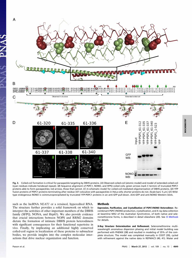
Extended C-Terminal Coiled-Coil Motif Is Essential for ParaspeckleLocalization. The final 40–50 aa observed in each protein (270–320in PSPC1) form an antiparallel coiled-coil, clamped at both endsby interactions with RRM2 and NOPS’ domains. A pronouncedright-handed twist (Figs. 1B and 3A) is consistent with the ob-served hendecad (11mer) sequence repeat, rather than a heptadrepeat observed in more common left-handed coiled coils (Fig. 3A and B). To our knowledge, this right-handed antiparallel het-erodimeric coiled-coil is unique. Although coiled coils play a vari-ety of roles in protein structure and cellular function (32), onlya limited number of proteins use the coiled-coil as a brace to placepaired nucleic acid binding domains. A particularly striking ex-ample is the pseudosymmetric arrangement observed in a Myco-plasma type 1 restriction enzyme (PDB ID code 1YDX) (33). Thestructure described here is an example of such an arrangementbeing observed in an RRM-containing protein.What of the coiled-coil beyond that observed in our structure?
It was necessary for us to test a number of C-terminal truncationsof the proteins to obtain crystals (34), and we were aware thatthe coiled-coil was predicted to resume, after a five-residuestutter, for 40 residues beyond the crystallized C terminus (Fig.3B). Furthermore, the first 42 aa of this additional region arehighly conserved across the family (81% identity among humanparalogues compared with ∼70% for the entire conserved re-gion). Thus, we chose to investigate the significance of C-ter-minal truncation of PSPC1 to its localization to paraspeckles inHeLa cells with an established fluorescence microscopy assay(18) (Fig. 3D). Having previously shown that PSPC1 truncated atposition 358 locates to paraspeckles (18), we investigated thesame protein as crystallized (PSPC1 61–320) and found that itwas not located in paraspeckles (Fig. 3D), suggesting a key rolefor the extended coiled-coil domain in paraspeckle targeting. Apanel of PSPC1 truncation mutants, with increasingly longcoiled-coil domains, revealed a sharp transition from diffusenuclear localization to paraspeckle localization: PSPC1 61–337 isfound in paraspeckles, whereas PSPC1 61–336 is not (Fig. 3Dand Fig. S6). Nevertheless, coimmunoprecipitation experimentsconfirmed that all PSPC1 truncations, regardless of localization,interact with NONO (Fig. 3E), presumably as dimers.Although a number of reasons may explain the sharp transi-
tion in localization, including disruption of a posttranslationalmodification, or a binding site for another protein or RNA, wehypothesize that a specific coiled-coil-mediated oligomerizationis being interrupted by these truncations. Given that we pre-viously showed that proteins including only the NOPS andcoiled-coil of PSPC1 form nonparaspeckle nuclear fibrils (18),and that overexpressed protein samples incorporating this ad-ditional coiled-coil display poor solubility (34), it is possible thatan interaction between these extended coiled coils allows DBHSdimers to form longer oligomers (Fig. 3C). Thus, paraspeckleassembly may be controlled in a manner consistent with othernuclear bodies, stress granules, and processing bodies for whichprotein self-association is important (35–37).In conclusion, our structural and molecular biology charac-
terization of the core conserved domains of PSPC1 and NONOexpands the catalog of possible arrangements of RRM domains.The structure showcases the functional flexibility of this commonmodule, needed to allow binding of unusual RNA substrates,
Fig. 2. Conserved residues in the NOPS domain (NONO: Y267, W271; PSPC1:Y275, W279) are critical for dimerization and paraspeckle localization. (A)The NOPS domain of PSPC1 (yellow) with Y275 and W279 circled. (B) Con-served Tyr and Trp residues have radically different side-chain conformationsin NONO and PSPC1. (C) Wild-type Tyr and Trp, and not Ala, are required forparaspeckle formation by full-length YFP-NONO and mCherry-PSPC1 fusionproteins in HeLa cells. (Scale bars: 5 μm.) (D) Wild-type Tyr and Trp, and notAla, are necessary for hetero- and homodimer formation in yeast two-hybridanalyses of full-length PSPC1/NONO (EYFP as negative control).
4848 | www.pnas.org/cgi/doi/10.1073/pnas.1120792109 Passon et al.
such as the lncRNA NEAT1 or a retained, hyperedited RNA.The structure further provides a solid framework on which tointerpret the activities of other important members of the DBHSfamily (SFPQ, NONA, and Hrp65). We also provide evidencethat crucial interactions between NOPS and RRM2 domainsdictate the formation of intimate DBHS protein heterodimerswith significant consequences for their functional repertoire invivo. Finally, by implicating an additional highly conservedcoiled-coil region in localization of these proteins to subnuclearbodies, we provide insights into the complex molecular inter-actions that drive nuclear organization and function.
MethodsExpression, Purification, and Crystallization of PSPC1/NONO Heterodimer. Re-combinant PSPC1/NONO production, crystallization, and X-ray data collectionat beamline MX2 of the Australian Synchrotron, of both native and sele-nomethionine forms, is described in detail elsewhere (20). See SI Methodsfor details.
X-Ray Structure Determination and Refinement. Selenomethionine multi-wavelength anomalous dispersion phasing and initial model building wasperformed with PHENIX (38) and resulted in modeling of 35% of the com-plete structure. The model was completed manually in COOT (39), cycledwith refinement against the native data in REFMAC5 (40, 41). Water and
Fig. 3. Coiled-coil formation is critical for paraspeckle targeting by DBHS proteins. (A) Observed coiled-coil (atomic model) and model of extended coiled-coil(cyan residues indicate hendecad repeat). (B) Sequence alignment of PSPC1, NONO, and SFPQ coiled coils; green arrows mark C termini of truncated PSPC1proteins able to form paraspeckles; red arrows, those that cannot. (C) A schematic model for coiled-coil-mediated oligomerization of DBHS proteins. (D) YFPfusion proteins of PSPC1 proteins terminating after residue 337 colocalize with paraspeckles in HeLa cells; shorter proteins do not. (Scale bars: 5 μm.) (E) Wild-type endogenous NONO is coimmunoprecipitated by truncated YFP-PSPC1 proteins in an anti-GFP pull-down. Anti-GFP and anti-NONO Western blots.
Passon et al. PNAS | March 27, 2012 | vol. 109 | no. 13 | 4849
BIOCH
EMISTR
Y
ethylene glycol molecules were added before final rounds of refinementwith BUSTER-TNT (42). Progress of refinement was monitored using R-free,the validation tools available in COOT, and MOLPROBITY (43). Refinementagainst native data to 1.9-Å resolution converged with residuals R = 0.183and Rfree = 0.228. The asymmetric unit contains a single heterodimer, resi-dues 66–320 of PSPC1 and residues 66–304 of NONO (494 aa), 470 waters,and 11 ethylene glycol molecules. The model has excellent geometry, with98.6% of residues in most favored regions of the Ramachandran plot (TableS1 and Fig. S7).
Molecular graphical analysis was performed using PyMol (version 1.2r3pre;Schrödinger, LLC) Sequence alignments were produced with ALINE (44) andtopology diagrams with TOPDRAW (45).
SAXS. SAXS experiments were carried out at the SAXSWAXS beamline of theAustralian Synchrotron on a concentration series (0.375–12 mg/mL) of pro-tein sample. Data were analyzed with the ATSAS suite (46, 47). See SIMethods and Fig. S2 for details.
Plasmids for Fluorescent Protein Fusions. Plasmids for fluorescent pro-tein fusion experiments were generated using the pEYFP-C1 plasmid
(Clontech) and standard molecular biology techniques. See SI Methodsfor details.
Localization Assays and Immunoprecipitation. PSPC1 localization assays andimmunoprecipitation experiments were carried out as previously described(18). See SI Methods for details.
Yeast Two-Hybrid Analyses. Yeast two-hybrid experiments were carried outusing the GAL4 DNA-binding domain fusions in pGBK-RC and GAL4 activationdomain fusions in pGAD-RC, respectively, using the lithium acetate methodaccording to Gietz and Woods (48). See SI Methods for details.
ACKNOWLEDGMENTS. We thank Tom Caradoc-Davies, Nathan Cowieson,Christine Gee, Nigel Kirby, and Adrian Hawley. Aspects of this research wereundertaken on the Macromolecular Crystallography and SAXSWAXS beam-lines at the Australian Synchrotron (Victoria, Australia). Yeast two-hybridplasmids and yeast strains were a kind gift from T. Ito (University of Tokyo).Support for this work was provided by National Health and Medical ResearchCouncil of Australia Project Grant 513880 (to C.S.B. and A.H.F.) and a fellowship(M.L.); a fellowship from the Western Australian Institute for Medical Research(A.H.F.); and a University of Western Australia scholarship (D.M.P.).
1. Shav-Tal Y, Zipori D (2002) PSF and p54(nrb)/NonO—multi-functional nuclear pro-teins. FEBS Lett 531:109–114.
2. Brown SA, et al. (2005) PERIOD1-associated proteins modulate the negative limb ofthe mammalian circadian oscillator. Science 308:693–696.
3. Duong HA, Robles MS, Knutti D, Weitz CJ (2011) A molecular mechanism for circadianclock negative feedback. Science 332:1436–1439.
4. Wang G, Cui Y, Zhang G, Garen A, Song X (2009) Regulation of proto-oncogenetranscription, cell proliferation, and tumorigenesis in mice by PSF protein and a VL30noncoding RNA. Proc Natl Acad Sci USA 106:16794–16798.
5. Stanewsky R, Rendahl KG, Dill M, Saumweber H (1993) Genetic and molecular analysisof the X chromosomal region 14B17-14C4 in Drosophila melanogaster: Loss of func-tion in NONA, a nuclear protein common to many cell types, results in specificphysiological and behavioral defects. Genetics 135:419–442.
6. Kuwahara S, et al. (2006) PSPC1, NONO, and SFPQ are expressed in mouse Sertoli cellsand may function as coregulators of androgen receptor-mediated transcription. BiolReprod 75:352–359.
7. Fox AH, et al. (2002) Paraspeckles: A novel nuclear domain. Curr Biol 12:13–25.8. Clemson CM, et al. (2009) An architectural role for a nuclear noncoding RNA: NEAT1
RNA is essential for the structure of paraspeckles. Mol Cell 33:717–726.9. Sasaki YT, Ideue T, Sano M, Mituyama T, Hirose T (2009) MENepsilon/beta noncoding
RNAs are essential for structural integrity of nuclear paraspeckles. Proc Natl Acad SciUSA 106:2525–2530.
10. Sunwoo H, et al. (2009) MEN epsilon/beta nuclear-retained non-coding RNAs are up-regulated upon muscle differentiation and are essential components of paraspeckles.Genome Res 19:347–359.
11. Souquere S, Beauclair G, Harper F, Fox A, Pierron G (2010) Highly ordered spatialorganization of the structural long noncoding NEAT1 RNAs within paraspeckle nu-clear bodies. Mol Biol Cell 21:4020–4027.
12. Bond CS, Fox AH (2009) Paraspeckles: Nuclear bodies built on long noncoding RNA.J Cell Biol 186:637–644.
13. Mao YS, Sunwoo H, Zhang B, Spector DL (2011) Direct visualization of the co-tran-scriptional assembly of a nuclear body by noncoding RNAs. Nat Cell Biol 13:95–101.
14. Chen LL, DeCerbo JN, Carmichael GG (2008) Alu element-mediated gene silencing.EMBO J 27:1694–1705.
15. Prasanth KV, et al. (2005) Regulating gene expression through RNA nuclear retention.Cell 123:249–263.
16. Zhang Z, Carmichael GG (2001) The fate of dsRNA in the nucleus: A p54(nrb)-con-taining complex mediates the nuclear retention of promiscuously A-to-I edited RNAs.Cell 106:465–475.
17. Kiesler E, Miralles F, Ostlund Farrants AK, Visa N (2003) The Hrp65 self-interaction ismediated by an evolutionarily conserved domain and is required for nuclear importof Hrp65 isoforms that lack a nuclear localization signal. J Cell Sci 116:3949–3956.
18. Fox AH, Bond CS, Lamond AI (2005) P54nrb forms a heterodimer with PSP1 that lo-calizes to paraspeckles in an RNA-dependent manner. Mol Biol Cell 16:5304–5315.
19. Maris C, Dominguez C, Allain FH (2005) The RNA recognition motif, a plastic RNA-binding platform to regulate post-transcriptional gene expression. FEBS J 272:2118–2131.
20. Passon DM, Lee M, Fox AH, Bond CS (2011) Crystallization of a paraspeckle proteinPSPC1-NONO heterodimer. Acta Crystallogr Sect F Struct Biol Cryst Commun 67:1231–1234.
21. Krissinel E, Henrick K (2007) Inference of macromolecular assemblies from crystallinestate. J Mol Biol 372:774–797.
22. Nooren IM, Thornton JM (2003) Diversity of protein–protein interactions. EMBO J 22:3486–3492.
23. Lee JH, Rangarajan ES, Yogesha SD, Izard T (2009) Raver1 interactions with vinculinand RNA suggest a feed-forward pathway in directing mRNA to focal adhesions.Structure 17:833–842.
24. Wang X, Tanaka Hall TM (2001) Structural basis for recognition of AU-rich elementRNA by the HuD protein. Nat Struct Biol 8:141–145.
25. Ding J, et al. (1999) Crystal structure of the two-RRM domain of hnRNP A1 (UP1)complexed with single-stranded telomeric DNA. Genes Dev 13:1102–1115.
26. Crichlow GV, et al. (2008) Dimerization of FIR upon FUSE DNA binding suggestsa mechanism of c-myc inhibition. EMBO J 27:277–289.
27. Song J, McGivern JV, Nichols KW, Markley JL, Sheets MD (2008) Structural basis forRNA recognition by a type II poly(A)-binding protein. Proc Natl Acad Sci USA 105:15317–15322.
28. Cléry A, Blatter M, Allain FH (2008) RNA recognition motifs: Boring? Not quite. CurrOpin Struct Biol 18:290–298.
29. Middleton CL, Parker JL, Richard DJ, White MF, Bond CS (2004) Substrate recognitionand catalysis by the Holliday junction resolving enzyme Hje. Nucleic Acids Res 32:5442–5451.
30. Katsamba PS, Bayramyan M, Haworth IS, Myszka DG, Laird-Offringa IA (2002) Com-plex role of the beta 2-beta 3 loop in the interaction of U1A with U1 hairpin II RNA.J Biol Chem 277:33267–33274.
31. Tsuda K, et al. (2009) Structural basis for the sequence-specific RNA-recognitionmechanism of human CUG-BP1 RRM3. Nucleic Acids Res 37:5151–5166.
32. Lupas AN, Gruber M (2005) The structure of alpha-helical coiled coils. Adv ProteinChem 70:37–78.
33. Calisto BM, et al. (2005) Crystal structure of a putative type I restriction-modification Ssubunit from Mycoplasma genitalium. J Mol Biol 351:749–762.
34. Lee M, Passon DM, Hennig S, Fox AH, Bond CS (2011) Construct optimization forstudying protein complexes: Obtaining diffraction-quality crystals of the pseudo-symmetric PSPC1-NONO heterodimer. Acta Crystallogr D Biol Crystallogr 67:981–987.
35. Kaiser TE, Intine RV, Dundr M (2008) De novo formation of a subnuclear body. Science322:1713–1717.
36. Ozgur S, Chekulaeva M, Stoecklin G (2010) Human Pat1b connects deadenylationwith mRNA decapping and controls the assembly of processing bodies. Mol Cell Biol30:4308–4323.
37. Sun Z, et al. (2011) Molecular determinants and genetic modifiers of aggregation andtoxicity for the ALS disease protein FUS/TLS. PLoS Biol 9:e1000614.
38. Adams PD, et al. (2010) PHENIX: A comprehensive Python-based system for macro-molecular structure solution. Acta Crystallogr D Biol Crystallogr 66:213–221.
39. Emsley P, Cowtan K (2004) COOT: Model-building tools for molecular graphics. ActaCrystallogr D Biol Crystallogr 60(Pt 12 Pt 1):2126–2132.
40. Murshudov GN, Vagin AA, Dodson EJ (1997) Refinement of macromolecular struc-tures by the maximum-likelihood method. Acta Crystallogr D Biol Crystallogr 53:240–255.
41. Collaborative Computational Project, Number 4 (1994) The CCP4 suite: Programs forprotein crystallography. Acta Crystallogr D Biol Crystallogr 50:760–763.
42. Blanc E, et al. (2004) Refinement of severely incomplete structures with maximumlikelihood in BUSTER-TNT. Acta Crystallogr D Biol Crystallogr 60(Pt 12 Pt 1):2210–2221.
43. Chen VB, et al. (2010) MolProbity: All-atom structure validation for macromolecularcrystallography. Acta Crystallogr D Biol Crystallogr 66:12–21.
44. Bond CS, Schüttelkopf AW (2009) ALINE: A WYSIWYG protein-sequence alignmenteditor for publication-quality alignments. Acta Crystallogr D Biol Crystallogr 65:510–512.
45. Bond CS (2003) TopDraw: A sketchpad for protein structure topology cartoons. Bio-informatics 19:311–312.
46. Konarev PV, Volkov VV, Sokolova AV, Koch MHJ, Svergun DI (2003) PRIMUS—aWindows-PC based system for small-angle scattering data analysis. J Appl Cryst 36:1277–1282.
47. Svergun DI (1992) Determination of the regularization parameter in indirect-trans-form methods using perceptual criteria. J Appl Cryst 25:495–503.
48. Gietz RD, Woods RA (2006) Yeast transformation by the LiAc/SS Carrier DNA/PEGmethod. Methods Mol Biol 313:107–120.
4850 | www.pnas.org/cgi/doi/10.1073/pnas.1120792109 Passon et al.