Embed Size (px)
Citation preview
John LowengrubDept Math, UCI
Nonlinear Modeling of Tumor Growth I:Basic Models
P. Macklin, M.S. 2003, Ph.D. 2007 (expected); X. Li Ph.D. 2007 (expected)
V. Cristini (Dept Biomed Eng, UCI); Q. Nie (Dept Math, UCI)
Motivation
• Provide biophysically justified in silicovirtual system to study
• Help experimental investigations; design new experiments
• Therapy protocols
Outline
•Introduction to tumor growthMultiscale complex soft matter problem
•Mathematical Models, Simplifications andAnalysis
•Numerical Methods
•Results
(limited biophysics)
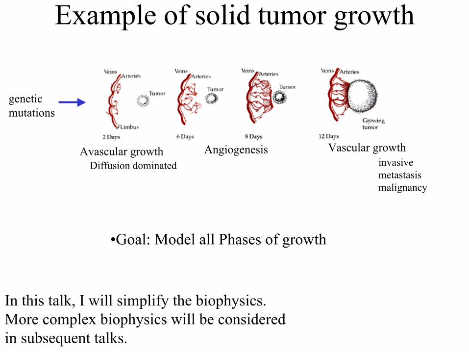
Example of solid tumor growth
geneticmutations
Avascular growth Angiogenesis Vascular growthinvasivemetastasismalignancy
Diffusion dominated
•Goal: Model all Phases of growth
In this talk, I will simplify the biophysics.More complex biophysics will be consideredin subsequent talks.
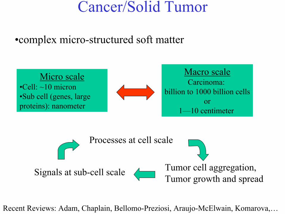
Cancer/Solid Tumor
Micro scale•Cell: ~10 micron•Sub cell (genes, large proteins): nanometer
Macro scaleCarcinoma:
billion to 1000 billion cellsor
1—10 centimeter
Processes at cell scale
Signals at sub-cell scale Tumor cell aggregation,Tumor growth and spread
•complex micro-structured soft matter
Recent Reviews: Adam, Chaplain, Bellomo-Preziosi, Araujo-McElwain, Komarova,…
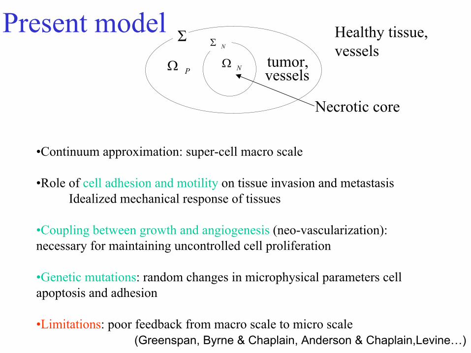
Present model
•Continuum approximation: super-cell macro scale
•Role of cell adhesion and motility on tissue invasion and metastasisIdealized mechanical response of tissues
•Coupling between growth and angiogenesis (neo-vascularization): necessary for maintaining uncontrolled cell proliferation
•Genetic mutations: random changes in microphysical parameters cell apoptosis and adhesion
•Limitations: poor feedback from macro scale to micro scale
Ω pΩ N
ΣN
Σ Healthy tissue, vessels
tumor,
Necrotic core
Σ
PΩ
NΣ
NΩvessels
(Greenspan, Byrne & Chaplain, Anderson & Chaplain,Levine…)
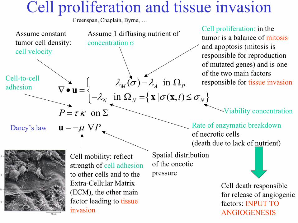
Cell proliferation and tissue invasion
( ) in
in | ( , )
on
M A P
N N Nt
PP
λ σ λλ σ σ
τ κµ
− Ω∇• = − Ω = ≤= Σ= − ∇
ux x
u
Cell proliferation: in the tumor is a balance of mitosisand apoptosis (mitosis is responsible for reproduction of mutated genes) and is one of the two main factors responsible for tissue invasion
Spatial distribution of the oncotic pressure
Cell mobility: reflect strength of cell adhesionto other cells and to the Extra-Cellular Matrix (ECM), the other main factor leading to tissue invasion
Assume constant tumor cell density: cell velocity
Darcy’s law
Cell death responsible for release of angiogenic factors: INPUT TO ANGIOGENESIS
Assume 1 diffusing nutrient of concentration σ
Cell-to-celladhesion
Greenspan, Chaplain, Byrne, …
Rate of enzymatic breakdownof necrotic cells (death due to lack of nutrient)
Viability concentration
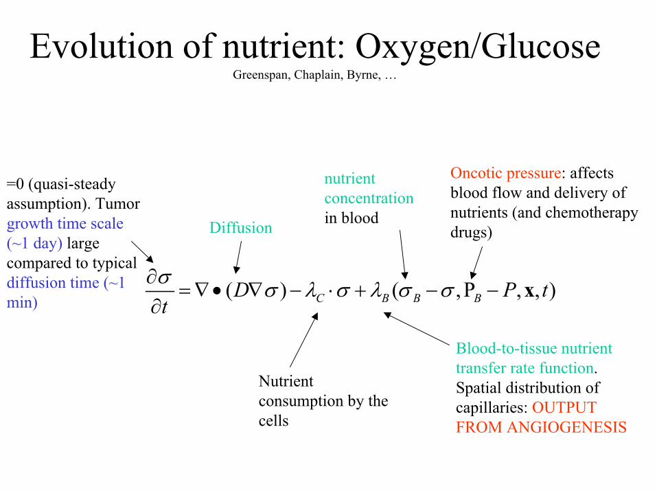
Evolution of nutrient: Oxygen/GlucoseGreenspan, Chaplain, Byrne, …
( ) ( ,P , , )C B B BD P ttσ σ λ σ λ σ σ∂= ∇• ∇ − ⋅ + − −
∂x
Oncotic pressure: affects blood flow and delivery of nutrients (and chemotherapy drugs)
Blood-to-tissue nutrient transfer rate function.Spatial distribution of capillaries: OUTPUT FROM ANGIOGENESIS
Nutrient consumption by the cells
Diffusion
=0 (quasi-steady assumption). Tumor growth time scale (~1 day) large compared to typical diffusion time (~1 min)
nutrientconcentrationin blood
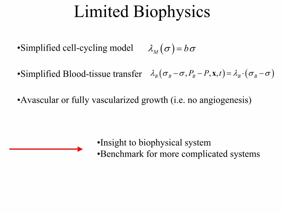
Limited Biophysics
•Simplified Blood-tissue transfer ( ) ( ), , ,B B B B BP P tλ σ σ λ σ σ− − = ⋅ −x
•Avascular or fully vascularized growth (i.e. no angiogenesis)
•Simplified cell-cycling model ( )M bλ σ σ=
•Insight to biophysical system•Benchmark for more complicated systems
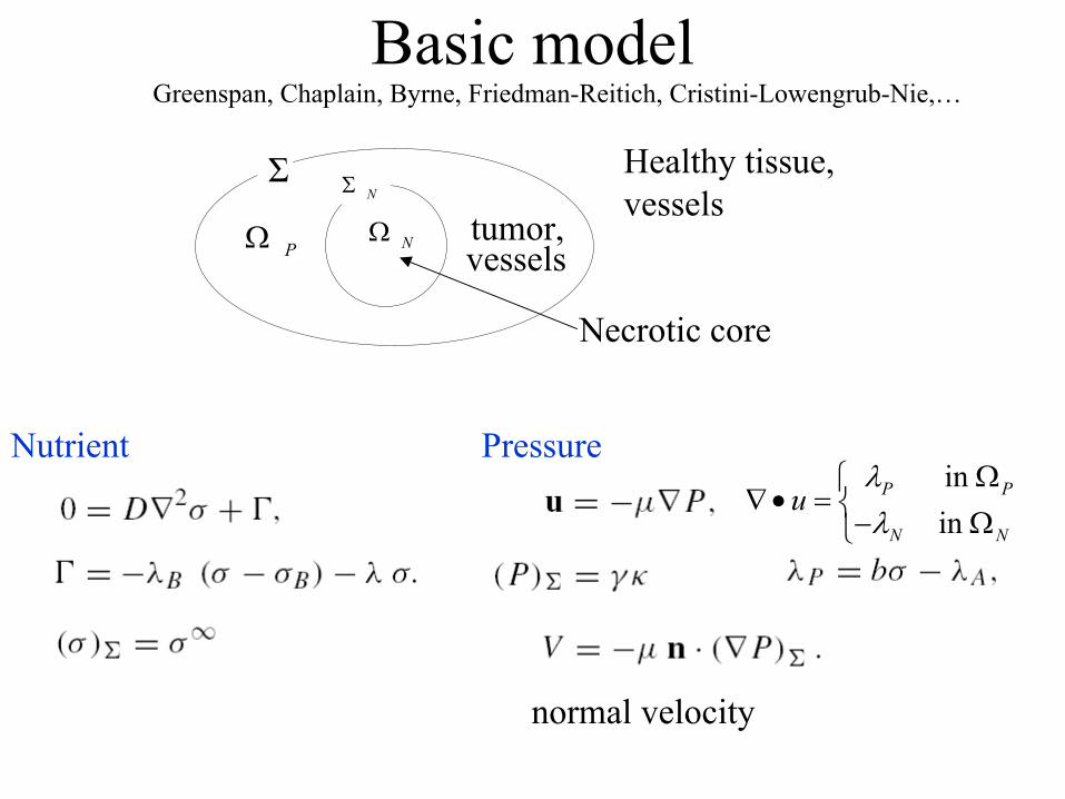
Basic modelGreenspan, Chaplain, Byrne, Friedman-Reitich, Cristini-Lowengrub-Nie,…
Ω pΩ N
ΣN
Σ Healthy tissue, vessels
tumor,
Necrotic core
Σ
PΩ
NΣ
NΩvessels
in in
P P
N N
uλλ
Ω∇• = − Ω
normal velocity
Nutrient Pressure
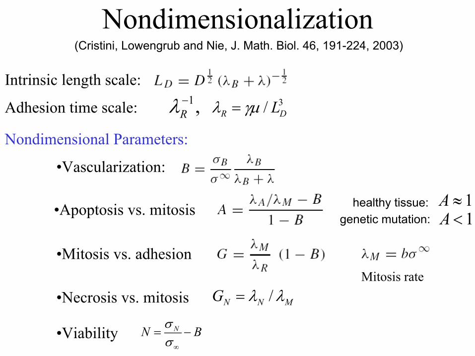
Nondimensionalization(Cristini, Lowengrub and Nie, J. Math. Biol. 46, 191-224, 2003)
Intrinsic length scale:
Adhesion time scale: 1,Rλ−
Nondimensional Parameters:
•Apoptosis vs. mitosis healthy tissue: 1A ≈genetic mutation: 1A <
•Vascularization:
•Mitosis vs. adhesion
•Necrosis vs. mitosis /N N MG λ λ=
•Viability NN Bσσ∞
= −
3/R DLλ γµ=
Mitosis rate
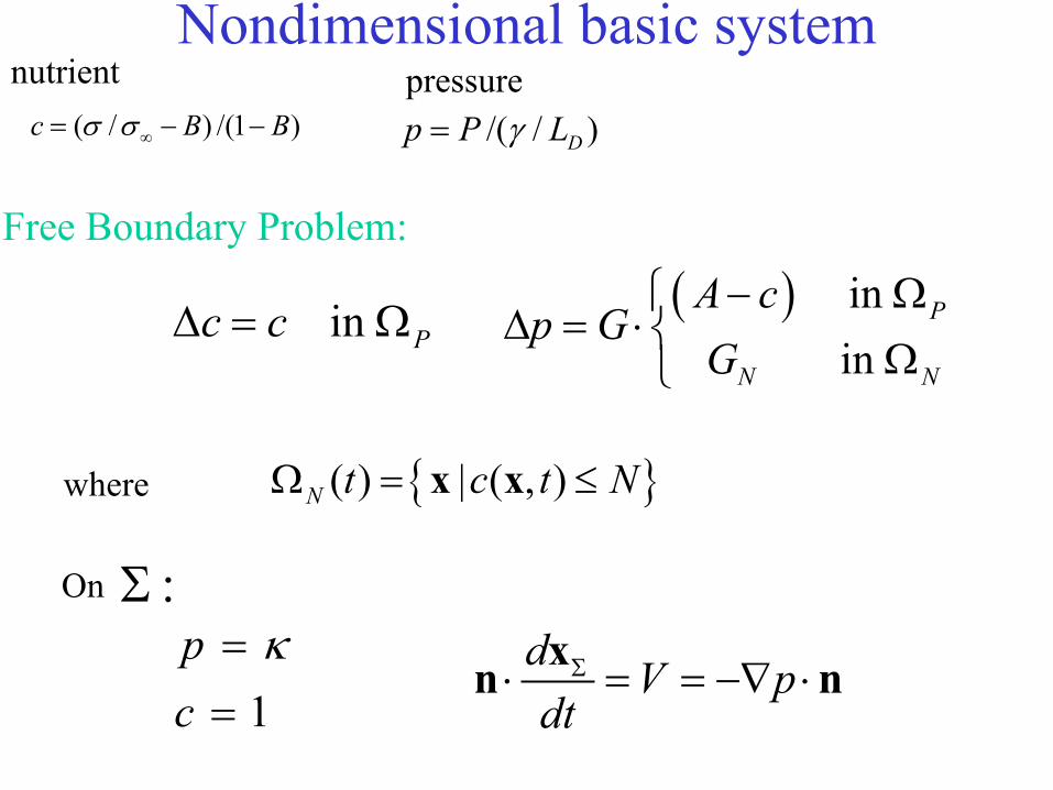
Nondimensional basic system
( ) in in
P
N N
A cp G
G − Ω
∆ = ⋅Ω
d V pdt
Σ⋅ = = −∇ ⋅xn n
1pc
κ==
On :Σ
( ) | ( , )N t c t NΩ = ≤x x
Free Boundary Problem:
in Pc c∆ = Ω
where
( / ) /(1 )c B Bσ σ∞= − − /( / )Dp P Lγ=
nutrient pressure
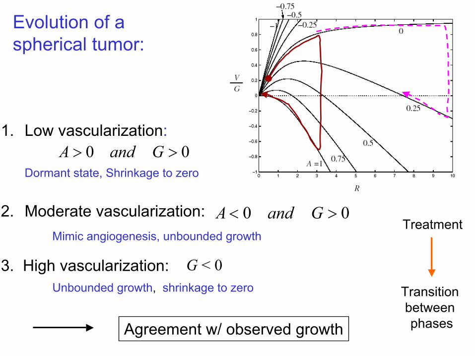
Evolution of a spherical tumor:
Transition between phases
Treatment
1. Low vascularization:
Dormant state, Shrinkage to zero
0 0A and G> >
2. Moderate vascularization: 0 0A and G< >Mimic angiogenesis, unbounded growth
3. High vascularization:Unbounded growth, shrinkage to zero
G < 0
Agreement w/ observed growth
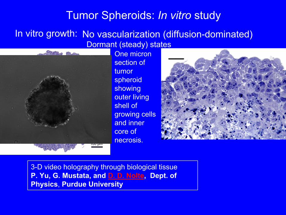
Tumor Spheroids: In vitro studyIn vitro growth: No vascularization (diffusion-dominated)
Dormant (steady) states
3-D video holography through biological tissueP. Yu, G. Mustata, and D. D. Nolte, Dept. of Physics, Purdue University
One micron section of tumor spheroid showing outer living shell of growing cells and inner core of necrosis.
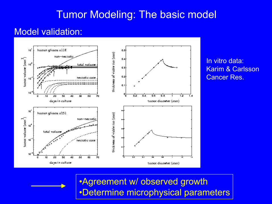
Tumor Modeling: The basic modelModel validation:
•Agreement w/ observed growth•Determine microphysical parameters
In vitro data:Karim & CarlssonCancer Res.
Growth of tumor Viable rim
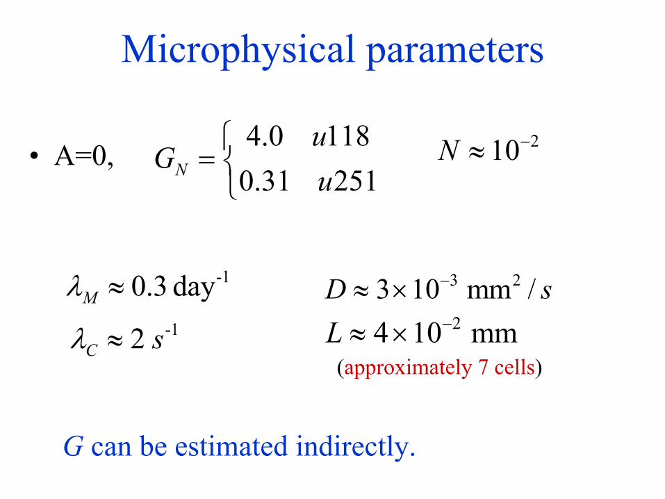
Microphysical parameters
• A=0,4.0 1180.31 251N
uG
u
=
210N −≈
-10.3 dayMλ ≈24 10 mmL −≈ ×
(approximately 7 cells)
-12C sλ ≈
G can be estimated indirectly.
3 23 10 mm /D s−≈ ×
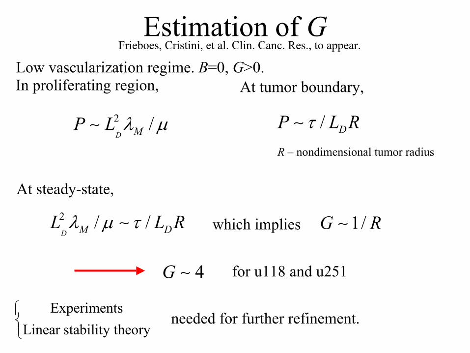
Estimation of GFrieboes, Cristini, et al. Clin. Canc. Res., to appear.
2 /D MP L λ µ∼
In proliferating region, At tumor boundary,
/ DP L Rτ∼R – nondimensional tumor radius
At steady-state,2 / /D M DL L Rλ µ τ∼ which implies 1/G R∼
4G ∼ for u118 and u251
needed for further refinement.Experiments
Linear stability theory
Low vascularization regime. B=0, G>0.
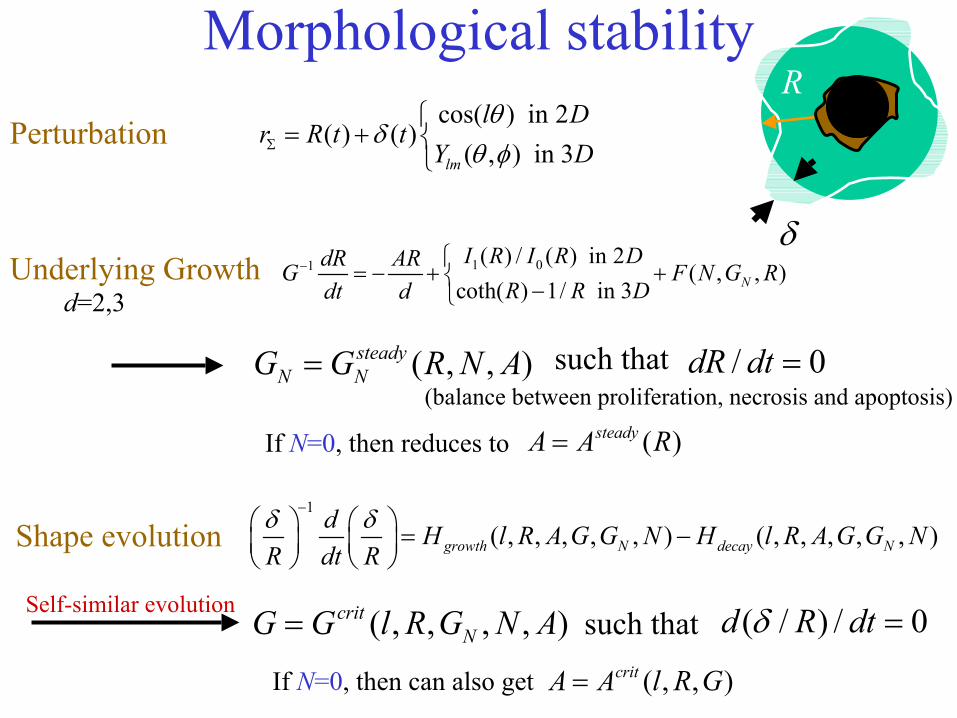
Morphological stabilitycos( ) in 2
( ) ( )( , ) in 3lm
l Dr R t t
Y Dθ
δθ φΣ
= +
R
δ1 01 ( ) / ( ) in 2
( , , )coth( ) 1/ in 3 N
I R I R DdR ARG F N G RR R Ddt d
− = − + + −
Perturbation
Underlying Growthd=2,3
( , , )steadyN NG G R N A= such that / 0dR dt =
(balance between proliferation, necrosis and apoptosis)
Shape evolution1
( , , , , , ) ( , , , , , )growth N decay Nd H l R A G G N H l R A G G N
R dt Rδ δ−
= −
( , , , , )critNG G l R G N A= such that ( / ) / 0d R dtδ =
If N=0, then can also get
Self-similar evolution
( , , )critA A l R G=
If N=0, then reduces to ( )steadyA A R=
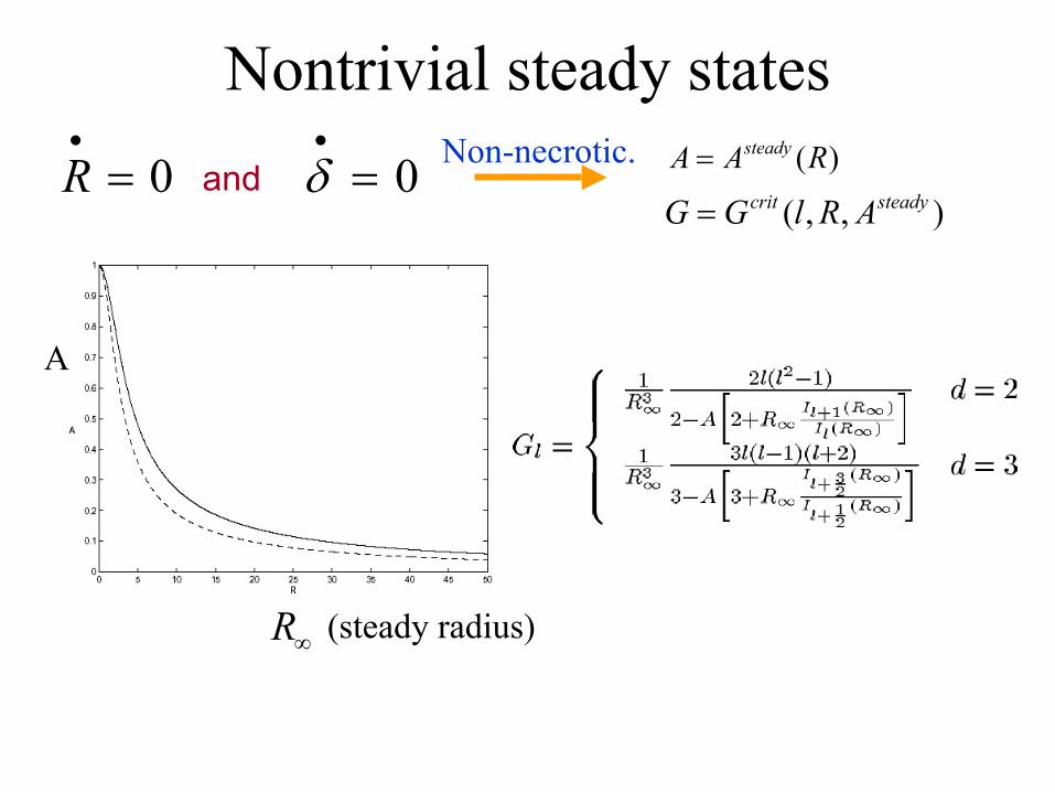
Nontrivial steady states
R∞ (steady radius)
0δ•
=0R•
= and ( )steadyA A R=
( , , )crit steadyG G l R A=
A
Non-necrotic.
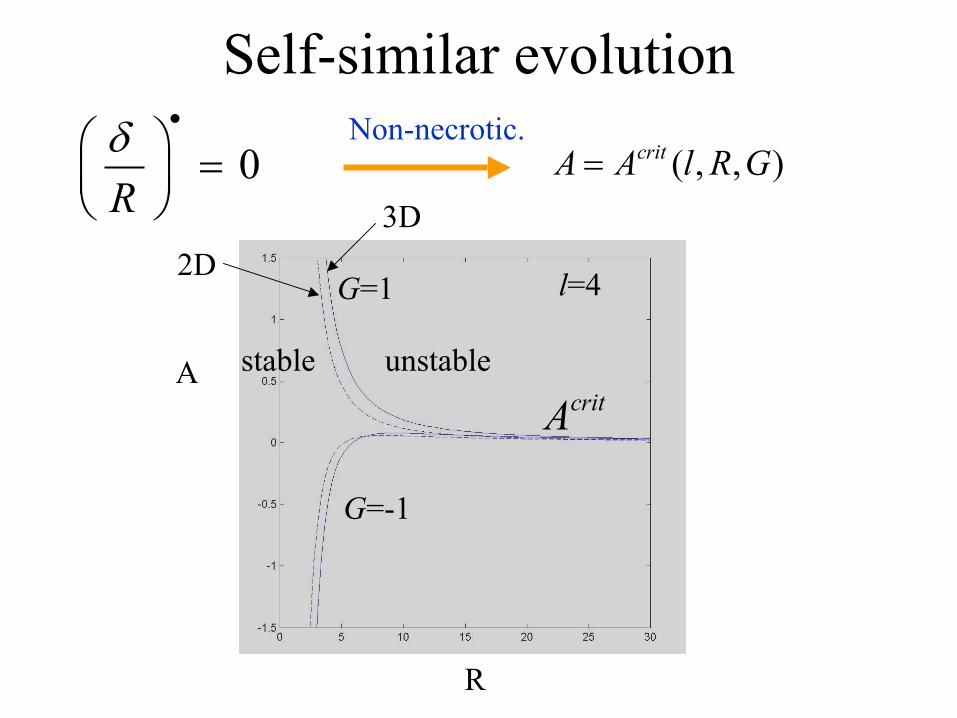
Self-similar evolution
0Rδ •
=
( , , )critA A l R G=
A
R
critAstable unstable
l=4
G=-1
G=12D
3D
Non-necrotic.
Vascular/mechanicalinhomogeneity
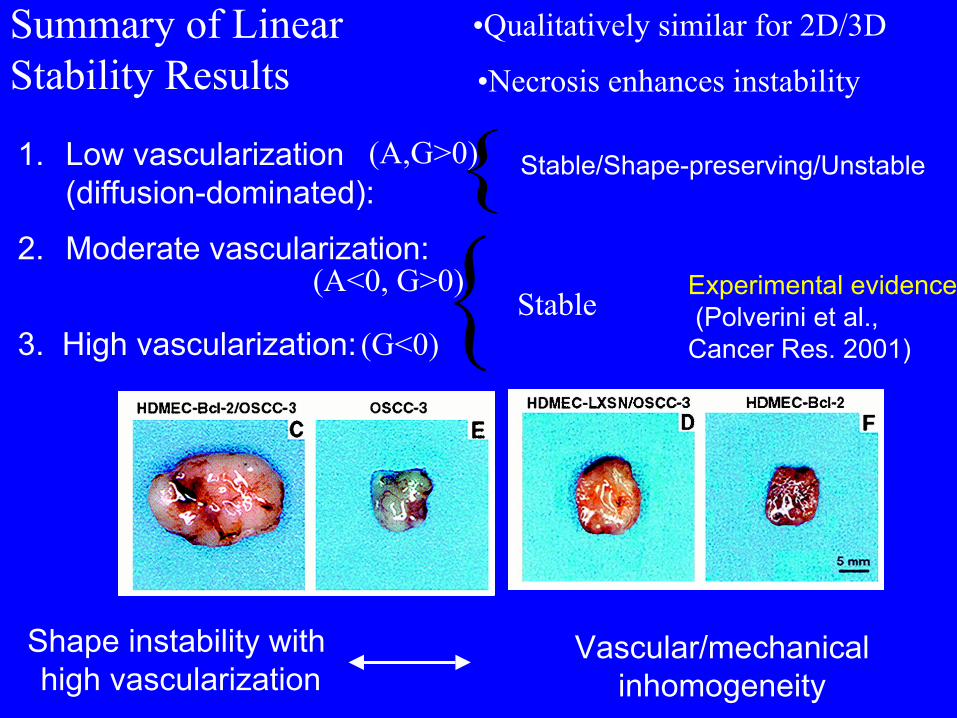
1. Low vascularization (diffusion-dominated):
2. Moderate vascularization:
3. High vascularization:
Stable/Shape-preserving/Unstable
Experimental evidence(Polverini et al., Cancer Res. 2001)
Shape instability withhigh vascularization
•Qualitatively similar for 2D/3D
•Necrosis enhances instability
(A,G>0)
(A<0, G>0)
(G<0)Stable
Summary of LinearStability Results
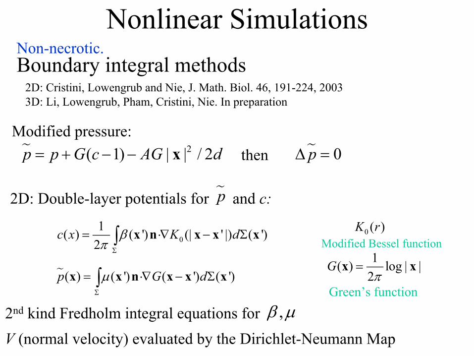
Nonlinear SimulationsBoundary integral methods
2D: Cristini, Lowengrub and Nie, J. Math. Biol. 46, 191-224, 20033D: Li, Lowengrub, Pham, Cristini, Nie. In preparation
2( 1) | | / 2p p G c AG d= + − − xModified pressure:
0p∆ =then
2D: Double-layer potentials for and c:p
01( ) ( ') (| ' |) ( ')
2c x K dβ
π Σ
= ⋅∇ − Σ∫ x n x x x
( ) ( ') ( ') ( ')p G dµΣ
= ⋅∇ − Σ∫x x n x x x1( ) log | |
2G
π=x x
Green’s function
0 ( )K rModified Bessel function
2nd kind Fredholm integral equations for V (normal velocity) evaluated by the Dirichlet-Neumann Map
,β µ
Non-necrotic.
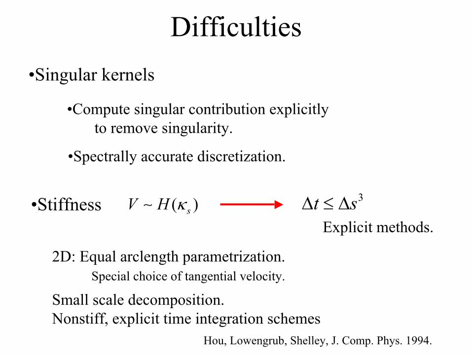
Difficulties•Singular kernels
•Stiffness
•Compute singular contribution explicitly to remove singularity.
•Spectrally accurate discretization.
( )sV H κ∼ 3t s∆ ≤ ∆Explicit methods.
2D: Equal arclength parametrization. Special choice of tangential velocity.
Small scale decomposition. Nonstiff, explicit time integration schemes
Hou, Lowengrub, Shelley, J. Comp. Phys. 1994.
Numerical Results
•Steady-states
•Self-similar evolution
•Stable evolution
•Diffusional Instability
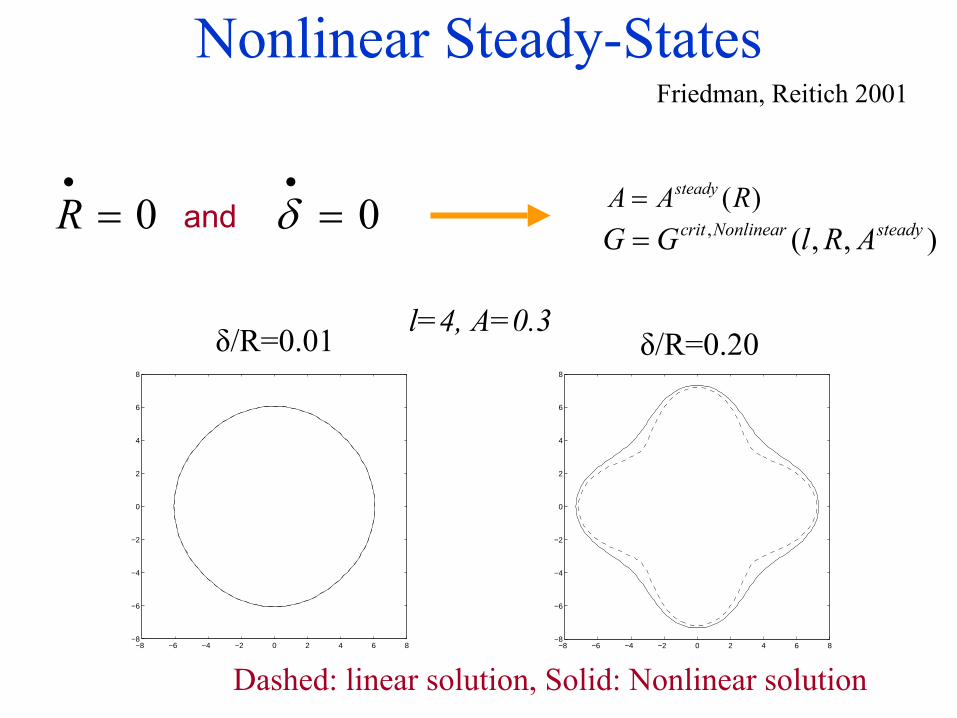
Nonlinear Steady-States
−8 −6 −4 −2 0 2 4 6 8−8
−6
−4
−2
0
2
4
6
8
−8 −6 −4 −2 0 2 4 6 8−8
−6
−4
−2
0
2
4
6
8
l=4, A=0.3δ/R=0.01 δ/R=0.20
Dashed: linear solution, Solid: Nonlinear solution
0δ•
=0R•
= and ( )steadyA A R=, ( , , )crit Nonlinear steadyG G l R A=
Friedman, Reitich 2001
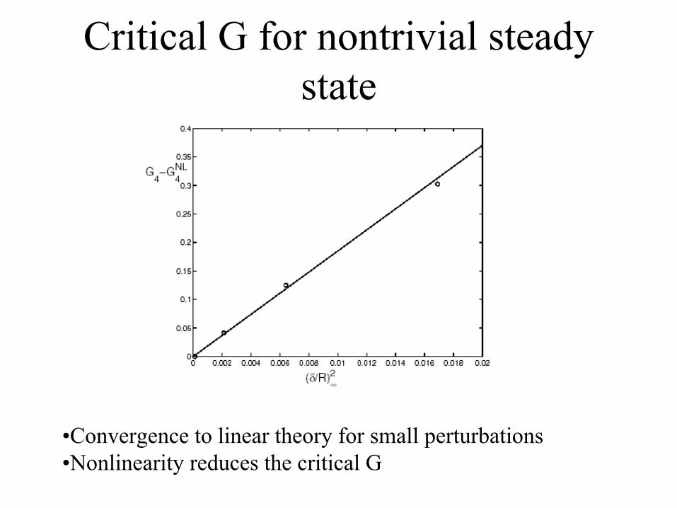
Critical G for nontrivial steady state
•Convergence to linear theory for small perturbations•Nonlinearity reduces the critical G
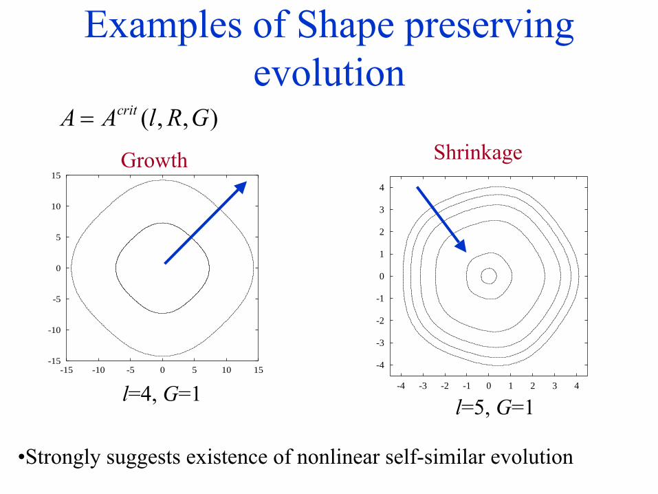
Examples of Shape preserving evolution
Growth
-15
-10
-5
0
5
10
15
-15 -10 -5 0 5 10 15
l=4, G=1-4
-3
-2
-1
0
1
2
3
4
-4 -3 -2 -1 0 1 2 3 4
Shrinkage
l=5, G=1
( , , )critA A l R G=
•Strongly suggests existence of nonlinear self-similar evolution
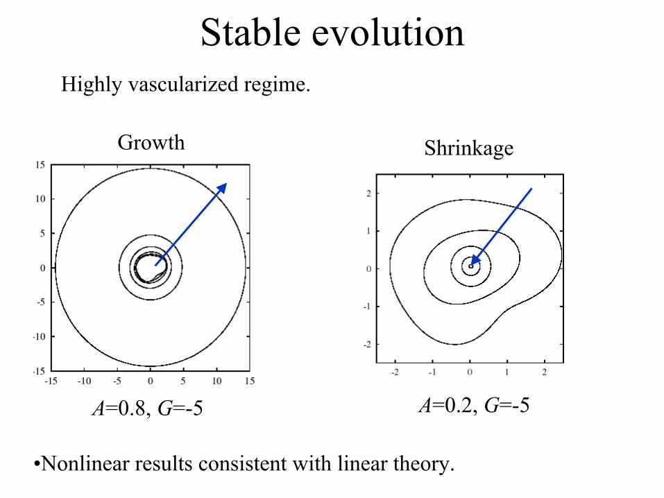
Stable evolutionHighly vascularized regime.
Growth
A=0.8, G=-5
Shrinkage
A=0.2, G=-5
•Nonlinear results consistent with linear theory.
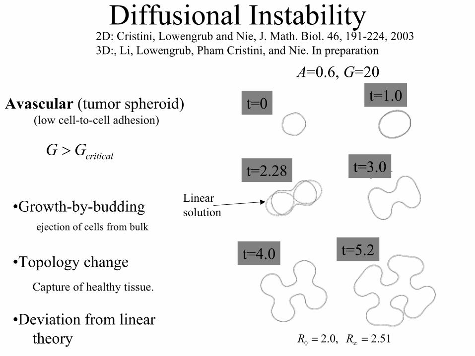
Diffusional Instability
Avascular (tumor spheroid) (low cell-to-cell adhesion)
•Growth-by-budding
criticalG G>
ejection of cells from bulk
2D: Cristini, Lowengrub and Nie, J. Math. Biol. 46, 191-224, 20033D:, Li, Lowengrub, Pham Cristini, and Nie. In preparation
•Topology changeCapture of healthy tissue.
t=1
t=1t=2.28
t=0 t=1.0
t=3.0
t=4.0 t=5.2
A=0.6, G=20
•Deviation from lineartheory
Linearsolution
2.51R∞ =0 2.0,R =
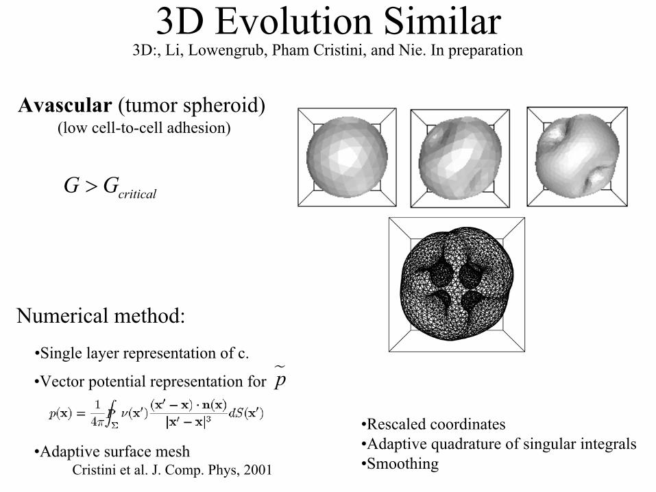
3D Evolution Similar
Avascular (tumor spheroid) (low cell-to-cell adhesion)
criticalG G>
3D:, Li, Lowengrub, Pham Cristini, and Nie. In preparation
•Single layer representation of c.
Numerical method:
•Vector potential representation for p
•Adaptive surface meshCristini et al. J. Comp. Phys, 2001
•Rescaled coordinates•Adaptive quadrature of singular integrals•Smoothing
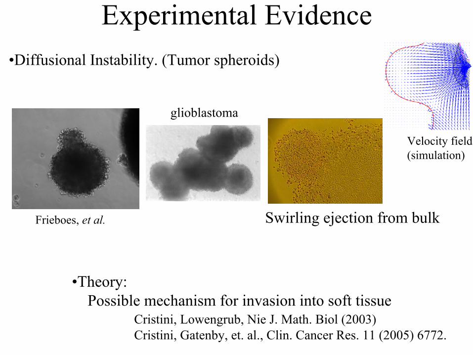
Experimental Evidence•Diffusional Instability. (Tumor spheroids)
Swirling ejection from bulkFrieboes, et al.
•Theory:Possible mechanism for invasion into soft tissue
Velocity field(simulation)
glioblastoma
Cristini, Lowengrub, Nie J. Math. Biol (2003)Cristini, Gatenby, et. al., Clin. Cancer Res. 11 (2005) 6772.
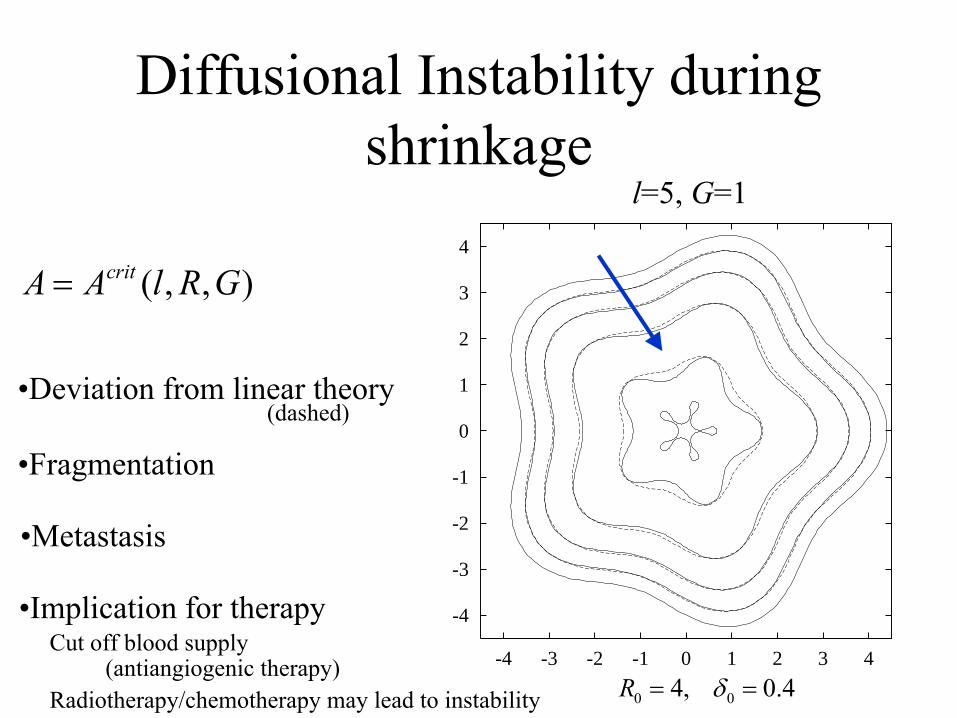
Diffusional Instability duringshrinkage
-4
-3
-2
-1
0
1
2
3
4
-4 -3 -2 -1 0 1 2 3 4
l=5, G=1
•Fragmentation
•Implication for therapy
•Metastasis
( , , )critA A l R G=
•Deviation from linear theory(dashed)
0 04, 0.4R δ= =
Cut off blood supply(antiangiogenic therapy)
Radiotherapy/chemotherapy may lead to instability
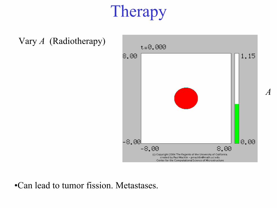
TherapyVary A (Radiotherapy)
•Can lead to tumor fission. Metastases.
A
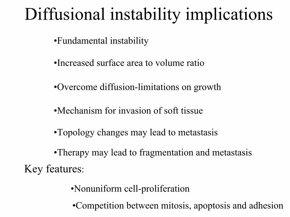
Diffusional instability implications
•Mechanism for invasion of soft tissue
•Increased surface area to volume ratio
•Overcome diffusion-limitations on growth
•Topology changes may lead to metastasis
Key features:
•Fundamental instability
•Nonuniform cell-proliferation
•Competition between mitosis, apoptosis and adhesion
•Therapy may lead to fragmentation and metastasis
Conclusions
•Basic model is able to capture basic qualitative/quantitativefeatures of tumor growth
•Instability in high vascularization regime requiresvascular or mechanical inhomogeneity
•Diffusional instability provides a mechanism to overcomediffusional limitations on growth and can lead to invasive growth and metastasis
Next Steps•More complex/realistic biophysics
•Effect of tissue inhomogeneities
•Going beyond fragmentation/tissue capture
•Angiogenesis
•More realistic mechanical response
•Multiphase/Multiscale models
•Requires new, robust numerical methods
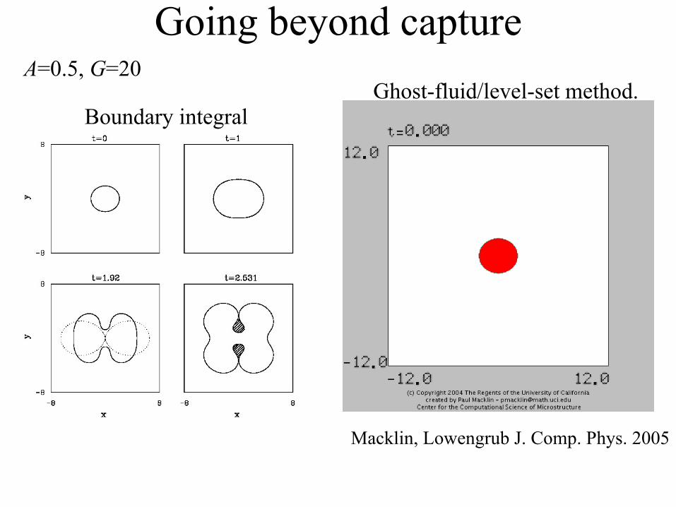
Going beyond captureA=0.5, G=20
Macklin, Lowengrub J. Comp. Phys. 2005
Ghost-fluid/level-set method.Boundary integral