Embed Size (px)
Citation preview

ORIGINAL RESEARCHpublished: 19 October 2016
doi: 10.3389/fncel.2016.00237
C1q/Tumor Necrosis Factor-RelatedProtein-3 Attenuates Brain Injuryafter Intracerebral Hemorrhage viaAMPK-Dependent Pathway in RatShaohua Wang 1,2†, Yang Zhou 1,2†, Yang Bo 1,2, Lingyu Li 1,2, Shanshan Yu 1,2,Yanlin Chen 1,2, Jin Zhu 1,2 and Yong Zhao 1,2*
1 Department of Pathology, Chongqing Medical University, Chongqing, China, 2 Key Laboratory of Neurobiology, ChongqingMedical University, Chongqing, China
Edited by:Daniela Tropea,
Trinity College Dublin, Ireland
Reviewed by:Dennis Qing Wang,
Third Affiliated Hospital of SunYat-Sen University, China
Tatsuro Mutoh,Fujita Health University, Japan
*Correspondence:Yong Zhao
†These authors have contributedequally to this work.
Received: 26 July 2016Accepted: 29 September 2016Published: 19 October 2016
Citation:Wang S, Zhou Y, Yang B, Li L, Yu S,
Chen Y, Zhu J and Zhao Y (2016)C1q/Tumor Necrosis Factor-Related
Protein-3 Attenuates Brain Injury afterIntracerebral Hemorrhage via
AMPK-Dependent Pathway in Rat.Front. Cell. Neurosci. 10:237.
doi: 10.3389/fncel.2016.00237
C1q/tumor necrosis factor (TNF)-related protein-3 (CTRP3) is a recently discoveredadiponectin paralog with established metabolic regulatory properties. However, therole of CTRP3 in intracerebral hemorrhage (ICH) is still mostly unresolved. The aimof the present report was to explore the possible neuroprotective effect of CTRP3 inan ICH rat model and to elucidate the fundamental mechanisms. ICH was inducedin rats by intracerebral infusion of autologous arterial blood. The effects of exogenousCTRP3 (recombinant or lentivirus CTRP3) on brain injury were explored on day 7.Treatment with CTRP3 reduced brain edema, protected against disruption of theblood-brain barrier (BBB), improved neurological functions and promoted angiogenesis.Furthermore, CTRP3 greatly intensified phosphorylation of AMP-activated protein kinase(AMPK) in addition to expression of hypoxia inducing factor-1α (HIF-1α) and vascularendothelial growth factor (VEGF). Finally, the protective effects of CTRP3 could beblocked by either AMPK or VEGF inhibitors. Our findings give the first evidence thatCTRP3 is a new proangiogenic and neuroprotective adipokine, which may exert itsprotective effects at least partly through an AMPK/HIF-1α/ VEGF-dependent pathway,and suggest that CTRP3 may provide a new therapeutic strategy for ICH.
Keywords: CTRP3, intracerebral hemorrhage, brain edema, blood-brain barrier disruption, neuroprotection,angiogenesis
INTRODUCTION
Intracerebral hemorrhage (ICH) is responsible for about 10–15% of stroke cases. Nevertheless,it is the most alarming type due to its large fatality rate and poor functional outcome (Qureshiet al., 2009; Manaenko et al., 2013). ICH results from rupture of blood vessels in the brain.The quick release of blood into the parenchyma leads to substantial mechanical damagethat may only be somewhat ameliorated by limiting hematoma volume (Mayer et al., 2008).Meanwhile, secondary damage ensues due to toxic effects of released blood components, suchas thrombin, as well as erythrocyte rupture (for example, iron-catalyzed free radical reactions).These events, including disruption of the blood-brain barrier (BBB) as well as growth of edemaand inflammation, are currently therapeutic targets for ICH (Fingas et al., 2009; Keep et al.,2012).
Frontiers in Cellular Neuroscience | www.frontiersin.org 1 October 2016 | Volume 10 | Article 237
Wang et al. Role of CTRP3 in ICH
Therapies such as angiogenesis have been suggested forICH. Growing evidence suggests that after ICH, angiogenesis isupregulated in damaged brain tissue of the peri-hematoma area,leading to compensatory cerebral vascular network remodeling(Teng et al., 2008; Lei et al., 2015). Past reports showed that manyangiogenic aspects could ease ischemic brain injury, elevate focalblood flow and enhance neurological results, which indicate thatrecently formed microvessels are indeed functional (Hao et al.,2011; Shen et al., 2013). Blood vessels are a significant scaffoldingfactor that assist with the migration of neurons to damagedbrain areas and supply trophic material to new neurons (Kojimaet al., 2010; Lei et al., 2013). The molecules that encourageneurogenesis and angiogenesis after brain injuries (e.g., ICH) arestill unidentified.
Recently, a highly conserved family of adiponectin paralogs,C1q/tumor necrosis factor(TNF)-related proteins (CTRPs), wasidentified. Each of the 15 known members (CTRP1–CTRP15)is made up of four separate domains including an N-terminalsignal peptide, a short variable domain, a collagen-like domain,and a C-terminal C1q-like globular domain (Ahima et al.,2006). Both CTRPs and adiponectin are a part of the C1q/TNFprotein superfamily, which proceeds to increase in size asmore C1q domain proteins are identified (Yi et al., 2012). TheCTRP family members exhibit broadly diverse physiologicalfunctions, including regulation of metabolism, protection againstendothelial dysfunction and angiogenesis.
CTRP3 is ubiquitously expressed in adipocytes,cartilagocytes, monocytes, fibroblasts, placenta, osteosarcoma,chondroblastoma, giant cell tumor, colon, small intestine,pancreas, kidney, thymus, ovary and in brain (Schaffler andBuechler, 2012). Most importantly, CTRP3 is the only onewhose biological functions have been identified (Petersonet al., 2010). It was found that CTRP3 can encourage in vitroendothelial cell proliferation and migration (Akiyama et al.,2007). But, the role of CTRP3 in promoting angiogenesisin ICH-induced brain injury is not yet known. Further,whether or not CTRP3, an important member of the mostrecently discovered adipokine family, works as a mediator orinhibitor of ICH has not been studied previously. Therefore,the goals of this research were: (1) to investigate the effectsof exogenous CTRP3 in an ICH rat model; (2) to determinewhether CTRP3 administration promotes angiogenesis afterICH; and (3) to elucidate the role of CTRP3 in pathogenesisof ICH.
MATERIALS AND METHODS
Experimental AnimalsAll animal studies were given approval by the ChongqingMedical University Biomedical Ethics Committee. Allexperimental procedures were done in accordance with theNational Institutes of Health Guide for the Care and Use ofLaboratory Animals. All efforts were made to minimize thenumber of animals used and their suffering. A total of 115 adultmale Sprague-Dawley rats (60–80 d old, 240–300 g) were usedfor the in vivo study.
Establishment of IntracerebralHemorrhage ModelICH was induced by an intrastriatal blood infusion method asdescribed previously (Ni et al., 2015). Briefly, rats were deeplyanesthetized with chloral hydrate (350 mg/kg, intraperitonealinjection) and placed in a stereotaxic frame (Kopf Instruments,Tujunga, CA, USA). After removing the hair and cleaning thescalp, the skin was incised. A burr hole was drilled 0.2 mmanterior and 3.0 mm lateral right of bregma. Whole blood(50 µL), which was drawn from the femoral artery, was infusedmanually over 10 min via a 26 G needle inserted into the striatumat a depth of 5.8 mm below the surface of the skull. After 10 min,the needle was steadily taken out for 5 min followed by thesealing of the burr hole with a sterilized medical bone wax. Thewound was cleaned, and the scalp was sutured. The animals weregiven time to heal in their cages. During the recovery period, theanimals had unlimited access to nourishment.
In vivo ExperimentsRats were given free access to food and water in an optimalenvironment preceding the operation. Three in vivo experimentswere performed as described below.
Experiment 1Adult rats were split at random into the following fourgroups: sham-operated (sham) group, ICH group, ICH + vehiclegroup and ICH + recombinant CTRP3 (rCTRP3, Chimerigen,USA) group. rCTRP3 was injected intracerebroventricularly(80 µg/kg) 30 min after ICH and then three times perweek until the animals were killed. Neurological deficits(assessed by a modified Garcia test, beam walking test andwire hanging test), hematoma volume, BBB integrity andbrain edema were measured 7 days after ICH, and samplesfor western blot, qRT-PCR and immunohistochemistry werecollected.
Experiment 2Adult rats were split at random into the following four groups:sham-operated (sham) group, ICH group, ICH + null vectorcontrol (Lenti.null) group, ICH + lentivirus overexpression ofCTRP3 (Lenti.CTRP3) group. Fourteen days after Lenti.CTRP3intracerebroventricular injection, the rats underwent ICH.Neurological deficits, hematoma volume, BBB integrity andbrain edema were measured 7 days after ICH, and samplesfor western blot, qRT-PCR and immunohistochemistry werecollected.
Experiment 3Adult rats were split at random into the following fourgroups: ICH group, ICH + rCTRP3 group, ICH + rCTRP3 +compound C (Com.C) group (AMP-activated protein kinase(AMPK) axis inhibitor, 20 µg/kg, intracerebroventricularinjection, 3 times per week), and ICH + rCTRP3 + CBO-P11 (CBO) group (vascular endothelial growth factor (VEGF)inhibitor 40 µg/kg, intracerebroventricular injection, 3 timesper week). Neurological deficits and BBB integrity were
Frontiers in Cellular Neuroscience | www.frontiersin.org 2 October 2016 | Volume 10 | Article 237

Wang et al. Role of CTRP3 in ICH
measured 7 days after ICH, and samples for western blot andimmunohistochemistry were collected.
Lentivirus-CTRP3 Gene Transfer in the RatBrainAdult rats were anesthetized with chloral hydrate (350 mg/kgintraperitoneal injection) and then placed in a Kopf stereotacticframe. A burr hole was bored in the pericranium 0.9 mm lateralto the sagittal suture and 1.9 mm posterior to the coronal suture.A 10 µL microinjection pump (WPI Inc., Sarasota, FL, USA)was stereotactically inserted 3.5 mm deeper into the cortex. A5 µL viral suspension consisting of 1 × 10⁹ genomic copies ofthe lentivirus-CTRP3 (Lenti-CTRP3) gene, which was injectedipsilaterally into the right lateral cerebral ventricle at a rate of0.2 µL/min. The needle was taken out after 15 min of injection.The animals then were allowed to heal and brought back to theircages.
Behavioral AssessmentBehavioral functions were measured using a modified Garciatest, beam walking test, and wire hanging test 7 days afterICH in a blind fashion (Chen et al., 2013). In the modifiedGarcia test, rats were given a score of 0–18. The scoringsystem consisted of six tests, with possible scores of 0–3(0 = worst; 3 = best). The minimum score was 0 andthe maximum was 18. Beam walking and wire hanging testsutilized bridges (550 cm wire or 590 cm beam) betweentwo platforms on which the rats were placed in the center.Rats were assessed according to six criteria that described ifthe animal could reach the platform and use its limbs ina symmetrical manner and were then given a score of 0–5(normal). The average of three trials per test for each animal wascalculated.
BBB PermeabilitySeven days after ICH, rats were intravenously injected with2% Evans blue dye (4 mL/kg; Sigma-Aldrich, St. Louis, MO,USA). Three hours later, the amount of extravasated Evans bluedye in the hemorrhagic brain hemispheres was evaluated byspectrophotometry (Thermo Scientific, MA, USA) at 620 nm(Cai et al., 2015).
Brain Water ContentSeven days after ICH, the cerebral hemisphere was cut into4-mm thick blocks around the needle track. Brain tissues wereimmediately weighed using an analytical balance and heated at100◦C for 24 h to obtain the dry weight. The water contentwas calculated using the following formula: (wet weight– dryweight)/wet weight× 100%.
Hematoma VolumeHematoma volume was evaluated using a spectrophotometrichemoglobin assay 7 days after the ICH operation (Ma et al.,2014).
Western Blot AnalysisTotal protein was extracted from the peri-hematoma area of therat striatum using cell lysis buffer supplemented with proteinaseand phosphatase inhibitors. Cell lysates were split by 10% SDS-PAGE and transferred to polyvinylidene fluoride membranes.Then, the membranes were blocked in 5% non-fat milk TBSTbuffer for 1.5 h at room temperature. The membranes wereincubated in primary antibody overnight at 4◦C and in secondaryantibody for 1 h at room temperature. Dilutions for primaryantibodies were the following: anti-vascular endothelial growthfactor (VEGFA; 1:1000, Abcam, Cambridge, MA, USA), anti-hypoxia inducing factor-1α (HIF-1α; 1:500, Abcam, Cambridge,MA, USA), and anti-AMPK (phospho-thr172; 1:500, Bioworld,Dublin, OH, USA). The secondary antibody was diluted1:5000 (Sangon Biotech, Shanghai, Co., Ltd.). The densityof bands was detected using an imaging densitometer (Bio-Rad, Foster City, CA, USA), and the gray value of bandswas quantified using Quantity One 1-D analysis software(Bio-Rad).
qRT-PCRTotal RNA was removed with RNAiso Plus (TaKaRaBiotechnology, Dalian, China) using the manufacturer’sinstructions. Reverse transcription was done with a cDNA
FIGURE 1 | Effects of CTRP3 treatment on brain water content 7 daysafter ICH. (A) Brain water content in ipsilateral (Ipsi) and contralateral (Contra)hemispheres 7 days after ICH in rats treated with rCTRP3. (B) Brain watercontent in ipsilateral (Ipsi) and contralateral (Contra) hemispheres 7 days afterICH in rats treated with Lenti-CTRP3. Values are mean ± SEM. n = 8 pergroup. ∗p < 0.05 vs. sham; #p < 0.05 vs. vehicle or Lenti.null. CTRP3,C1q/tumor necrosis factor-related protein-3; rCTRP3, recombinant CTRP3;Lenti.CTRP3, Lentivirus overexpression of CTRP3; ICH, intracerebralhemorrhage.
TABLE 1 | Primers used in qRT-PCR.
Gene product Forward primer Reverse primer Fragment size (bp)
CTRP3 5′-ATGGAGGTGAGCAGAAGAGC-3′ 5′CACAGTCCCCGTTTTAGCAT-3′ 126HIF-1α 5′-CTCCCTTTTTCAAGCAGCAG-3′ 5′GCTCCATTCCATCCTGTTCA-3′ 125VEGFA 5′-CGTCCTGTGTGCCCCTAAT-3′ 5′TGGCTTTGGTGAGGTTTGAT-3′ 121β-actin 5′-TGTTTGAGACCTTCAACACC-3′ 5′-CGCTCATTGCCGATAGTGAT-3′ 207
Frontiers in Cellular Neuroscience | www.frontiersin.org 3 October 2016 | Volume 10 | Article 237

Wang et al. Role of CTRP3 in ICH
synthesis kit (TaKaRa Biotechnology). Real-time PCRreactions were performed with TaKaRa SYBR Premix ExTaq II (TliRnaseH Plus, TaKaRa Biotechnology) on a PCRamplifier (CFX-96 Content Real-time System). Primers (SangonBiotech) are recorded in Table 1.
ImmunohistochemistryRats were killed 7 days after ICH induction by intraperitonealinjection of chloral hydrate. Immunohistochemistry wasperformed as previously described (Lei et al., 2013; Lapi et al.,2015). The primary antibody used was rabbit anti-rat CD31(1:100, Abcam, USA). Total vessel densities were computed bycounting 4 areas in 3 sections through the stroke region foreach animal. Sections were stained with antibody against newvessel marker CD31; positive staining appeared brown. Standardquantitation was done as percent CD31-positive in the regionbordering the hematoma.
FIGURE 2 | Effects of CTRP3 treatment on neurological functions 7days after ICH. All animals after ICH showed significant neurological deficitsbased on performance on the modified Garcia (A), beam balance (B) and wirehanging (C) tests. Rats treated with rCTRP3 showed reduced neurologicaldeficits in all three tests. Treatment with Lenti-CTRP3 starting 2 weeks beforeinduction of ICH showed a tendency to ameliorate neurological deficits(D–F). Values are mean ± SEM. n = 9 per group. ∗p < 0.05 vs. sham;∗∗p < 0.01 vs. sham; #p < 0.05 vs. vehicle or Lenti.null. CTRP3, C1q/tumornecrosis factor-related protein-3; rCTRP3, recombinant CTRP3; Lenti-CTRP3,Lentivirus overexpression of CTRP3; ICH, intracerebral hemorrhage.
Statistical AnalysisAll data are given as mean± S.E.M. One-way analysis of variance(ANOVA) followed by Student’s t test was utilized to collateoutcomes among all groups. The SPSS 17.0 software package wasutilized to do all statistics. P < 0.05 was considered statisticallysignificant.
RESULTS
CTRP3 Reduced Brain Edema andImproved Functional Outcomes after ICHQuantification of brain water content showed that ICH ratshad significantly greater edema in the ipsilateral hemispherethan sham-operated rats (Figure 1A). Brain edema in theipsilateral hemisphere was significantly less in rCTRP3-treated rats than in vehicle-treated rats (Figure 1A). Inthe contralateral hemisphere, rCTRP3 failed to affectbrain water content (Figure 1A). Subsequently, we testedfunctional outcomes using a battery of behavioral tests inrats treated with rCTRP3 or vehicle. Neurological deficitswere significantly more severe in all ICH vs. sham animals7 days after ICH as tested by the modified Garcia test(Figure 2A), wire hanging test (Figure 2B), and beam balancetest (Figure 2C). A statistically significant advancementwas seen in all three neurobehavioral tests after rCTRP3treatment.
Similarly, Lenti.CTRP3 treatment decreased brain watercontent in ICH rats (Figure 1B). Statistically significantneurological shortfalls were observed in all ICH vs. shamanimals 7 days after ICH. Treatment with Lenti-CTRP3tended to ameliorate neurological deficits 7 days after ICH(Figures 2D–F). Both the rCTRP3 and Lenti.CTRP3 treatmentswere effective.
FIGURE 3 | Effects of CTRP3 treatment on ICH-induced blood-brainbarrier (BBB) damage 7 days after ICH. (A) Quantification of Evans bluedye extravasation (blue staining) in the ipsilateral (Ipsi) and contralateral(Contra) hemispheres 7 days after ICH in rats treated with rCTRP3.(B) Quantification of Evans blue dye extravasation (blue staining) in ipsilateral(Ipsi) and contralateral (Contra) hemispheres 7 days after ICH in rats treatedwith Lenti-CTRP3. Values are mean ± SEM. n = 6 per group. ∗∗p < 0.01 vs.sham; #p < 0.05 vs. vehicle or Lenti.null. CTRP3, C1q/tumor necrosisfactor-related protein-3; rCTRP3, recombinant CTRP3; Lenti-CTRP3,Lentivirus overexpression of CTRP3; ICH, intracerebral hemorrhage.
Frontiers in Cellular Neuroscience | www.frontiersin.org 4 October 2016 | Volume 10 | Article 237

Wang et al. Role of CTRP3 in ICH
FIGURE 4 | CTRP3 encourages angiogenesis in the hematoma border zone 7 days after ICH. (A) Capillary density computed by immunohistochemicalstaining for CD31 (brown) in the perifocal region in ICH (b), rCTRP3 (c) and Lenti-CTRP3 (d) 7 days after ICH. A control brain section demonstrates a normaldistribution of microvessels in the same region (a). Bar = 100 µm. (B) Bar graph showing the number of brown stained capillaries 7 days after ICH. Values aremean ± SEM. n = 5 per group. ∗p < 0.05 vs. sham; ∗∗p < 0.01 vs. sham; #p < 0.05 vs. ICH. CTRP3, C1q/tumor necrosis factor-related protein-3; rCTRP3,recombinant CTRP3; Lenti-CTRP3, Lentivirus overexpression of CTRP3; ICH, intracerebral hemorrhage; CD31, platelet endothelial cell adhesion molecule-1.
FIGURE 5 | CTRP3 upregulates pAMPK/HIF-1α/vascular endothelial growth factor (VEGF) axis protein expression in the hematoma border zone 7days after ICH. (A) Western blot analysis of the effect of rCTRP3 on CTRP3, phosphorylated (p) AMP-activated protein kinase (AMPK; Thr172)/AMPK, HIF-1α andVEGF-A. (B–E) Representative ratios of CTRP3, p-AMPK, HIF-1α and VEGF-A protein expression. (F) Western blot analysis of the effect of Lenti-CTRP3 on CTRP3,phosphorylated (p) AMPK (Thr172)/AMPK, HIF-1α and VEGF-A. (G–J) Representative ratios of CTRP3, p-AMPK, HIF-1α and VEGF-A protein expression. Values aremean ± SEM. n = 4 per group. ∗p < 0.05 vs. sham; ##p < 0.01 vs. vehicle or Lenti.null. CTRP3, C1q/tumor necrosis factor-related protein-3; rCTRP3, recombinantCTRP3; Lenti-CTRP3, Lentivirus overexpression of CTRP3; ICH, intracerebral hemorrhage.
Frontiers in Cellular Neuroscience | www.frontiersin.org 5 October 2016 | Volume 10 | Article 237
Wang et al. Role of CTRP3 in ICH
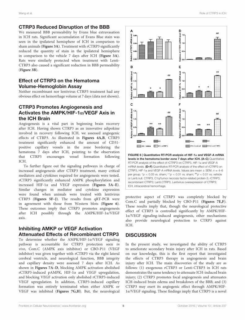
CTRP3 Reduced Disruption of the BBBWe measured BBB permeability by Evans blue extravasationin ICH rats. Significant accumulation of Evans Blue stain wasseen in the ipsilateral hemisphere of ICH in comparison tosham animals (Figure 3A). Treatment with rCTRP3 significantlyreduced the quantity of stain in the ipsilateral hemispherein comparison to the vehicle 7 days after ICH (Figure 3A).Rats were similarly protected when treatment with Lenti-CTRP3 also caused a significant reduction in BBB permeability(Figure 3B).
Effect of CTRP3 on the HematomaVolume-Hemoglobin AssayNeither recombinant nor lentivirus CTRP3 treatment had anyobvious effect on hemorrhage volume at 7 days (data not shown).
CTRP3 Promotes Angiogenesis andActivates the AMPK/HIF-1α/VEGF Axis inthe ICH BrainAngiogenesis is a vital part in beginning brain recoveryafter ICH. Having shown CTRP3 as an innovative adipokineinvolved in recovery following ICH, we assessed angiogeniceffects of CTRP3. As illustrated in Figures 4A,B, CTRP3treatment significantly enhanced the amount of CD31-positive capillary vessels in the zone bordering thehematoma 7 days after ICH, pointing to the observationthat CTRP3 encourages vessel formation followingICH.
To further figure out the signaling pathways in charge ofincreased angiogenesis after CTRP3 treatment, many criticalmediators and cytokines required for angiogenesis were tested.rCTRP3 significantly enhanced AMPK phosphorylation andincreased HIF-1α and VEGF expression (Figures 5A–E).Similar changes in mediator and cytokine expressionwere found when animals were treated with lentivirusCTRP3 (Figures 5F–J). The results from qRT-PCR werein agreement with those from Western blots (Figure 6).These outcomes imply that CTRP3 promotes angiogenesisafter ICH possibly through the AMPK/HIF-1α/VEGFaxis.
Inhibiting AMKP or VEGF ActivationAttenuated Effects of Recombinant CTRP3To determine whether the AMPK/HIF-1α/VEGF signalingpathway is accountable for CTRP3 protection seen invivo, Com.C (AMPK axis inhibitor) or CBO-P11 (VEGFinhibitor) was given together with rCTRP3 via the right lateralcerebral ventricle, and neurological function, BBB integrityand capillary density were assessed 7 days after ICH. Asshown in Figures 7A–D, blocking AMPK activation abolishedrCTRP3-induced pAMPK, HIF-1α and VEGF upregulation,and blocking VEGF activation only abolished rCTRP3-inducedVEGF upregulation. In addition, CTRP3-induced capillaryformation was entirely terminated when either AMPK orVEGF was inhibited (Figures 7G,H). But, the neurological
FIGURE 6 | Quantitative RT-PCR analysis of HIF-1α and VEGF-A mRNAlevels in the hematoma border zone 7 days after ICH. (A–C) QuantitativeRT-PCR analysis of the effect of rCTRP3 on CTRP3, HIF-1α and VEGF-AmRNA levels. (D–F) Quantitative RT-PCR analysis of the effect of rCTRP3 onCTRP3, HIF-1α and VEGF-A mRNA levels. Values are mean ± SEM. n = 4–6per group. ∗p < 0.05 vs. sham; ∗∗p < 0.01 vs. sham; ##p < 0.01 vs. vehicleor Lenti.null. CTRP3, C1q/tumor necrosis factor-related protein-3; rCTRP3,recombinant CTRP3; Lenti.CTRP3, Lentivirus overexpression of CTRP3;ICH, intracerebral hemorrhage.
protective aspect of CTRP3 was completely blocked byCom.C and partially blocked by CBO-P11 (Figures 7E,F).These results imply that, though the neurological protectiveeffect of CTRP3 is controlled significantly by AMPK/HIF-1α/VEGF signaling-induced angiogenesis, other mechanismsalso provide neurological protection to CTRP3 againstICH.
DISCUSSION
In the present study, we investigated the ability of CTRP3to ameliorate secondary brain injury after ICH in rats. Basedon our knowledge, this is the first report that investigatedthe effects of CTRP3 therapy in angiogenesis and braininjury after ICH. The main discoveries of the study are asfollows: (1) exogenous rCTRP3 or Lenti-CTRP3 in ICH ratsdemonstrates the same tendency to attenuate ICH-induced braininjury; (2) CTRP3 promotes focal angiogenesis and attenuatesICH-induced brain edema and breakdown of the BBB; and (3)CTRP3 may exert its angiogenic effect through AMPK/HIF-1α/VEGF signaling. These findings imply that CTRP3 is a novel
Frontiers in Cellular Neuroscience | www.frontiersin.org 6 October 2016 | Volume 10 | Article 237

Wang et al. Role of CTRP3 in ICH
FIGURE 7 | CTRP3 promotes angiogenesis via an AMPK-dependent signaling mechanism. (A) Western blot analysis of the effects of COM.C and CBO onrCTRP3-induced p-AMPK, HIF-1α and VEGF-A expression. (B–D) Representative ratios of p-AMPK, HIF-1α and VEGF-A protein expression. (E) Modified Garcia testanalysis of the effects of COM.C and CBO on neurological function improvement induced by rCTRP3. (F) Evans blue dye extravasation analysis of the effects ofCOM.C and CBO on ICH-induced BBB damage reduced by rCTRP3. (G) Immunohistochemical staining analysis of the effects of COM.C and CBO on angiogenesisinduced by rCTRP3. Capillary density measured by immunohistochemical staining for CD31 (brown) in the perifocal region in ICH (a), rCTRP3 (b), COM.C (c) andCBO (d) 7 days after ICH. (H) Bar graph showing the number of brown stained capillaries. Values are mean ± SEM. n = 4–6 per group. ∗∗p < 0.01 vs. ICH;#p < 0.05 vs. rCTRP3; ##p < 0.01 vs. rCTRP3; &p < 0.05 vs. COM.C. CTRP3, C1q/tumor necrosis factor-related protein-3; rCTRP3, recombinant CTRP3;Lenti-CTRP3, Lentivirus overexpression of CTRP3; ICH, intracerebral hemorrhage; COM.C, compound C; CBO, CBO-P11.
angiogenic factor that might perform a key part in encouragingangiogenesis by activating the AMPK signaling pathway duringICH.
The prognosis of ICH is affected by multiple factors (Brownet al., 1996). Neurotrophin family, anti-oxidative mediators,anti-mitochondrial impairment or anti-inflammatory drugscontribute to functional recovery and promote neuronal survivalin the central nervous system (Ip et al., 1993; Chung et al.,2013; Xu et al., 2013; Chen et al., 2015; Wei et al., 2015).Angiogenesis induced in the ischemic penumbra (Risau, 1997)as well as rapid recovery of reperfusion and oxygen supplyin injured brain tissues are critical prognostic factors (Mayer
et al., 1998). In this study, we provide new evidence thatCTRP3 has strong angiogenic and neuroprotective aspects,implying that CTRP3 could be an innovative therapeutic targetof ICH.
It is known that compensatory angiogenesis can happen inthe peri-hematoma region after ICH. Angiogenesis is a stepwiseprocedure that includes an increase in vascular permeability,degradation of the surrounding matrix, proliferation andmigration of endothelial cells, and stabilization of freshly createdmicrovessels (Conway et al., 2001). Concerted actions of manyangiogenic molecules are necessary in this procedure, andVEGF is the most vital factor during each step of angiogenesis
Frontiers in Cellular Neuroscience | www.frontiersin.org 7 October 2016 | Volume 10 | Article 237

Wang et al. Role of CTRP3 in ICH
(Rosenstein et al., 1998; Yancopoulos et al., 2000). A numberof animal experimental studies have shown that VEGF andfibroblast growth factor treatment encourages angiogenesis withideal efficacy and increases capillary numbers (Lavu et al.,2011; Ye, 2016). However, endogenous angiogenesis followinga stroke is insufficient to reverse brain injury. Our studyshows that CTRP3 successfully encourages angiogenesis andupregulates VEGF expression in the striatum ipsilateral to thehemorrhage, which leads to increased vessel density. BecauseCTRP3 has been established to directly encourage endothelialcell proliferation and migration but not increase tube formation(a procedure that requires complexity surpassing proliferationand migration), angiogenic factors other than VEGF are likelyinvolved.
These findings contribute to the increasing literature onthe vital part of VEGF in brain injury. In fact, VEGF levelsare increased during a plethora of pathological events in thebrain, implicating its essential role in brain repair processes(Cristofaro and Emanueli, 2009). VEGF binds to two receptors,VEGF receptor-1 (VEGFR-1) and VEGF receptor-2 (VEGFR-2), through which it encourages revascularization and themending of the BBB and re-establishes metabolic and trophicassistance to injured tissue (Krum et al., 2008; Shimotake et al.,2010). Future work in this field should aim to illuminatewhether CTRP3 interacts with one or both receptors duringICH.
Because ICH-induced stress can change the composition,structure and distribution of the extracellular matrix, which hasa vital role in creating normal brain tissue structures and isclosely linked with brain injury-induced brain edema formation(Keep et al., 2012; Chung et al., 2013; Turner and Sharp,2016), we investigated the effect of CTRP3 on BBB integrityby measuring brain water content and extravasation of Evansblue dye. We showed that angiogenesis via CTRP3 treatmentresulted in preservation of the BBB. This observation in ICHextends past discoveries from other models of brain injury.In a model of cerebral ischemia, VEGF bound to VEGFR-2,which is expressed predominantly on activated astrocytes in thecentral nervous system, and encouraged revascularization andrepair of the BBB by giving metabolic and trophic assistanceto injured tissue (Krum et al., 2008; Shimotake et al., 2010;Hao et al., 2011). In a rat model of stroke, VEGF enhancedangiogenesis in the ischemic brain and reduced neurologicaldeficits during recovery (Zhang et al., 2000). But, we were notable to demonstrate any effect of CTRP3 treatment on hematomavolume. We speculate that this may be due to use of a blood-induced ICH model.
It is important to note that the CTRP3-treated rats manifestednot only greatly strengthened angiogenesis and enhanced BBBpreservation but also diminished neurological deficits. Thereare several possibilities for this improvement. First, the greatangiogenic effect of CTRP3 in the border zone may helpin restoring the blood flow, thereby rescuing dying neuronsaround the hematoma. Second, angiogenesis may assist newneurons migrating to damaged brain regions and give trophicmaterial to these cells (Kojima et al., 2010; Lei et al., 2013).Finally, CTRP3 may promote differentiation of neural stem
cells into neurons. Such intriguing possibilities all merit expressstudy.
Around the hematoma, ischemia and hypoxia are apparentin ICH and this stress activates the AMPK and HIF1 signalingpathway, which induces the AMPK phosphorylation in humanumbilical vein endothelial cells and promotes the recruitment,migration, proliferation and differentiation of endothelial cells(Nagata et al., 2003). In ischemic mice hindlimbs, AICAR,an AMPK antagonist induces expression of endogenous HIFtarget gene VEGF, but dominant-negative AMPK abolishes thisexpression at both steady state mRNA and protein levels (Ouchiet al., 2005). These data suggest that AMPK signaling is likely toregulate the expression of VEGF and promote angiogenesis inresponse to ischemic injury.
CTRP3 is a paralog of adiponectin, and it is well receivedthat adiponectin encourages angiogenesis through activation ofAMPK signaling (Shimano et al., 2010). Yet, whether CTRP3encourages angiogenesis in cerebral tissue through the samepathway is not yet known. More important, inhibiting AMPKphosphorylation by Com.C greatly eradicated CTRP3-inducedHIF-1α and VEGF expression and blocked the angiogenic effectof CTRP3. These findings defend the existence of an AMPK-dependent mechanism for angiogenic effects of CTRP3.
CONCLUSION
We conclude that CTRP3, a key member of a newly identifiedadipokine family, upregulates expression of angiogenic cytokinesand induces robust angiogenesis, which led to enhancedpreservation of the BBB and reduction of neurological deficitsafter ICH. The effect is mediated by the AMPK signalingpathway. These findings suggest that CTRP3 plays a positive roleduring ICH and has therapeutic potential.
AUTHOR CONTRIBUTIONS
SW, YaZ, YB, LL and YoZ: conceived and designed theexperiments. SW, YaZ, YB and SY: conducted the experiments.SW and YaZ: analyzed the results. SW, YC and JZ: contributedmaterials and analysis tools. SW and YaZ: wrote the article. SWand YaZ: contributed equally to this study. All authors reviewedthe manuscript.
FUNDING
This work was supported by The National Natural ScienceFoundation of China (81271460 and 81671158), Natural ScienceYouth Foundation of China (No. 81301125), the Medicalscientific research projects of Chongqing (20120221) andthe Natural Science Foundation of Chongqing Science andTechnology Committee, China (No. cstc2015jcyjA10048).
ACKNOWLEDGMENTS
We are grateful to Key Laboratory of Neurobiology (Chongqing,China) for technical guidance and partial sponsorship.
Frontiers in Cellular Neuroscience | www.frontiersin.org 8 October 2016 | Volume 10 | Article 237

Wang et al. Role of CTRP3 in ICH
REFERENCES
Ahima, R. S., Qi, Y., Singhal, N. S., Jackson, M. B., and Scherer, P. E. (2006). Brainadipocytokine action andmetabolic regulation.Diabetes 55, S145–S154. doi: 10.2337/db06-s018
Akiyama, H., Furukawa, S., Wakisaka, S., and Maeda, T. (2007). CTRP3/cartducinpromotes proliferation and migration of endothelial cells. Mol. Cell. Biochem.304, 243–248. doi: 10.1007/s11010-007-9506-6
Brown, R. D., Whisnant, J. P., Sicks, J. D., O’Fallon, W. M., and Wiebers, D. O.(1996). Stroke incidence, prevalence and survival: secular trends in Rochester,Minnesota, through 1989. Stroke 27, 373–380.
Cai, P., Luo, H., Xu, H., Zhu, X., Xu, W., Dai, Y., et al. (2015). RecombinantADAMTS 13 attenuates brain injury after intracerebral hemorrhage. Stroke 46,2647–2653. doi: 10.1161/STROKEAHA.115.009526
Chen, S., Ma, Q., Krafft, P. R., Chen, Y., Tang, J., Zhang, J., et al. (2013).P2X7 receptor antagonism inhibits p38 mitogen-activated protein kinaseactivation and ameliorates neuronal apoptosis after subarachnoid hemorrhagein rats. Crit. Care Med. 41, e466–e474. doi: 10.1097/CCM.0b013e31829a8246
Chen, D., Wei, X., Zou, J., Wang, R., Liu, X., Xu, X., et al. (2015). Contra-directional expression of serum homocysteine and uric acid as importantbiomarkers of multiple system atrophy severity: a cross-sectional study. Front.Cell. Neurosci. 9:247. doi: 10.3389/fncel.2015.00247
Chung, C. Y., Yang, J. T., and Kuo, Y. C. (2013). Polybutylcyanoacrylatenanoparticles for delivering hormone response element-conjugatedneurotrophin-3 to the brain of intracerebral hemorrhagic rats. Biomaterials 34,9717–9727. doi: 10.1016/j.biomaterials.2013.08.083
Conway, E. M., Collen, D., and Carmeliet, P. (2001). Molecular mechanismsof blood vessel growth. Cardiovasc. Res. 49, 507–521. doi: 10.1016/s0008-6363(00)00281-9
Cristofaro, B., and Emanueli, C. (2009). Possible novel targets for therapeuticangiogenesis. Curr. Opin. Pharmacol. 9, 102–108. doi: 10.1016/j.coph.2008.11.006
Fingas, M., Penner, M., Silasi, G., and Colbourne, F. (2009). Treatment ofintracerebral hemorrhage in rats with 12 h, 3 days and 6 days of selectivebrain hypothermia. Exp. Neurol. 219, 156–162. doi: 10.1016/j.expneurol.2009.05.007
Hao, Q., Su, H., Palmer, D., Sun, B., Gao, P., Yang, G. Y., et al. (2011).Bone marrow-derived cells contribute to vascular endothelial growth factor-induced angiogenesis in the adult mouse brain by supplying matrixmetalloproteinase-9. Stroke 42, 453–458. doi: 10.1161/STROKEAHA.110.596452
Ip, N. Y., McClain, J., Barrezueta, N. X., Aldrich, T. H., Pan, L., Li, Y., et al. (1993).The alpha component of the CNTF receptor is required for signaling anddefines potential CNTF targets in the adult and during development. Neuron10, 89–102. doi: 10.1016/0896-6273(93)90245-m
Keep, R. F., Hua, Y., and Xi, G. (2012). Intracerebral haemorrhage: mechanisms ofinjury and therapeutic targets. Lancet Neurol. 11, 720–731. doi: 10.1016/s1474-4422(12)70104-7
Kojima, T., Hirota, Y., Ema, M., Takahashi, S., Miyoshi, I., Okano, H., et al.(2010). Subventricular zone-derived neural progenitor cells migrate along ablood vessel scaffold toward the post-stroke striatum. Stem Cells 28, 545–554.doi: 10.1002/stem.306
Krum, J. M., Mani, N., and Rosenstein, J. M. (2008). Roles of the endogenousVEGF receptors flt-1 and flk-1 in astroglial and vascular remodeling after braininjury. Exp. Neurol. 212, 108–117. doi: 10.1016/j.expneurol.2008.03.019
Lapi, D., Vagnani, S., Sapio, D., Mastantuono, T., Boscia, F., Pignataro, G.,et al. (2015). Effects of bone marrow mesenchymal stem cells (BM-MSCs) on rat pial microvascular remodeling after transient middle cerebralartery occlusion. Front. Cell. Neurosci. 9:329. doi: 10.3389/fncel.2015.00329
Lavu, M., Gundewar, S., and Lefer, D. J. (2011). Gene therapy for ischemicheart disease. J. Mol. Cell. Cardiol. 50, 742–750. doi: 10.1016/j.yjmcc.2010.06.007
Lei, C., Lin, S., Zhang, C., Tao, W., Dong, W., Hao, Z., et al. (2013). Activationof cerebral recovery by matrix metalloproteinase-9 after intracerebralhemorrhage. Neuroscience 230, 86–93. doi: 10.1016/j.neuroscience.2012.11.008
Lei, C., Zhang, S., Cao, T., Tao, W., Liu, M., and Wu, B. (2015). HMGB1 mayact via RAGE to promote angiogenesis in the later phase after intracerebralhemorrhage. Neuroscience 295, 39–47. doi: 10.1016/j.neuroscience.2015.03.032
Ma, Q., Chen, S., Hu, Q., Feng, H., Zhang, J. H., and Tang, J. (2014). NLRP3inflammasome contributes to inflammation after intracerebral hemorrhage.Ann. Neurol. 75, 209–219. doi: 10.1002/ana.24070
Manaenko, A., Lekic, T., Ma, Q., Zhang, J. H., and Tang, J. (2013). Hydrogeninhalation ameliorated mast cell-mediated brain injury after intracerebralhemorrhage in mice. Crit. Care Med. 41, 1266–1275. doi: 10.1097/CCM.0b013e31827711c9
Mayer, S. A., Brun, N. C., Begtrup, K., Broderick, J., Davis, S., Diringer, M. N.,et al. (2008). Efficacy and safety of recombinant activated factor VII foracute intracerebral hemorrhage. N. Engl. J. Med. 358, 2127–2137. doi: 10.1056/NEJMoa0707534
Mayer, S. A., Lignelli, A., Fink, M. E., Kessler, D. B., Thomas, C. E., Swarup, R.,et al. (1998). Perilesional blood flow and edema formation in acute intracerebralhemorrhage: a SPECT study. Stroke 29, 1791–1798. doi: 10.1161/01.str.29.9.1791
Nagata, D., Mogi, M., and Walsh, K. (2003). AMP-activated protein kinase(AMPK) signaling in endothelial cells is essential for angiogenesis inresponse to hypoxic stress. J. Biol. Chem. 278, 31000–31006. doi: 10.1074/jbc.M300643200
Ni, W., Okauchi, M., Hatakeyama, T., Gu, Y., Keep, R. F., Xi, G., et al.(2015). Deferoxamine reduces intracerebral hemorrhage-induced white matterdamage in aged rats. Exp. Neurol. 272, 128–134. doi: 10.1016/j.expneurol.2015.02.035
Ouchi, N., Shibata, R., and Walsh, K. (2005). AMP-activated protein kinasesignaling stimulates VEGF expression and angiogenesis in skeletal muscle.Circ.Res. 96, 838–846. doi: 10.1161/01.res.0000163633.10240.3b
Peterson, J. M., Wei, Z., and Wong, G. W. (2010). C1q/TNF-related protein-3(CTRP3), a novel adipokine that regulates hepatic glucose output. J. Biol. Chem.285, 39691–39701. doi: 10.1074/jbc.M110.180695
Qureshi, A. I., Mendelow, A. D., and Hanley, D. F. (2009). Intracerebralhaemorrhage. Lancet 373, 1632–1644. doi: 10.1016/S0140-6736(09)60371-8
Risau, W. (1997). Mechanisms of angiogenesis. Nature 386, 671–674. doi: 10.1038/386671a0
Rosenstein, J. M., Mani, N., Silverman, W. F., and Krum, J. M. (1998). Patternsof brain angiogenesis after vascular endothelial growth factor administration invitro and in vivo. Proc. Natl. Acad. Sci. U S A 95, 7086–7091. doi: 10.1073/pnas.95.12.7086
Schaffler, A., and Buechler, C. (2012). CTRP family: linking immunity tometabolism. Trends Endocrinol. Metab. 23, 194–204. doi: 10.1016/j.tem.2011.12.003
Shen, L., Miao, J., Yuan, F., Zhao, Y., Tang, Y., Wang, Y., et al. (2013).Overexpression of adiponectin promotes focal angiogenesis in the mouse brainfollowing middle cerebral artery occlusion. Gene Ther. 20, 93–101. doi: 10.1038/gt.2012.7
Shimano, M., Ouchi, N., Shibata, R., Ohashi, K., Pimentel, D. R., Murohara, T.,et al. (2010). Adiponectin deficiency exacerbates cardiac dysfunction followingpressure overload through disruption of an AMPK-dependent angiogenicresponse. J. Mol. Cell. Cardiol. 49, 210–220. doi: 10.1016/j.yjmcc.2010.02.021
Shimotake, J., Derugin, N.,Wendland,M., Vexler, Z. S., and Ferriero, D.M. (2010).Vascular endothelial growth factor receptor-2 inhibition promotes cell deathand limits endothelial cell proliferation in a neonatal rodent model of stroke.Stroke 41, 343–349. doi: 10.1161/STROKEAHA.109.564229
Teng, H., Zhang, Z. G., Wang, L., Zhang, R. L., Zhang, L., Morris, D., et al. (2008).Coupling of angiogenesis and neurogenesis in cultured endothelial cells andneural progenitor cells after stroke. J. Cereb. Blood Flow Metab. 28, 764–771.doi: 10.1038/sj.jcbfm.9600573
Turner, R. J., and Sharp, F. R. (2016). Implications of MMP9 for blood brainbarrier disruption and hemorrhagic transformation following ischemic stroke.Front. Cell. Neurosci. 10:56. doi: 10.3389/fncel.2016.00056
Wei, X., Gao, H., Zou, J., Liu, X., Chen, D., Liao, J., et al. (2015). Contra-directionalcoupling of Nur77 and Nurr1 in neurodegeneration: a novel mechanism formemantine-induced anti-inflammation and anti-mitochondrial impairment.Mol. Neurobiol. doi: 10.1007/s12035-015-9477-7 [Epub ahead of print].
Frontiers in Cellular Neuroscience | www.frontiersin.org 9 October 2016 | Volume 10 | Article 237

Wang et al. Role of CTRP3 in ICH
Xu, Y. Q., Long, L., Yan, J. Q., Wei, L., Pan, M. Q., Gao, H. M., et al.(2013). Simvastatin induces neuroprotection in 6-OHDA-lesioned PC12 viathe PI3K/AKT/caspase 3 pathway and anti-inflammatory responses. CNSNeurosci. Ther. 19, 170–177. doi: 10.1111/cns.12053
Yancopoulos, G. D., Davis, S., Gale, N. W., Rudge, J. S., Wiegand, S. J., andHolash J. (2000). Vascular-specific growth factors and blood vessel formation.Nature 407, 242–248. doi: 10.1038/35025215
Ye, W. (2016). The complexity of translating anti-angiogenesis therapy frombasic science to the clinic. Dev. Cell 37, 114–125. doi: 10.1016/j.devcel.2016.03.015
Yi,W., Sun, Y., Yuan, Y., Lau,W. B., Zheng, Q., Wang, X., et al. (2012). C1q/tumornecrosis factor-related protein-3, a newly identified adipokine, is a novelantiapoptotic, proangiogenic and cardioprotective molecule in the ischemicmouse heart. Circulation 125, 3159–3169. doi: 10.1161/CIRCULATIONAHA.112.099937
Zhang, Z. G., Zhang, L., Jiang, Q., Zhang, R., Davies, K., Powers, C., et al. (2000).VEGF enhances angiogenesis and promotes blood-brain barrier leakage in theischemic brain. J. Clin. Invest. 106, 829–838. doi: 10.1172/jci9369
Conflict of Interest Statement: The authors declare that the research wasconducted in the absence of any commercial or financial relationships that couldbe construed as a potential conflict of interest.
Copyright © 2016 Wang, Zhou, Bo, Li, Yu, Chen, Zhu and Zhao. This is an open-access article distributed under the terms of the Creative Commons AttributionLicense (CC BY). The use, distribution and reproduction in other forums ispermitted, provided the original author(s) or licensor are credited and that theoriginal publication in this journal is cited, in accordance with accepted academicpractice. No use, distribution or reproduction is permitted which does not complywith these terms.
Frontiers in Cellular Neuroscience | www.frontiersin.org 10 October 2016 | Volume 10 | Article 237


![Tumor Necrosis Factor-aSensitizes Prostate …cancerres.aacrjournals.org/content/canres/59/7/1606.full.pdf[CANCER RESEARCH 59, 1606–1614, April 1, 1999] Tumor Necrosis Factor-aSensitizes](https://img.pdfslide.us/doc/110x75/5af72b157f8b9ae9488fbe33/tumor-necrosis-factor-asensitizes-prostate-cancer-research-59-16061614.jpg)